Article

The Arabidopsis altered in stress response2 is Impaired in Resistance to Root and Leaf Necrotrophic Fungal Pathogens

by
Louise F. Thatcher
1,* and
Karam B. Singh
1,2,3
1
CSIRO Agriculture and Food, Centre for Environment and Life Sciences, Wembley, Western Australia 6913, Australia
2
The Institute of Agriculture, The University of Western Australia, 35 Stirling Highway, Crawley, Western Australia 6009, Australia
3
Centre for Crop and Disease Management, Department of Environment and Agriculture, Curtin University, Bentley, Western Australia 6102, Australia
*
Author to whom correspondence should be addressed.
Plants 2019, 8(3), 60; https://doi.org/10.3390/plants8030060
Submission received: 14 February 2019 / Revised: 4 March 2019 / Accepted: 8 March 2019 / Published: 11 March 2019

Abstract

:
The Arabidopsis thaliana Glutathione S-transferase Phi8 (GSTF8) gene is recognised as a marker for early defence and stress responses. To identify regulators of these responses, a forward genetic screen for Arabidopsis mutants with up-regulated GSTF8 promoter activity was conducted by screening a mutagenized population containing a GSTF8 promoter fragment fused to the luciferase reporter gene (GSTF8:LUC). We previously identified several enhanced stress response (esr) mutants from this screen that conferred constitutive GSTF8:LUC activity and increased resistance to several pathogens and/or insects pests. Here we identified a further mutant constitutively expressing GSTF8:LUC and termed altered in stress response2 (asr2). Unlike the esr mutants, asr2 was more susceptible to disease symptom development induced by two necrotrophic fungal pathogens; the root pathogen Fusarium oxysporum, and the leaf pathogen Alternaria brassicicola. The asr2 allele was mapped to a 2.1 Mbp region of chromosome 2 and narrowed to four candidate loci.

1. Introduction

Crop damage caused by pathogen and pest attack costs the global economy billions of dollars annually, with the Food and Agriculture Organization of the United Nations (FAO) estimating global annual yield reductions in the range of 20% to 40% [1]. The identification of disease resistance-associated genes with quantitative effects and an understanding of their function is a necessity to mitigate the ongoing effects of biotic stresses.
A large gene family associated with responses to an array of biotic stresses are the Glutathione S-transferases (GST) [2,3]. Through a combination of catalytic or non-catalytic functions, repertoires of GST protein products function in response to pathogen attacks by regulating oxidative bursts or the accumulation of defence-related metabolites [4]. In addition to involvement in biotic stress, some plant GSTs are also well known for their roles in response to abiotic stresses and the detoxification of xenobiotic compounds such as herbicides [5]. Of the 55 members in Arabidopsis, the GSTPHI8 (GSTF8) gene is comparatively well studied and has emerged as a marker gene for early stress and defence responses [3,6,7,8]. Its expression is induced by multiple biotic elicitors including of fungal or bacterial origin, and phytohormones or signalling molecules such as salicylic acid (SA) or H2O2 [6,7,9,10,11,12,13].
Based on the well-characterised GSTF8 expression profile [3,10,13,14], an Arabidopsis line containing the GSFT8 promoter linked to the Firefly Luciferase reporter gene (GSTF8:LUC) has been used to non-invasively monitor a plant’s stress status and response to defence cues such as to plant hormones or challenge with fungal pathogens [6,11,12,15]. Coupling this line with a mutagenesis approach facilitated the discovery of genes involved in the positive or negative regulation of stress responses and the discovery of mutants altered in resistance to pathogens or pests [7,8,16]. One mutant termed disrupted in stress responses1 (dsr1) encodes a positive regulator [7]. It exhibited a loss of salicylic acid (SA)-inducible GSTF8:LUC activity and increased susceptibility to several fungal and bacterial pathogens. The causal mutation results in a single amino acid change in a subunit of the mitochondrial energy machinery (complex II subunit SDH1-1), causing a reduction in induced reactive oxygen species (ROS) production from mitochondria.
Complementing the discovery of dsr1, several mutants with constitutive GSTF8:LUC expression were also discovered and designated as an enhanced stress response (esr). Three mutants (esr1-1, esr1-3, esr1-4) encoded alleles of the K homology (KH) domain containing RNA-binding protein At5g53060 and conferred increased resistance to the root-infecting fungal pathogen Fusarium oxysporum [8]. Fusarium oxysporum is considered a hemibiotrophic pathogen, living part of its life as a biotroph (a pathogen that derives nutrients from living host cells), and the other part, often associated with the later stages of infection, as a necrotroph (a pathogen that derives nutrients from dead cells) [17]. The esr1 mutations conferred a STOP codon substitution or splicing defects at splice site junctions. Global transcriptome analysis of the esr1-1 mutant compared to wild-type plants identified altered expression of genes involved in responses to biotic and abiotic stress [8]. A second negative defence-regulator, whose protein product interacts with ESR1/KH-domain, was identified from the same screen. Two mutant alleles of this gene, Enhanced Stress Response3/RNA Polymerase II C-Terminal Domain (CTD) Phosphatase-Like1 (CPL1) (At4g21670) were cloned and also result from an induced stop codon change or splicing defect [16]. The cpl1-7 (esr3-1) and cpl1-8 (esr3-2) mutants displayed strong resistance to F. oxysporum, as well as increased resistance to the leaf necrotrophic fungal pathogen Alternaria brassicicola and to an aphid pest. Global transcriptome analysis of cpl1-8 compared to wild-type plants identified up-regulated expression of genes related to defence and oxidative stress/redox state processes [16].
Here we extend on our analysis of mutants from the GSTF8:LUC mutagenesis screen and isolate a second class of constitutively expressing GSTF8:LUC mutant. This mutant termed altered in stress response2 (asr2) differs from the constitutive GSTF8:LUC esr1 and cpl1 mutants and confers increased susceptibility to both the root fungal pathogen F. oxysporum and the leaf fungal pathogen A. brassicicola, suggesting ASR2 encodes a positive regulator of defence responses. The causal mutation was narrowed to four candidate loci not previously implicated in resistance to fungal pathogens.

2. Results

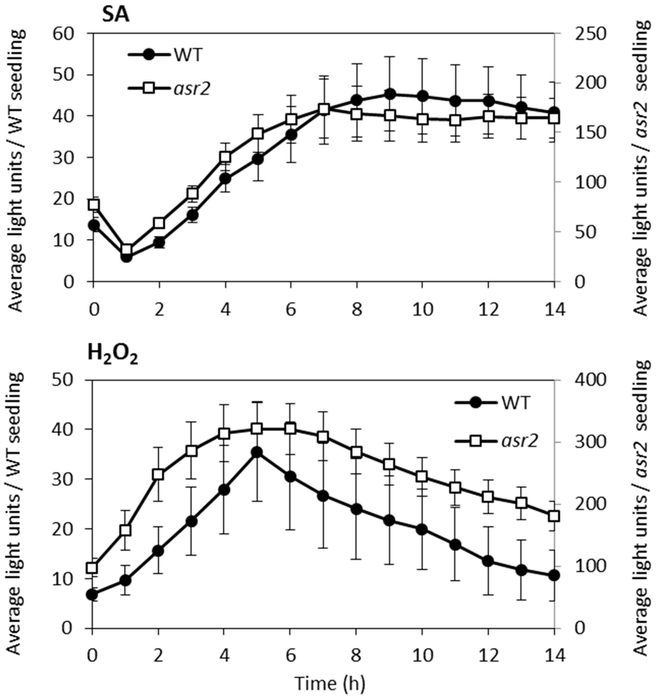
2.1. Identification of asr2

From our previous screen that identified the esr1 and cpl1 (esr3) mutants [8,16], a further 40 constitutively expressing GSTF8:LUC mutants were identified including one M2 mutant labelled asr2 that, following esr1 and cpl1, had the next highest levels of basal promoter expression (Figure 1a). Reciprocal crosses between asr2 and esr1-1, cpl1-7 (esr3-1) or cpl1-8 (esr3-2), and assessment of complementing constitutive luciferase phenotypes in F1 populations determined asr2 was not an allele of either esr1 or cpl1 (esr3) (data not shown). For cloning and heritability studies, an M3 asr2 was out-crossed to the Landsberg erecta ecotype (Ler). All F1 plants showed the wild-type phenotype, and the F2 plants displayed a ~3:1 (wild-type:mutant) segregation suggesting the asr2 phenotype was due to a recessive, monogenetic trait (59:21, χ2 test p = 0.80).

2.2. Basal Defence Gene Expression is Altered in asr2

GSTF8:LUC expression was confirmed in the M3 asr2 generation by in vivo quantification and a biochemical luciferase assay (Figure 1b). Quantitative real-time PCR (qRT-PCR) identified a small (2-fold) but non-significant increase in endogenous asr2 GSTF8 expression relative to wild-type GSTF8:LUC seedlings (Figure 1c). Since enhanced pathogen or pest resistance in the esr1 and cpl1 mutants is linked to altered defence-associated pathway expression, we hypothesised that asr2 may also be altered in its defence gene expression. We, therefore, assessed the expression of two defence marker genes; Pathogenesis Related1 (PR1), an SA pathway marker; and Plant Defensin1.2 (PDF1.2), a jasmonic acid (JA) pathway marker. In a simplistic model, defences dependent on the SA-signalling pathway are generally required for resistance against biotrophs, while the JA-signalling pathway is required for resistance against necrotrophic pathogens [18,19]. A small (2.3-fold) but non-significant increase in PR1 expression was measured in asr2 (Figure 1c). PDF1.2 expression, on the other hand, was significantly decreased by nearly 12-fold compared to wild-type GSTF8:LUC plants (Figure 1c). This suggests a potential imbalance in asr2 defence signalling.
The higher levels of the SA-inducible GSTF8 and PR1 genes in asr2 prompted us to determine whether GSTF8:LUC promoter activity in this mutant was hyper-responsive to SA [10,11,13]. Induction of GSTF8:LUC activity after SA treatment was seen in both wild-type and asr2, with the asr2 mutant achieving a higher level of expression but a smaller fold change in activation (asr2 2.2-fold, and wild-type 3.3-fold). (Figure 2). A similar result was obtained following H2O2 treatment (wild-type 5.3-fold; asr2 3.3-fold). The lower fold change in asr2 is likely due to its higher basal level of GSTF8:LUC expression and may represent an upper limit of activation potential of this promoter. In both treatments, the temporal GSTF8:LUC expression profile in asr2 closely resembled that in wild-type. For subsequent assays, a twice-backcrossed (to wild-type) asr2 line was developed to remove other ethyl methansulfonate (EMS)-induced mutations. This line had the same phenotypes as the M3 asr2 line (data not shown).

2.3. ASR2 is Necessary for Resistance Against the Necrotrophic Fungal Pathogens Fusarium oxysporum and Alternaria brassicicola

The esr1 and cpl1 mutants both had increased resistance to the root fungal pathogen F. oxysporum, and also showed altered responses to the leaf fungal pathogen A. brassicicola. We hypothesised that asr2 may also display an altered response to these pathogens considering its down-regulated expression of the JA-marker gene PDF1.2 and this gene’s association with resistance to necrotrophic fungal pathogens [20,21,22]. Following inoculation with F. oxysporum, disease symptom development progressed more quickly in asr2 compared to wild-type plants. Within 14 days post inoculation (dpi), on average 5.3 leaves per asr2 plant showed Fusarium disease symptoms compared to 3.8 leaves per wild-type plant. Of these diseased leaves, the number of necrotic leaves on asr2 plants was significantly higher than the number on wild-type (Figure 3a,b). By 21 dpi, the survival rate of asr2 plants was significantly lower than wild-type plants—23% compared to 60% (Figure 3c). On assessment of responses to A. brassicicola, significantly larger lesions developed on asr2 leaves compared to wild-type (Figure 4a,b). The disease assay also induced chlorosis symptom development on inoculated leaves where the region of chlorosis on asr2 plants was observed to also be significantly larger compared to wild-type (Figure 4b,c). Overall, the Fusarium and Alternaria disease phenotypes suggest ASR2 is required for defence responses against necrotrophic fungal pathogens.

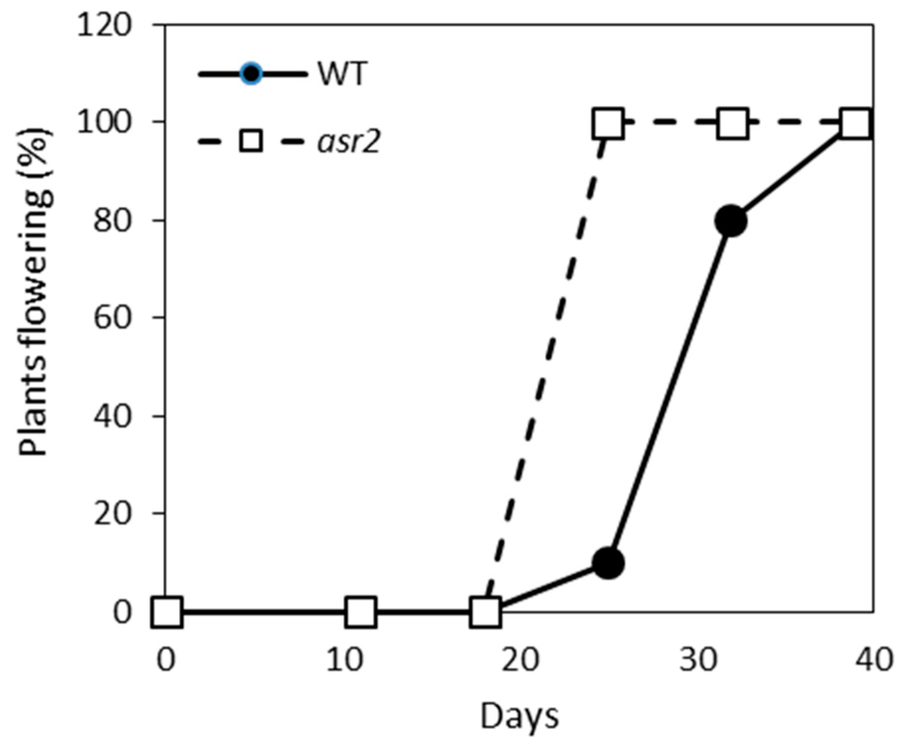
2.4. asr2 Has an Accelerated Flowering Phenotype

Abnormalities in basal defence gene expression are often linked to altered developmental phenotypes or other pleiotropic effects (reviewed in References [23,24]). To determine if the imbalance in SA–JA defence gene expression was reflective of other abnormalities in asr2 plants, we assessed their growth and flowering over an eight-week period. asr2 plants were phenotypically normal apart from an early flowering phenotype. Within three and a half weeks post-germination, all asr2 plants had transitioned to the flowering phase compared to only 10% of wild-type plants (Figure 5). A further two weeks were required for all wild-type plants to enter flowering.

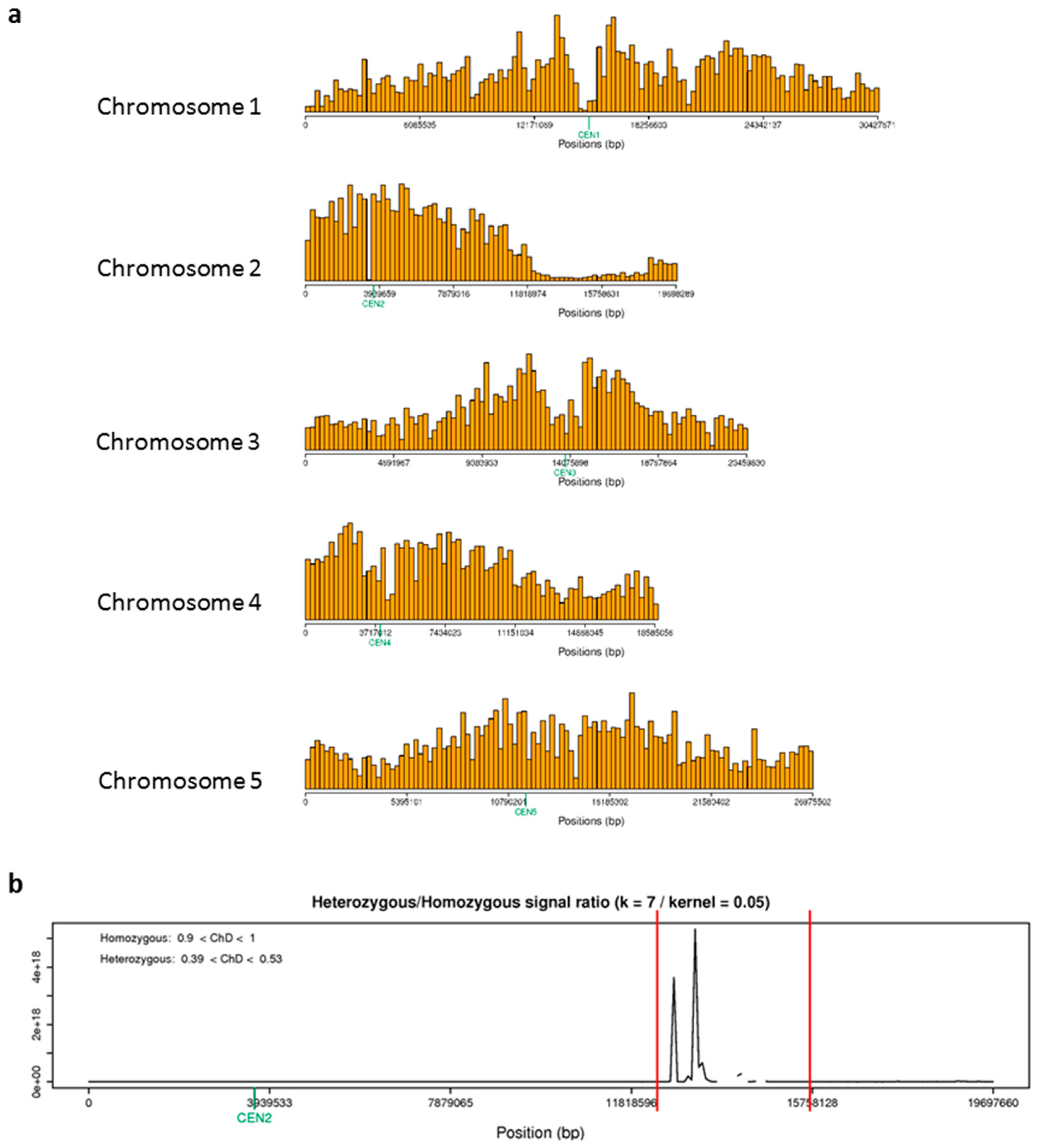
2.5. Fine Mapping Localises asr2 to Four Genes on Chromosome 2

To map the loci encompassing asr2 we employed a Next-Generation Mapping (NGM) approach that we had previously successfully used to map several esr1 and esr3/cpl1 alleles and narrow their causal loci to a handful of genes [8,16,25]. The process of NGM identifies the mutation of interest by quantifying the contribution of single nucleotide polymorphisms (SNPs) in a pooled F2 population between a mutant and its mapping population. We generated a mapping population between asr2 (in Col-0 background) and the Ler ecotype and selected 60 homozygous asr2 F2s based on their constitutive GSTF8:LUC phenotype. DNA was extracted from tissue pooled from the 60 F2s and sequenced on an Illumina HiSeq Platform. Reads were mapped to the Arabidopsis TAIR10 genome, and SNPs were identified and processed through the web-based NGM tool [25].
SNP desserts, corresponding to linkage to the asr2 mutation, were identified on chromosome 2 (Figure 6a). The proportion of reads at a polymorphic genomic position that differed from the reference genome sequence was calculated via the NGM tool and a discordant chastity statistic provided. A discordant chastity statistic of 0 represents genomic positions where the reference base is observed, 0.5 for equal mutant and mapping parental genotype, and a value of 1.0 for all positions that are homozygous for a base that differs from the Col-0 genome, that is, an induced mutation. Closer inspection of chromosome 2 with a discordant chastity cut-off value of 0.85 narrowed the asr2 locus to a 2,093,087 bp region that encompassed eight candidate mutations in coding regions (Figure 6b and Table 1). All mutations encoded non-synonymous codons with one encoding a stop codon change (At2g35170 Histone H3 K4-specific methyltransferase). These were At2g31320 POLY(ADP-RIBOSE) POLYMERASE 1 (PARP1), At2g31400 GENOMES UNCOUPLED 1 (GUN1), At2g31990 an Exostosin family protein, At2g33170 a Leucine-rich repeat receptor-like protein, At2g35170 a Histone H3 K4-specific methyltransferase, At2g35740 NOSITOL TRANSPORTER 3 (ATINT3/INT3), At2g36420 TON1 RECRUITING MOTIF 27 (TRM27), and At2g36835 an unknown protein.
We undertook the same genetic complementation studies used to clone the esr1 and esr3/cpl1 alleles [8,16] by searching for publicly available mutant or T-DNA knockout lines in the candidate ASR2 genes. Firstly, a search of the SALK Arabidopsis T-DNA insertion collection identified insertions in coding regions of three candidate genes. These were At2g33170 Leucine-rich repeat receptor-like, At2g36420 TRM27, and At2g36835 encoding the unknown protein. No SALK insertion lines in the coding regions for the five remaining candidates were available. We obtained homozygous lines of the three available coding region mutants and challenged them with F. oxysporum which provided a strong and relevant phenotype representative of the asr2 mutant. The SALK_092719C insertion line in Leucine-rich repeat receptor-like had poor germination, and we were unable to successfully screen for disease response. Neither of the two other T-DNA lines (SALK_049785C, At2g36420, TRM27; SALK_043358C, At2g36835, unknown protein) differed from wild-type plants in their F. oxysporum disease phenotype (diseased plants P > 0.05), suggesting ASR2 is not encoded by these candidate genes.
Of the five candidates for which no SALK T-DNA knockout lines were available, we ranked At2g31400 GUN1 as a strong candidate as it encodes a member of the RNA-binding pentatricopeptide-repeat protein family which are involved in RNA processing roles related to the functions of ESR1 and ESR3/CPL1. For example, pentatricopeptide-repeat proteins can facilitate processing, splicing, editing, stability, and translation of RNAs [26]. We searched the literature and obtained an EMS-induced stop-codon gun1 mutant termed gun1-9 [27]. Screening of gun1-9 with F. oxysporum did not reveal enhanced susceptibility that mimicked the asr2 phenotype (diseased plants P > 0.05). We also undertook genetic complementation studies with gun1-9. The two asr2 and gun1-9 homozygous recessive mutants were crossed, and the F1 progeny screened for complementation of the constitutive GSTF8:LUC phenotype. Neither of the reciprocal crosses between asr2 and gun1-9 displayed constitutive luciferase expression (data not shown), suggesting a wild-type copy of ASR2 in gun1-9 had restored the wild-type GSTF8:LUC phenotype and that ASR2 equivalently does not encode GUN1. For the four remaining candidate genes for which no suitable mutant lines were available, molecular complementation tests have been prioritised.

3. Discussion

Facilitated through a forward genetic screen for mutants altered in GSTF8:LUC defence/stress reporter expression, we previously identified several alleles of the ESR1/KH-domain and CPL1 genes that conferred constitutive GSTF8:LUC activity and enhanced resistance to fungal pathogens. In this study, we identified asr2 as a second class of mutant that also conferred constitutive GSTF8:LUC activity but was more susceptible than wild-type to necrotrophic fungal pathogens—the root-infecting pathogen F. oxysporum and the leaf-infecting pathogen A. brassicicola.
The asr2 plants displayed contrasting expression of two classical SA- or JA-mediated defence marker genes where the JA-marker gene PDF1.2 was highly down-regulated. A functional JA-pathway is required for resistance against A. brassicicola [22,28], and the asr2 PDF1.2 phenotype may contribute to its susceptibility towards this pathogen. PDF1.2 is also associated with resistance against F. oxysporum. For example, in ERF4 transcription factor overexpression lines, their impairment in JA-induced PDF1.2 expression is linked to increased F. oxysporum susceptibility [29]. JA signalling can however also play contrasting roles in F. oxysporum disease outcome. It is proposed that non-defensive aspects of JA-signalling such as senescence processes strongly contribute [30,31,32,33,34]. Consequently, mutants with reduced defensive and non-defensive JA-signalling such as esr1-1 are more resistant [8]. This disease-resistance phenotype is even more pronounced in mutants with abolished or severely impaired JA-signalling such as the coronatine insensitive1 (coi1) or mediator complex mutants med25/pft1, med18, or med20 [30,32,33].
The asr2 mutant also displayed an early flowering phenotype. A significant correlation between flowering time and F. oxysporum disease outcome was established through disease assays on a large Arabidopsis ecotype collection that differed in their flowering response [35]. Early flowering ecotypes were more susceptible while late-flowering ecotypes showed enhanced resistance. Increased expression of the negative regulator of flowering time FLOWERING LOCUS C (FLC) in cpl1 and several med mutants was linked to their delayed flowering and enhanced disease resistance [16,30,33]. However, a positive correlation between reduced FLC expression (accelerated flowering time) and increased susceptibility to F. oxysporum did not hold true [35]. Once the ASR2 gene is cloned, functional studies using ASR2 knockout or ASR2 overexpression lines will be used to dissect the role of ASR2 in defence and flowering.
Next-Generation Mapping narrowed the causal asr2 loci to eight candidate genes. Fusarium disease assays on publicly available EMS or SALK T-DNA insertion mutants eliminated at least three genes as candidates. Of the remaining candidates, POLY(ADP-RIBOSE) POLYMERASE 1 (PARP1) is one of three PARPs in Arabidopsis whose activity is activated in response to stress such as ROS. Their post-translational modification of nuclear proteins activates DNA repair machinery (reviewed in Reference [36]). The defined role of PAR1 in stress is unclear as plants with reduced PAR activity by means of chemical inhibitors or gene silencing are tolerant of a broad range of abiotic stresses, but individual, double, or triple PARP GABI-Kat knockout lines do not differ from wild-type [36,37]. A parp1/parp2 double mutant is slightly more susceptible to virulent Pseudomonas bacteria [38]. The individual role of PARP1 and PARP2 in this response was not tested due to potential functional redundancy. However, the minor Pseudomonas susceptibility response in the double mutant suggests individual mutants may show no phenotype. NOSITOL TRANSPORTER 3 (ATINT3/INT3) may affect stress responses. The Brassica napus homologue of this gene is up-regulated following infection with the necrotrophic fungal pathogen Sclerotinia sclerotiorum [39]. The two remaining ASR2 candidate genes, exostosin family protein and histone H3 K4-specific methyltransferase, are functionally uncharacterised and to our knowledge, no link to biotic stress has been reported. Of the detected SNPs in the mapped asr2 loci, the mutation in At2g35170 (histone H3 K4-specific methyltransferase) is the only predicted stop codon change. Based on the above characteristics, PARP1, ATINT3, and the methyltransferase have been prioritised for molecular complementation studies in the asr2 background, and the parp1/parp2 T-DNA double mutant will be obtained for Fusarium disease assessment.
In summary, the high-throughput GSTF8:LUC forward genetic screen facilitated the discovery of the asr2 mutant accelerated in flowering and heightened in susceptibility to root- and leaf-infecting necrotrophic fungal pathogens. The susceptibility to pathogens of dissimilar lifecycles suggests ASR2 is an important component of the host defence response and its functional characterisation will be important to our combined knowledge of plant disease resistance mechanisms.

4. Materials and Methods

4.1. Plant Material and Growth Conditions

The Arabidopsis thaliana Columbia-0 transgenic line (JC66/GSTF8:LUC) containing 791 bp of the GSTF8 promoter fused to a luciferase reporter [12,15] was used in all experiments unless otherwise noted. Agar plate assays were performed using surface-sterilised seeds plated onto Murashige and Skoog (MS) salt agar plates as described previously and supplemented with 50 uM luciferin (Biosynth AG) for luciferase assays [6,11]. Plate- and soil-grown plants were incubated under a 16-h light/8-h dark cycle at 22 °C.

4.2. Bioluminescence and Luciferase Assays

Biochemical luciferase assays and imaging of in vivo seedling bioluminescence in an EG & G Berthold molecular light imager were conducted as described previously [6,8,15].

4.3. Genetic and Mapping Studies

For allelism tests, reciprocal crosses between asr2 and esr1 or esr3/cpl1 plants were conducted. Bioluminescence activity in progeny was assessed. Next-Generation mapping was performed as described previously [8]. Briefly, a two-times backcrossed asr2 line was crossed with Ler, and pooled DNA (CTAB extraction) from 60 homozygous asr2 F2 plants (exhibiting constitutive GSTF8:LUC activity) were sequenced by the Australian Genome Research Facility (AGRF) using an Illumina HiSeq Platform to provide 9.25 Gb of raw data that would be sufficient to provide ~50× genome coverage. A total of 85.9 million paired-end reads (100 bp in length) were cleaned, trimmed, and mapped to the Arabidopsis TAIR10 genome reference sequence, SNPs called using the recommended SAMtools mpileup script, and processed through the NGM tool http://bar.utoronto.ca/ngm/ [25].

4.4. Pathogen and Flowering Time Assays

Fungal pathogen inoculations were performed according to Thatcher et al. and Gleason et al. [7,32]. For F. oxysporum (Fo5176), root-dip inoculations on 4-week-old plants were carried out using a 1×10⁶ cell/mL spore suspension. For A. brassicicola (UQ4273), a 1×10⁶ cell/mL spore suspension was applied to leaves of 3- to 4-week-old plants. Lesion size was measured with ImageJ [40]. Flowering time assays were conducted under long day conditions of a 16-h light/8-h dark cycle at 22 °C (n = 10).

4.5. RNA Isolation and qRT-PCR

For qRT-PCR experiments, tissue was collected from whole 7-day-old seedlings germinated and grown on MS media. Two separate biological replicates were taken for each genotype, with each replicate consisting of tissue pooled from 20 seedlings grown at the same time in the same environment, then frozen in liquid nitrogen and stored at −80 °C. RNA extractions, DNase treatment, cDNA synthesis, and qRT-PCR was performed on a MyiQ (Bio-Rad) system as described previously [6,14]. Gene expression was calculated using the Elongation Factor1 reference gene (EF-1-F 5’ ATGCCCCAGGACATCGTGATTTCAT 3’; EF-1-R 5’ TTGGCGGCACCCTTAGCTGGATCA 3’) according to the equation: relative ratio gene of interest/EF-1 = (2−Ct gene)/(2−Ct EF-1) where Ct is the cycle threshold value. Gene-specific primer sequences used are GSTF8 (GSTF8-F 5’ CCCCGTCGATATGAGAGC 3’; GSTF8-R 5’ GAGAGAGGGTCACTACTGCTTCTGG 3’), PR1 (PR1-F 5’ TTCTTCCCTCGAAAGCTCAA 3’; PR1-R 5’ AAGGCCCACCAGAGTGTATG 3’) and PDF1.2 (PDF1.2-F 5’ TGTTCTCTTTGCTGCTTTCGACG 3’; PDF1.2-R 5’ GCATGATCCATGTTTGGCTCCT 3’).

Author Contributions

Conceptualization, methodology, and data analysis, L.F.T. and K.B.S.; performed experiments and data curation, L.F.T.; writing—original draft preparation, L.F.T.; writing—review and editing, L.F.T. and K.B.S.

Funding

This research was funded by the Commonwealth Scientific and Industrial Research Organisation.

Acknowledgments

The research was undertaken with the assistance of resources from the Australian Genome Research Facility (AGRF) which is supported by the Australian Government. We thank the ABRC for seeds of Arabidopsis T-DNA mutants, Joanne Chory and the SALK Institute for Biological Sciences for the gun1-9 seed, Hayley Casarotto for technical support and thank Drs Jonathan Anderson and Brendan Kidd for critical reading of the manuscript and useful discussions.

Conflicts of Interest

The authors declare no conflict of interest. The funders had no role in the design of the study; in the collection, analyses, or interpretation of data; in the writing of the manuscript, or in the decision to publish the results.

References

  1. FAO. Food and Agriculture Organization of the United Nations. Available online: http://www.fao.org/home/en (accessed on 19 November 2018).
  2. Moons, A. Regulatory and functional interactions of plant growth regulators and plant glutathione s-transferases (GSTs). Vitam. Horm. 2005, 72, 155–202. [Google Scholar] [PubMed]
  3. Dixon, D.P.; Edwards, R. Glutathione transferases. Arabidopsis Book 2010, 8, e0131. [Google Scholar] [CrossRef] [PubMed]
  4. Nianiou-Obeidat, I.; Madesis, P.; Kissoudis, C.; Voulgari, G.; Chronopoulou, E.; Tsaftaris, A.; Labrou, N.E. Plant glutathione transferase-mediated stress tolerance: Functions and biotechnological applications. Plant Cell Rep. 2017, 36, 791–805. [Google Scholar] [CrossRef] [PubMed]
  5. Frova, C. The plant glutathione transferase gene family: Genomic structure, functions, expression and evolution. Physiol. Plant. 2003, 119, 469–479. [Google Scholar] [CrossRef]
  6. Thatcher, L.F.; Carrie, C.; Andersson, C.R.; Sivasithamparam, K.; Whelan, J.; Singh, K.B. Differential gene expression and subcellular targeting of Arabidopsis glutathione s-transferase F8 is achieved through alternative transcription start sites. J. Biol. Chem. 2007, 282, 28915–28928. [Google Scholar] [CrossRef] [PubMed]
  7. Gleason, C.; Huang, S.; Thatcher, L.F.; Foley, R.C.; Anderson, C.R.; Carroll, A.J.; Millar, A.H.; Singh, K.B. Mitochondrial complex ii has a key role in mitochondrial-derived reactive oxygen species influence on plant stress gene regulation and defense. Proc. Natl. Acad. Sci. USA 2011, 108, 10768–10773. [Google Scholar] [CrossRef] [PubMed]
  8. Thatcher, L.F.; Kamphuis, L.G.; Hane, J.K.; Onate-Sanchez, L.; Singh, K.B. The Arabidopsis KH-domain rna-binding protein ESR1 functions in components of jasmonate signalling, unlinking growth restraint and resistance to stress. PLoS ONE 2015, 10, e0126978. [Google Scholar] [CrossRef] [PubMed]
  9. Jones, A.M.E.; Thomas, V.; Truman, B.; Lilley, K.; Mansfield, J.; Grant, M. Specific changes in the Arabidopsis proteome in response to bacterial challenge: Differentiating basal and R-gene mediated resistance. Phytochemistry 2004, 65, 1805–1816. [Google Scholar] [CrossRef] [PubMed]
  10. Chen, W.; Chao, G.; Singh, K.B. The promoter of a H2O2-inducible, Arabidopsis glutathione s-transferase gene contains closely linked OBF- and OBP1-binding sites. Plant J. 1996, 10, 955–966. [Google Scholar] [CrossRef] [PubMed]
  11. Foley, R.C.; Sappl, P.G.; Perl-Treves, R.; Millar, A.H.; Singh, K.B. Desensitization of GSTF8 induction by a prior chemical treatment is long lasting and operates in a tissue-dependent manner. Plant Physiol. 2006, 142, 245–253. [Google Scholar] [CrossRef] [PubMed]
  12. Perl-Treves, R.; Foley, R.C.; Chen, W.; Singh, K.B. Early induction of the Arabidopsis GSTF8 promoter by specific strains of the fungal pathogen Rhizoctonia solani. Mol. Plant Microbe Interact. 2004, 17, 70–80. [Google Scholar] [CrossRef] [PubMed]
  13. Sappl, P.G.; Carroll, A.J.; Clifton, R.; Lister, R.; Whelan, J.; Harvey Millar, A.; Singh, K.B. The arabidopsis glutathione transferase gene family displays complex stress regulation and co-silencing multiple genes results in altered metabolic sensitivity to oxidative stress. Plant J. 2009, 58, 53–68. [Google Scholar] [CrossRef] [PubMed]
  14. Sappl, P.G.; Oñate-Sánchez, L.; Singh, K.B.; Millar, A.H. Proteomic analysis of glutathione s-transferases of Arabidopsis thaliana reveals differential salicylic acid-induced expression of the plant-specific phi and tau classes. Plant Mol. Biol. 2004, 54, 205–219. [Google Scholar] [CrossRef] [PubMed]
  15. Chen, W.; Singh, K.B. The auxin, hydrogen peroxide and salicylic acid induced expression of the Arabidopsis GST6 promoter is mediated in part by an ocs element. Plant J. 1999, 19, 667–677. [Google Scholar] [CrossRef] [PubMed] [Green Version]
  16. Thatcher, L.F.; Foley, R.; Casarotto, H.J.; Gao, L.-L.; Kamphuis, L.G.; Melser, S.; Singh, K.B. The Arabidopsis RNA polymerase II carboxyl terminal domain (CTD) phosphatase-like1 (CPL1) is a biotic stress susceptibility gene. Sci. Rep. 2018, 8, 13454. [Google Scholar] [CrossRef] [PubMed]
  17. Agrios, G.N. Plant diseases caused by fungi. In Plant Pathology, 5th ed.; Agrios, G.N., Ed.; Academic Press: San Diego, CA, USA, 2005; Chapter Eleven; pp. 385–614. [Google Scholar]
  18. Glazebrook, J. Contrasting mechanisms of defense against biotrophic and necrotrophic pathogens. Annu. Rev. Phytopathol. 2005, 43, 205–227. [Google Scholar] [CrossRef] [PubMed]
  19. Spoel, S.H.; Dong, X. Making sense of hormone crosstalk during plant immune responses. Cell Host Microbe 2008, 3. [Google Scholar] [CrossRef] [PubMed]
  20. Dombrecht, B.; Xue, G.P.; Sprague, S.J.; Kirkegaard, J.A.; Ross, J.J.; Reid, J.B.; Fitt, G.P.; Sewelam, N.; Schenk, P.M.; Manners, J.M.; et al. MYC2 differentially modulates diverse jasmonate-dependent functions in Arabidopsis. Plant Cell 2007, 19, 2225–2245. [Google Scholar] [CrossRef] [PubMed]
  21. Anderson, J.P.; Badruzsaufari, E.; Schenk, P.M.; Manners, J.M.; Desmond, O.J.; Ehlert, C.; Maclean, D.J.; Ebert, P.R.; Kazan, K. Antagonistic interaction between abscisic acid and jasmonate-ethylene signaling pathways modulates defense gene expression and disease resistance in Arabidopsis. Plant Cell 2004, 16, 3460–3479. [Google Scholar] [CrossRef] [PubMed]
  22. Thomma, B.P.; Eggermont, K.; Penninckx, I.A.; Mauch-Mani, B.; Vogelsang, R.; Cammue, B.P.; Broekaert, W.F. Separate jasmonate-dependent and salicylate-dependent defense-response pathways in arabidopsis are essential for resistance to distinct microbial pathogens. Proc. Natl. Acad. Sci. USA 1998, 95, 15107–15111. [Google Scholar] [CrossRef] [PubMed]
  23. van Schie, C.C.N.; Takken, F.L.W. Susceptibility genes 101: How to be a good host. Annu. Rev. Phytopathol. 2014, 52, 551–581. [Google Scholar] [CrossRef] [PubMed]
  24. Thatcher, L.F.; Anderson, J.P.; Singh, K.B. Plant defence responses: What have we learnt from arabidopsis? Funct. Plant Biol. 2005, 32, 1–19. [Google Scholar] [CrossRef]
  25. Austin, R.S.; Vidaurre, D.; Stamatiou, G.; Breit, R.; Provart, N.J.; Bonetta, D.; Zhang, J.; Fung, P.; Gong, Y.; Wang, P.W.; et al. Next-generation mapping of Arabidopsis genes. Plant J. 2011, 67, 715–725. [Google Scholar] [CrossRef] [PubMed] [Green Version]
  26. Manna, S. An overview of pentatricopeptide repeat proteins and their applications. Biochimie 2015, 113, 93–99. [Google Scholar] [CrossRef] [PubMed] [Green Version]
  27. Koussevitzky, S.; Nott, A.; Mockler, T.C.; Hong, F.; Sachetto-Martins, G.; Surpin, M.; Lim, J.; Mittler, R.; Chory, J. Signals from chloroplasts converge to regulate nuclear gene expression. Science 2007, 316, 715–719. [Google Scholar] [CrossRef]
  28. van Wees, S.C.M.; Chang, H.-S.; Zhu, T.; Glazebrook, J. Characterization of the early response of Arabidopsis to Alternaria brassicicola infection using expression profiling. Plant Physiol. 2003, 132, 606–617. [Google Scholar] [CrossRef] [PubMed]
  29. McGrath, K.C.; Dombrecht, B.; Manners, J.M.; Schenk, P.M.; Edgar, C.I.; Maclean, D.J.; Scheible, W.R.; Udvardi, M.K.; Kazan, K. Repressor- and activator-type ethylene response factors functioning in jasmonate signaling and disease resistance identified via a genome-wide screen of Arabidopsis transcription factor gene expression. Plant Physiol. 2005, 139, 949–959. [Google Scholar] [CrossRef] [PubMed]
  30. Kidd, B.N.; Edgar, C.I.; Kumar, K.K.; Aitken, E.A.; Schenk, P.M.; Manners, J.M.; Kazan, K. The mediator complex subunit PFT1 is a key regulator of jasmonate-dependent defense in Arabidopsis. Plant Cell 2009, 21, 2237–2252. [Google Scholar] [CrossRef] [PubMed]
  31. Thatcher, L.F.; Kidd, B.N.; Kazan, K. Belowground defence strategies against Fusarium oxysporum. In Belowground Defence Strategies in Plants; Vos, C.M.F., Kazan, K., Eds.; Springer International Publishing: Cham, Switzerland, 2016; Part II; pp. 71–98. [Google Scholar]
  32. Thatcher, L.F.; Manners, J.M.; Kazan, K. Fusarium oxysporum hijacks coi1-mediated jasmonate signaling to promote disease development in Arabidopsis. Plant J. 2009, 58, 927–939. [Google Scholar] [CrossRef] [PubMed]
  33. Fallath, T.; Kidd, B.N.; Stiller, J.; Davoine, C.; Bjorklund, S.; Manners, J.M.; Kazan, K.; Schenk, P.M. Mediator18 and Mediator20 confer susceptibility to Fusarium oxysporum in Arabidopsis thaliana. PLoS ONE 2017, 12, e0176022. [Google Scholar] [CrossRef] [PubMed]
  34. Kidd, B.N.; Aitken, E.A.; Schenk, P.M.; Manners, J.M.; Kazan, K. Plant mediator: Mediating the jasmonate response. Plant Signal. Behav. 2010, 5, 718–720. [Google Scholar] [CrossRef] [PubMed]
  35. Lyons, R.; Rusu, A.; Stiller, J.; Powell, J.; Manners, J.M.; Kazan, K. Investigating the association between flowering time and defense in the Arabidopsis thaliana-Fusarium oxysporum interaction. PLoS ONE 2015, 10, e0127699. [Google Scholar] [CrossRef] [PubMed]
  36. Rissel, D.; Heym, P.; Thor, K.; Brandt, W.; Wessjohann, L.; Peiter, E. No silver bullet—Canonical poly(adp-ribose) polymerases (parps) are no universal factors of abiotic and biotic stress resistance of Arabidopsis thaliana. Front. Plant Sci. 2017, 8, 59. [Google Scholar] [CrossRef] [PubMed]
  37. Block, M.D.; Verduyn, C.; Brouwer, D.D.; Cornelissen, M. Poly(adp-ribose) polymerase in plants affects energy homeostasis, cell death and stress tolerance. Plant J. 2005, 41, 95–106. [Google Scholar] [CrossRef] [PubMed]
  38. Feng, B.; Liu, C.; de Oliveira, M.V.V.; Intorne, A.C.; Li, B.; Babilonia, K.; de Souza Filho, G.A.; Shan, L.; He, P. Protein poly(adp-ribosyl)ation regulates arabidopsis immune gene expression and defense responses. PLoS Genet. 2015, 11, e1004936. [Google Scholar] [CrossRef] [PubMed]
  39. Zhao, J.; Wang, J.; An, L.; Doerge, R.W.; Chen, Z.J.; Grau, C.R.; Meng, J.; Osborn, T.C. Analysis of gene expression profiles in response to Sclerotinia sclerotiorum in Brassica napus. Planta 2007, 227, 13–24. [Google Scholar] [CrossRef] [PubMed]
  40. Schneider, C.A.; Rasband, W.S.; Eliceiri, K.W. Nih image to imagej: 25 years of image analysis. Nat. Methods 2012, 9, 671–675. [Google Scholar] [CrossRef] [PubMed]
Figure 1. asr2 causes basal hyper-expression of GSTF8::LUC and altered defence marker gene expression. (a) Comparison of basal GSTF8:LUC expression in four-day-old wild-type (WT), asr2, esr1-1, and cpl1-8 (esr3-2) seedlings. (b) Quantification of bioluminescence via in vivo light emission (relative light units/seedling; values are averages ± SE (n = 30) from four-day-old seedlings) and in vitro biochemical assays (units/20sec/mg protein; values are averages ± SE (n = 30) from nine-day-old seedlings). (c) GSTF8, PR1, and PDF1.2 expression in seven-day-old seedlings (values are averages ± SE of two biological replicates consisting of pools of 20 seedlings). Gene expression levels are expressed logarithmically relative to the normalised expression levels in WT plants. For normalisation, the internal control EF-1 gene was used. Asterisks indicate values that are significantly different (** p < 0.01, * p < 0.05 Student’s t-test) from WT.
Plants 08 00060 g001
Figure 2. GSTF8:LUC response in asr2 is maintained following salicylic acid (SA) or H2O2 induction. Average GSTF8:LUC expression per wild-type (WT) and asr2 seedling per hour after treatment with 1 mM salicylic acid (SA) (upper panel) or 1 mM H2O2 (lower panel). Values are averages ± SE (n = 10) from four-day-old seedlings. WT values are plotted on the primary (left) axis and asr2 values on the secondary (right) axis.
Plants 08 00060 g002
Figure 3. asr2 has increased susceptibility to the necrotrophic fungal pathogen Fusarium oxysporum. (ac) Disease phenotypes of F. oxysporum inoculated plants with (a) necrotic leaves and (b) representative images of plants at 14 days post inoculation (dpi), and (c) survival of plants at 21 dpi. Values are averages ± SE (n = 30). Asterisks indicate values that are significantly different (** p < 0.01, * p < 0.05 Student’s t-test) from wild-type (WT). Similar results were obtained in independent experiments.
Plants 08 00060 g003
Figure 4. asr2 had increased susceptibility to the necrotrophic fungal pathogen Alternaria brassicicola. (ac) A. brassicicola induced lesions at 3 dpi with (a) representative images of leaves, (b) size of Alternaria induced lesions, and (c) size of chlorotic regions. Values are averages ± SE of three biological replicates consisting of lesions measured from five inoculated leaves per plant. Asterisks indicate values that are significantly different (** p < 0.01, * p < 0.05 Student’s t-test) from wild-type (WT). Similar results were obtained in independent experiments.
Plants 08 00060 g004
Figure 5. asr2 has an early flowering phenotype. The number of flowering wild-type (WT) and asr2 plants were measured over a 40-day time-course (n = 10). Similar results were obtained in an independent experiment.
Plants 08 00060 g005
Figure 6. Fine mapping of asr2 locates it to a region of chromosome 2. Next-Generation Mapping (NGM) was applied to an asr2 mapping population by sequencing homozygous asr2 F2 plants from an asr2 and Ler outcross, and processing SNPs that deviated from the Arabidopsis TAIR10 genome reference sequence through the NGM tool http://bar.utoronto.ca/ngm/ [25]. (a) The tool identified SNP desserts (depression on chromosome 2) corresponding to asr2 linkage. Shown are genome-wide SNP frequencies (y-axis) plotted as a function of chromosomal position (x-axis) using a bin size of 250 kb. (b) Closer inspection of chromosome 2 narrowed the asr2 locus to a 2,093,087 bp region indicated by peaks on the y-axis (ratio of homozygous to heterozygous signals) and bordered by two red bars.
Plants 08 00060 g006
Table 1. Candidate asr2 genes. Shown are predicted EMS-induced mutations in coding regions of genes in the asr2 loci. Reference base refers to the Col-0 reference and SNP base to the detected SNP. DC refers to the discordant chastity value.
Chrom.PositionRef. BaseSNP BaseDepthDCAccessionStrandRef. CodonSNP CodonAA Change
213358132CT550.91AT2G31320-GGAGAAG- > E
213389698CT570.98AT2G31400-GAGAAGE- > K
213612057CT410.92AT2G31990-GCTACTA- > T
214057731CT460.91AT2G33170-GGAGAAG- > E
214829201CT200.85AT2G35170+CAATAAQ- > *
215025500CT320.93AT2G35740-GGTAGTG- > S
215288977CT490.96AT2G36420+TCTTTTS- > F
215451219GA410.95AT2G36835-CCGCTGP- > L

