Article

Phytophthora pseudocryptogea, P. nicotianae and P. multivora Associated to Cycas revoluta: First Report Worldwide

by
Francesco Aloi
,
Rossana Parlascino
,
Sebastiano Conti Taguali
,
Roberto Faedda
*,
Antonella Pane
* and
Santa Olga Cacciola
Department of Agriculture, Food and Environment, University of Catania, 95123 Catania, Italy
*
Authors to whom correspondence should be addressed.
These authors contributed equally to this work.
Plants 2023, 12(5), 1197; https://doi.org/10.3390/plants12051197
Submission received: 30 January 2023 / Revised: 28 February 2023 / Accepted: 3 March 2023 / Published: 6 March 2023
(This article belongs to the Special Issue Crop Fungal, Oomycetic Disease and Control)

Abstract

:
A dieback was observed on three-year-old pot-grown plants of Cycas revoluta in Sicily (Italy). Symptoms, including stunting, yellowing and blight of the leaf crown, root rot and internal browning and decay of the basal stem, closely resembled the Phytophthora root and crown rot syndrome, common in other ornamentals. Isolations from rotten stem and roots, using a selective medium, and from rhizosphere soil of symptomatic plants, using leaf baiting, yielded three Phytophthora species, P. multivora, P. nicotianae and P. pseudocryptogea, were obtained. Isolates were identified by both morphological characters and DNA barcoding analysis, using three gene regions: ITS, β-tub and COI. Phytophthora pseudocryptogea was the sole species isolated directly from the stem and roots. The pathogenicity of the isolates of the three Phytophthora species was tested on one-year-old potted plants of C. revoluta, using both stem inoculation by wounding, and root inoculation through infested soil. Phytophthora pseudocryptogea was the most virulent and, like P. nicotianae, reproduced all the symptoms of natural infections, while P. multivora was the least virulent and induced solely very mild symptoms. Phytophthora pseudocryptogea was identified as the causal agent of the decline of C. revoluta, as it was re-isolated from both the roots and stems of artificially infected symptomatic plants, thus fulfilling Koch’s postulates.

1. Introduction

Cycads (order Cycadales) are slow-growing, long-living, perennial, dioecious plants with a palm-like habit, robust stems and large evergreen leathery pinnate leaves. Geographically, cycads, in the wild, are restricted to tropical and subtropical or warm, temperate regions of the world, with predominantly summer rainfalls. This group of gymnosperms is often cited as one of the most ancient lineages of living seed plants with an evolutionary history dating back to more than 300 million years ago [1,2,3]. In Asia, cycads are grown in gardens since ancient times and after their first discovery by Europeans in the 18th century, have become highly appreciated as ornamentals worldwide. Many of them are regarded as a threatened species, mainly because they have been massively removed from their natural range for private collections or botanical gardens, landscaping or other commercial purposes, such as pharmaceutical and food uses [1,4]. The living cycads can be divided into three families: Cycadaceae, Stangeriaceae, and Zamiaceae. The current World List of Cycads, which is periodically updated to reflect changes in cycad nomenclature and taxonomy, comprises a total of 10 accepted genera and 370 accepted species [5]. The number of recognized species within each genus is as follows: Bowenia (2), Ceratozamia (32), Cycas (118), Dioon (16), Encephalartos (65), Lepidozamia (2), Macrozamia (41), Microcycas (1), Stangeria (1) and Zamia (81). These taxa are distributed throughout the warmer areas of North and South America, Africa, Asia, and Australia, as well as a large number of oceanic islands. Centers of cycad diversity occur in southern Africa, Australia, and the tropical New World. All three cycad families are represented in Australia and Africa, whereas only Cycadaceae and Zamiaceae are endemic in Asia and the New World, respectively. At the generic level, the New World and Australia show the greatest diversity, each with four genera (Ceratozamia, Dioon, Microcycas, and Zamia in New World; Bowenia, Cycas, Macrozamia and Lepidozamia in Australia). Three genera are endemic in Africa (Cycas, Encephalartos, and Stangeria) and only Cycas occurs in Asia [1]. Cycas is the genus type and the only genus currently recognized in the family Cycadaceae. Diversification analyses based on time-calibrated phylogenetic trees point at Indocina as the ancestral geographic area of the origin of Cycas, which evolved more recently than other members of Cycadales [3,6]. The most popular species of this genus is Cycas revoluta, known with diverse names, the most common of which is the Sago Palm or King Sago Palm, referring to both its palm-like appearance and sago, a starch extract used as a traditional food by populations of Southeast Asia. This cycad is believed to be native to Japan and southern China, but is cultivated as an ornamental and landscape plant in warm and temperate climates all over the world. It is common in botanical and private gardens and is easily propagated by seeds or offshoots.
In April 2010, for the first time, 30% of a production block of approximately 8000 three-year-old potted sago palm plants, grown in open air in a commercial nursery of ornamentals in the Catania province (Eastern Sicily, Italy), showed severe symptoms of dieback. Overall, symptoms closely resembled the Phytophthora root rot (PRR) syndrome, which is common in nurseries of ornamentals worldwide [7,8,9,10,11,12,13,14,15,16,17,18]. For instance, Phytophthora nicotianae and P. palmivora were responsible for a similar disease of potted ornamental palms reported a few years ago in Sicily [19,20]. Moreover, P. cinnamomi was identified as the causal agent of a severe dieback associated with stem and root rot of Modjiadi cycad (Encephalartos transvenosus), within the native range of this cycad in South Africa [21]. The same Phytophthora species was reported as a pathogen of other cycads, including species of Cycas in New Zealand, Macrozamia in Australia and Zamia in Taiwan [21], and Johnson’s cycad (Macrozamia johnsonii) is in the list of native plant species threatened by P. cinnamomi in New South Wales (Australia) [22]. Since the first record in 2010, the disease has been under observation and has occurred sporadically and with a low incidence in several nurseries in the same production area of Sicily. However, in May 2021, it appeared again in a severe form and with an incidence comparable with its first record, in a stock of 15,000 three-year-old potted plants of sago palm grown in open air, in a nursery where the disease had not been noticed before. The severity of this new disease outbreak prompted us to investigate a possible involvement of Phytophthora in its etiology.

2. Results

2.1. Symptoms of the Disease

Symptoms consisted of stunting, chlorosis and blight of the leaflets or entire leaves, initially affecting only part of the leaf crown and, subsequently, expanding to the other leaves of the crown and resulting in the withering of the entire plant in most severe cases. Foliar symptoms were associated with the internal browning and the decay of the stem base and root rot (Figure 1).

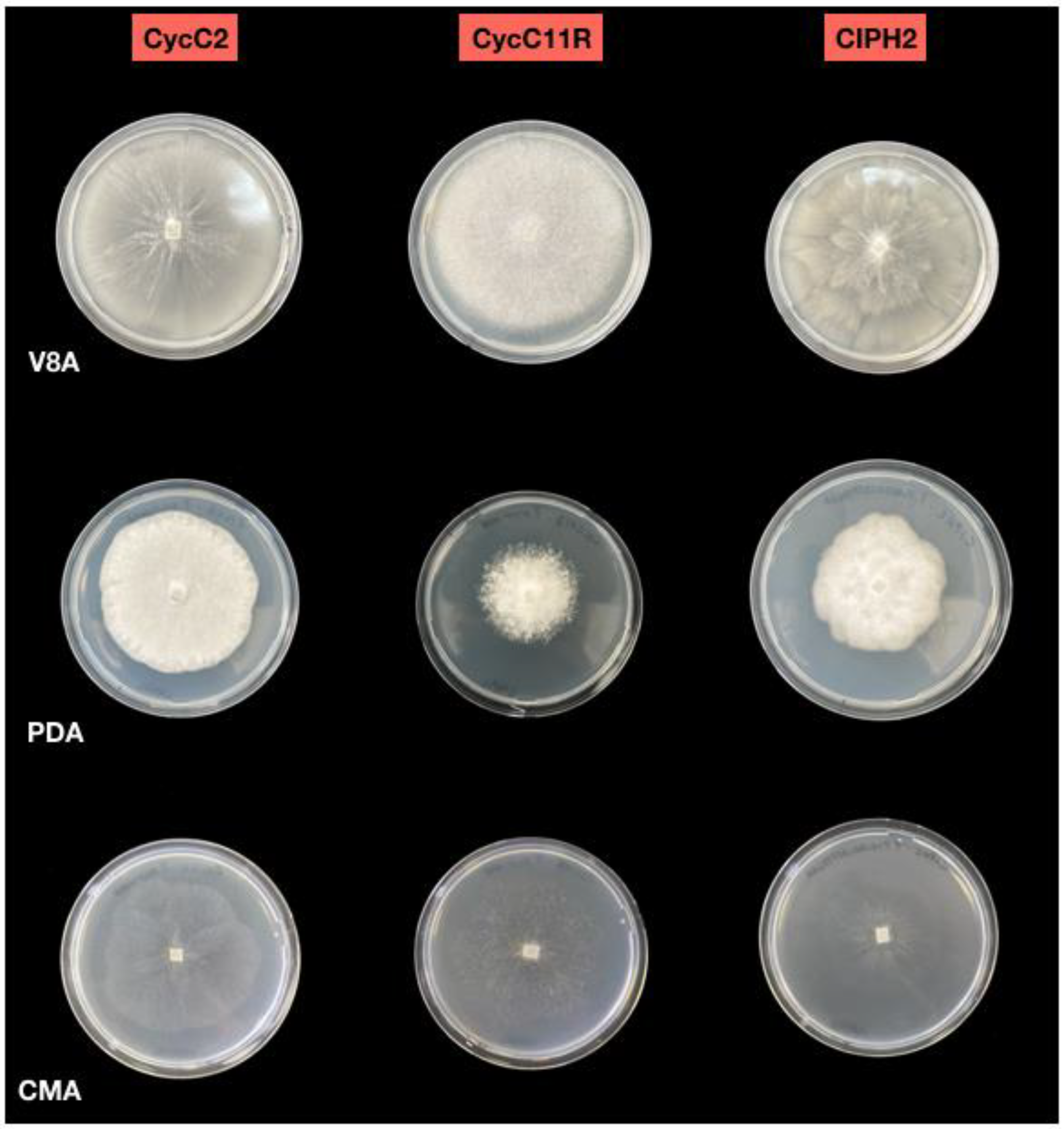
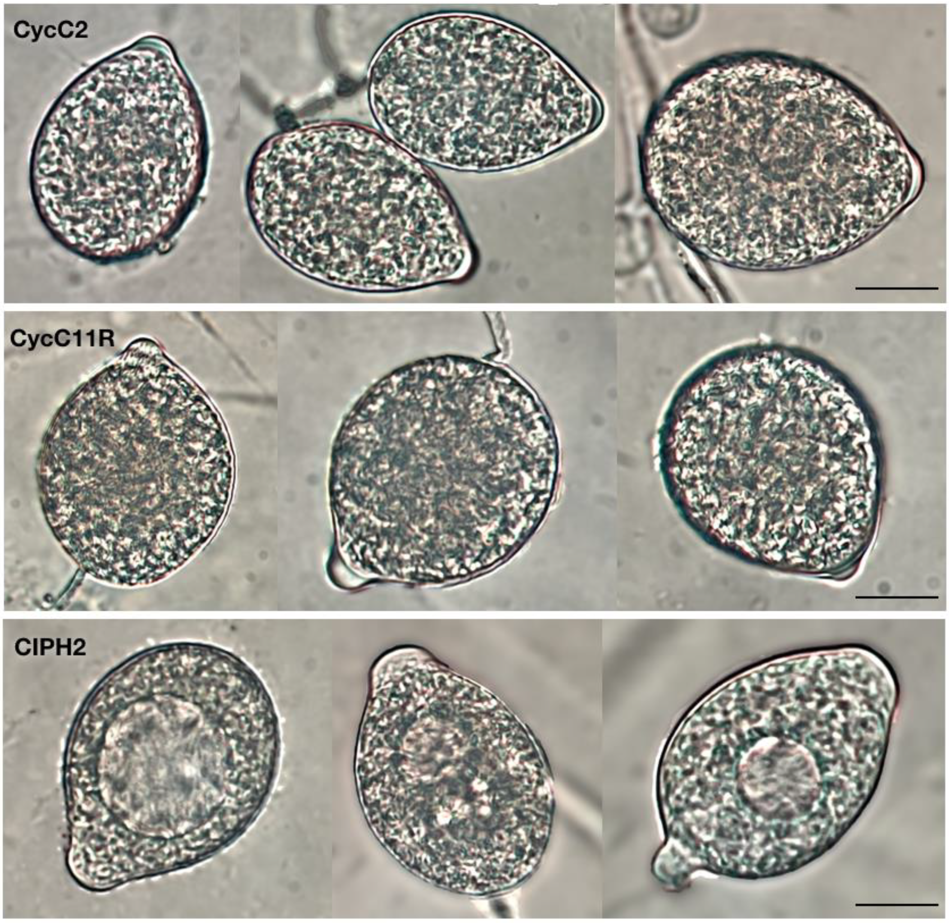
2.2. Morphological Identification of Isolates

In agreement with a provisional diagnosis based on the symptoms, isolates of a Phytophthora sp. were consistently obtained from rotten tissues of diseased sago palm plants from the nursery in the province of Catania, where the disease was first noticed and the isolates were preserved as living cultures in the collection of the Molecular Plant Pathology Laboratory of the Department of Agriculture, Food and Environment of the University of Catania, Catania, Italy. Since then, the disease has been under observation and has occurred sporadically and with a low incidence in several nurseries. However, in May 2021, it appeared again in a severe form and with an incidence comparable to its first record, in a stock of 15,000 three-year-old potted plants of sago palm grown in open air, in a nursery where the disease had not been noticed before.
Direct isolation from rotten roots and stems of sago palm plants consistently produced only one species of Phytophthora, as tentatively determined based on the morphology of colonies, with a mean isolation frequency of 75%. Overall, representative single-hypha isolates (isolates from roots and stems) sourced in 2021 were selected and characterized together with isolates obtained from roots and stems of symptomatic plants in 2010, which shared the same culture morphology (Table 1).
All the isolates formed colonies with chrysanthemum pattern on V8A and grew between 3 °C and 35 °C on CMA, with the optimum at 25 °C. Sporangia were non-papillate, persistent, terminal, ovoid and ellipsoid to obpyriform (23–38 × 35–62 µm), originating in unbranched or simple sympodial sporangiophores (Figure 2 and Figure 3). Hyphal swellings were globose to irregular and produced in visible networks. Chlamydospores were absent. In dual cultures, all isolates produced gametangia and oospores only when mated with the A2 tester strain of P. cryptogea, indicating they were all of A1 mating type. Oogonia were globose with a smooth wall, some with tapered bases (diameter ranging from 27.3 to 37.0 μm). Antheridia were amphigynous, and oospores aplerotic to nearly plerotic (diameter ranging from 24 to 32 µm), with thick walls (2.5 µm).
By contrast, three different species of Phytophthora were recovered by leaf-baiting from rhizosphere soil of symptomatic plants. Two species were heterothallic and did not form gametangia in the single culture, whereas the third species was homothallic and produced gametangia and oospores in the single culture. The two heterothallic species were recovered from the soil of 20 and 18 pots, respectively, of the 20 sampled pots, while the homothallic species was recovered from 11 out of 20 pots. Overall, the proportions of isolates of each species recovered from potting soil were 48%, 33% and 19%, respectively.
The first heterothallic species, the dominant one and the only one recovered from all pots, showed the same colony morphology of the species recovered from the roots and stems. All isolates were of A1 mating type. The second heterothallic species was distinguishable from the previous one as it formed coralloid colonies on V8A and grew between 9 °C and 33 °C on CMA, with the optimum at 28 °C (Figure 2). Sporangia were persistent, papillate, spherical to ovoid, ellipsoid and obpyriform (24–63 × 15–46 µm) (Figure 3). Hyphal swellings were globose, sub-globose and intercalary, with radiating hyphae. The majority (66%) of the isolates of this species were A2 mating type, while 34% were of A1 mating type. Oogonia were spherical with smooth walls (mean diameter 26 ± 2 µm), amphigynous antheridia and aplerotic oospores (20–27 µm diam.). Spherical, intercalary chlamydospores were produced. The third species was homothallic and produced gametangia and oospores in the single cultures. It formed stellate colonies on V8A and grew on CMA at the optimum temperature of 25 °C, minimum 5 °C and maximum 32.5 °C (Figure 2). Sporangia were semi-papillate, often with two or three papillae, persistent, and ovoid, limoniform, or ellipsoid (37–56 × 13–31 µm) (Figure 3). Hyphal swellings and chlamydospores were absent. Oogonia were globose, with smooth walls (mean diameter 26.3 ± 1.7 µm), oospores were nearly plerotic, globose with thick walls (17–30 µm diam.), and antheridia paragynous.

2.3. Molecular Identification

ITS, β-tub and COI sequences of all isolates obtained from the stems and roots, and from the prevailing species among the three Phytophthora species recovered from rhizosphere soil of C. revoluta, and separated preliminarily based on the colony morphology, revealed 100% identity with the corresponding sequences of P. pseudocryptogea ex-type isolate CBS139749 = VHS16118 (Accession Nos. KP288376: ITS, KP288392: β-tubulin, KP288342: COI) and other reference isolates of the same species. Interestingly, the isolates obtained from symptomatic C. revoluta plants sampled in 2010, when the disease was noticed for the first time, belonged to this species. The isolates of the other two species of Phytophthora recovered from rhizosphere soil showed 99/100% sequence identity for all three barcoding genes with those of well-authenticated specimens of P. nicotianae (e.g., Accession Nos. MG865550: ITS, MH493986: β-tubulin, MH136943: COI) and the ex-type CBS 124094 and other reference isolates of P. multivora (Accession Nos. FJ237521: ITS, FJ665260: β-tubulin, MH136939: COI), respectively. Since the isolates belonging to the same species were all identical, only three isolates, one per species, were used for the pathogenicity assays.
The sequences generated in this study have been deposited in the NCBI-GenBank and accession numbers are given in Table 1.

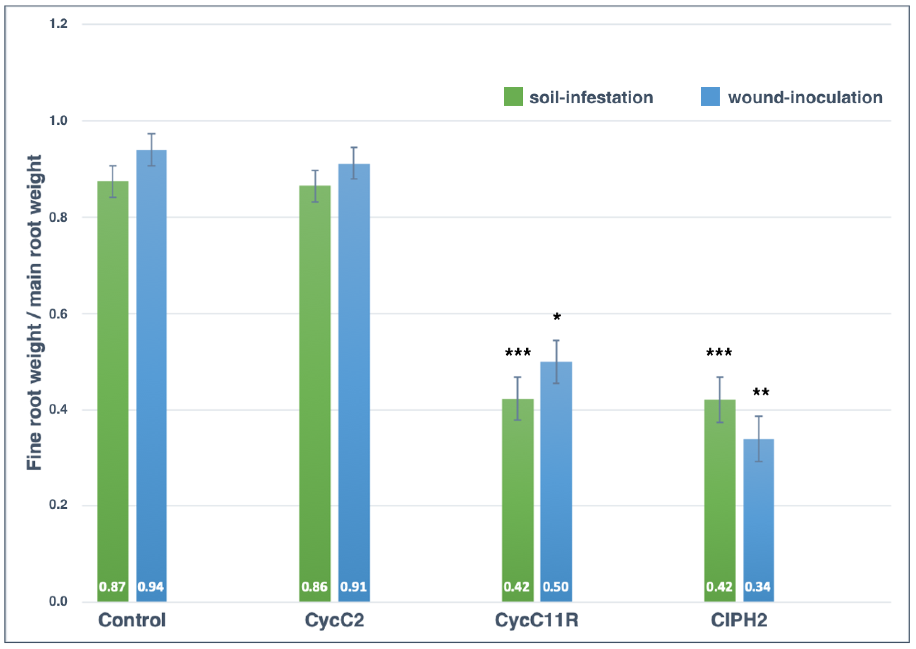
2.4. Pathogenicity Tests

Both the progression and severity of symptoms did not differ significantly between plants inoculated on the stem by wounding and plants inoculated on roots through infested soil.
The first symptoms consisting of leaf yellowing were observed on plants inoculated with the isolate CIPH2 of P. pseudocryptogea, six days post-inoculation (dpi). Later, at 24 dpi, the chlorotic leaves withered and the value of the disease severity index (DSI) ranged between 2 and 3. Sixty dpi in plants inoculated with P. pseudocryptogea, symptoms further evolved and the DSI values ranged between 3 and 4.
On plants inoculated with the isolate CycC11R of P. nicotianae, the leaves turned yellow at 24 dpi (DSI ranging from 1 to 2) and were withered at 36 dpi (mean DSI 2). Conversely, the plants inoculated with the isolate CycC2 of P. multivora showed only mild leaf symptoms and the control plants remained completely asymptomatic for the duration of the test (Figure 4).
All plants of C. revoluta, tested both by wound-inoculation and soil-infestation with the isolates CIPH2 of P. pseudocryptogea and CycC11R of P. nicotianae, developed symptoms of leaf chlorosis, root and stem rot within two months after the transplanting, (Figure 5), although symptoms were more severe on plants inoculated with P. pseudocryptogea. Conversely, plants inoculated with the isolate CycC2 of P. multivora and the control plants did not show symptoms of stem and root rot.
The stem damage class for control plants was zero in both experiments (soil-infestation and wound-inoculation) and the mean value of fine root/main root weight (frw/mrw) ratio in soil-infestation and stem wound-inoculation tests were 0.87 ± 0.12 and 0.94 ± 0.21, respectively (Figure 6, Figure 7 and Figure 8). Phytophthora pseudocryptogea (CIPH2) was the most aggressive species, causing withering of 70% of sago palm plants within two months after the transplanting. It had a frw/mrw ratio of 0.42/0.34 (soil-infestation/wound-inoculation method), (51.7% and 63.8% reduction compared to the control) and a stem damage class of 3.5 (i.e., 76.5% extensive stem dieback) in the soil-infestation test and 3 (i.e., 65% advanced stem dieback with large necrotic lesions) in the wound-inoculation test (Figure 6, Figure 7 and Figure 8). Phytophthora nicotianae (CycC11R) caused withering of 30% of the plants, a frw/mrw ratio of 0.42/0.50 (51.7% and 42.5% reduction compared to the control), and a stem damage class of 1.5 in the soil-infestation test and 1 in the wound-inoculation test (Figure 6, Figure 7 and Figure 8). Differences in the stem damage class and frw/mrw ratio compared to the control were statistically significant for both isolates CIPH2 (P. pseudocryptogea) (stem damage class: p < 0.01 in both test; frw/mrw ratio: p < 0.001 in the soil-infestation test, p < 0.01 in the wound-inoculation test) and CycC11R (P. nicotianae) (stem damage class: p < 0.001 in the soil-infestation test, p < 0.05 in the wound-inoculation test). In plants inoculated with P. multivora (CycC2), the stem damage class and frw/mrw ratio values were not statistically significant compared to the control.
Phytophthora pseudocryptogea was re-isolated consistently, from both symptomatic roots and stems, thus fulfilling Koch’s postulates. Phytophthora nicotianae was re-isolated consistently only from the roots and only very sporadically from the stems. Phytophthora multivora was re-isolated in a very low proportion and solely from the roots. Each species of Phytophthora was re-isolated from the infested potting soil used in root-inoculation tests. The identity of isolates obtained from necrotic roots and stem rot of symptomatic, artificially inoculated sago palm plants, was determined by the colony morphology, microscopic observations and rDNA ITS sequencing.
In summary, P. pseudocryptogea proved to be pathogenic to C. revoluta plants, causing stem and root rot, and was the most virulent among the three species tested. Additionally, P. nicotiane caused stem and root rot, but was less virulent than P. pseudocryptogea. Phytophthora multivora resulted in being only weakly pathogenic to this host and induced mild symptoms on the leaf crown, while no significant effect of inoculation was evident on the stem and roots compared to the non-inoculated control.

3. Discussion

In this study, three diverse Phytophthora species, P. multivora (ITS clade 2c), P. nicotianae (ITS clade 1) and P. pseudocryptogea (ITS clade 8a), were found to be associated with the dieback syndrome of pot-grown plants of C. revoluta observed in Sicily. Of the three species, only P. pseudocryptogea was isolated from infected tissues of stems and roots of C. revoluta plants with natural infections, while P. multivora and P. nicotianae were recovered solely from the rhizosphere soil. Moreover, P. pseudocryptogea was isolated from symptomatic plants when the disease was first recorded, and again 11 years later when it reappeared in a distinct nursery with the same incidence and severity as before. This was also the Phytophthora species recovered consistently and most frequently from the rhizosphere soil of symptomatic plants. In pathogenicity tests, P. pseudocryptogea was the most virulent among the three species recovered from potting soil of sago palm plants and induced typical symptoms of the disease on leaves, stems and roots. Finally, it was re-isolated consistently from symptomatic, artificially infected plants, thus fulfilling Koch’s postulates. Based on this experimental evidence, P. pseudocryptogea was identified as the pathogen responsible for the dieback syndrome of C. revoluta occurring in Sicilian nurseries. Phytophthora pseudocryptogea has been segregated as a separate species from the P. cryptogea species complex, based prevalently on multiple gene phylogenetic analyses and described formally in 2015 as a new species in clade 8a distinct from P. cryptogea sensu stricto [23]. Previous studies demonstrated that P. pseudocryptogea isolates from diverse continents and hosts are genetically uniform [23,24]. It was hypothesized that this homogeneity may be due to the global distribution of a single clone and the lack of sexual reproduction since, consistently with the present study, all isolates of this species so far characterized are of the A1 mating type [23]. In addition, P. pseudocryptogea isolates from C. revoluta characterized in this study and those examined by Safaiefarahani et al. [23], who first described P. pseudocryptogea as a distinct species, share identical double bases at the same position in ITS sequences, strengthening the hypothesis that they belong to the same clonal population and confirming that P. pseudocryptogea is an introduced pathogen in Sicily. Until recently, the geographic distribution of P. pseudocryptogea was believed to be restricted to Asia (Iran), Australia and South America (Ecuador) [25]. However, in the last years, this species has been repeatedly recovered from nurseries and forest stands in the two major Italian islands (Sardinia and Sicily), Spain and Turkey, indicating it has established and is widespread in the Mediterranean region [18,26,27,28,29,30,31]. The known host range of this Phytophthora species is being rapidly expanding and presently includes species of the Aquifoliaceae, Asparagaceae, Asphodeliaceae, Cupressaceae, Fagaceae, Lauraceae, Oleaceae, Platanaceae, Proteaceae, Salicaceae and Solanaceae families [18,25,26,27,28,29,30,31]. Therefore, this is not the first report of P. pseudocryptogea on a gymnosperm, but the first one as a pathogen of a cycad worldwide. Possibly, reports of P. cryptogea on other ornamentals in Sicily, prior to the introduction of multiple gene phylogenetic analyses as a taxonomic and diagnostic criterion and the definition of P. pseudocryptogea as a distinct species within the P. cryptogea complex, have to be critically reconsidered [32,33,34]. This study confirms that P. pseudocryptogea has entered the nursery supply chain in Italy, and like other Phytophthora species introduced recently, such as P. niederhauserii [12,35,36], it may be regarded as an emerging pathogen in the Italian nursery industry [37].
Phytophthora nicotianae, the second species recovered most frequently from rhizosphere soil of symptomatic C. revoluta plants in this study, is a cosmopolitan and very polyphagous pathogen, as well as one of the most prevalent Phytophthora species in ornamental nurseries in Italy [13,37,38,39,40,41,42,43,44]. Like other soilborne Phytophthora species, even though it is prevalently a root pathogen, in ornamental nurseries under favorable environmental conditions, it may adapt temporarily to an aerial life style, causing aboveground infections [45,46]. Differently from P. pseudocryptogea, both A1 and A2 mating types of P. nicotianae were recovered from the potting soil of C. revoluta plants, in agreement with previous reports, indicating that both mating types of this species occur in nurseries of ornamentals in Italy [39,40,47]. The presence of both mating types in the same nursery is a prerequisite for this heterothallic species to reproduce sexually and the demonstration of its ability to give rise spontaneously to interspecific hybrids confirm sexual reproduction is not a rare event in the nursery of ornamentals [48,49]. Genetic recombination through sexual reproduction might favor the emergence of new genotypes and the expansion of the already broad host range of this pathogen [50]. Moreover, the ability to form resting structures, such as oospores or chlamydospores, may condition other aspects of the biology of P. nicotianae, such as the tolerance of extreme environmental conditions. The ecological plasticity of this species and its polyphagy may explain why it is so common in the nursery industry of southern Italy. In pathogenicity tests, P. nicotianae induced decay symptoms in both stems and roots of artificially inoculated plants of C. revoluta, but was relatively more virulent to roots than stems, indicating sago palm as a potential host this Phytophthora species, thus expanding the list of its hosts, and confirming it is prevalently a root pathogen [44,51].
Phytophthora multivora was formally described as a separate species of the P. citricola species complex in 2009 [52]. Later, it was found to be common in natural ecosystems in South Africa [53]. However, there it did not cause any severe disease outbreaks, suggesting a long-term co-evolution with the native tree species. Presently, P. multivora has a wide geographical distribution, encompassing Western Australia, South Africa, New Zealand, USA, Europe and the Canary Islands, probably as a consequence of the global trade of living plants and plant material [53]. The genetic analysis of a large collection of isolates from all over the world, using two diverse sets of molecular markers, pointed at South Africa as the center of origin of this Phytophthora species [53]. In Sicily, P. multivora is common in protected natural areas, vegetation conservation sites and ornamental nurseries [26,29,30,54]. Although this Phytophthora species is regarded an emerging pathogen worldwide and has a broad host range, also including Macrozamia riediei, a cycad native to Australia [25], in pathogenicity tests on C. revoluta, it was weakly pathogenic, causing only mild symptoms in leaves and no visible decay of the stem and roots, confirming it was not the causal agent of the dieback syndrome of this cycad recorded in Sicilian nurseries. The presence of recently introduced exotic species, such as P. multivora and P. pseudocryptogea, or a species preferentially associated with agricultural systems, such as P. nicotianae, in natural ecosystems and nurseries in southern Italy, would confirm the role of the nursery industry in the introduction and spread of invasive Phytophthora species, as evidenced for P. ramorum [55]. All three Phytophthora species recovered from potting soil in this study are polyphagous and the nursery where the disease was detected was not specialized in the production of sage palm. Consequently, any hypothesis concerning the introduction and spread of these pathogens in the nurseries would be speculative.
In general, the contemporary presence of diverse Phytophthora species, including species known as aggressive pathogens, is common in ornamental nurseries. However, in many cases, not all the species cause disease and some species, despite their polyphagia, privilege certain hosts, although evidence of host specialization has been provided only in a few cases. The association of multiple Phytophthora species with a specific host inspired the concept that in nurseries of ornamental plants, PRR has to be regarded as a complex disease [56]. However, potentially pathogenic Phytophthora species may be recovered from soil of asymptomatic plants and not all Phytophthora species detected in the soil of symptomatic plants are causing disease. To make PRR management strategies in nurseries more effective, it would be useful to complement the analysis of the Phytophthora species community associated with the nursery industry, with the study of the lifestyle of these species.

4. Materials and Methods

4.1. Isolation and Morphological Characterization of Isolates

Isolations were made from 20 symptomatic sago palm (C. revoluta) plants with different disease severities, collected in 2021 from a single nursery in the Catania province, Sicily (Southern Italy). Plants were uprooted, washed with tap water and cut off. Rotten roots and stems were washed again with running tap water, immersed in 1% NaClO for 2 min and then in 70% EtOH for 30 s, rinsed in sterile distilled water, and dipped dry before being cut into 5 mm pieces. Subterranean stem and root tissue pieces were plated onto selective BNPRAH V8 agar medium [57] in Petri dishes (five pieces per dish), and then incubated for 24–72 h in the dark at 25 °C. Pure cultures of the same colony pattern were obtained by subculturing single hyphae onto V8 juice agar (V8A) [58]. Potting-soil samples were collected from the same 20 pots with symptomatic plants used for direct isolations from tissues. Subsamples of ca. 200 mL soil per pot were used for the leaf baiting test, which was performed in a walk-in growth chamber with 12 h natural day light at 20 °C, according to the protocol described by Riolo et al. [59]. Young leaves of native plant species (Ceratonia siliqua and Quercus spp.) were used as baits. Necrotic segments (2 × 2 mm) from infected leaves were plated onto selective PARPNH agar [60]. Petri dishes were incubated at 20 °C in the dark. Outgrowing Phytophthora hyphae were transferred onto V8A under the stereomicroscope. All isolates were maintained on the V8A and stored at 6 °C in the dark. Isolates were in-depth characterized based on the colony morphology. Isolates obtained from symptomatic sago palm in 2010 and preserved in the collection of the Molecular Plant Pathology Laboratory, at the University of Catania, were also included in this study. All Phytophthora isolates characterized in this study are listed in Table 1. However, since all isolates of the same species showed the same features, only a few of them are listed in the table.
Morphological features of isolates, including the morphology and dimensions of reproductive structures, and colony shape, were determined on V8A and PDA at 25 ± 1 °C in the dark, according to standard procedures [58]. Sporangia production was stimulated following the method described by Jung et al. [61]. Small fragments (size 2 mm) were cut from the growing edge of 5- to 7-day-old cultures grown in Petri dishes (15 mm diam) on V8A at 20 °C in the dark. They were then placed in a 5 cm diameter Petri dish and flooded with non-sterile soil extract water (200 g soil suspended in 1 L of de-ionized water for 24 h at room temperature and then filtered). After incubation at 20 °C in the dark for 24–72 h, dimensions and morphological features of 50 mature sporangia of each isolate were determined at ×400 magnification. For cardinal temperatures, the isolates were grown on CMA (Corn Meal Agar; Oxoid Limited, Basingstoke, UK) dishes and incubated at 1, 3, 5, 10, 15, 20, 25, 30, 35 and 37 °C, and radial colony growth was measured after 2–10 days. There were three replicate plates for each isolate and temperature.
The sexual compatibility type of P. pseudocryptogea isolates from cycas was determined by crossing Phytophthora isolates with A1 and A2 mating types of P. cryptogea (CBS 113.119) and P. drechsleri (CBS 292.35), respectively. Mycelial plugs (5 mm diameter) of the unknown mating type isolate and either CBS 113.119 or CBS 292.35 isolates were placed 4 cm apart from each other on 10% clarified V8A medium. The plates were incubated for 10 days at 20 °C in the dark or until oospores were produced. The mating type of P. nicotianae isolates from cycas was determined using the same method, and the P. nicotianae isolates IMI 268688 (A1) and Albicocco9 (=IMI 396203) (A2), as testers [62,63]. All the mating type tests were repeated in duplicate.

4.2. Molecular Identification

DNA-based identification of 16 representative Phytophthora isolates from cycas (Table 1) was performed by sequence analysis of internally transcribed spacer of ribosomal DNA (ITS-rDNA) region, β-tubulin (β-tub) and cytochrome c oxidase subunit 1 (COI). DNA was extracted from 7-day-old cultures grown on V8A at 20 °C, by using the PowerPlant Pro DNA isolation Kit (MO BIO Laboratories, Inc., Carlsbad, CA, USA), following the manufacturer’s instructions. ITS-rDNA was amplified using forward primers ITS6 [64] and the reverse primer ITS4 [65]. All PCRs were carried out in a 25 µL reaction mix containing the PCR buffer (1X), dNTP mix (0.2 mM), MgCl2 (1.5 mM), forward and reverse primers (0.5 mM each), Taq DNA Polymerase (1 U) and 100 ng of DNA. The thermocycler conditions were as follows: 94 °C for 3 min, followed by 35 cycles of 94 °C for 30 s, 55 °C for 30 s, and 72 °C for 30 s, and then 72 °C for 10 min [64]. β-tubulin and COI were amplified by using the primer pairs TUBUF2/TUBUR1 and COXF4N/COXR4N, respectively [66]. The thermocycle sequence was as follows: an initial denaturation at 94 °C for 2 min, 35 cycles consisting of denaturation at 94 °C for 30 s, annealing for 30 s, and extension at 72 °C for 60 s, and a final extension at 72 °C for 10 min. Annealing temperatures were 60 °C for β-tubulin and 52 °C for COI.
Amplicons were detected in 1% agarose gel and sequenced in both directions by an external service (Macrogen, Amsterdam, The Netherlands). All resultant chromatograms were checked for correct base-calling using FinchTV v.1.4.0. The consensus sequences were first compared by the BLAST program to the gene sequences in the NCBI databases, and then with those of the ex-types and other well-authenticated specimens of Phytophthora, using the Clustal 2.1 software.

4.3. Pathogenicity Tests

The pathogenicity of Phytophthora spp. isolates from sago palm plants was tested in two separate experiments, using the soil-infestation method and the method of stem inoculation by wounding, as described by Aloi et al. [67]. The isolates of P. pseudocryptogea (CIPH2), P. nicotianae (CycC11R) and P. multivora (CycC2) were used to inoculate six-month-old potted plants of sago palm (10 plants for each isolate). Pathogenicity tests by wound inoculation were performed by inserting a mycelial plug of 7-day-old colonies, grown on V8A, into a wound made with a scalpel in the subterranean stem and the tissue patch was sealed with Parafilm®. Control plants were inoculated with sterile agar plugs. In soil-infestation pathogenicity assays, cycas plants were transplanted into 12 cm diameter pots containing a mixture of 1:1 steam-sterilized, sandy loam (vol/vol) with 4% inoculum produced on autoclaved wheat kernel seeds. Inoculum consisted of a 21-day-old culture of the isolates grown in the dark at 25 °C in 750 mL Erlenmeyer flasks, containing a sterilized medium made of 50 mL of wheat seeds and 50 mL V8 juice broth. Ten control plants were transplanted in free-draining pots containing non-infested potting mixtures. For both inoculation methods, all plants were maintained in saturated soil for 48 h and then transferred to a growth chamber at 23 °C, 80% relative humidity, and a photoperiod of 16 h of light and 8 h of dark. The disease severity index (DSI) was assessed visually at 6-day intervals, since the appearance of first symptoms as frond yellowing or wilting using a 0–4 rating scale (0 = no symptoms, 1 = 1–33% frond yellowing or wilting, 2 = 34–66% frond yellowing or wilting, 3 = 67–100% frond yellowing or wilting, 4 = dead plant). Symptoms were evaluated at four time intervals after inoculation, i.e., 12, 24, 36, and 60 days post-inoculation (dpi). The trials were considered concluded when the plants showed severe symptoms of withering (60 dpi). Data were analyzed by using a one-way ANOVA, followed by a Tukey ‘s HSD test (honestly significant difference) as a post-hoc test (R software). Differences at p ≤ 0.05 were considered significant. At the end of the tests, from all plants showing necrosis of the stem, the severity of stem rot was rated according to a scale of five stem damage classes: 0 = healthy stem; 1 =< 25% slight stem rot; 2 = 26–50% stem rot, beginning stem dieback with small necrotic lesions; and 3 = 51–75% stem rot, advanced stem dieback with large necrotic lesions; 4 = 76–100% stem rot, extensive stem dieback. The roots were dried for 72 h at 65 °C and the dry weights of the main roots (diam. 2–10 mm) and fine roots (diam < 2 mm) were recorded for each plant. Data were analyzed by one-way analysis of variance (ANOVA), followed by a Dunnett’s multiple comparisons test. Statistical analysis was carried out by the R software and considered as significant at p < 0.05. Re-isolations from necrotic stems and roots were performed on the BNPRAH V8A medium and the identity of each Phytophthora species was confirmed by morphological and molecular analyses.

5. Conclusions

Three Phytophthora species, P. multivora, P. nicotianae and P. pseudocryptogea, were recovered from the rhizosphere soil of pot-grown sago palm (Cycas revoluta) with symptoms of PRR. Of the three Phytophthora species recovered from the potting soil of sago palm plants, Koch’s postulates were completed only for P. pseudocryptogea, which was identified as the etiological agent of the disease. Conversely, neither of the other two species was isolated from infected tissues of symptomatic plants with natural infections, and in artificial inoculation tests on sago palms, only P. nicotianae was proven to be an aggressive pathogen, while P. multivora induced very mild symptoms. The role of both P. multivora and P. nicotianae in PRR of sago palm deserves to be investigated further. It is noteworthy that PRR outbreaks were observed exclusively in plant blocks grown in open air, suggesting the hypothesis that they were triggered by conducive environmental conditions occurring outside the greenhouse, such as soil waterlogging due to heavy rains in winter and aerial dispersal of the inoculum via splashing water.

Author Contributions

Conceptualization, A.P., S.O.C. and R.F.; methodology, A.P., R.F., R.P., S.C.T. and F.A.; analysis of results, A.P., R.F., R.P., S.C.T. and F.A.; resources, S.O.C., A.P.; original—draft preparation, A.P., R.F. and S.O.C.; editing, A.P., S.O.C. and R.F.; supervision, A.P. and S.O.C. All authors have read and agreed to the published version of the manuscript.

Funding

This research was funded by the University of Catania, Italy “Investigation of phytopathological problems of the main Sicilian productive contexts and eco-sustainable defense strategies (ME-DIT-ECO)” PiaCeRi-PIAno di inCEntivi per la Ricerca di Ateneo 2020-22 linea 2” “5A722192155”, by PON “RICERCA E INNOVAZIONE” 2014–2020, Azione II-Obiettivo Specifico 1b-Progetto “Miglioramento delle produzioni agroalimentari mediterranee in condizioni di carenza di risorse idriche-WATER4AGRIFOOD”, B64I2000016000500, by the project “Smart and innovative packaging, postharvest rot management, and shipping of organic citrus fruit (BiOrangePack)” under the Partnership for Research and Innovation in the Mediterranean Area (PRIMA)—H2020 (E69C20000130001), by the “Italie–Tunisie Cooperation Program 2014–2020” project “PROMETEO «Un village transfrontalier pour protéger les cultures arboricoles méditerranéennes en partageant les connaissances» cod. C-5-2.1-36, CUP 453E25F2100118000 and by the by European Union (NextGeneration EU), through the MUR-PNRR project SAMOTHRACE (ECS00000022).

Institutional Review Board Statement

Not applicable.

Informed Consent Statement

Not applicable.

Data Availability Statement

Data will be available upon specific request to the authors.

Acknowledgments

The authors are endlessly grateful to Giovanni Granata for his helpfulness and for providing the Cycas plants for pathogenicity tests.

Conflicts of Interest

The authors declare no conflict of interest.

References

  1. Donaldson, J.S.; Hill., K.D.; Stevenson, D.W. Cycads of the World: An Overview. In Cycads; Donaldson, J.S., Ed.; IUCN/SSC Cycad Specialist Group: Gland, Switzerland; Cambridge, UK, 2003; pp. 3–8. [Google Scholar]
  2. Nagalingum, N.S.; Marshall, C.R.; Quental, T.B.; Rai, H.S.; Little, D.P.; Mathews, S. Recent synchronous radiation of a living fossil. Science 2011, 334, 796–799. [Google Scholar] [CrossRef] [PubMed]
  3. Mankga, L.T.; Yessoufou, K.; Mugwena, T.; Citakira, M. The cycad genus Cycas may have diversified from Indochina and occupied its current ranges through vicariance and dispersal events. Front. Ecol. Evol. 2020, 8, 44. [Google Scholar] [CrossRef] [Green Version]
  4. Cousins, S.R.; Williams, V.L.; Witkowski, E.T.F. Uncovering the cycad taxa (Encephalartos species) traded for traditional medicine in Johannesburg and Durban, South Africa. S. Afr. J. Bot. 2012, 78, 129–138. [Google Scholar] [CrossRef] [Green Version]
  5. Calonje, M.; Stevenson, D.; Osborne, R. The World List of Cycads Online Edition 2013–2023. Montgomery Botanical Center, Coral Gables, FL, USA. Available online: http://www.cycadlist.org (accessed on 12 January 2023).
  6. Condamine, F.L.; Nagalingum, N.S.; Marshall, C.R.; Morlon, H. Origin and diversification of living cycads: A cautionary tale on the impact of the branching process prior in Bayesian molecular dating. BMC Evol. Biol. 2015, 15, 65. [Google Scholar] [CrossRef] [PubMed] [Green Version]
  7. Hardy, G.E.; Sivasithamparam, K. Phytophthora spp. associated with container-grown plants in nurseries in Western Australia. Plant Dis. 1988, 72, 435–437. [Google Scholar] [CrossRef] [Green Version]
  8. Dart, N.L.; Riley, K.L.; Rugarber, E.F.; Castagner, G.A. Recovery frequency of Phytophthora ramorum and other Phytophthora spp. in the soil profile of ornamental retail nurseries. Plant Dis. 2007, 91, 1419–1422. [Google Scholar]
  9. Schwingle, B.W.; Smith, J.A.; Blanchette, R.A. Phytophthora species associated with diseased woody ornamentals in Minnesota nurseries. Plant Dis. 2007, 91, 97–102. [Google Scholar] [CrossRef] [Green Version]
  10. Moralejo, E.; Pérez-Sierra, A.M.; Álvarez, L.A.; Belbahri, L.; Lefort, F.; Descals, E. Multiple alien Phytophthora taxa discovered on diseased ornamental plants in Spain. Plant Pathol. 2009, 58, 100–110. [Google Scholar] [CrossRef]
  11. Leonberger, A.J.; Speers, C.; Ruhl, G.; Creswell, T.; Beckerman, J.L. A survey of Phytophtohora spp. in Midwest nurseries, greenhouses, and landscapes. Plant Dis. 2013, 97, 635–640. [Google Scholar] [CrossRef] [Green Version]
  12. Abad, Z.G.; Abad, J.A.; Cacciola, S.O.; Pane, A.; Faedda, R.; Moralejo, E.; Pérez-Sierra, A.; Abad-Campos, P.; Alvarez-Bernaola, L.A.; Bakonyi, J.; et al. Phytophthora niederhauserii sp. nov., a polyphagous species associated with ornamentals, fruit trees and native plants in 13 countries. Mycologia 2014, 106, 431–447. [Google Scholar] [CrossRef]
  13. Prigigallo, M.I.; Mosca, S.; Cacciola, S.O.; Cooke, D.E.L.; Schena, L. Molecular analysis of Phytophthora diversity in nursery-grown ornamental and fruit plants. Plant Pathol. 2015, 64, 1308–1319. [Google Scholar] [CrossRef] [Green Version]
  14. Jung, T.; Orlikowski, L.; Henricot, B.; Abad-Campos, P.; Aday, A.G.; Aguín Casal, O.; Bakonyi, J.; Cacciola, S.O.; Cech, T.; Chavarriaga, D.; et al. Widespread Phytophthora infestations in European nurseries put forest, semi-natural and horticultural ecosystems at high risk of Phytophthora diseases. For. Pathol. 2016, 46, 134–163. [Google Scholar] [CrossRef] [Green Version]
  15. Junker, C.; Goff, P.; Wagner, S.; Werres, S. Occurrence of Phytophthora species in commercial nursery production. Plant Health Prog. 2016, 17, 64–75. [Google Scholar] [CrossRef]
  16. Patel, J.S.; Vitoreli, A.; Palmateer, A.J.; El-Sayed, A.; Norman, D.J.; Goss, E.M.; Brennan, M.S.; Ali, G.S. Characterization of Phytophthora spp. isolated from ornamental plants in Florida. Plant Dis. 2016, 100, 500–509. [Google Scholar] [CrossRef] [PubMed] [Green Version]
  17. Riddell, C.E.; Frederickson-Matika, D.; Armstrong, A.C.; Elliot, M.; Forster, J.; Hedley, P.E.; Morris, J.; Thorpe, P.; Cooke, D.E.L.; Pritchard, L.; et al. Metabarcoding reveals a high diversity of woody host associated Phytophthora spp. in soils at public gardens and amenity woodlands in Britain. PeerJ 2019, 7, e6931. [Google Scholar] [CrossRef] [Green Version]
  18. Mora-Sala, B.; León, M.; Pérez-Sierra, A.; Abad-Campos, P. New reports of Phytophthora species in plant nurseries in Spain. Pathogens 2022, 11, 826. [Google Scholar] [CrossRef]
  19. Pane, A.; Allatta, C.; Sammarco, G.; Cacciola, S.O. First report of bud rot of Canary Island date palm caused by Phytophthora palmivora in Italy. Plant Dis. 2007, 91, 1059. [Google Scholar] [CrossRef]
  20. Faedda, R.; Pane, A.; Granata, G.; Magnano di San Lio, G. First report of Phytophthora nicotianae as pathogen of blue Mediterranean fan palm. New Dis. Rep. 2011, 23, 3. [Google Scholar] [CrossRef] [Green Version]
  21. Nesamari, R.; Coutinho, T.A.; Roux, J. Investigations into Encephalartos insect pests and diseases in South Africa and identification of Phythopthora cinnamomi as a pathogen of Modjiadi cycad. Plant Pathol. 2017, 66, 612–622. [Google Scholar] [CrossRef]
  22. NSW Department of Planning, Industry and Environment. Infection of Native Plants by Phytophthora Cinnamomi. Available online: https://www.environment.nsw.gov.au/savingourspeciesapp/project.aspx?ProfileID=20026 (accessed on 13 January 2023).
  23. Safaiefarahani, B.; Mostowfizadeh-Ghalamfarsa, R.; Hardy, G.; Burgess, T.I. Re-evaluation of the Phytophthora cryptogea species complex and the description of a new species, Phytophthora pseudocryptogea sp. nov. Mycol. Prog. 2015, 14, 108. [Google Scholar] [CrossRef]
  24. Martin, F.N.; Blair, J.E.; Coffey, M.D. Combined mitochondrial and nuclear multilocus phylogeny of the genus Phytophthora. Fungal Genet. Biol. 2014, 66, 19–32. [Google Scholar] [CrossRef]
  25. Abad, G.Z.; Burgess, T.; Redford, A.J.; Bienapfl, J.C.; Mathew, R.; Srivastava, S.K.; Jennings, K.C. IDphy: An international online resource for molecular and morphological identification of Phytophthora. Plant Dis. 2022, 10, 259. [Google Scholar] [CrossRef]
  26. Jung, T.; La Spada, F.; Pane, A.; Aloi, F.; Evoli, M.; Jung, M.H.; Scanu, B.; Faedda, R.; Rizza, C.; Puglisi, I.; et al. Diversity and distribution of Phytophthora species in protected natural areas in Sicily. Forests 2019, 10, 259. [Google Scholar] [CrossRef] [Green Version]
  27. Seddaiu, S.; Brandano, A.; Ruiu, P.A.; Sechi, C.; Scanu, B. An overview of Phytophthora species inhabiting declining Quercus suber stands in Sardinia (Italy). Forests 2020, 11, 971. [Google Scholar] [CrossRef]
  28. Bregant, C.; Mulas, A.A.; Rossetto, G.; Deidda, A.; Maddau, L.; Piras, G.; Linaldeddu, B.T. Phytophthora mediterranea sp. nov., a new species closely related to Phytophthora cinnamomi from nursery plants of Myrtus communis in Italy. Forests 2021, 12, 682. [Google Scholar] [CrossRef]
  29. Riolo, M.; La Spada, F.; Aloi, F.; Giusso del Galdo, G.; Santilli, E.; Pane, A.; Cacciola, S.O. Phytophthora diversity in a sentinel arboretum and in a nature reserve area. Biol. Life Sci. Forum 2021, 4, 51. [Google Scholar]
  30. La Spada, F.; Cock, P.J.A.; Randall, E.; Pane, A.; Cooke, D.E.L.; Cacciola, S.O. DNA metabarcoding and isolation by baiting complement each other in revealing Phytophthora diversity in anthropized and natural ecosystems. J. Fungi 2022, 8, 330. [Google Scholar] [CrossRef]
  31. Kurbetli, İ.; Woodward, S.; Aydoğdu, M.; Sülü, G.; Özben, S. Phytophthora plurivora and Phytophthora pseudocryptogea isolated from soils supporting declining oaks (Quercus robur L.) in İstanbul, Turkey. For. Pathol. 2022, 56, e12782. [Google Scholar]
  32. Cacciola, S.O.; Magnano di San Lio, G.; Greco, G.; Pane, A. Electrophoretic study of three related Phytophthora species. EPPO Bull. 1990, 20, 47–58. [Google Scholar] [CrossRef]
  33. Cacciola, S.O.; Pane, A.; Raudino, F.; Davino, S. First report of root and crown rot of sage caused by Phytophthora cryptogea in Italy. Plant Dis. 2002, 86, 1176. [Google Scholar] [CrossRef]
  34. Cacciola, S.O.; Chimento, A.; Pane, A.; Cooke, D.E.L.; Magnano di San Lio, G. Root rot of lantana caused by Phytophthora cryptogea. Plant Dis. 2005, 89, 909. [Google Scholar] [CrossRef]
  35. Cacciola, S.O.; Scibetta, S.; Martini, P.; Rizza, C.; Pane, A. Phytophthora taxon niederhauserii, a new root and crown pathogen of Banksia spp. in Italy. Plant Dis. 2009, 93, 1216. [Google Scholar] [CrossRef]
  36. Faedda, R.; Cacciola, S.O.; Pane, A.; Martini, P.; Odasso, M.; Magnano di San Lio, G. First report of Phytophthora taxon niehederhauserii causing root and stem rot of mimosa in Italy. Plant Dis. 2013, 97, 688. [Google Scholar] [CrossRef]
  37. Antonelli, C.; Biscontri, M.; Tabet, D.; Vettraino, A.M. The never-ending presence of Phytophthora species in Italian nurseries. Pathogens 2023, 12, 15. [Google Scholar] [CrossRef]
  38. Pane, A.; Martini, P.; Chimento, A.; Rapetti, S.; Savona, S.; Grasso, F.M.; Cacciola, S.O. Phytophthora species on ornamental plants in Italy. J. Plant Pathol. 2005, 87, 301. [Google Scholar]
  39. Pane, A.; Cacciola, S.O.; Chimento, A.; Allatta, C.; Scibetta, S.; Magnano di San Lio, G. First report of Phytophthora spp. as pathogens of Pandorea jasminoides in Italy. Plant Dis. 2008, 92, 313. [Google Scholar] [CrossRef]
  40. Rizza, C.; Faedda, R.; Pane, A.; Cacciola, S.O. First report of root and basal stem rot caused by Phytophthora nicotianae on tree aeonium (Aeonium arboreum) in Italy. Plant Dis. 2011, 92, 362. [Google Scholar] [CrossRef] [PubMed]
  41. Kamoun, S.; Furzer, O.; Jones, J.D.G.; Judelson, H.S.; Ali, G.S.; Dalio, R.J.D.; Roy, S.G.; Schena, L.; Zambounis, A.; Panabières, F.; et al. The top 10 oomycete pathogens in molecular plant pathology. Mol. Plant Pathol. 2015, 16, 413–434. [Google Scholar] [CrossRef] [PubMed]
  42. Migliorini, D.; Ghelardini, L.; Tondini, E.; Luchi, N.; Santini, A. The potential of symptomless potted plants for carrying invasive soilborne plant pathogens. Divers. Distrib. 2015, 21, 1218–1229. [Google Scholar] [CrossRef] [Green Version]
  43. Panabières, F.; Ali, G.S.; Allagui, M.B.; Dalio, R.J.D.; Gudmestad, N.C.; Kuhn, M.L.; Guha Roy, S.; Schena, L.; Zampounis, A. Phytophthora nicotianae diseases worldwide: New knowledge of a long-recognised pathogen. Phytopathol. Mediterr. 2016, 55, 20–40. [Google Scholar]
  44. Prigigallo, M.I.; Abdelfataah, A.; Cacciola, S.O.; Faedda, R.; Sanzani, S.M.; Cooke, D.E.L.; Schena, L. Metabarcoding analysis of Phytophthora diversity using genus-specific primers and 454 pyrosequencing. Phytopathology 2016, 196, 305–313. [Google Scholar] [CrossRef] [PubMed] [Green Version]
  45. Cacciola, S.O.; Pane, A.; Faedda, R.; Rizza, C.; Badalà, F.; Magnano di San Lio, G. Bud and root rot of windmill palm (Trachycarpus fortunei) caused by simultaneous infections of Phytophthora palmivora and P. nicotianae in Sicily. Plant Dis. 2011, 95, 769. [Google Scholar] [CrossRef] [PubMed]
  46. Baysal-Gurel, F.; Bika, R.; Simmons, T.; Avin, F. Identification and management of Phytophthora aerial blight caused by Phytophthora nicotianae on Catharanthus roseus. Plant Dis. 2022, 106, 1271–1277. [Google Scholar] [CrossRef] [PubMed]
  47. Biasi, A.; Martin, F.N.; Cacciola, S.O.; Magnano di San Lio, G.; Grünwald, N.G.; Schena, L. Genetic analysis of Phytophthora nicotianae populations from different hosts using microsatellite markers. Phytopathology 2016, 106, 1006–1014. [Google Scholar] [CrossRef] [PubMed] [Green Version]
  48. Faedda, R.; Cacciola, S.O.; Pane, A.; Szigethy, A.; Bakonyi, J.; Man In’t Veld, W.; Martini, P.; Schena, L.; Magnano di San Lio, G. Phytophthora × pelgrandis causes root and collar rot of Lavandula stoechas in Italy. Plant Dis. 2013, 97, 1091–1096. [Google Scholar] [CrossRef] [PubMed] [Green Version]
  49. Szigethy, A.; Nagy, Z.Á.; Vettraino, A.M.; Józsa, A.; Cacciola, S.O.; Faedda, R.; Bakonyi, J. First Report of Phytophthora × pelgrandis causing root rot and lower stem necrosis of common box, lavender and Port-Orford-cedar in Hungary. Plant Dis. 2013, 97, 152. [Google Scholar] [CrossRef]
  50. Cacciola, S.O.; Gullino, M.L. Emerging and re-emerging fungus and oomycete soil-borne plant diseases in Italy. Phytopathol. Mediterr. 2019, 58, 451–472. [Google Scholar]
  51. Puglisi, I.; De Patrizio, A.; Schena, L.; Jung, T.; Evoli, M.; Pane, A.; Van Hoa, N.; Van Tri, M.; Wright, S.; Ramstedt, M.; et al. Two previously unknown Phytophthora species associated with brown rot of pomelo (Citrus grandis) fruits in Vietnam. PLoS ONE 2017, 12, e0172085. [Google Scholar] [CrossRef] [Green Version]
  52. Scott, P.M.; Burgess, T.I.; Barber, P.A.; Shearer, B.L.; Stukely, M.J.; Hardy, G.E.; Jung, T. Phytophthora multivora sp. nov., a new species recovered from declining Eucalyptus, Banksia, Agonis and other plant species in Western Australia. Pers.-Mol. Phylogeny Evol. Fungi 2009, 22, 1–13. [Google Scholar] [CrossRef] [Green Version]
  53. Tsykun, T.; Prospero, S.; Schoebel, C.N.; Rea, A.; Burgess, T.I. Global invasion history of the emerging plant pathogen Phytophthora multivora. BMC Genom. 2022, 23, 153. [Google Scholar] [CrossRef]
  54. Riolo, M.; Aloi, F.; La Spada, F.; Sciandrello, S.; Moricca, S.; Santilli, E.; Pane, A.; Cacciola, S.O. Diversity of Phytophthora communities across different types of Mediterranean vegetation in a nature reserve area. Forests 2020, 11, 853. [Google Scholar] [CrossRef]
  55. Sims, L.L.; Garbelotto, M. Phytophthora species repeatedly introduced in Northern California through restoration projects can spread into adjacent sites. Biol. Invasions 2021, 23, 2173–2190. [Google Scholar] [CrossRef]
  56. Weiland, J.E. The challenges of managing Phytophthora root rot in the nursery industry. Plant Health Prog. 2021, 22, 332–342. [Google Scholar] [CrossRef]
  57. Masago, H.; Yoshikawa, M.; Fukada, M.; Nakanishi, N. Selective inhibition of Pythium spp. on a medium for direct isolation of Phytophthora spp. from soils and plants. Phytopathology 1977, 67, 425–428. [Google Scholar] [CrossRef]
  58. Erwin, D.C.; Ribeiro, O.K. Phytophthora Diseases Worldwide; APS Press—The American Phytopathological Society: St. Paul, MN, USA, 1996; pp. 84–95, 96–144, 456–463. [Google Scholar]
  59. Riolo, M.; Aloi, F.; Conti Taguali, S.; Pane, A.; Franco, M.; Cacciola, S.O. Phytophthora × cambivora as a major factor inciting the decline of European beech in a stand within the southernmost limit of its natural range in Europe. J. Fungi 2022, 8, 973. [Google Scholar] [CrossRef] [PubMed]
  60. Jung, T.; Blaschke, H.; Neumann, P. Isolation, identification and pathogenicity of Phytophthora species from declining oak stands. Eur. J. For. Pathol. 1996, 26, 253–272. [Google Scholar] [CrossRef]
  61. Jung, T.; Cooke, D.E.L.; Blaschke, H.; Duncan, J.M.; Oßwald, W. Phytophthora quercina sp. nov., causing root rot of European oaks. Mycol. Res. 1999, 103, 785–798. [Google Scholar] [CrossRef]
  62. Pane, A.; Cacciola, S.O.; Scibetta, S.; Bentivenga, G.; Magnano di San Lio, G. Four Phytophthora species causing foot and root rot of apricot in Italy. Plant Dis. 2009, 83, 844. [Google Scholar] [CrossRef]
  63. Mammella, M.A.; Cacciola, S.O.; Martin, F.; Schena, L. Genetic characterization of Phytophthora nicotianae by the analysis of polymorphic regions of the mitochondrial DNA. Fungal Biol. 2011, 115, 432–442. [Google Scholar] [CrossRef]
  64. Cooke, D.E.L.; Drenth, A.; Duncan, J.M.; Wagels, G.; Brasier, C.M. A molecular phylogeny of Phytophthora and related oomycetes. Fungal Genet. Biol. 2000, 30, 17–32. [Google Scholar] [CrossRef]
  65. White, T.J.; Bruns, T.; Lee, S.; Taylor, J.W. Amplification and direct sequencing of fungal ribosomal RNA genes for phylogenet-ics. In PCR Protocols: A Guide to Methods and Applications; Innis, M.A., Gelfand, D.H., Sninsky, J.J., White, T.J., Eds.; Academic Press Inc.: San Diego, CA, USA, 1990; Volume 18, pp. 315–322. [Google Scholar]
  66. Kroon, L.P.N.M.; Bakker, F.T.; Van Den Bosch, G.B.M.; Bonants, P.J.M.; Flier, W.G. Phylogenetic analysis of Phytophthora species based on mitochondrial and nuclear DNA sequences. Fungal Genet. Biol. 2004, 41, 766–782. [Google Scholar] [CrossRef] [PubMed]
  67. Aloi, F.; Riolo, M.; La Spada, F.; Bentivenga, G.; Moricca, S.; Santilli, E.; Pane, A.; Faedda, R.; Cacciola, S.O. Phytophthora root and collar rot of Paulownia, a new disease for Europe. Forests 2021, 12, 1664. [Google Scholar] [CrossRef]
Figure 1. Symptoms of chlorosis, (A) followed by desiccation (B) of the older leaves and root rot, associated with the internal browning of the subterranean stem on three-year-old potted sago palm plants (CE).
Plants 12 01197 g001
Figure 2. Colony morphologies of Phytophthora multivora (CycC2), P. nicotianae (CycC11R) and P. pseudocryptogea (CIPH2) after 7 days of growth at 20 °C in the dark on V8A, PDA and CMA.
Plants 12 01197 g002
Figure 3. Semi-papillate, persistent, ovoid, limoniform, or ellipsoid sporangia of Phytophthora multivora (CycC2). Papillate, persistent, spherical to ovoid, ellipsoid, obpyriform sporangia of P. nicotianae (CycC11R). Non-papillate, persistent, terminal, ovoid, ellipsoid to obpyriform sporangia of P. pseudocryptogea (CIPH2). Scale bar: 25 μm.
Plants 12 01197 g003
Figure 4. Symptom progression in Cycas revoluta plants inoculated (soil-infestation (S) and wound-inoculation method (W)) with Phytophthora multivora (CycC2), P. nicotianae (CycC11R) and P. pseudocryptogea (CIPH2). Non-inoculated plants served as a control. Symptom severity was expressed as the mean value of the disease severity index (DSI) at 12, 24, 36, and 60 days post-inoculation (dpi). Images show the type of symptoms at each time interval post-inoculation. At each time interval, values sharing the same letters are not significantly different according to the Tukey’s honestly significant difference (HSD) test (p ≤ 0.05).
Plants 12 01197 g004
Figure 5. Symptoms of root rot and leaf chlorosis on Cycas revoluta plants, 2 months after transplanting from two separate experiments, soil-infestation (S) and wound-inoculation method (W) with Phytophthora pseudocryptogea (CIPH2) and P. nicotianae (CycC11R); no symptoms appear in the sago palm plants transplanted in both test with P. multivora (CycC2). Control plants remained asymptomatic in the two tests.
Plants 12 01197 g005
Figure 6. Internal stem necrosis from representative samples of Cycas revoluta plants from two separate experiments, the soil-infestation test (A) and the wound-inoculation test (B) with Phytophthora pseudocryptogea (CIPH2) and P. nicotianae (CycC11R), observed at the end of the two experiments (60 dpi). No symptoms appear in the stems inoculated with P. multivora (CycC2) in both tests. Stem from the control plants (control) remained asymptomatic.
Plants 12 01197 g006
Figure 7. The mean stem damage class of Cycas revoluta plants two months after transplanting in the soil-infestation and wound-inoculation tests with non-infested soil/non-inoculated control (control), P. multivora (CycC2), P. nicotianae (CycC11R) and P. pseudocryptogea (CIPH2). Bars show standard deviations; asterisks represent statistical significances (** = p < 0.01; * = p < 0.05).
Plants 12 01197 g007
Figure 8. The mean fine root weight/main root weight ratio of Cycas revoluta plants 2 months after transplanting in the soil-infestation and wound-inoculation tests with non-infested soil/non-inoculated control (control), P. multivora (CycC2), P. nicotianae (CycC11R) and P. pseudocryptogea (CIPH2). Bars show standard deviations; asterisks represent statistical significances (*** = p < 0.001; ** = p < 0.01; * = p < 0.05).
Plants 12 01197 g008
Table 1. Isolates of Phytophthora from the basal stem, roots and rhizosphere soil of sago palm characterized in this study.
SpeciesIsolate CodeSourceMating TypeYear of IsolationGenbank Accession No.
ITSTUBCOI
P. pseudocryptogeaCIPHStemA12010HM627524OP629924OP563125
CIPH2OP557985OP629925OP563126
CIPH3RRootsOP557986OP629926OP563127
CIPH4ROP557987OP629927OP563128
CycS1Stem2021OP557988OP629928OP563129
CycS2OP557989OP629929OP563130
CycR1RootsOP557990OP629930OP563131
CycR2OP557991OP629931OP563132
CycC14RRhizosphere soilOP557992OP629932OP563133
CycC15ROP557993OP629933OP563134
P. multivoraCycC2H aOP557994OP629934OP563135
CycC3OP557995OP629935OP563136
CycC4OP557996OP629936OP563137
P. nicotianaeCycC11RA2OP557997OP629937OP563138
CycC12ROP557998OP629938OP563139
CycC13RA1OP557999OP629939OP563140
a Homothallic species.
Disclaimer/Publisher’s Note: The statements, opinions and data contained in all publications are solely those of the individual author(s) and contributor(s) and not of MDPI and/or the editor(s). MDPI and/or the editor(s) disclaim responsibility for any injury to people or property resulting from any ideas, methods, instructions or products referred to in the content.

