Article

In Vitro Interactions of Moroccan Propolis Phytochemical’s on Human Tumor Cell Lines and Anti-Inflammatory Properties

by
Soraia I. Falcão
1,*,
Ricardo C. Calhelha
1,
Soumaya Touzani
2,
Badiaâ Lyoussi
2,
Isabel C. F. R. Ferreira
1 and
Miguel Vilas-Boas
1,*
1
Centro de Investigação de Montanha (CIMO), Instituto Politécnico de Bragança, Campus de Santa Apolónia, 5300-253 Bragança, Portugal
2
Laboratory of Physiology, Pharmacology & Environmental Health, Faculty of Science, University Sidi Mohamed Ben Abdellah, Fez 30050, Morocco
*
Authors to whom correspondence should be addressed.
Biomolecules 2019, 9(8), 315; https://doi.org/10.3390/biom9080315
Submission received: 6 July 2019 / Revised: 22 July 2019 / Accepted: 23 July 2019 / Published: 29 July 2019
(This article belongs to the Special Issue Multidrug Combinations)

Abstract

:
Propolis is a resin manufactured by bees through the mixture of plant exudates and waxes with secreted substances from their metabolism, resulting in a complex mixture of natural substances of which quality depends on the phytogeographic and climatic conditions around the hive. The present study investigated the contribution of phenolic compounds to the cytotoxic and anti-inflammatory activities of propolis. The phenolic composition was evaluated by liquid chromatography with diode-array detection coupled to electrospray ionization tandem mass spectrometry (LC/DAD/ESI-MSn) analysis after phenolic extraction. The cytotoxicity of the extracts was checked using human tumor cell lines (MCF7- breast adenocarcinoma, NCI-H460- non-small cell lung carcinoma, HeLa- cervical carcinoma, HepG2- hepatocellular carcinoma, and MM127- malignant melanoma), as well as non-tumor cells (a porcine liver primary culture-PLP2). The anti-inflammatory activity was assessed using the murine macrophage (RAW 264.7) cell line. The results showed a composition rich in phenolic acids, such as caffeic and p-coumaric acid, as well as flavonoids, such as pinocembrin, pinobanksin, and pinobanksin-3-O-butyrate. Samples MP2 from Sefrou and MP3 from Moulay Yaâcoub presented a high concentration in phenolic compounds, while MP1 and MP4 from Boulemane and Immouzzer Mermoucha, respectively, showed similar composition with low bioactivity. The higher concentration of phenolic compound derivatives, which seems to be the most cytotoxic phenolic class, can explain the pronounced antitumor and anti-inflammatory activity observed for sample MP2.

1. Introduction

Plants and plant-derived products have proven to be rich sources of natural compounds, inducing many applications as new pharmaceutical agents. Nowadays, more than 60% of anticancer compounds come from natural products or substances derived from them, confirming interesting effects on cell lines and murine models and yielding higher activity compared to synthetic compounds [1,2]. Additionally, the combination of toxic natural products and monoclonal antibodies or polymeric carriers can lead to more effective targeted therapies, enabling synergistic or additive effects, which allow lower treatment concentrations and avoid multi-drug resistance, commonly developed when single drug molecules are used [2,3,4]. Natural products or natural product-derivatives may provide such combinations for use in chemotherapy and chemoprevention [3]. In recent years, modified natural products produced by bees, such as propolis, have been receiving great attention due to its wide spectrum of biological and pharmaceutical properties [2].
Propolis is a bee product that worker honey bees (Apis mellifera L.) prepare from resins that they collect from different plants and mix with beeswax. In some circumstances, bees may also cut fragments of vegetative tissues to release the resin used in propolis production [5]. Bees make use of propolis as a building material but also to defend the hive against pathogens [6]. It plays an important role in the colony-level immunity defense since the sources of the resin are complex plant secretions with high antimicrobial properties [7]. The chemical variability of propolis is strongly reliant on the plant sources available around the hive, which depends on the geographic and climatic conditions of the site, although bees show some preference for specific resin sources. This preference seems to be linked to the sticky and resinous properties of the material, but there is also some evidence that bees select plant resins rich in biologically active phytochemicals that fulfill their needs [5,7].
Propolis contains approximately 50% resin and vegetable balsam (phenolic compounds), 30% of wax, 10% essential and aromatic oils, 5% pollen, and 5% other substances (amino acids, vitamins, mineral salts), including organic debris [8,9]. It is known to possess valuable biological properties such as antimicrobial, anti-inflammatory, antitumor and antioxidant activities, which are due to its rich composition in phenolic compounds [10,11]. Within the most abundant compounds, we may find flavonoids such as pinocembrin, pinobanksin, galangin and chrysin, common in propolis from temperate regions where poplar are the main source of the resin, while prenylated phenylpropanoids, like artepillin C, are found in Brazilian propolis with origin in Baccharis spp [5,12]. Numerous scientific works have focused their research on propolis phenolic compounds as a potential tool for the development of new drugs to use against cancer [10]. Several mechanisms of action against tumor cells are described for propolis phenolic extracts, including induction of apoptosis, cell-cycle arrest, inhibition of matrix metalloproteinases, anti-angiogenesis effect, and prevention of metastasis and invasion [2,10,13,14].
Previous studies on Moroccan propolis extracts were shown to have in vitro and in vivo antitumor potential against three mammalian tumor cell lines: BSR (hamster renal adenocarcinoma), Hep-2 (human laryngeal carcinoma) and P815 (murin mastocytoma) [15]. Other biological activities such as antioxidant, antimicrobial [16], anti-inflammatory and acetylcholinesterase inhibitory activities [17] were also reported. Biological properties research of such a complex matrix as propolis should always be accompanied by chemical composition characterization of its botanical source, in order to explore its bioactivity and interaction with its constituents. Therefore, the aim of this study was to investigate the contribution of Moroccan propolis phenolic compounds to the bioactive properties of Moroccan propolis, in particular the anti-inflammatory activity and the cytotoxicity on human tumor cell lines (MCF-7—breast adenocarcinoma, NCI-H460—non-small cell lung carcinoma, HeLa—cervical carcinoma, HepG2—hepatocellular carcinoma, MM127—human malignant melanoma) and non-tumor primary cells (PLP2).

2. Materials and Methods

2.1. Standards and Reagents

Caffeic acid, p-coumaric acid, pinocembrin, chrysin, and caffeic acid phenylethyl ester (CAPE) were purchased from Sigma Chemical Co (St Louis, MO, USA). Kaempferol and apigenin were acquired from Extrasynthese (Genay, France). HPLC-grade methanol, ethanol, and acetonitrile were purchased from Fisher Scientific (Leics, UK). Water was treated in a Milli-Q water purification system (TGI pure system, Houston, TX, USA).
Fetal bovine serum (FBS), L-glutamine, Hank’s balanced salt solution (HBSS), trypsin- ethylenediaminetetraacetic acid (EDTA), penicillin/streptomycin solution (100 U/mL and 100 mg/mL, resp.), RPMI-1640, and DMEM media were purchased from Hyclone (Logan, UT, USA). Acetic acid, ellipticine, dexamethasone, sulforhodamine B (SRB), trypan blue, trichloroacetic acid (TCA), and tris were obtained from Sigma Chemical Co. (Saint Louis, MO, USA).

2.2. Propolis Samples

The study was performed on propolis samples collected, by scratching, from four different geographical locations, within the Fez region in Morocco: MP1 is from Boulemane (main vegetation: Bupleurum, Ceratonia, Eucalyptus, Thymus and Rosmarinus), MP2 from Sefrou (main vegetation: Olea, Pinus, Quercus, Juniperus, Rosmarinus, Cistus, Lavandula and Pistacia), MP3 from Moulay Yaâcoub (main vegetation: Ceratonia, Citrus, Eucalyptus and Silybum) and MP4 from Immouzzer Mermoucha (main vegetation: Populus, Ceratonia, Eucalyptus, Rosmarinus and Quercus). All the samples, with a similar brownish colour, were gathered in 2018 and kept at −20 °C until analysis.

2.3. Propolis Phenolic Compounds Extraction

The phenolic compounds were obtained through a hydro-ethanolic extraction procedure [11]. Briefly, 1 g of sample was mixed with 10 mL of 80% of ethanol/water and kept at 70 °C for 1 h under stirring. The resulting mixture was filtered and re-extracted in the same conditions. Finally, the resulting extracts were combined, concentrated, and freeze-dried.

2.4. Chemical Characterization of the Samples by LC/DAD/ESI-MSn

The LC/DAD/ESI-MSn analyses were performed on a Dionex Ultimate 3000 UPLC instrument (Thermo Scientific, San Jose, CA, USA) equipped with a diode-array detector and coupled to a mass detector. The column used for high-performance liquid chromatography (HPLC) was a Macherey-Nagel Nucleosil C18 (250 mm × 4 mm id; 5 mm particle diameter, end-capped) and its temperature was maintained at 30 °C. The LC conditions used followed our previous work [12]. A flow rate of 1 mL/min was applied; with an injection volume of 10 µL. Spectral data for all peaks were accumulated in the range of 190–600 nm.
The mass spectrometer was operated in the negative ion mode using Linear Ion Trap LTQ XL mass spectrometer (Thermo Scientific, CA, USA) equipped with an electrospray ionization (ESI) source. ESI source parameters were as follows: Source voltage, 5 kV; capillary voltage, −20 V; tube lens voltage, −65 V; capillary temperature, 325 °C; and sheath and auxiliary gas flow (N2) set as 50 and 10 (arbitrary units), respectively. Mass spectra were acquired on full range acquisition covering 100–1000 m/z. For the fragmentation study, a data dependent scan was performed by deploying collision-induced dissociation (CID). The normalized collision energy of CID cell was set at 35 (arbitrary units). Data acquisition was carried out with Xcalibur® data system (Thermo Scientific, San Jose, CA, USA).
The elucidation of the phenolic compounds was achieved by comparison of their chromatographic behavior, UV spectra, and MS information, to those of reference compounds. When standards were not available, the structural information was confirmed with UV data combined with MS fragmentation patterns previously reported in the literature.
Quantification was achieved using calibration curves for caffeic acid (0.002–0.35 mg/mL; y = 3 × 10⁷x − 78,726; R2 = 0.999), p-coumaric acid (0.02–0.15 mg/mL; y = 5 × 10⁷x − 94,095; R2 = 0.999), chrysin (0.0025–0.16 mg/mL; y = 2 × 10⁷x − 7021; R2 = 0.999), kaempferol (0.005–0.075 mg/mL; y = 6 × 10⁶x − 2761; R2 = 0.999), and pinocembrin (0.005–1 mg/mL; y = 2 × 10⁷x − 247,019; R2 = 0.999). When the standard was not available, the compound quantification was expressed in equivalent terms of the structurally closest compound. The assays were performed in duplicate and the results expressed as mg/g of extract.

2.5. Cytotoxic Activity

The cytotoxic effects were evaluated in five human tumor cell lines: MCF-7 (breast adenocarcinoma), NCI-H460 (non-small cell lung cancer), HeLa (cervical carcinoma) and HepG2 (hepatocellular carcinoma), obtained from DSMZ (Leibniz-Institut DSMZ–Deutsche Sammlung von Mikroorganismen und Zellkulturen GmbH), and MM127 (human malignant melanoma) from the ECACC General Cell Collection. All the cells were routinely maintained as adherent cell cultures in RPMI-1640 medium, containing 10% FBS, 2 mM glutamine, 100 U/mL penicillin, and 100 µg/mL streptomycin, and kept at 37 °C in a humidified air incubator containing 5% CO2. The cytotoxic potential of the propolis samples was evaluated by the sulforhodamine B (SRB) assay, according to the procedure described previously [18]. For hepatotoxicity evaluation, a cell culture was prepared from a freshly harvested porcine liver obtained from a local slaughterhouse, according to an established procedure, and designated as PLP2 (porcine liver primary culture). The cell cultivation was maintained, with direct monitoring, every two to three days using a phase contrast microscope. Before confluence was reached, cells were subcultured and plated in 96-well plates at a density of 1.0 × 10⁴ cells/well, and cultivated in DMEM medium with 10% FBS, 100 U/mL penicillin, and 100 µg/mL streptomycin. The propolis samples were dissolved in 50% DMSO:water to obtain a stock solution of 10 mg/mL. Cells were treated with different concentrations of the samples (400–6.25 µg/mL) and SRB assay was performed as previously described. The results were expressed as GI50 values in µg/mL (sample concentration that inhibited 50% of the net cell growth). Ellipticine was used as a positive control.

2.6. Anti-Inflammatory Activity

The anti-inflammatory activity was evaluated using the murine macrophage (RAW 264.7) cell line, obtained from ECACC General Cell Collection. Cells were routinely maintained as adherent cell cultures in DMEM medium containing 10% FBS, 2 mM glutamine, 100 U/mL penicillin, and 100 µg/mL streptomycin, and kept at 37 °C in a humidified air incubator containing 5% CO2 [19]. The extracts were dissolved in DMSO/water (1:1) obtaining a stock solution with a final concentration of 10 mg/mL. Nitric oxide (NO) production was studied with a Griess Reagent System kit. Results were expressed as IC50 values (μg/mL), equal to the sample concentration providing a 50% inhibition of NO production. The cells treatment and the nitric oxide determination was carried out as previously described [19] and compared to the standard calibration curve (y = 0.073 + 0.1509; R2 = 0.999). Dexamethasone was used as a positive control.

2.7. Statistical Analysis

All experiments were performed in duplicate. Results are expressed as mean values and standard deviation (SD). The differences between samples were checked using analysis of variance (ANOVA) followed by Tukey post hoc significant difference test with α = 0.05. Additionally, a multivariate statistical analysis (Spearman rank correlation analysis) was performed to establish links between propolis chemical composition and the biological activities, and to assess the relationship between variables. The statistical analysis was performed using the program SPSS v. 20.0.

3. Results and Discussion

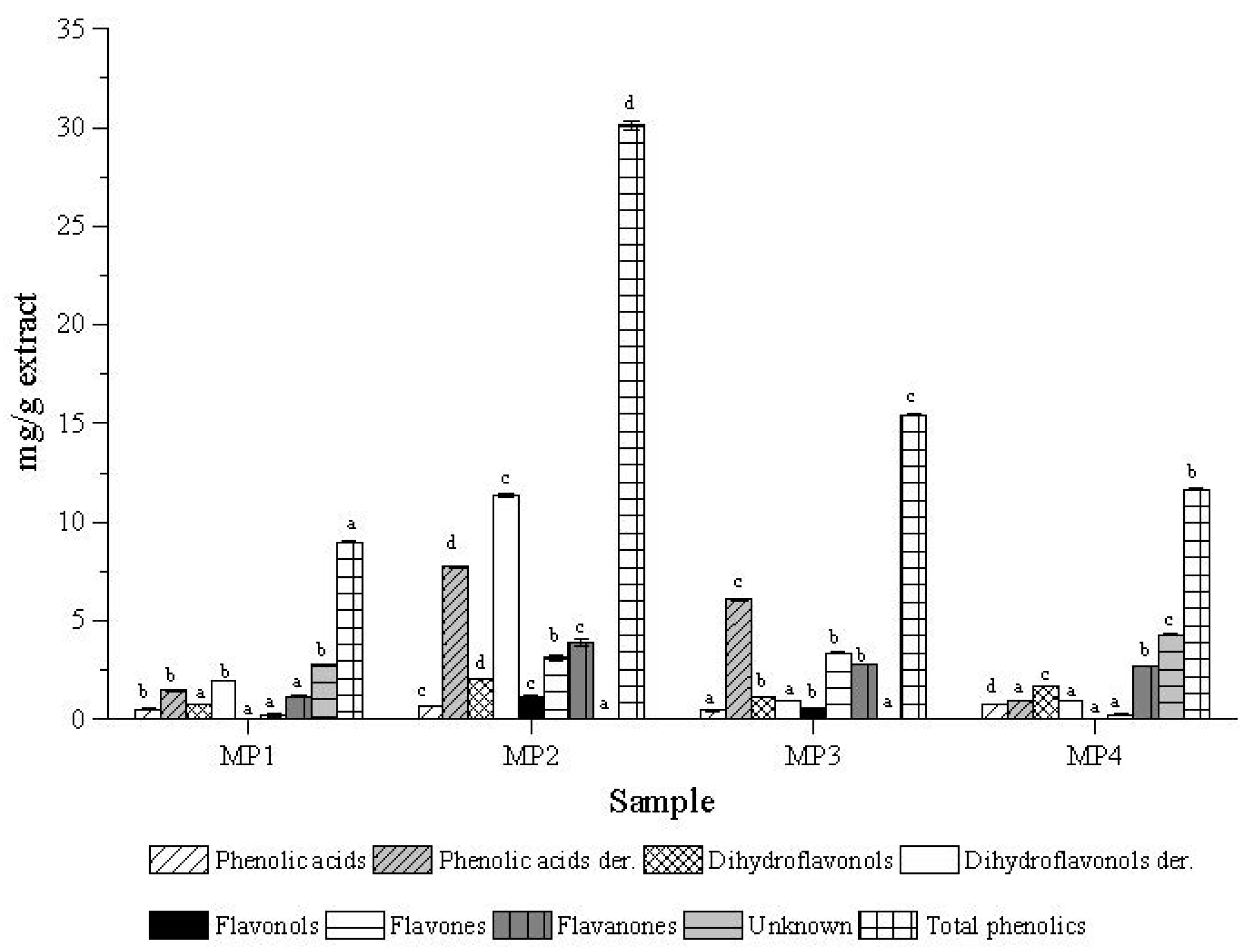
3.1. Moroccan Propolis Chemical Characterization

The chemical complex nature of propolis is linked to the resinous materials gathered by honey bees from different floral sources available around the hive, which have a direct impact on the quality and bioactivity of the propolis. Liquid chromatography coupled with mass spectrometry is a powerful tool that can be used to overcome the difficult task of propolis chemical profiling [6].
The phenolic composition of the four propolis samples from Morocco was evaluated by LC/DAD/ESI-MSn, with the identification of twenty-six phenolic compounds. Negative ion mode was used in this study because of its higher analytical sensitivity for different polyphenol classes [12]. Caffeic acid, p-coumaric acid, pinobanksin, pinocembrin, chrysin, and pinobanksin-3-O-acetate (Table 1) were present in all of the samples, and most likely arose from the common source of the resin, the poplar tree. However, other species like Ulmus, Pinus, Quercus, Salix e Acacia, can be also adequate sources for temperate propolis, particularly when poplars are unavailable [5]. Some compounds were specific to only one sample, such as sterubin, 3-prenyl-p-coumaric acid, dihydrokampferide, capillartimisin A, and isosakuranetin found in sample MP4 from Immouzzer Mermoucha; pinobanksin-5-methyl-ether (3, m/z 285) present in sample MP1 from Boulemane; apigenin, caffeic acid pentyl ester, pinobanksin-3-O-propionate, pinobanksin-3-O-butyrate, and pinobanksin-3-O-pentanoate observed in MP2 from Sefrou; and benzoyl hydroxyphenyl acetic acid only present in MP3 from Moulay Yaâcoub. Phenolic acid derivatives such as caffeic acid isoprenyl ester, caffeic acid benzyl ester, and caffeic acid phenylethyl ester (CAPE) were observed in samples MP1, MP2, and MP3, while flavonols such as kaempferol-methyl-ether, galangin and 6-methoxychrysin were characteristic of MP2 and MP3 samples (Table 1). Besides the compounds mentioned above, the composition of samples MP1 and MP4 included a chemical structure with m/z 377, which was not possible to identify, and therefore described as unknown (Table 1). Overall, the phenolic composition of the Moroccan propolis resembles other studies from the group [20], with a resin rich in flavonoid aglycones and phenolic acid esters with similarities to the poplar propolis.
Figure 1 displays the quantification analysis for the different samples. There is an evident difference between samples MP2 (30 mg/g of total phenolics) and MP3 (15mg/g of total phenolics), with MP2 containing twice the amount of phenolics. Samples MP1 and MP4 presented the poorest phenolic profile, with only twelve compounds identified and a total phenolic content of 9 and 12 mg/g extract, respectively.
In respect to the individual classes, dihydroflavonol derivatives, amongst them pinobanksin-3-O-acetate, were the main compounds present in these Moroccan propolis samples, especially in sample MP2. This was followed by the phenolic acids derivatives, where the phenolic acids esters emerge as the major compounds (Figure 1). Flavonols, such as kaempferol-methyl-ether and galangin were found as minor compounds, present only in samples MP2 and MP3 (Table 1).

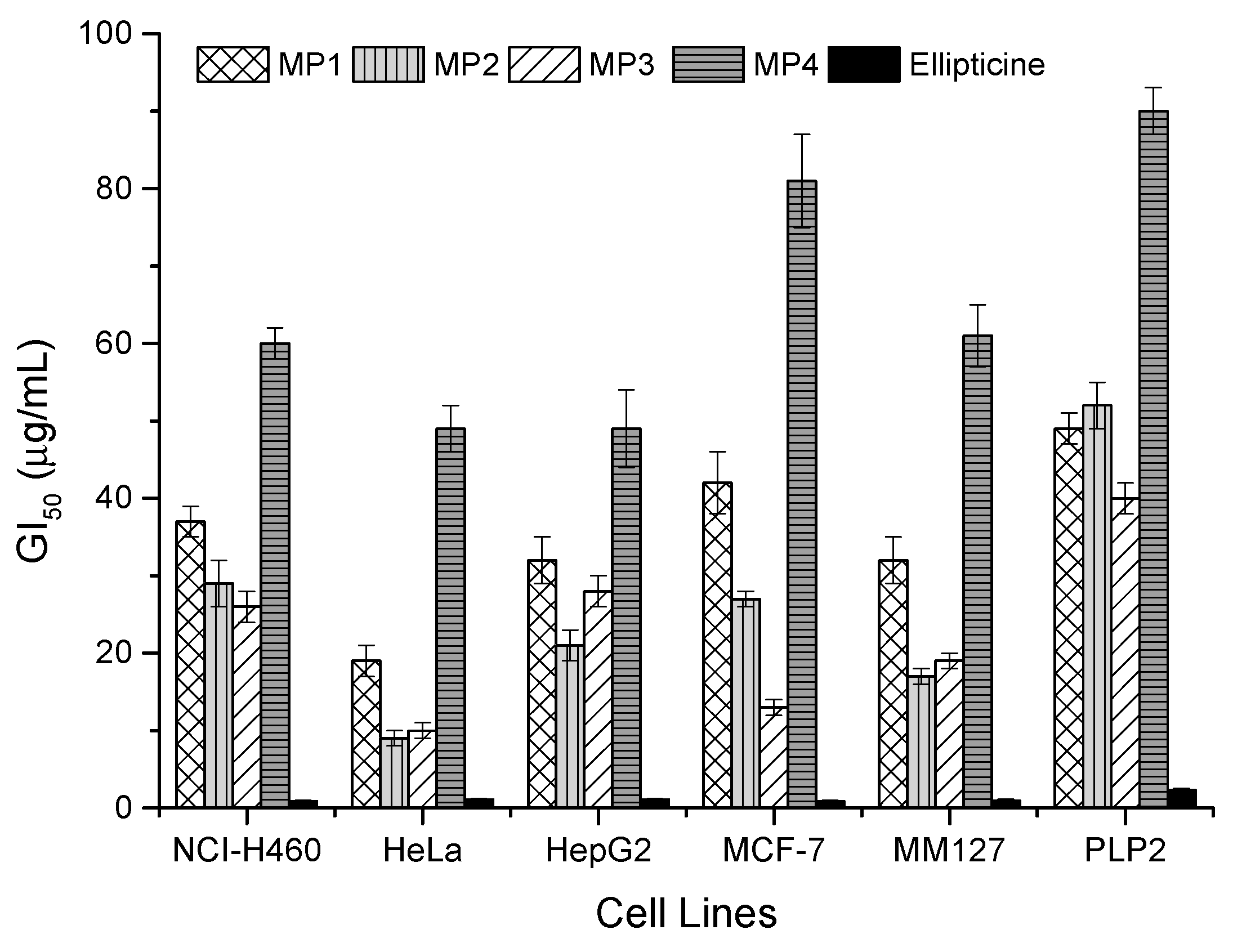
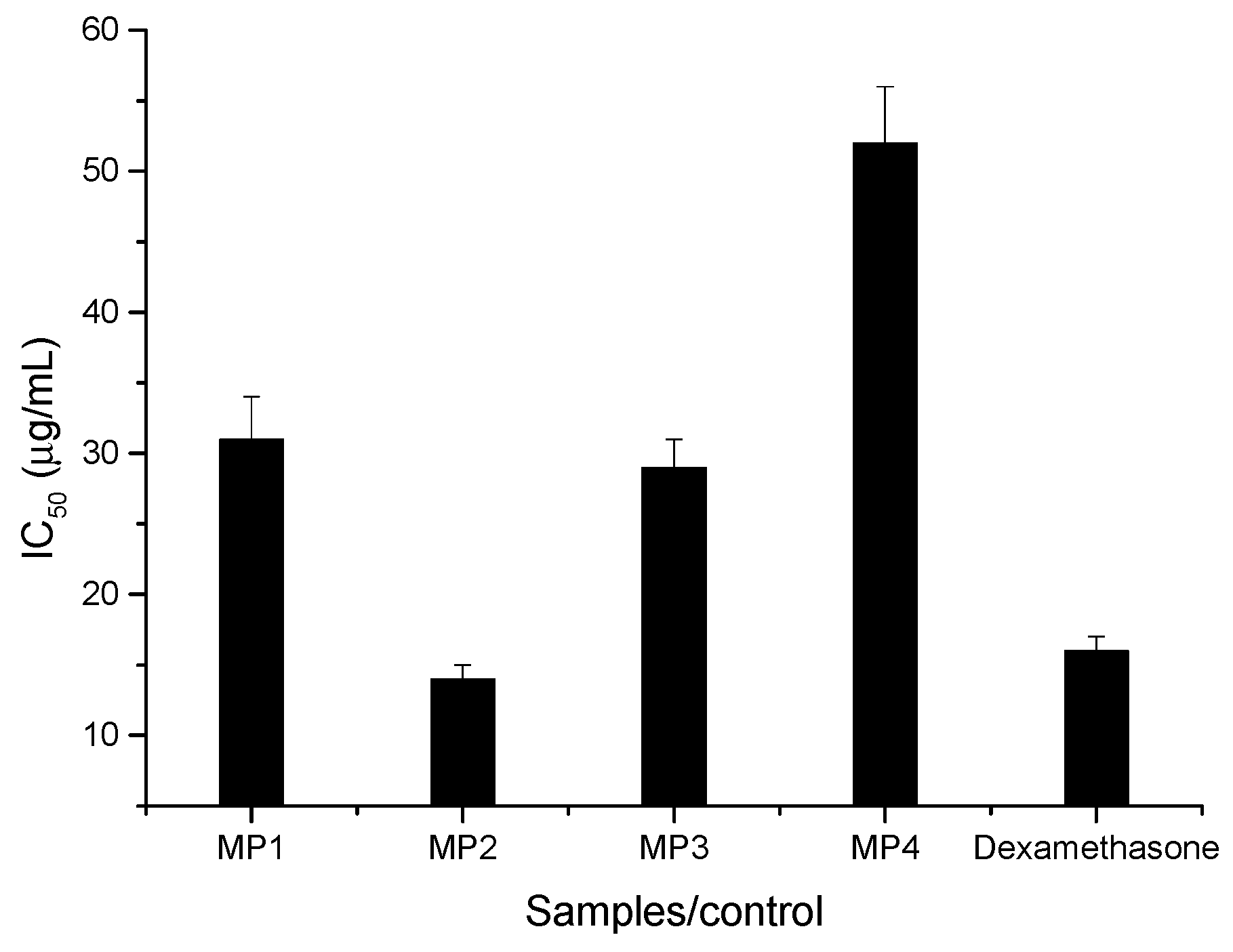
3.2. Cytotoxicity and Anti-Inflammatory Activity

The chemical compounds presented in the resin, which bees collect from plants to prepare propolis, are, in general, powerful bioactive metabolites used by plants for protection against pathogens, and so, are used by bees to immunize the hive environment [13]. Therefore, they may also function well as a relevant source of bioactive substances for pharmaceutical purposes. With this in mind, the cytotoxicity of the Moroccan propolis phenolic extracts was tested in five human tumor cell lines, including MCF-7 (breast adenocarcinoma), NCI-H460 (non-small cell lung cancer), HeLa (cervical carcinoma), HepG2 (hepatocellular carcinoma), and MM127 (human malignant melanoma) as well as in a porcine liver primary cell culture (PLP2), established by us. Also, the anti-inflammatory activity was evaluated using the murine macrophage (RAW 264.7) cell line. The growth inhibition of the tumor cells was observed in all the experiments, but particularly on sample MP2, which had the lowest cytotoxicity GI50 values in three of the five cell lines (Figure 2), followed by MP3 with the best performance for breast adenocarcinoma and lung cancer cells.
Overall, growth inhibition GI50 results were lower compared to the ones reported by us in Portuguese propolis [25]. The activity against HeLa cell line is particularly interesting for samples MP2 and MP3, with GI50 values of 9 μg/mL and 10 μg/mL, respectively (Figure 2), which is not as far from the activity of the synthetic standard elliptimicine as we would expect for a mixture of natural substances. On the opposite side is the sample MP4 from Immouzzer, with the weakest performance against all the tumor cell lines tested. Although it shows a richer composition in total phenolic compounds than MP1, the poor performance can be explained by the low concentration of specific bioactive compounds, such as those derived from the phenolic acids. Regardless of the high cytotoxicity exhibited by the Moroccan propolis against the studied tumor cell lines, all the extracts displayed some toxicity for non-tumor (normal) liver primary culture (PLP2). However, the values obtained for the tumor cells were always below the ones for non-tumor cells, highlighted in the results obtained for HeLa, MCF-7 and MM127, especially in the case of samples MP2 and MP3 (Figure 2).
The reported bioactivity is most probably correlated with the phenolic composition of propolis. Indeed, MP2 and MP3 as the samples with higher phenolic content were the ones with higher cytotoxicity. Several mechanisms of action are described for the interaction of phenolic compounds with tumor cells, including induction of apoptosis, cell-cycle arrest, inhibition of matrix metalloproteinases, anti-angiogenesis effect, and prevention of metastasis and invasion [2,10,13,14].
From Table 2 it is possible to retrieve additional information with respect to the correlation between the bioactivity against the different cell lines, and the phenolic classes. Without any doubt, the phenolic acid derivatives are the most active class, exhibiting a negative correlation (higher cytotoxicity) for all the cell lines, and particularly against HeLa and MM127.
This class of compounds, which includes phenolics such as caffeic acid phenylethyl ester (CAPE), observed in higher amounts in samples MP2 and MP3, is reported to play a key role in the bioactivity of propolis extracts, including against rat glioma C6, human squamocosal carcinoma, breast murine B16F10, human breast adenocarcinoma, and different leukemia cells [26]. The cytotoxicity of flavones seems to be more selective, with greater correlation to lung and hepatocellular carcinoma (Table 2). Indeed, compounds such as chrysin, found in higher amounts in propolis, have been shown to act against Murine B16F10, human colon carcinoma HCT-15 and human hepatoma Hep-3B cells [14].
One of the classes most represented in the Moroccan propolis samples under study, the dihydroflavonols derivatives, seems to correlate only with cervical carcinoma HeLa and malignant melanoma, MM127. These findings corroborate previous studies on Moroccan propolis extracts, which also observed an anti-proliferative effect against other skin tumor cells such as P815 (murine mastocytoma), in addition to effects against BSR (hamster renal adenocarcinoma) and Hep-2 (Human laryngeal carcinoma) [15]. Recently, dihydroflavonols such as pinobanksin and its derivatives, for example pinobanksin-3-O-acetate, were described to exert an anti-proliferative effect, induced through apoptosis in a B-cell lymphoma cancer cell line [27]. Furthermore, it was reported that the anti-metastatic effectiveness of the entire propolis extract was higher than that presented by its individual constituents, resulting in a synergistic effect between constituents [13]. Overall, these compounds are described to target various genetic and biochemical pathways of cancer progression like induction of apoptosis, cell-cycle arrest, inhibition of matrix metalloproteinases, anti-angiogenesis effect, and prevention of metastasis and invasion [10,14,26].
The anti-inflammatory effect, whose results are summarized in Figure 3, was assessed using murine macrophage-like RAW 264.7 cells and quantified through the nitric oxide (NO) production.
All propolis extracts under study showed anti-inflammatory capacity, with IC50 values between 14 and 52 µg/mL. The highest activity was observed for sample MP2, which contains a higher quantity of bioactive compounds such as phenolic acid derivatives and flavones, followed by the sample MP3, with an IC50 value of 29 µg/mL. It is worth mentioning the great performance of sample MP2, which displayed an even higher anti-inflammatory activity than the positive control dexamethasone. Compounds such as phenolic acid derivatives, flavonols, and dihydroflavonols derivatives, seem to be the major responsible substances, as considering their significant negative correlation with anti-inflammatory activity (Table 2). This also may explain the fact that, despite the low concentration of phenolic compounds in sample MP1, its anti-inflammatory activity was similar to that of sample MP3, which can be attributed to the higher concentration of dyhidroflavonols derivatives in the chemical composition of sample MP1 as compared to sample MP3 (Figure 1).

4. Conclusions

Four samples of Moroccan propolis from the Fez region were evaluated to determine their phenolic composition, cytotoxicity against tumor cell lines, and anti-inflammatory activity. The data presented here indicates that Moroccan propolis is composed of a complex phenolic composition, with 26 phenolic compounds identified. According to the cytotoxicity analysis, Moroccan propolis presents high toxicity against tumor cell lines, in particular HeLa (cervical carcinoma) and MM127 (human malignant melanoma). Compounds such as phenolic acid derivatives and flavonols seem to be the phenolic classes that contribute most to the cytotoxic effect. In some cases, the closeness of the in vitro doses for tumor and normal cell lines should be taken into consideration and confirmed by in vivo tests. Additionally, the propolis extracts displayed high anti-inflammatory activity, which was evaluated using murine macrophage-like RAW 264.7 cells, and the activity explained through the presence of phenolic acid and dihydroflavonols derivatives. Further studies concerning the isolation of major phenolic compounds and its biological properties against different tumor and non-tumor cell lines should be investigated. Also, phenolic compounds are described to have different mechanisms of action against tumor cells—they can target human cellular receptors, enzymes, transcription factors, and DNA. Future work with these samples will complement this study and develop further understandings of the mechanism of action of phenolic compounds in anticancer treatment. Moreover, propolis action should be compared to antitumor drugs, or even be tested in association with them, to investigate a possible synergistic action. These results support the hypothesis that specific phenolic compounds contribute greatly to the bioactive properties of propolis.

Author Contributions

S.I.F., B.L., I.C.F.R.F., M.V.-B. conceived and designed the experimental; S.I.F., R.C.C performed experiments; S.I.F., R.C.C, M.V.-B analyzed and interpreted the data; S.T., B.L., I.C.F.R.F., M.V.-B drafted and revised manuscript. All authors have read and approved the final manuscript.

Funding

The authors are grateful to the Foundation for Science and Technology (FCT, Portugal) and FEDER under Program PT2020 for financial support to CIMO (UID/AGR/00690/2013).

Acknowledgments

S.I.F. and R.C.C. thanks National funding by FCT—Foundation for Science and Technology, P.I., through the institutional scientific employment program-contract.

Conflicts of Interest

The authors declare no conflicts of interest.

References

  1. Carocho, M.; Ferreira, I. The Role of Phenolic Compounds in the Fight against Cancer—A Review. Anticancer Agents Med. Chem. 2013, 13, 1236–1258. [Google Scholar] [CrossRef]
  2. Watanabe, M.A.E.; Amarante, M.K.; Conti, B.J.; Sforcin, J.M. Cytotoxic constituents of propolis inducing anticancer effects: A review. J. Pharm. Pharmacol. 2011, 63, 1378–1386. [Google Scholar] [CrossRef]
  3. Parhi, P.; Mohanty, C.; Sahoo, S.K. Nanotechnology-based combinational drug delivery: An emerging approach for cancer therapy. Drug Dis. Today 2012, 17, 1044–1052. [Google Scholar] [CrossRef]
  4. Karikas, G.A. Anticancer and chemopreventing natural products: Some biochemical and therapeutic aspects. J. BUON 2010, 15, 627–638. [Google Scholar]
  5. Salatino, A.; Fernandes-Silva, C.C.; Righi, A.A.; Salatino, M.L.F. Propolis research and the chemistry of plant products. Nat. Prod. Rep. 2011, 28, 925–936. [Google Scholar] [CrossRef]
  6. Bankova, V.; Bertelli, D.; Borba, R.; Conti, B.J.; da Silva Cunha, I.B.; Danert, C.; Eberlin, M.N.; Falcão, S.I.; Isla, M.I.; Moreno, M.I.N.; et al. Standard methods for Apis mellifera propolis research. J. Apic. Res. 2016, 8839, 1–49. [Google Scholar] [CrossRef]
  7. Bankova, V.; Popova, M.; Trusheva, B. Phytochemistry The phytochemistry of the honeybee. Phytochemistry 2018, 155, 1–11. [Google Scholar] [CrossRef]
  8. Falcão, S.I.; Tomás, A.; Vale, N.; Gomes, P.; Freire, C.; Vilas-Boas, M. Phenolic quantification and botanical origin of Portuguese propolis. Ind. Crops Prod. 2013, 49, 805–812. [Google Scholar] [CrossRef] [Green Version]
  9. Burdock, G.A. Review of the Biological Properties and Toxicity of Bee Propolis (Propolis). Food Chem. Toxicol. 1998, 36, 347–363. [Google Scholar] [CrossRef]
  10. Sforcin, J.M. Biological Properties and Therapeutic Applications of Propolis. Phytother. Res. 2016, 905, 894–905. [Google Scholar] [CrossRef]
  11. Falcão, S.I.; Vilas-Boas, M.; Estevinho, L.M.; Barros, C.; Domingues, M.R.M.; Cardoso, S.M. Phenolic characterization of Northeast Portuguese propolis: Usual and unusual compounds. Anal. Bioanal. Chem. 2010, 396, 887–897. [Google Scholar] [CrossRef]
  12. Falcão, S.I.; Vale, N.; Gomes, P.; Domingues, M.R.M.; Freire, C.; Cardoso, S.M.; Vilas-Boas, M. Phenolic profiling of Portuguese propolis by LC-MS spectrometry: Uncommon propolis rich in flavonoid glycosides. Phytochem. Anal. 2013, 24, 309–318. [Google Scholar] [CrossRef]
  13. Maurício, J.; Bankova, V. Propolis: Is there a potential for the development of new drugs? J. Ethnopharmacol. 2011, 133, 253–260. [Google Scholar]
  14. Patel, S. Emerging Adjuvant Therapy for Cancer: Propolis and its Constituents. J. Diet. Suppl. 2015, 13, 1–24. [Google Scholar] [CrossRef]
  15. Mouse, H.A.; Tilaoui, M.; Jaafari, A.; Aboufatima, R.; Zyad, A. Anticancer properties of Moroccan propolis extracts. Rev. Bras. Farmacogn. 2012, 22, 558–567. [Google Scholar] [CrossRef]
  16. Touzani, S.; Al-waili, N.; Menyiy, N.E.; Filipic, B.; Pereyra, A.; Arabi, I.E.L.; Al-waili, W.; Lyoussi, B. Chemical analysis and antioxidant content of various propolis samples collected from different regions and their impact on antimicrobial activities. Asian Pac. J. Trop. Med. 2018, 11, 436–442. [Google Scholar]
  17. Miguel, G.; Doughmi, O.; Aazza, S.; Antunes, D.; Lyoussi, B. Antioxidant, Anti-inflammatory and Acetylcholinesterase Inhibitory Activities of Propolis from Different Regions of Morocco. Food Sci. Biotechnol. 2014, 23, 313–322. [Google Scholar] [CrossRef]
  18. Alonso-esteban, J.I.; Pinela, J.; Barros, L.; Ana, Ć.; Sokovi, M.; Calhelha, R.C.; Torija-isasa, E.; Sánchez-mata, M.D.C.; Ferreira, I.C.F.R. Phenolic composition and antioxidant, antimicrobial and cytotoxic properties of hop (Humulus lupulus L.) Seeds. Ind. Crops Prod. 2019, 134, 154–159. [Google Scholar] [CrossRef]
  19. Mandim, F.; Barros, L.; Calhelha, R.C.; Abreu, R.M.V.; Pinela, J.; Alves, M.J.; Heleno, S.; Santos, P.F.; Ferreira, I.C.F.R. (Calluna vulgaris L.) Hull: Chemical characterization, evaluation of its bioactive properties and effect on the vaginal microbiota. Food Funct. 2019, 10, 78–89. [Google Scholar] [CrossRef]
  20. Popova, M.; Smail, A.; Bankova, V. Plant sources of propolis: An update from a chemist’s point of view. Nat Prod Commun. 2015, 28–29. [Google Scholar]
  21. Yang, J.; Liang, Q.; Wang, M.; Je, C.; Smithson, D.; Tu, Y.; Boulos, N.; Jacob, M.R.; Shelat, A.A.; Wu, Y.; et al. UPLC-MS-ELSD-PDA as a Powerful Dereplication Tool to Facilitate Compound Identi fi cation from Small-Molecule Natural Product Libraries. J. Nat. Prod. 2014, 77, 902–909. [Google Scholar] [CrossRef]
  22. Coelho, J.; Falcão, S.I.; Vale, N.; Almeida-Muradian, L.B.; Vilas-Boas, M. Phenolic composition and antioxidant activity assessment of southeastern and south Brazilian propolis. J. Apic. Res. 2017, 56, 21–31. [Google Scholar] [CrossRef] [Green Version]
  23. Saleh, K.; Zhang, T.; Fearnley, J.; Watson, D.G. A Comparison of the Constituents of Propolis from Different Regions of the United Kingdom by Liquid Chromatography-high Resolution MassSpectrometry Using a Metabolomics Approach A Comparison of the Constituents of Propolis from Different Regions of the Un. Curr. Metabol. 2015, 3, 42–53. [Google Scholar] [CrossRef]
  24. Andrade, A.; Finger, D.; Schinieder, C.; Morgado, E.; Marçal, P.; Paula, A.; Nunes, N.; Maria, T.; Morais, F.; Goretti, M.; et al. In vivo antitumoural activity and composition of an oil extract of Brazilian propolis. Food Chem. 2011, 126, 1239–1245. [Google Scholar]
  25. Calhelha, R.C.; Falcão, S.; Queiroz, M.J.R.P.; Vilas-Boas, M.; Ferreira, I.C.F.R. Cytotoxicity of portuguese propolis: The proximity of the in vitro doses for tumor and normal cell lines. BioMed Res. Int. 2014, 2014, 7. [Google Scholar] [CrossRef]
  26. Premratanachai, P.; Chanchao, C. R eview of the anticancer activities of bee products. Asian Pac. J. Trop. Biomed. 2014, 4, 337–344. [Google Scholar] [CrossRef]
  27. Alday, E.; Valencia, D.; Carreño, A.L.; Picerno, P.; Lisa, A.; Rastrelli, L.; Robles-zepeda, R.; Hernandez, J. Apoptotic induction by pinobanksin and some of its ester derivatives from Sonoran propolis in a B-cell lymphoma cell line. Chem. Biol. Interact. 2015, 242, 35–44. [Google Scholar] [CrossRef]
Figure 1. Principal phenolic classes presented in the composition of propolis from different Morocco origins.
Biomolecules 09 00315 g001
Figure 2. Cytotoxic activity of propolis samples on cell lines expressed in GI50 values (µg/mL) (mean ± SD, n = 2). GI50 values correspond to the extract concentration achieving 50% of growth inhibition in human tumor cell lines or in liver primary culture PLP2.
Biomolecules 09 00315 g002
Figure 3. Anti-inflammatory activity of propolis samples expressed in IC50 values (µg/mL) of NO production inhibition (mean ± SD, n = 2). IC50 values correspond to the extract concentration achieving 50% of the inhibition of the NO production.
Biomolecules 09 00315 g003
Table 1. Phenolic compounds in Moroccan propolis identified and quantified by LC/DAD/ESI-MSn (each value is the mean ± standard deviation, n = 2).
PeakProposed CompoundRT (min)λmax
(nm)
[M − H]
m/z
MSn
(% Base Peak)
MP1
mg/g Extract
MP2
mg/g Extract
MP3
mg/g Extract
MP4
mg/g Extract
1Caffeic acid a,b10.9322179MS2 [179]: 1350.31 ± 0.000.50 ± 0.000.31 ± 0.000.30 ± 0.00
2p-Coumaric acid a,b15.8310163MS2 [163]: 1190.22 ± 0.000.21 ± 0.000.16 ± 0.000.50 ± 0.00
3Pinobanksin-5-methyl-ether b,c36.9286285MS2 [285]: 267 (100), 253 (13), 239 (27)0.77 ± 0.01
4Sterubin b,d47.3284301MS2 [301]: 286 (100), 165 (8); MS3 [286]: 258 (37), 195 (17), 165 (100) 0.83 ± 0.01
5Pinobanksin b,c47.3292271MS2 [271]: 253 (100), 225 (26), 151 (10)0.83 ± 0.002.04 ± 0.001.14 ± 0.030.83 ± 0.01
6Dihydrokaempferide b,e50.4291301MS2 [301]: 283 (100), 151 (22); MS3 [283]: 268 (100), 255 (40), 227 (41) 0.86 ± 0.01
7Apigenin a,b54.9268, 337269MS2 [269]: 225 (100), 151 (29) 0.27 ± 0.00
8Kaempferol-methyl ether b,c57.7265, 352299MS2 [299]: 284 0.15 ± 0.010.26 ± 0.00
93-prenyl-p-coumaric acid b,e63.6315231MS2 [231]: 187; MS3 [187]: 132 0.71 ± 0.00
10Caffeic acid isoprenyl ester a,b65.2325247MS2 [247]: 179 (100), 135 (13)0.34 ± 0.001.73 ± 0.011.40 ± 0.00
11Caffeic acid isoprenyl ester b,c66.3325247MS2 [247]: 179 (100), 135 (13)0.35 ± 0.013.00 ± 0.012.59 ± 0.00
12Capillartimisin A b,e66.4309315MS2 [315]: 285 (60), 271 (100), 241 (67); MS3 [271]: 253 (41), 241 (100) 0.21 ± 0.00
13Caffeic acid benzyl ester b,c66.7325269MS2 [269]: 178 (100), 161 (12), 134 (32)0.43 ± 0.011.18 ± 0.030.90 ± 0.02
14Pinocembrin a,b67.6289255MS2 [255]: 213 (100), 211 (32), 151 (48)1.17 ± 0.043.91 ± 0.152.80 ± 0.031.81 ± 0.00
15Isosakuranetin b,f,g68.1292285MS2 [285]: 270 (100), 243 (25), 164 (17), 151 (4) MS3 [270]: 242 (41), 165 (100), 164 (70) 0.92 ± 0.00
16Benzoyl hydroxyphenyl acetic acid b,f68.4280257MS2 [257]: 213; MS3 [213]: 169 (100), 122 (49) 0.61 ± 0.02
17Chrysin a,b69.6268, 313253MS2 [253]: 225 (17), 209 (100), 151 (5)0.25 ± 0.081.99 ± 0.142.88 ± 0.010.27 ± 0.03
18Pinobanksin-3-O-acetate b,c69.6292313MS2 [313]: 271 (20), 253 (100)1.22 ± 0.026.82 ± 0.061.01 ± 0.000.96 ± 0.02
19Caffeic acid phenylethyl ester a,b69.9325283MS2 [283]: 179 (100), 135 (22)0.37 ± 0.021.24 ± 0.030.57 ± 0.00
20Galangin a,b70.3265, 300sh, 358269MS2 [269]: 269 (100), 241 (61), 227 (20), 197 (22), 151 (20) 1.04 ± 0.020.35 ± 0.00
21Caffeic acid pentyl ester b71.1325249MS2 [249]: 179 (100), 161 (47), 135 (32) 0.62 ± 0.00
226-Methoxychrysin b,c72.3265, 300sh, 350sh283MS2 [283]: 269 0.88 ± 0.050.48 ± 0.04
23Pinobanksin-3-O-propionate b,c75.2289327MS2 [327]: 271 (9), 253 (100) 1.08 ± 0.01
24Unknown79.5239377MS2 [377]: 359 (100), 331 (84), 313 (8)2.76 ± 0.04 4.31 ± 0.03
25Pinobanksin-3-O-butyrate or isobutyrate b,c79.9292341MS2 [341]: 271 (2), 253 (100) 2.66 ± 0.01
26Pinobanksin-3-O-pentanoate or 2-methylbutyrate b,c84.1292355MS2 [355]: 271 (3), 253 (100) 0.80 ± 0.00
a Confirmed with standard; b Confirmed with MSn fragmentation; c Confirmed with references: [12]; d [21]; e [22]; f [23]; g [24].
Table 2. Spearman’s correlations between the propolis phenolic composition (mg/g of extract) and cytotoxicity and anti-inflammatory activity GI50/IC50 values (μg/mL).
NCI-H460HeLaHepG2MCF-7MM127PLP2RAW264.7
Phenolic acids0.786 a0.3810.786 a0.4520.3570.881 a0.357
Phenolic acid derivatives−0.810 a−0.976 b−0.762 a−0.833 a−0.952 b−0.500−0.905 b
Dihydroflavonols0.024−0.3810.024−0.310−0.3570.429−0.381
Dihydroflavonols derivatives−0.405−0.810 a−0.357−0.619−0.738 a−0.310−0.857 b
Flavonols−0.710 a−0.913 b−0.710 a−0.862 b−0.888 b−0.317−0.875 b
Flavones−0.881 b−0.619−0.881 b−0.690−0.690−0.548−0.500
Flavanones−0.619−0.786 a−0.571−0.643−0.762 a−0.167−0.667
Total phenolics−0.595−0.762 a−0.548−0.667−0.786 a−0.143−0.643
Spearman’s correlation significance levels: a Correlation is significant at p < 0.05; b Correlation is significant at p < 0.01.

