Article

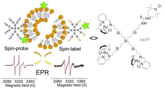
Exploring the Interactions of Ruthenium (II) Carbosilane Metallodendrimers and Precursors with Model Cell Membranes through a Dual Spin-Label Spin-Probe Technique Using EPR

by
Riccardo Carloni
1,†,
Natalia Sanz del Olmo
2,3,4,†,
Paula Ortega
2,3,4,
Alberto Fattori
1,
Rafael Gómez
2,3,4,
Maria Francesca Ottaviani
1,
Sandra García-Gallego
2,3,4,
Michela Cangiotti
1,* and
F. Javier de la Mata
2,3,4,*
1
Department of Pure and Applied Sciences, University of Urbino “Carlo Bo”, 61029 Urbino, Italy
2
Department of Organic and Inorganic Chemistry, and Research Institute in Chemistry “Andrés M. del Río” (IQAR), University of Alcalá, 28805 Madrid, Spain
3
Networking Research Center on Bioengineering, Biomaterials and Nanomedicine (CIBER-BBN), 088034 Barcelona, Spain
4
Institute Ramón y Cajal for Health Research (IRYCIS), 28034 Madrid, Spain
*
Authors to whom correspondence should be addressed.
Both authors contributed equally to this work.
Biomolecules 2019, 9(10), 540; https://doi.org/10.3390/biom9100540
Submission received: 7 August 2019 / Revised: 17 September 2019 / Accepted: 23 September 2019 / Published: 27 September 2019

Abstract

:
Dendrimers exhibit unique interactions with cell membranes, arising from their nanometric size and high surface area. To a great extent, these interactions define their biological activity and can be reported in situ by spin-labelling techniques. Schiff-base carbosilane ruthenium (II) metallodendrimers are promising antitumor agents with a mechanism of action yet to explore. In order to study their in situ interactions with model cell membranes occurring at a molecular level, namely cetyltrimethylammonium bromide micelles (CTAB) and lecithin liposomes (LEC), electron paramagnetic resonance (EPR) was selected. Both a spin probe, 4-(N,N-dimethyl-N-dodecyl)ammonium-2,2,6,6-tetramethylpiperidine-1-oxyl bromide (CAT12), able to enter the model membranes, and a spin label, 2,2,6,6-tetramethylpiperidine-1-oxyl (TEMPO) covalently attached at newly synthesized heterofunctional dendrimers, were used to provide complementary information on the dendrimer–membrane interactions. The computer-aided EPR analysis demonstrated a good agreement between the results obtained for the spin probe and spin label experiments. Both points of view suggested the partial insertion of the dendrimer surface groups into the surfactant aggregates, mainly CTAB micelles, and the occurrence of both polar and hydrophobic interactions, while dendrimer–LEC interactions involved more polar interactions between surface groups. We found out that subtle changes in the dendrimer structure greatly modified their interacting abilities and, subsequently, their anticancer activity.

Graphical Abstract

1. Introduction

Current anticancer therapies are poorly efficient because of the non-specific drug distribution, the development of multidrug resistance, and the intrinsic heterogeneity of cancer. In the cancer field, and in many other fields, nanotechnology provides new tools that increase the therapeutic efficiency and minimize side-effects. Nanotechnology provides multifunctional platforms to be used either as therapeutic agents, as drug delivery systems [1,2], or as contrast agents with longer circulation time for the early detection and diagnosis of diseases [3]. These multifunctional platforms may further be designed to simultaneously present two or more activities in a single molecule, the so-called nanotheranostics. Nanotheranostics are cutting-edge nanoparticles (NPs) designed for simultaneously providing accurate diagnosis together with effective therapeutic activity, monitoring drug release and distribution in real time, and have been already used in different fields including cancer [4,5] and neurological disorders [6]. The different nanotheranostic agents reported in the literature [7]—metal nanoparticles, polymeric nanoparticles, and dendrimers—are useful tools for optimizing treatment outcomes in cancer and other diseases.
In particular, dendrimers are excellent platforms for accurate heterofunctionalization. They are the flagship of precision nanoparticles, whose monodisperse scaffold enables a controlled design of the groups located at the periphery or within the interior [8]. They allow a precise structure-to-activity relationship, unattainable by other nanoparticles, exhibiting different biological activity depending on both their functional groups and the nature of their scaffold. As an example, carbosilane dendrimers rely on a hydrophobic scaffold, comprising multiple C–C and C–Si bonds, which provide extraordinary flexibility and stability and enhance the interaction with biological membranes. Many biomedical applications have been reported for this dendritic family, such as antiviral and antibacterial agents, non-viral vectors for nucleic acid delivery, and antitumor drugs, among others [9].
Dendrimers interact well with membranes and cells [10,11], and thus trigger multiple biological activities. The nanoparticle–cell interactions are modulated by both the physicochemical properties of the NPs and cell-specific parameters [12], and a thorough understanding of this interaction can provide valuable information for the design of effective drugs. Among the available tools for studying nanoparticle–cell interactions, electron paramagnetic resonance (EPR) spectroscopy has excelled in providing on-site structural and dynamical information. The spin-probe/spin-label-based EPR technique has already been demonstrated to be a powerful tool in characterizing the interactions of dendrimers with model membranes and cells [10,13,14,15,16,17,18,19,20,21,22,23,24], and has even been used in living systems [25]. Importantly, EPR spectroscopy shares many of the features of magnetic resonance imaging (MRI), including the underlying principles, and exhibits superior detection sensitivity. The recent developments in EPR instrumentation, probes, and methods offer opportunities in potential areas of clinical application [26], such as oximetry and dosimetry [27].
We have recently reported the use of Schiff-base containing carbosilane dendrimers as efficient chelators of copper (II) [28,29] and ruthenium (II) [30,31] complexes. The inclusion of metallodrugs in nanostructures improves their delivery and penetration—mainly by endocytosis—thus increasing the concentration in cells and subsequently the anticancer effect [32]. Accordingly, the carbosilane metallodendrimers exhibited promising antitumor activity in both in vitro and in vivo assays. Further insight into the antitumor activity of the Cu(II) metallodendrimers was obtained through the EPR analysis of the dendrimers’ interaction with model membranes [29]. We found out that the dendritic generation, as well as the metal counter-ion, tuned the strength of interaction of the dendrimer with the membrane and affected the toxicity and selectivity towards cancer cells.
With the aim of obtaining more information about the interactions occurring at a molecular level in the biological environment of Schiff-base carbosilane dendrimers and their Ru(II) complexes, we selected the EPR technique to study the site–site interactions between the dendrimers and model cell membranes, namely cetyltrimethylammonium bromide micelles (indicated as CTAB) and lecithin liposomes (indicated as LEC). To characterize these interactions in situ, both a spin probe, 4-(N,N-dimethyl-N-dodecyl)-ammonium-2,2,6,6-tetramethylpiperidine-1-oxyl bromide (CAT12) able to enter the model membranes, and a spin label, 2,2,6,6-tetramethylpiperidine-1-oxyl (TEMPO) covalently attached to the dendrimer surface, were used to provide complementary information on the dendrimer–model membrane interactions from both sides. A comparative EPR analysis was performed on the labelled dendrimers in the absence and presence of the model membranes. Relevant insight was obtained related to the influence of different parameters—generation, equilibration time, peripheral groups, and presence of heterofunctional ligands—on the nanoparticle–membrane interactions, which can explain their biological activity.

2. Materials and Methods

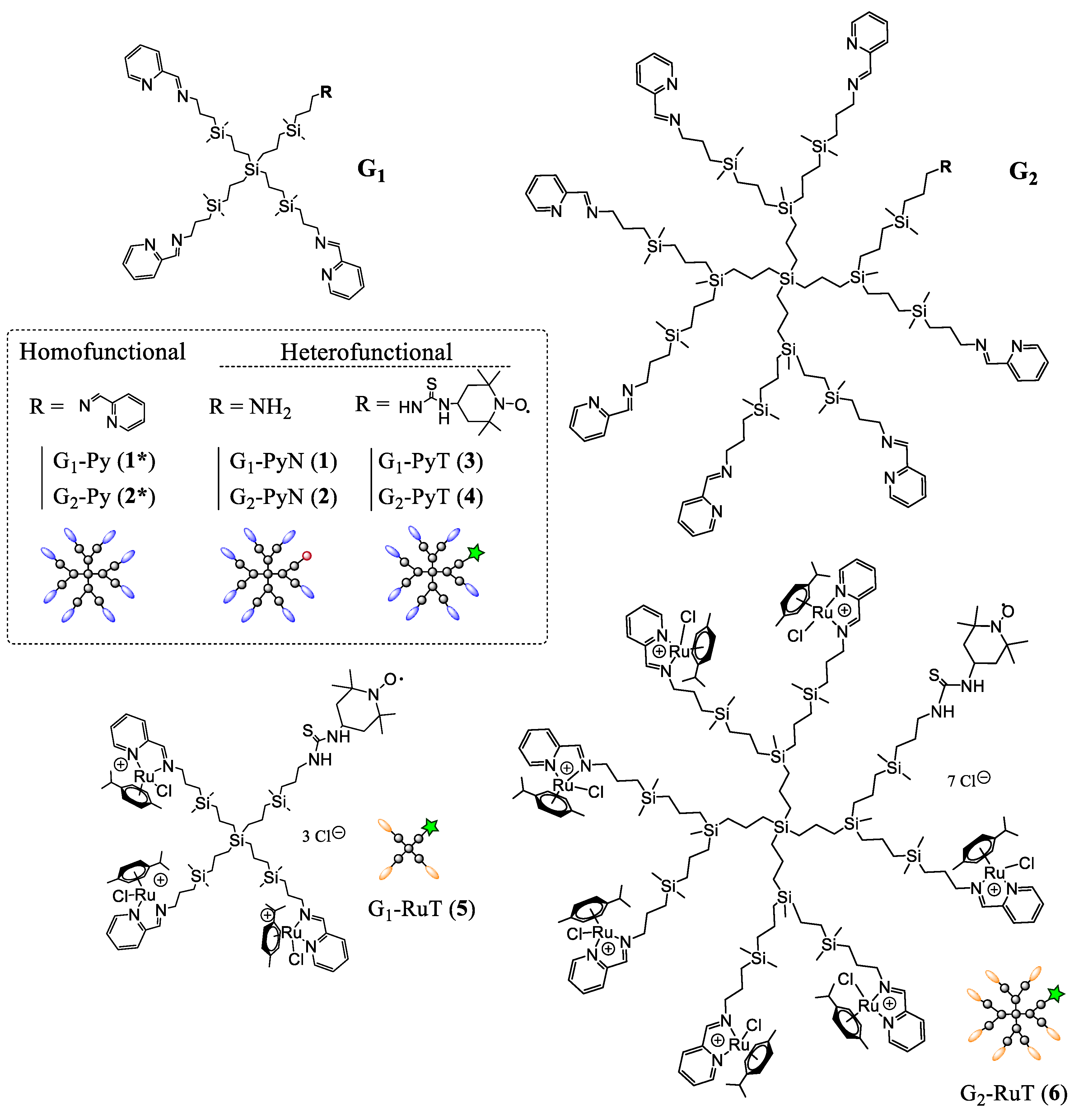
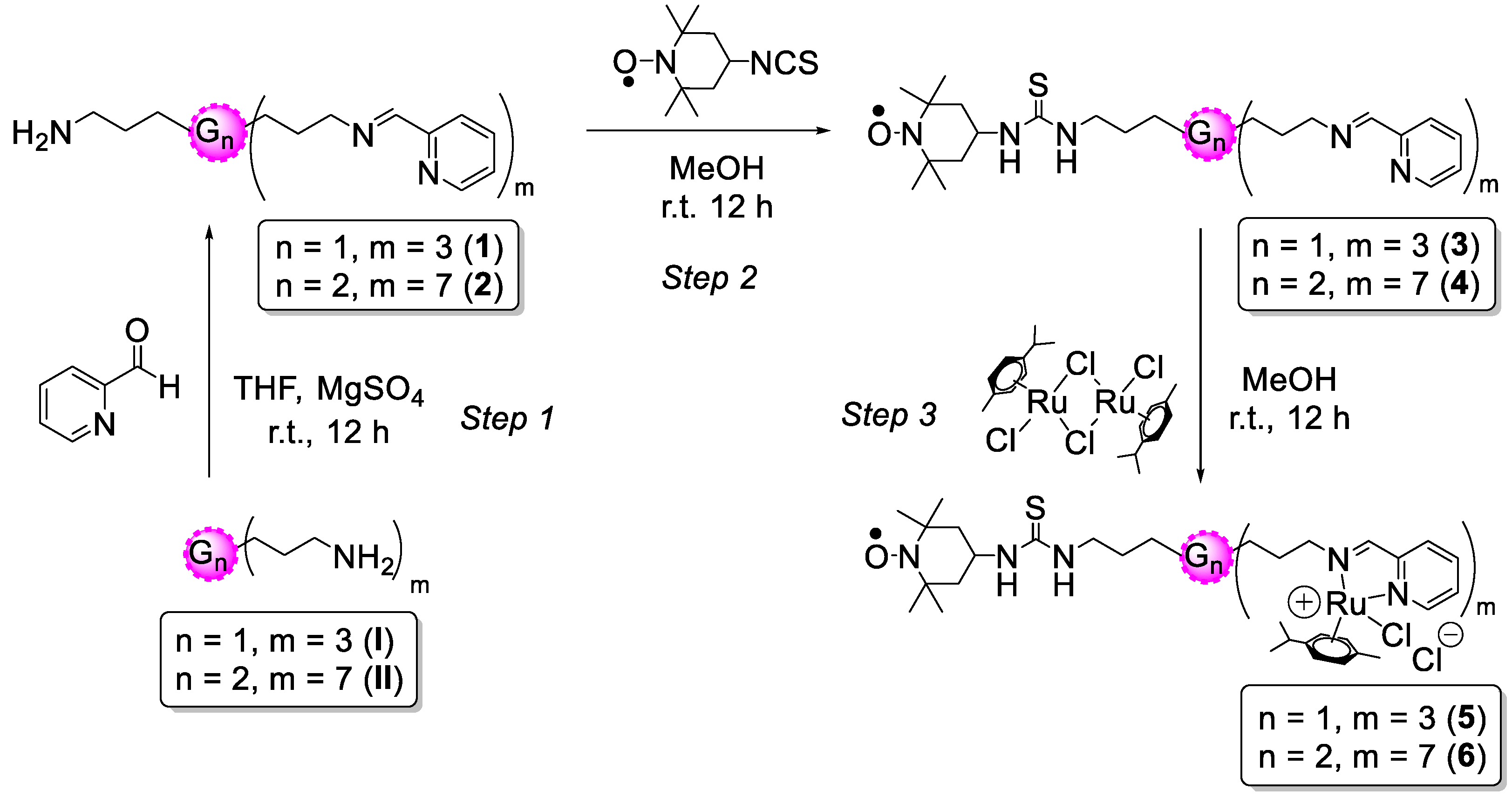
2.1. Dendrimers and Metallodendrimers

In order to evaluate the dendrimer–membrane interactions from the dendrimer point of view, novel labelled iminopyridine dendrimers were prepared (Figure 1). The three-step synthetic route included the preparation of the heterofunctional dendrimers Gn-{[NCPh(o-N)]m-1[NH2]} [17] (for simplicity Gn-PyN; n = 1, m = 4 (1); n = 2, m =8 (2)), Gn-{[NCPh(o-N)]m-1[NHC(S)NH-TEMPO]} (Gn-PyT, n = 1, m = 4 (3); n = 2, m = 8 (4)), and Gn-{[NCPh(o-N)Ru(η6-p-cymene)Cl2]m-1[NHC(S)NH-TEMPO]} (Gn-RuT; n = 1, m = 4 (5); n = 2, m = 8 (6)). The synthetic protocols and characterization details are described below for each dendrimer.
For comparison, iminopyridine homofunctional dendrimers Gn-{[NCPh(o-N)]m (Gn-Py; n = 1, m = 4 (1*); n = 2, m = 8 (2*) and the mononuclear counterpart n = 0, m = 1 (0*)) were used in EPR evaluation. Overall, these dendrimers provided information about the effect of dendritic generation (G1 vs. G2), the functional groups (Gn-Py vs. Gn-PyN), the labelling (Gn-Py vs. Gn-PyT) and the metal complexation (Gn-PyT vs. Gn-RuT), the latter known to enhance anticancer drug properties [32].
G1-{[NCPh(o-N)]3[NH2]} (G1-PyN, 1). To a solution of precursor G1-[NH2]4 (I) (313.1 mg, 0.47 mmol) in anhydrous tetrahydrofuran (THF), 2-pyridinecarboxaldehyde (152.1 mg, 1.42 mmol) was added. The mixture was stirred under inert atmosphere at room temperature in the presence of anhydrous MgSO4 for 12 h. Subsequently, the solution was filtered and the solvent was evaporated to obtain compound 1 as brown oil in quantitative yield. 1H-NMR (CDCl3): δ -0.06 (s, 24H, -(CH3)2Si-); 0.43 (m, 2H, (-CH2CH2CH2NH2); 0.51 (m, 24H, -SiCH2-); 1.29 (m, 8H, -SiCH2CH2CH2Si); 1.37 (m, 2H, (-SiCH2CH2CH2NH2); 1.68 (m, 6H, -SiCH2CH2CH2NCPh); 2.62 (m, 2H, (-CH2NH2); 3.62 (m, 6H,-CH2NCPh); 7.28 (m, 3H, Ar); 7.70 (m, 3H, Ar); 7.95 (m, 3H, Ar); 8.60 (m, 3H, Ar); 8.33 (s, 3H, -N=CHimine). 13C{1H}-NMR (CDCl3): δ -3.33 (-(CH3)2Si-); 1.00 (-(CH3)2SiCH2CH2CH2NH2); 12.1 (-SiCH2CH2CH2NH2); 13.1 (-SiCH2CH2CH2NCPh); 17.5, 18.5, 20.2 (-SiCH2CH2CH2Si-); 25.3 (-SiCH2CH2CH2NCPh); 28.3 (-SiCH2CH2CH2NH2); 45.5 (-CH2NH2); 52.9 (-CH2NCPh); 121.2, 124.5, 136.5, 149.3, 161.7 (CAr); 161.7 (-N=CHimine). Elemental analysis (%): calculated for C50H89N7Si5 (928.7 g/mol): C, 64.66; H, 9.66; N, 10.56; found: C, 64.30; H, 9.00; N, 10.48.
G2-{[NCPh(o-N)]7[NH2]} (G2-PyN, 2). Dendrimer G2-PyN was prepared through the same procedure as compound 1, using the following reagents: G2-[NH2]8 (II) (113.0 mg, 0.069 mmol), 2-pyridinecarboxaldehyde (51.73 mg, 0.483 mmol). Dendrimer 2 was isolated as brown oil in quantitative yield. 1H-NMR (CDCl3): δ -0.12 (s, 12H, -(CH3)Si(CH2CH2CH2Si)2); -0.06 (s, 48H, -(CH3)2Si-); 0.51 (m, 64H, -SiCH2-); 1.26 (m, 24H, -SiCH2CH2CH2Si- and -SiCH2CH2CH2NH2); 1.68 (m, 16H, (-SiCH2CH2CH2NCPh); 2.62 (m, 2H, (-CH2NH2); 3.63 (m, 14H,-CH2NCPh); 7.28 (m, 7H, Ar); 7.71 (m, 7H, Ar); 7.97 (m, 7H, Ar); 8.62 (m, 7H, Ar); 8.35 (s, 7H, -N=CHimine). Elemental analysis (%): calculated for C122H217N15Si13 (2259.3 g/mol): C, 64.86; H, 9.68, N, 9.30; found: C, 64.28; H, 8.51; N, 8.62.
G1-{[NCPh(o-N)]3[NHC(S)NH-TEMPO]} (G1-PyT, 3). To a solution of G1-PyN (1) (41.8 mg, 0.045 mmol) in methanol, the radical 4-isothiocyanate-TEMPO (7.7 mg, 0,036 mmol) was added. The mixture was stirred under inert atmosphere and protected from light at room temperature for 12 h. Subsequently, the solvent was evaporated to obtain dendrimer 3 as brown oil in quantitative yield. 1H-NMR (CD3OD): δ 0.00 (s, 24H, -(CH3)2Si-); 0.60 (m, 24H, -SiCH2-); 1.39 (m, 10H, -SiCH2CH2CH2Si- and -SiCH2CH2CH2NH-TEMPO); 1.74 (m, 6H, (-SiCH2CH2CH2NCPh); 3.68 (m, 6H,-CH2NCPh); 7.48 (m, 3H, Ar); 7.90 (m, 3H, Ar); 8.02 (m, 3H, Ar); 8.62 (m, 3H, Ar); 8.37 (s, 3H, -N=CHimine). Elemental analysis (%): calculated for C60H106N9OSSi5 (1142.1 g/mol): C, 63.10; H, 9.36; N, 11.04; S, 2.81; found: C, 61.46; H, 8.59; N, 10.00; S, 2.72.
G2-{[NCPh(o-N)]7[NHC(S)NH-TEMPO]} (G2-PyT, 4). Dendrimer G2-PyT was prepared through the same procedure as compound 3, using the following reagents: G2-PyN (2) (56.0 mg, 0.025 mmol), 4-isothiocyanate-TEMPO (4.2 mg, 0.020 mmol). Dendrimer 4 was isolated as brown oil in quantitative yield. 1H-NMR (CDCl3): δ -0.05 (s, 12H, -(CH3)Si(CH2CH2CH2Si)2); -0.02 (s, 48H, -(CH3)2Si-); 0.61 (m, 64H, -SiCH2-); 1.42 (m, 26H, -SiCH2CH2CH2Si- and -SiCH2CH2CH2NH-TEMPO); 1.75 (m, 14H, (-SiCH2CH2CH2NCPh); 3.68 (m, 14H,-CH2NCPh); 7.46 (m, 7H, Ar); 7.89 (m, 7H, Ar); 8.00 (m, 7H, Ar); 8.61 (m, 7H, Ar); 8.35 (s, 7H, -N=CHimine). Elemental analysis (%): calculated for C132H234N17OSSi13 (2472.6 g/mol): C, 64.12; H, 9.54; N, 9.63; S, 1.30; found: C, 62.99; H, 8.70; N, 8.62; S, 0.82.
G1-{[NCPh(o-N)Ru(η6-p-cymene)Cl2]3[NHC(S)NH-TEMPO]} (G1-RuT, 5). To a solution of [Ru(η6-p-cymene)Cl2]2 (59.8 mg, 0.052 mmol) in methanol, G1-PyT (3) (48.1 mg, 0.16 mmol) was added dropwise at 0 °C. The mixture was stirred under inert atmosphere at room temperature for 12 h and protected from light. Subsequently, the solvent was evaporated to obtain dendrimer 5 as brown oil in quantitative yield. 1H-NMR (CD3OD): δ 0.04 (s, 24H, -(CH3)2Si-); 0.64 (m, 24H, -SiCH2-); 1.03 (d, 9H, -(CH3)2CHcym); 1.15 (d, 9H, -(CH3)2CHcym); 1.39 (m, 10H, -SiCH2CH2CH2Si- and -SiCH2CH2CH2NH-TEMPO); 1.82 (m, 3H, (-SiCH2CH2CH2NCPh); 2.02 (m, 3H, (-SiCH2CH2CH2NCPh); 2.29 (s, 9H, -(CH3)cym); 2.69 (s, 3H, -(CH3)2CHcym); 4.27 (m, 3H,-CH2NCPh); 4.69 (m, 3H,-CH2NCPh); 5.84 (m, 7H, Arcym); 6.08 (m, 3H, Arcym); 6.19 (m, 3H, Arcym); 7.80 (m, 3H, Ar); 8.20 (m, 6H, Ar); 9.50 (s, 3H, Ar); 8.69 (s, 3H, -N=CHimine). Elemental analysis (%): calculated for C90H148Cl6N9ORu3SSi5 (2060.6 g/mol): C, 52.46; H, 7.24; N, 6.12; S, 1.56; found: C, 52.08; H, 7.12; N, 6.60; S, 1.62.
G2-{[NCPh(o-N)Ru(η6-p-cymene)Cl2]7[NHC(S)NH-TEMPO]} (G2-RuT, 6). Dendrimer G2-RuT was prepared through the same procedure as for compound 5, using the following reagents: G2-PyT (4) (29.3 mg, 0.012 mmol), [Ru(η6-p-cymene)Cl2]2 (25.7 mg, 0.084 mmol). Dendrimer 6 was isolated as brown oil in quantitative yield. 1H-NMR (CD3OD): δ -0.02 (s, 12H, -(CH3)Si(CH2CH2CH2Si)2); 0.06 (s, 48H, -(CH3)2Si-); 0.64 (m, 64H, -SiCH2-); 1.05 (m, 21H, -(CH3)2CHcym); 1.15 (m, 21H, -(CH3)2CHcym); 1.41 (m, 26H, -SiCH2CH2CH2Si- and -SiCH2CH2CH2NH-TEMPO); 1.88 (m, 7H, (-SiCH2CH2CH2NCPh); 2.01 (m, 7H, (-SiCH2CH2CH2NCPh); 2.29 (s, 21H, -(CH3)cym); 2.70 (s, 7H, -(CH3)2CHcym); 4.28 (m, 7H,-CH2NCPh); 4.69 (m, 7H,-CH2NCPh); 5.84 (m, 14H, Arcym); 6.08 (m, 7H, Arcym); 6.18 (m, 7H, Arcym); 7.79 (s, 7H, Ar); 8.19 (m, 14H, Ar); 9.49 (s, 7H, Ar); 8.68 (s, 7H, -N=CHimine).Elemental analysis (%): calculated for C202H332Cl14N17ORu7SSi13 (4615.9 g/mol): C, 52.56; H, 7.25; N, 5.16; S, 0.69; found: C, 52.11; H, 7.04; N, 5.68; S, 0.65.

2.2. Nuclear Magnetic Resonance

Proton and carbon nuclear magnetic resonance (1H-NMR, 13C-NMR) experiments were performed on Varian Unity-500, Unity Plus-300, and Mercury Plus-300 instruments (Varian, Palo Alto, CA, USA). Deuterated chloroform (CDCl3) and deuterated methanol (CD3OD) were used as solvents. Total correlation spectroscopy (TOCSY) experiments were performed on selected compounds for further characterization insight.

2.3. Fourier-Transform Infrared Spectroscopy

Fourier-transform infrared (FT-IR) spectra were measured using a PerkinElmer Frontier spectrometer (Waltham, MA, USA) over KBr solid samples in the range 4000–400 cm−1.

2.4. Elemental Analysis

C, H, and N elemental analysis were performed in a microanalyzer LECO CHNS-932 (LECO, St. Joseph, MI, USA).

2.5. Sample Preparation for Electron Paramagnetic Resonance Analysis

2.5.1. Dendrimer Stock Solution

Each dendrimer was dissolved in 1 mL of dimethyl sulfoxide DMSO:distilled water (1:9). This solution was then diluted in water to obtain a 1 mM stock solution with final 1% DMSO.

2.5.2. Liposome Stock Solution

Egg lecithin was dissolved in chloroform under magnetic stirring at room temperature (r.t.) for 15 min. After chloroform evaporation, a water:phosphate buffered saline (PBS) mixture (1:1, pH = 7.2) was added to obtain a solution at a concentration of 0.05 M. The evaporated sample was added with 10 mL of distilled water in a thermostatic bath at 37 °C under stirring for 15 min. Then, 10 mL of PBS was added and stirred for 45 min. Finally, the mixture was sonicated.

2.5.3. Micelle Stock Solution

Cetyltrimethylammonium bromide micelles (CTAB) were dissolved in a water/PBS buffer solution under stirring at 37 °C for 15 min.

2.5.4. Final Mixtures

Final molar ratios: Dendrimer in surface groups 0.5 mM (in distilled water 0.5% DMSO), CTAB or lecithin 25 mM in buffer (PBS; pH = 7.2), and CAT12 0.125 mM in distilled water. Each sample was prepared mixing 250 µL of dendrimer stock solution and 250 µL of membrane model (CTAB/LEC) stock solutions, then stirred at 37 °C overnight and finally analyzed. We verified the low impact of the spin probe and the spin label on the system properties by changing the spin probe and the spin label concentrations and, consequently, controlling the invariability of the spectral line shape. Furthermore, the CAT12 probe was specifically synthesized and selected to mimic the surfactant structure of the CTAB and lecithin used to make the membrane models. Therefore, the eventual line shape modifications were considered informative on the system properties.

2.6. Electron Paramagnetic Resonance Instrumentation

Electron paramagnetic resonance spectra were recorded by means of an EMX-Bruker spectrometer operating at X band (9.5 GHz) and interfaced with a PC software WinEPR for spectra acquisition and handing, from Bruker (Bruker BioSpin, Rheinstetten, Germany). The temperature was controlled with a Bruker ST3000 variable-temperature assembly cooled with liquid nitrogen. The spectra were recorded at 37 °C ± 1. The spectra were considered valid if reproducible in at least three repeated experiments on the same sample.

2.7. Computation and Analysis of the Electron Paramagnetic Resonance Spectra

The computational procedure proposed by Budil et al. [33] allowed us to obtain the following main parameters of computation:
(a) The Azz component of the A tensor for the coupling between the electron spin and the nuclear spin of the nitrogen nucleus of the nitroxide group. Axx = Ayy = 6 G were assumed constant for simplicity. The increase in Azz reflected an increase in the polarity of the nitroxide environment. For this reason, this parameter was henceforth called the polarity parameter. The error was in the second decimal and was calculated by computation; values exceeding the error produced a worse fitting between the experimental and the computed spectra, that is, we visually saw a discrepancy between the experimental line shape and the spectrum computed by using the NLSL program by Budil et al. [33]
(b) The correlation time for the rotational motion of the nitroxide group, τ, measured the microviscosity of the nitroxide environment, and, in turn, evaluated the strength of interaction occurring at the nitroxide site. This parameter was henceforth called the microviscosity parameter. The error was ± 0.001 ns for a narrow-lines spectrum and ± 0.01 ns for a broad spectrum, and was calculated by computation, that is, as indicated in (a), with values exceeding the error producing a worse fit between the experimental and the computed spectra.
In most cases, the EPR spectra were constituted by two components due to nitroxide radicals in different environments. In these cases, the experimental spectra containing the two components in different relative amounts were subtracted from each other to extract each of the components and also evaluate, by double integration of the components, their relative percentages. Then, each component was analyzed as described above.
Finally, the total intensity of well reproducible EPR spectra was evaluated by the double integral of the spectra and scaled to 100, assuming intensity = 100 for the spectrum at the highest intensity. Quantitative EPR measurements of spin concentration cannot be performed in the absence of an internal reference, but, in the present case, we trusted the intensity values only in a comparative way for a series of samples, for an indirect measure of the spin-probe solubility, and of the effect of antioxidants.

3. Results and Discussion

Schiff-base carbosilane dendrimers, especially those with the N,N-chelating 1-(2-pyridinyl)methanimine ligand, represent a promising tool for the synthesis of nanosized metallodrugs [28,29,30,31]. These dendrimers are air stable and stable in organic solvents for months; however, they are insoluble in water, methanol, and DMSO, preventing their biological evaluation through common techniques. After binding the metal complexes, they become water-soluble and biologically active. The resultant Ru(II) metallodendrimers exhibited promising antitumor activity in both in vitro and in vivo assays.
Herein, we pursue a thorough understanding about the site-site interactions between the dendrimers and the model membranes from two different points of view: the dendrimer point of view, by attaching the TEMPO spin label at the dendrimer surface; and the membrane point of view using the spin probe CAT12 that is able to enter the model membrane with the C12 chain, while the positively charged CAT group remains at the membrane surface, monitoring the interactions.

3.1. Synthesis and Characterization of Heterofunctional TEMPO-Labelled Ru(II) Metallodendrimers

In order to study the interactions between the dendrimers and membrane models by means of EPR, a paramagnetic species had to be introduced into the system, and a good approach was to spin label the dendritic molecule. The synthesis of the heterofunctional ruthenium metallodendrimers of carbosilane nature with the paramagnetic probe TEMPO was carried out using a three-step strategy (Scheme 1).
In the first step, we synthesized new heterofunctional dendrimers comprising multiple iminopyridine ligands, able to coordinate Ru(II) complexes, and a primary amino group, available for TEMPO binding. We decided to attach a single nitroxide group per dendrimer, regardless of the dendritic generation, for two reasons: a single radical can provide valuable information about the system, and rules out the possibility of spin–spin interactions between two proximal radicals. Furthermore, we wanted to maximize the number of iminopyridine groups and their metal complexes on the dendritic surface, and subsequently the anticancer activity. Using a statistical approach for the heterofunctionalization of the precursor dendrimers Gn-[NH2]m (n = 1, m = 4 (I), n = 2, m = 8 (II))[34], a condensation reaction was carried out between the amino groups present in the precursor dendrimers and 2-pyridinecarboxaldehyde, in a 1:3 ratio for the first-generation derivative, and a 1:7 ratio for the second-generation analogue, resulting in the formation of Gn-{[NCPh(o-N)](m-1)[NH2]}, (n = 1, m = 4 (G1-PyN, 1); n = 2, m = 8 (G2-PyN, 2)) with one free -NH2 group (Scheme 1) [30].
To control the partial functionalization of the precursor dendrimers, the reactions were monitored by 1H-NMR, adding the aldehyde portion-wise until the signal corresponding to the –CH2NH2 methylene group at 2.62 ppm integrated 25% in the case of the first-generation dendrimer and 12.5% for the second-generation compound, compared to the beginning of the reaction. The reactions were carried out under inert atmosphere using dry THF as solvent, and in the presence of MgSO4 as drying agent. It is important to note that the addition of the aldehyde produces a reaction color change, indicative of the formation of the new imine bond (-C=N). Once the reaction was completed, the mixture was filtered and the solvent was evaporated in vacuo obtaining Gn-PyN dendrimers 1 and 2 as brown oils in quantitative yields. The 1H-NMR spectra of the Schiff-base containing heterofunctional dendritic systems confirmed the presence of a new signal from the new methinic group bound to the iminopyridine at 3.62 ppm and another new signal at 8.34 ppm corresponding to the new imine formed (Figure S1). Moreover, it was possible to observe one signal at 2.62 ppm corresponding to the methinic group attached to the amino group of the non-functionalized branch. The presence of two different types of dendritic branches was corroborated also by NMR in the first-generation dendrimer 1 spectrum because of the presence of unfolded signals, which were assigned through TOCSY-1D experiments (Figure S2). Regarding G2-PyN (2), the higher number of branches hindered the observation of unfolded signals, but it was confirmed through the –CH2NH2 signal.
In the second step, a covalent thiourea bond was formed through the reaction between the -NH2 group in Gn-PyN dendrimers 1 and 2 and the isothiocyanate moiety in the TEMPO, leading to the labelled dendrimers Gn-{[NCPh(o-N)](m-1)[NHC(S)NHTEMPO]} (n = 1, m = 4 (G1-PyT, 3); n = 2, m = 8 (G2-PyT, 4)). In order to rule out the presence of non-bound TEMPO, which could hinder a clear EPR analysis, a 1:0.8 stoichiometry (dendrimer:radical) was used. This reaction was carried out under inert atmosphere, protected from light, and using methanol as solvent. After 12 h stirring at r.t., the solvent was evaporated and Gn-PyT dendrimers 3 and 4 were obtained as brown oils in quantitative yields.
The structural characterization of compounds 3 and 4 was carried out by 1H-NMR using CD3OD as a solvent and FT-IR. The most relevant signals in the 1H-NMR spectrum confirmed the binding of TEMPO radical by the attenuation of the methylene group signal closest to the amino group (Figure S3). As previously reported in the literature [35], the NMR signals assigned to the TEMPO radical could not be identified because of the paramagnetic nature of this molecule. Probably for the same reason, it was not possible to identify the new signal corresponding to the methylene groups directly bound to the thiourea group. In order to compare the signal of the –CH2NH2 properly, the characterization of the ligand precursors in methanol was needed, due to the fact that these compounds were previously characterized in CDCl3 because of the great resolution of the signals that we observed in this solvent. With the aim of corroborating the completion of this reaction, FT-IR experiments were performed. The high-intensity broad bands belonging to the isothiocyanate groups (-N=C=S), which appeared around 2188.43 cm−1 in the reagent 4-isothiocyanate TEMPO, disappeared once attached to our dendrimers (Figure S5).
In the final step, the ruthenium precursor [Ru(η6-p-cymene)Cl2]2 was coordinated to the N,N-chelate ligands present at the periphery of the dendrimers. Gn-PyN dendrimers 3 or 4 were slowly added to a methanol solution of the Ru(II) precursor at 0 °C. The reaction mixture was stirred for 12 h and then evaporated in vacuo, obtaining, in quantitative yields, the labelled metallodendrimers Gn-{[NCPh(o-N)](m-1)[NHC(S)NHTEMPO]} (n = 1, m = 4 (G1-RuT, 5); n = 2, m = 8 (G2-RuT, 6)) as water-soluble brown solids (Scheme 1).
The 1H-NMR spectra of final Ru(II) complexes, G1-RuT and G2-RuT (indicated as 5 and 6 in Scheme 1), revealed the formation of two new Ru–N bonds by the shift towards higher frequencies of the signal assigned to the methylene group closer to the imine nitrogen, the proton of the imine itself, as well as of the methinic groups present in the pyridine ring (Figure S4). The addition of the Ru(II) precursor was completely controlled by NMR in order to avoid having an excess of the metal precursor.

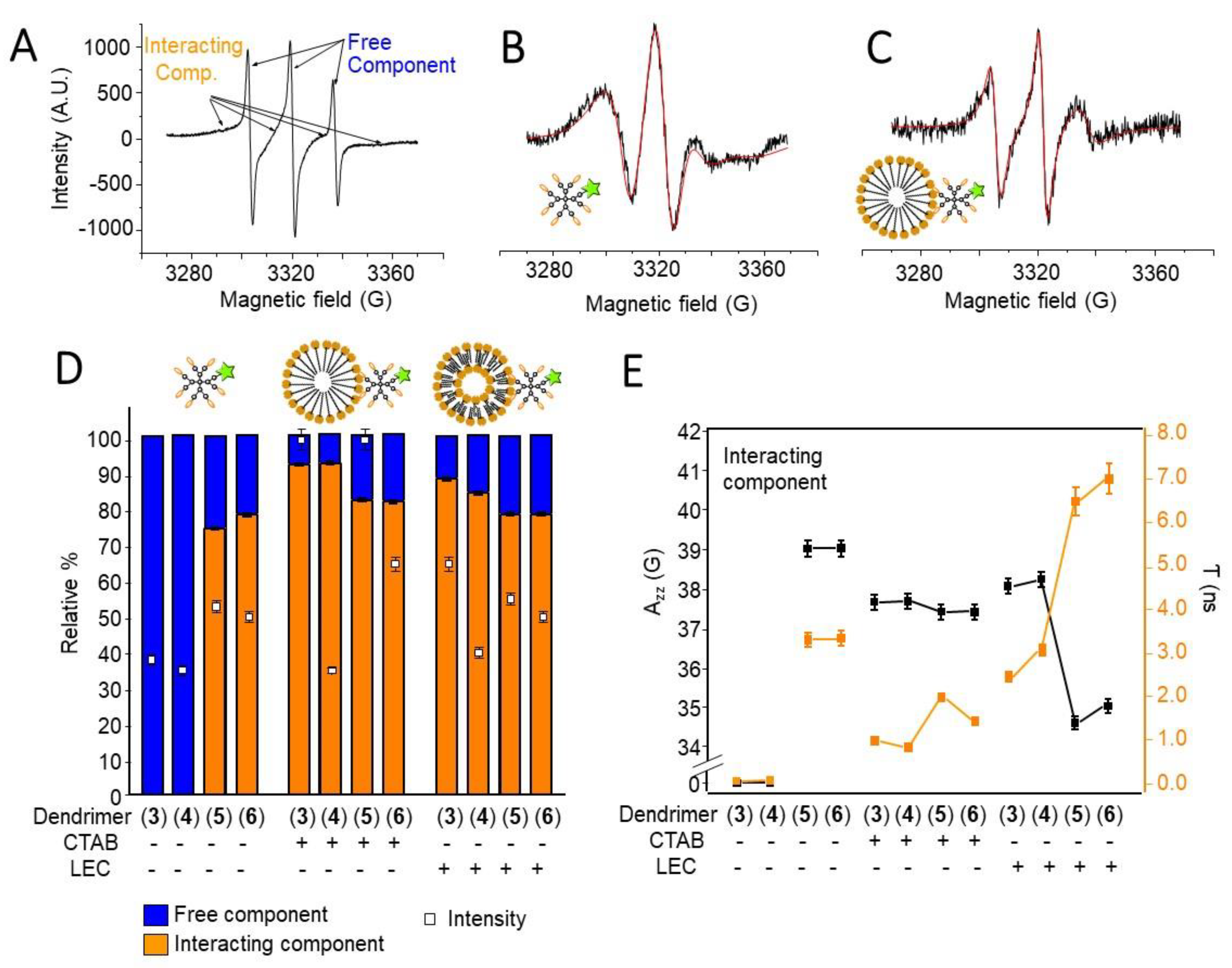
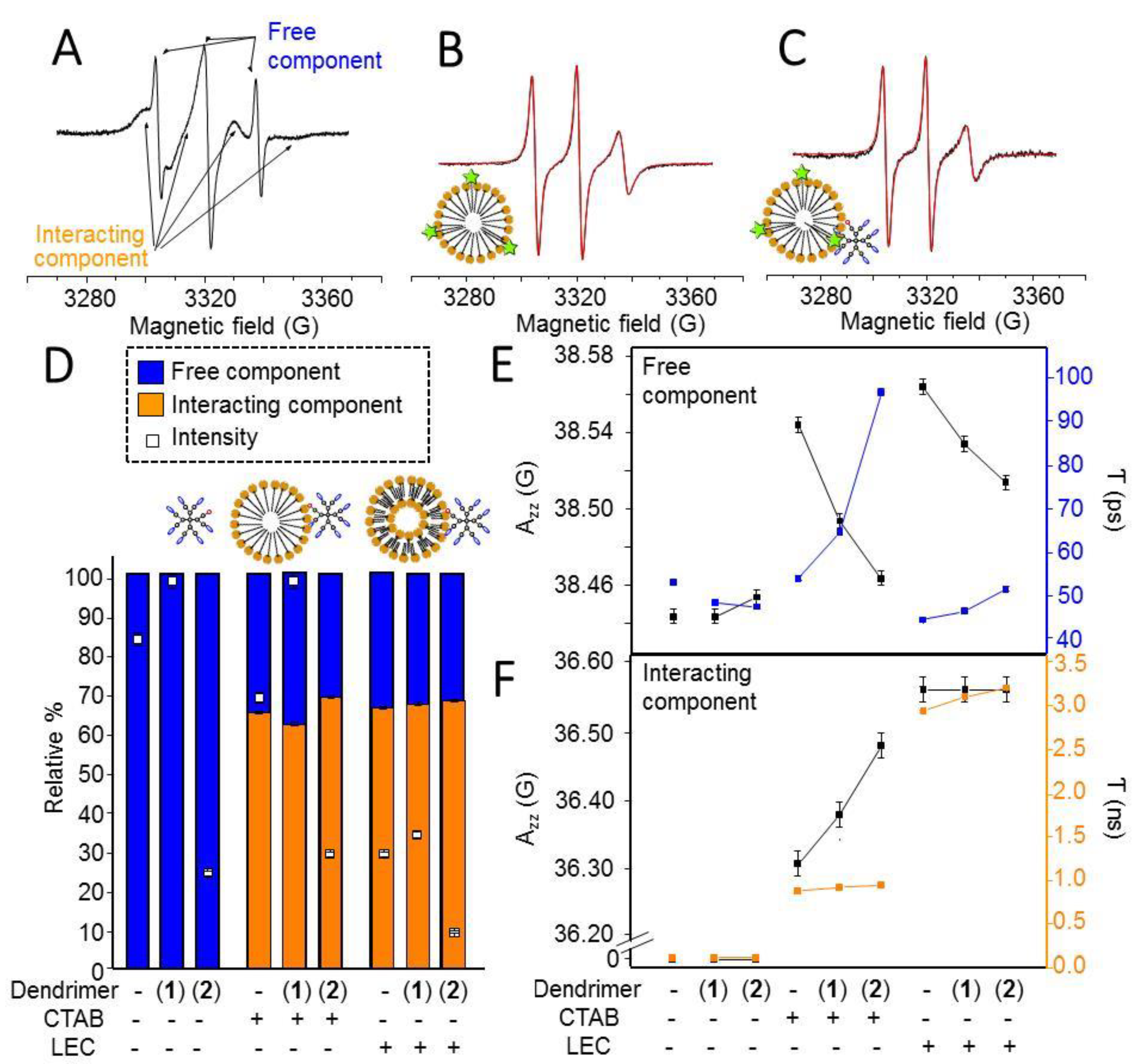
3.2. Electron Paramagnetic Resonance Study of TEMPO-Labelled Dendrimers in the Absence and Presence of Model Membranes

The spin-labelling technique is a very useful tool that offers an in situ reporter at the dendrimer surface about the eventually occurring interactions. An accurate computer-aided analysis of the EPR spectra allowed us to obtain specific information about the type and strength of interactions and the structural variations at the Gn-PyT and Gn-RuT dendrimer/model membrane interface.
Figure 2A shows, as an example, the EPR spectrum of G1-RuT (5) in PBS buffer. The arrows in the figure indicate the main features of the two components constituting the spectra. The first component is composed of three narrow lines, which are characteristic of fast moving nitroxide radicals, and it is henceforth called the Free component. The second broader component shows a partial resolution of the anisotropies, evidenced by the shift of the main peaks. The resolution of the anisotropies arose in the case of slow motion of the nitroxide group, due to an increase of the local microviscosity, which, in turn, was related to interactions occurring at the nitroxide environment. For these reasons, this component is henceforth called Interacting component. The occurrence of both these components for the labelled dendrimers in the absence of membrane models indicated that the free and interacting labels were already present at the surface of the labelled dendrimers, due to a different location of the labels themselves, one more external and the other more internal at the dendrimer interface; however, as we will discuss later, both components were affected by the interactions with the model membranes.
The occurrence of both components allowed us to perform subtractions between spectra to extract each of the components and also evaluate, by double integration of the components, their relative percentages. The components were then also computed to extract parameters providing structural and dynamical information (Table 1, see the experimental section for details). Figure 2B,C show as examples the experimental and computed Free and Interacting components for G1-RuT (5) alone and in the presence of CTAB. Further examples of experimental and computed spectra of labelled dendrimers are shown in Figure S6.
TEMPO-labelled dendrimers Gn-PyT (3 and 4, entries 1 and 2 in Table 1, Figure 2D) only showed a Free component, since the radical group was free to move at the dendrimer surface. The computation revealed a very polar environment and a high mobility (low microviscosity), almost equivalent to that found for TEMPO radicals in water. The minor differences in Azz and τ between these two dendrimers can be ascribed to the larger size of G2-PyT, which produced a small increase in microviscosity. The addition of CTAB micelles to these dendrimers (entries 3 and 4 in Table 1) altered the Free component, inducing a decrease of the polarity and mobility (increase in the microviscosity parameter τ) in agreement with a partial insertion of the nitroxide label into the micellar structure. Unexpectedly, the addition of liposomes provoked a lower change of the Free component (entries 5 and 6 in Table 1), indicating that the fast moving labels interact weakly with the liposome structure and mainly remained confined at the interface.
The attachment of Ru(II) complexes to the labelled dendrimers revealed a different interacting behavior. In TEMPO-labelled metallodendrimers Gn-RuT (5 and 6, entries 7 and 8 in Table 1, Figure 2D), the Free component already showed, in the absence of model membranes, a slightly lower polarity and a higher microviscosity if compared to Gn-PyT. The addition of the micelles and the liposomes poorly modified these values (Entries 9–12 in Table 1). Therefore, for the Ru-containing dendrimers, the external free labels were already partially hindered in their rotational mobility at the dendrimer surface, and the interactions with the model membranes provided only small changes in the microviscosity and polarity of the radical environment.
A different but complementary story was told by the computation parameters (the polarity parameter, Azz, and the microviscosity parameter, τ) of the Interacting component (Table 1 and Figure 2E). It is useful to discuss these parameters together with other parameters, i.e., (a) the total intensity, evaluated by the double integral of the spectra and scaled to 100 (assuming I = 100% for the labelled dendrimers in the absence of model membranes), reported in Figure 2D, squares; and (b) the relative percentage of the Interacting component, reported in Figure 2D, bars.
The total intensity of the EPR spectra measured the dendrimer solubility. In the absence of model membranes, the intensities for Gn-PyT were quite low, and slightly increased for Gn-RuT. However, the intensity increased in the presence of CTAB, revealing a significant improvement of solubility due to the interactions with micelles, mainly for first-generation dendrimers. Conversely, the presence of LEC showed no significant changes on the dendrimers’ solubility, except for G1-PyT. The relative percentage of Interacting component (Figure 2D, bars) quantifies the interacting capacity of the dendrimers. As previously mentioned, the Interacting component was absent for Gn-PyT when the model membranes are not present. Conversely, it appeared at a quite high relative extent in the presence of model membranes, mainly for CTAB. The Ru-containing dendrimers, Gn-RuT 5 and 6, showed an Interacting component already in the absence of model membranes, due to the interaction of the TEMPO label with the Ru complex. This percentage slightly increased by adding model membranes, especially CTAB micelles, monitoring the dendrimer/membrane interactions.
As shown in Figure 2E, the environmental polarity measured by Azz decreased from the Free to the Interacting component, and from the absence to the presence of model membranes, indicating that the label responsible of the Interacting component was partially inserted into the lipid core of the aggregate. This effect was quite significant for Gn-RuT with LEC, while the Gn-PyT dendrimers showed a better insertion into CTAB with respect to LEC. A perfect agreement was observed for the microviscosity parameter τ, giving the highest values for the Ru-metallodendrimers in the presence of LEC. Conversely, the CTAB micelles provided a more fluid environment with respect to the liposomes for the dendrimer surface groups partially entering the lipid structure, confirming a better interaction with the micelles.

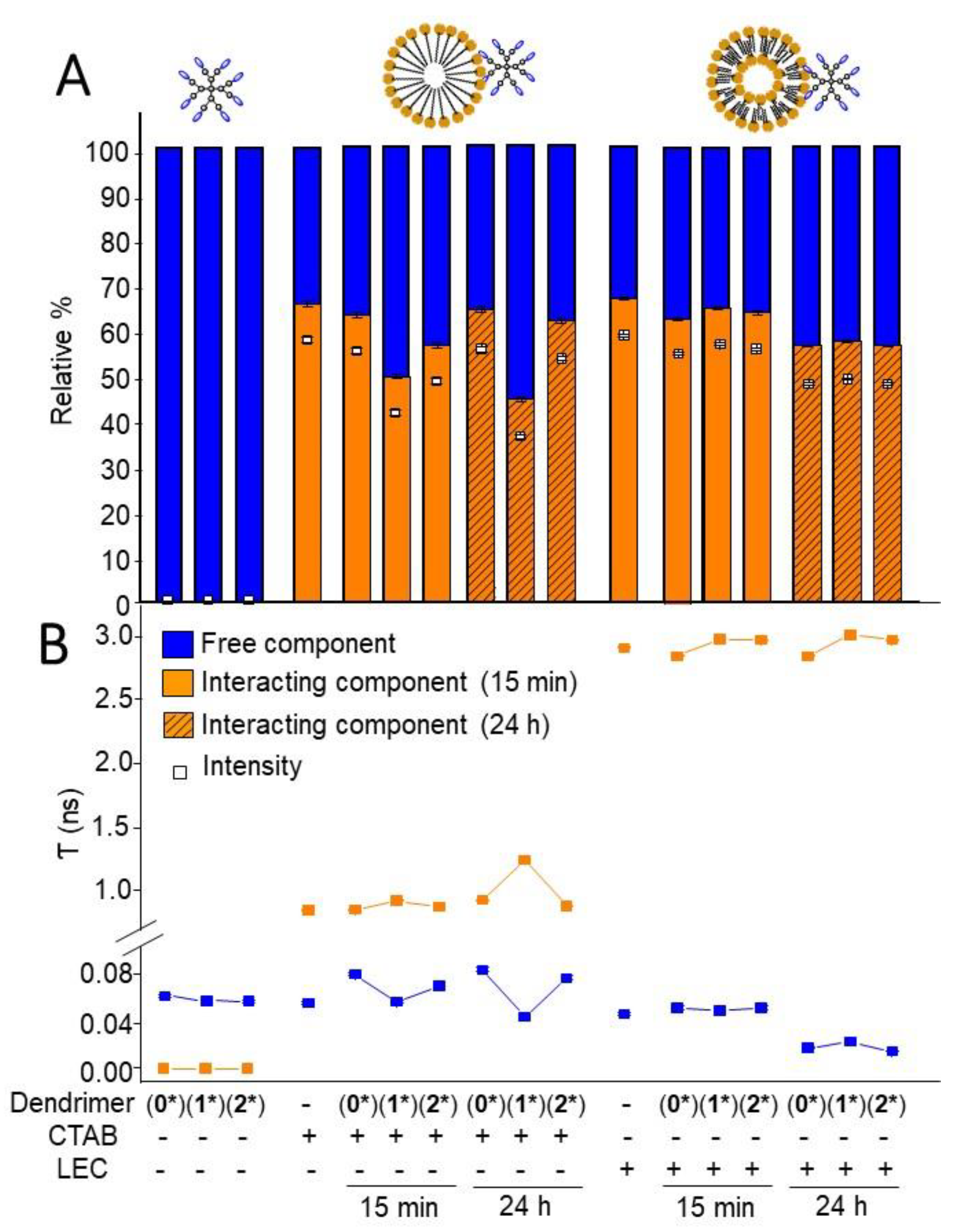
3.3. Electron Paramagnetic Resonance Study of Unlabelled Dendrimers Using CAT12 Probe in the Absence and Presence of Model Membranes: Comparison with TEMPO-Labelled Dendrimers

In order to gain further insight into the interacting abilities of the dendrimers, we selected the family of homofunctional dendrimers comprising iminopyridine groups in the periphery Gn-Py (first- and second-generation dendrimers 1* and 2*, Figure 1, as well as the mononuclear ligand 0*) [30]. Considering their diamagnetic properties, the use of a spin probe was required to analyze by EPR the interactions occurring between the dendrimers and the model membranes. The CAT12 probe was selected to provide complementary information with respect to those obtained by using labelled dendrimers. CAT12, being a surfactant, was expected to enter the model membrane with the carbon chain, while the positively charged CAT group localized at the membrane surface. This location allowed us to obtain information about the interactions between the dendrimer and the model membrane from the membrane side, while the labels at the dendrimer surface obviously provided information from the other side, the dendrimer surface.
The selection of the CAT12 probe was also based on the similarity between the EPR spectra obtained from the labelled dendrimers and CAT12 in the presence of both the dendrimer and the model membrane. The same line shape was obtained by using the homofunctional Gn-Py family and the heterofunctional Gn-PyN family, and, as shown in Figure 2A, the spectra were again constituted by the two components, Free and Interacting. In both cases, labelled and unlabelled dendrimers, the subtraction procedure between spectra allowed us to extract the two components, evaluate their relative percentages, and compute both of them to obtain the mobility and polarity parameters, which provided information about the dendrimer–model membrane interactions, as described above for the TEMPO-labelled dendrimers. Similarly, the same computation parameters were analyzed for the CAT12 probe as for the labelled dendrimers. In this case, we also found spectral variations over the equilibration time, and, therefore, results are shown and discussed for spectra recorded both after a short time (15 min) and a longer time (24 h) of mixture equilibration. The equilibration time impact was similar for the homofunctional Gn-Py and the heterofunctional Gn-PyN dendrimers. Then, the equilibration time effect will only be discussed for the homofunctional Gn-Py dendrimers, while, for simplicity, the results for the heterofunctional Gn-PyN dendrimers are only shown for the 15 min spectra to underline the differences between the two series of dendrimers. Further examples of experimental and computed spectra of non-labelled dendrimers in the absence and presence of model membranes are shown in Figure S7.

3.3.1. CAT12 Electron Paramagnetic Resonance Study of the Interactions between Homofunctional Gn-Py Dendrimers and Model Membranes

The relative intensity and relative percentage of interacting component of CAT12 in CTAB and LEC samples in the absence and presence of the Gn-Py dendrimers are shown in Figure 3A. The CAT12 probe is quite soluble in the PBS solution (intensity about 85%), but in the absence of model membranes the intensity decreased by adding G0-Py and G2-Py and increased by adding G1-Py, which was assumed as 100% of intensity. This behavior helped to clarify some properties of the different generation dendrimers in respect to their interacting ability, since CAT12 was interacting by means of both polar and hydrophobic interactions with the dendrimers, as already described in previous studies, and therefore followed the fate of the dendrimers [36]. It can be concluded that G1-Py showed the highest solubility and interacting ability with the medium, while G0-Py had the lowest solubility, probably due to the higher hydrophobicity. The opposite experiment, using a solution of model membranes in the absence of the dendrimers, revealed a decrease in the intensity with respect to the CAT12 probe in PBS. It can be explained by the entry and high concentration of the probes at the interface of the aggregates provoking strong spin–spin interactions, which led to very broad lines, no more visible in the spectra. The decrease in intensity was higher for LEC with respect to CTAB, probably due to the high packing of the LEC structure.
In the presence of both the dendrimer and the model membrane, the trend found by changing generation was also maintained in the presence of CTAB and LEC and at the different equilibration times. For CTAB dendrimer samples, the intensity was in between the ones for the dendrimer alone and for the CTAB alone in the case of G1-Py and G2-Py, while for G0-Py, the two components’ mixture further decreased the solubility. For LEC dendrimer samples, the intensity decreased (~30% maximum) as the radicals were concentrated at the dendrimer/liposome interface. The significant decrease in intensity from 15 min to 24 h in the presence of LEC indicated that the radicals progressively approached each other at the aggregate/dendrimer interface, while the opposite happened in CTAB micelles, where the radicals redistributed better over time into the micelles because of their fluidity when compared to the highly packed liposomes.
Figure 3A also shows the relative percentages of interacting components for CTAB and LEC samples in the presence of the various dendrimers at 15 min and 24 h equilibration times. While the dendrimers alone only showed a Free component, the probe in the model membranes in the absence and presence of the dendrimers showed both the Free and the Interacting components. For CTAB samples, the variation in relative percentage of Interacting component followed an opposite trend with respect to the intensity variation, being the lowest for G1-Py and the highest for G0-Py. The high solubility of CAT12 with G1-Py dendrimer well justified the low interacting component percentage, as this dendrimer, when interacting with the micelles, extracted the probes from the CTAB interface and promoted their solubilization, with this effect continuing over time. The percentage of Interacting component was the highest for G0-Py, almost comparable to the percentage for CTAB alone,. G1-Py showed an intermediate situation, however, for G0-Py and furthermore for G2-Py, the percentage of interacting component in the presence of CTAB increased over time, suggesting a progressive increase of interactions over time.
Similarly to CTAB, the relative percentage of interacting probes for LEC decreased from the absence to the presence of the dendrimers; however, in this case, G1-Py dendrimer showed a higher percentage with respect to the other generations. The LEC liposomes were well packed and the dendrimer–LEC interactions were poorly able to modify the liposome structure. However, over time, the percentage of interacting probes further decreased in line with the decrease in intensity, indicating an increased dendrimer/liposome interaction, which provided an increased local concentration of probes at the interface with consequent strong spin–spin interactions and the disappearance of these probes from contributing to the EPR spectrum.
Further information comes from the parameters Azz and τ obtained by spectral computation (Figure 3E and Figure S8). First of all, we noted that the Free component showed almost equivalent τ and Azz values for CTAB in the absence of the dendrimers and for the dendrimers in the absence of CTAB. This meant that the CAT12 heads were at the external surface of the micelles and the dendrimers, feeling the same environment (the water solution). The polarity for the Free probes almost did not change in function of generation and equilibration time. Conversely, the microviscosity for the Free probes increased for CTAB samples in the presence of G0-Py and G2-Py, while it decreased in the presence of G1-Py samples, mainly at 24 h. Since for G1-Py + CTAB samples the intensity was higher than for the other generations and the relative percentage of interacting probes decreased, it was clear that the interaction between G1-Py dendrimer and the micelles provoked the extraction of probes from the micelles to the solution, thus increasing both the relative quantity of Free probes and their freedom of motion. This behavior may be accounted for by a modification of the micellar structure due to interactions with G1-Py. The fact that the microviscosity of the Interacting component for G1-Py + CTAB increased, mainly after 24 h, and that the polarity decreased, further demonstrated the structural variations of the micelles when interacting with G1-Py. A higher packing of the surfactants accounted well for the extrusion of some probes from the micelles while the probes remaining inside the micelles more strongly interacted with the dendrimer surface, also partially approaching the less polar region. A quite different behavior was found for G0-Py and G2-Py—for them, the relative percentage of Interacting component changed poorly with respect to the micelles in the absence of the dendrimers, and the Free probes increased their microviscosity since they remained trapped at the dendrimer–micelle interphase. The interactions were weaker with G0-Py and G2-Py if compared to G1-Py because of an unfavorable balance between polar and hydrophobic forces, and the interacting probes showed a smaller decrease in polarity, more significant for G0-Py than for G2-Py.
For LEC liposomes, the differences as a function of generation were much lower. The LEC structure was preserved after 15 min, but after 24 h all the dendrimers provoked a decrease of both the percentage of interacting probes and the microviscosity of free probes, indicating a structural variation connected with the extraction of probes from the liposomes to the solution due to the dendrimer/liposome interactions. The interaction strength of the CAT group at the liposome interface with G0-Py was quite low, while it increased with both G1-Py and G2-Py because of the higher density of polar surface groups.

3.3.2. Electron Paramagnetic Resonance Study of the Interactions between Heterofunctional Gn-PyN Dendrimers and Model Membranes Using CAT12 as Spin Probe

Once we had established the interactions between homofunctional iminopyridine dendrimers Gn-Py and the model membranes, we addressed the influence of leaving one –NH2 group unmodified (Figure 1, compounds 1 and 2). Interesting variations of the EPR parameters were observed for the heterofunctional dendrimers Gn-PyN, compared to the homofunctional counterparts Gn-Py.
For a matter of clarity, an example of EPR spectrum obtained for CAT12 in G1-PyN+LEC is shown in Figure 4A, again constituted by the two components, Free and Interacting. Examples of computations of the interacting components are shown in Figure 4B,C, while the main parameters obtained from the analysis of the EPR spectra are shown in Figure 4D,E.
As already found for Gn-Py dendrimers, the intensity (Figure 4D, squares) measuring the probe solubility, was higher for the first-generation dendrimer when compared to the other generations. The solubility of CAT12 probes increased in the presence of G1-PyN because of a good balance between polar and hydrophobic interactions. This effect was maintained for G1-PyN in the presence of CTAB micelles. However, the absence of G1-PyN (CTAB alone) decreased the intensity, probably because some probes concentrated into the micelles provoking strong spin–spin interactions, which led to significant line broadening and the spectral contribution being no longer visible in the spectra. A similar effect may justify the significant decrease in intensity found for the liposomes in the absence of dendrimers, as already discussed in the previous section. For G2-PyN, the CAT12 solubility was low both in the absence and in the presence of CTAB, and reached a very low value (below 10%) in the presence of LEC. The interactions between G2-PyN and LEC provoked a further local increase in probe concentration, increasing spin–spin interactions.
After CAT12 aggregation into the micelles, which led to the intensity decrease in the presence of G2-PyN, the remaining non-aggregated probes showed an increase in the relative percentage of interacting component due to G2-PyN-micelle interactions (Figure 4D, bars). This effect, not observed for G2-Py, was undoubtedly due to the presence of the -NH2 group. Conversely, G1-PyN showed a decrease in the percentage of the interacting component, even if not as low as for G1-Py. Therefore, also in this case, G1-PyN interactions with CTAB produced an extraction of probes from the micelles to the solution, related to a restructuration of the micelles upon interaction with the dendrimer. However, the presence of the -NH2 group, instead of another iminopyridine ligand, decreased this effect, and promoted the dendrimer–micelle interactions preserving the micellar structure.
For LEC liposomes, the presence of the -NH2 group inverted the trend for the variation of the percentage of interacting component. Indeed, for Gn-Py dendrimers, the percentage diminished in the series LEC > LEC+G1-Py > LEC+G2-Py, while for Gn-PyN dendrimers the interacting percentage slightly increased in the series LEC < LEC+G1-PyN < LEC+G2-PyN. The interactions were preferentially at the polar surface of the liposomes, as also demonstrated by the polarity parameter Azz (Figure 4F).
For the polarity and microviscosity parameters, the Free component was also informative (Figure 4E). This Free component arose from probes in solution, but was affected by the surface of dendrimers and model membranes. By adding the dendrimers to CTAB micelles, the polarity decreased because of the approaching of the less polar dendrimer to the model membrane. This approaching partly reduced the mobility (increasing the microviscosity). For LEC, by adding the dendrimers, the freedom of motion and the polarity were reduced because of dendrimer/membrane interactions, and this effect was stronger by increasing generation.
In respect to the Interacting component (Figure 4F), both polarity and microviscosity increased for the model membranes from the absence to the presence of the dendrimers, by increasing generation, and from CTAB to LEC. Also in this case, some trends were inverted with respect to Gn-Py dendrimers. For instance, the microviscosity, measuring the strength of interaction, decreased from G1-Py to G2-Py, while it increased from G1-PyN to G2-PyN; in detail, in respect to CTAB, G1-Py was more interactive than G1-PyN, while G2-Py was less interactive than G2-PyN. For the polarity, G1-Py showed lower polarity than G1-PyN, while G2-Py showed higher polarity than G2-PyN. These results revealed a different interaction mechanism between the different dendrimers and CTAB micelles, being G1-Py able to partially enter the micelles, approaching less polar sites and providing a stronger interaction with the micelles. The presence of -NH2 at the G1-PyN surface hindered the penetration into the micelle, favoring interactions at the charged external surface. For G2-Py, the higher density of surface groups, the lower availability of the low polar dendrimer core, and the higher structural rigidity impeded the penetration into the micelles. Therefore, the interaction was only at the external surface, and the presence of -NH2 at the G2-PyN surface favored polar and electrostatic interactions with the micelles.
The higher Azz and τ parameters for LEC with respect to CTAB were due to the more dense and viscous liposome structure at the interface. The increased microviscosity and polarity parameters in the presence of the dendrimers were clearly reporting the occurrence of electrostatic interactions between the dendrimers and the liposome surface, which became stronger by increasing generation due to the higher density of surface groups for the higher generation. If compared to Gn-Py, Gn-PyN dendrimers showed higher strength of interaction and higher polarity. Furthermore, G2-PyN showed stronger interactions with respect to G1-PyN, while the opposite held for Gn-Py dendrimers. This confirmed that the dendrimer–liposome interactions were mainly polar/electrostatic between the polar/charged surfaces. Therefore, the -NH2 group favored these kinds of interactions.
The last interesting finding was the agreement between the results obtained for the labelled dendrimers (36) and for the unlabeled ones (12, 1*2*). Both points of view suggested the partial insertion of the dendrimer surface groups into the surfactant aggregates, mainly CTAB micelles, and the occurrence of both polar and hydrophobic interactions, while dendrimer–LEC interactions involved more polar interactions between surface groups. Finally, the G1-Py interactions were significantly favored, mainly with CTAB, but both the presence of -NH2 and TEMPO moieties perturbed these interactions, partially preventing the entry of dendrimer surface groups into the micellar core.

4. Conclusions

A selective and efficient anticancer therapy requires an efficient uptake of the drug by cancer cells, which in turn depends on the ability of the drug to interact and penetrate the cell membrane. Using model cell membranes (CTAB and LEC), we evaluated in situ the interactions performed by a new family of differently decorated carbosilane dendrimers through a computer-assisted EPR analysis. The analysis of the EPR spectra provided parameters informative of the interacting ability of the dendrimers, and the type and strength of interactions in function of the effect of a metal (Ru(II)), the type of model membrane, a partial functionalization, and generation.
In order to report the interactions from the dendrimer point of view, we synthesized new heterofunctional Ru(II) metallodendrimers labelled with a TEMPO radical. The new nanotheranostic Gn-RuT dendrimers can potentially exhibit an antitumor effect while monitoring in real time drug release and distribution. In the synthetic route, interesting precursors were prepared—The Gn-PyN family, with multiple iminopyridine ligands and a single –NH2 group, and the Gn-PyT family, with multiple iminopyridine ligands and a TEMPO label. These subtle changes of a single group later revealed important differences regarding the interactions between the dendrimers and the CTAB and LEC models. Initially, a comparative EPR study was performed on the labelled dendrimers Gn-PyT and Gn-RuT in the absence and presence of the model membranes. It was found that the Gn-PyT dendrimers partially penetrated the micelle structure, with G1-PyT being more interactive than G2-PyT. In the liposomes the interactions occurred at the external surface, with G2-PyT being more interactive than G1-PyT. In the metallodendrimers, the TEMPO group interacted with Ru(II) ions but gained freedom entering the micellar structure; otherwise their mobility was slowed down by interacting with the LEC surface. Therefore, for both Gn-PyT and Gn-RuT, the interactions were perturbative of the CTAB micellar structure, but preserve the LEC structure, mainly occurring at the external interface.
The EPR results from the spin-labelled dendrimers were compared and integrated with those obtained by both the unlabeled dendrimer precursors (Gn-PyN) and the homofunctional iminopyridine dendrimers (Gn-Py). In these last cases, the CAT12 probe demonstrated the provision of complementary information with respect to those obtained with the labelled dendrimers. Also in this case, the computer aided EPR analysis allowed us to extract similar parameters as those obtained by means of the TEMPO label, but the point of view provided by CAT12 was on the model membrane side, since this spin probe is deeply inserted into the surfactant aggregates. It was found, similar to Gn-PyT, that G1-Py interacts better with CTAB, while G2-Py interacts better with LEC. G1-Py and G1-PyN interactions with CTAB produced an extraction of probes from the micelles to the solution, which was related to a restructuration of the micelles in agreement with the results from the labelled dendrimers. The presence of the NH2 group at the G2-PyN surface promoted polar and electrostatic interactions with the micelles instead of penetration of dendrimer branches into the micellar structure. Therefore, the presence of NH2 at the G1-PyN dendrimer surface impeded the penetration into the micelle, favoring interactions at the charged external surface.
Clearly the EPR analysis demonstrated itself to be very useful in providing detailed information on the mechanism of interactions of the dendrimer with a model cell membrane, in view of the use of these new dendrimers as anticancer drugs. A second manuscript containing comparative EPR experiments and biological tests using both healthy and tumor cells in the absence and presence of the dendrimers is currently in preparation.

Supplementary Materials

The following are available online at https://www.mdpi.com/2218-273X/9/10/540/s1, Figure S1: 1H- and 13C{1H}-NMR spectra of compound 1. Figure S2: TOCSY 1D spectrum of compound 1. Figure S3: 1H-NMR spectrum of compound 3. Figure S4: 1H-NMR spectrum of compound 5. Figure S5: FT-IR spectra of precursor 4-isothiocyanateTEMPO and compound 3. Figure S6: Examples of experimental and computed spectra of labelled dendrimers in the absence and presence of model membranes. Figure S7: Examples of experimental and computed spectra of non-labelled dendrimers in the absence and presence of model membranes. Figure S8: Azz values obtained by computing the Free and the Interacting components for CTAB for homofunctional dendrimers Gn-Py.

Author Contributions

conceptualization, M.F.O. and F.J.d.l.M.; methodology, P.O. and S.G.G.; formal analysis, A.F. and M.C.; investigation, R.C., N.S.O., and A.F.; resources, M.F.O., R.G., and F.J.d.l.M.; data curation, R.C., N.S.O., and M.C.; writing—original draft preparation, S.G.G. and M.C.; writing—review and editing, M.F.O., S.G.G., and P.O.; supervision, M.C., R.G., and F.J.d.l.M.; funding acquisition, M.F.O., R.G., and F.J.d.l.M.

Funding

This research was funded by grants from CTQ2017-86224-P (MINECO), consortiums IMMUNOTHERCAN-CM B2017/BMD-3733 and NANODENDMED II-CM ref. B2017/BMD-3703, project SBPLY/17/180501/000358 Junta de Comunidades de Castilla-la Mancha (JCCM) and the Comunidad de Madrid Research Talent Attraction Program 2017-T2/IND-5243. CIBER-BBN is an initiative funded by the VI National R&D&I Plan 2008–2011, Iniciativa Ingenio 2010, Consolider Program, CIBER Actions and financed by the Instituto de Salud Carlos III with assistance from the European Regional Development Fund. N.S.O. wishes to thank JCCM for a predoctoral fellowship. M.F.O., M.C., R.C., and A.F. thank DiSPeA at the University of Urbino for funding. This article is based upon work from COST Action CA17140 “Cancer Nanomedicine from the Bench to the Bedside” supported by COST (European Cooperation in Science and Technology).

Conflicts of Interest

The authors declare no conflict of interest. The funders had no role in the design of the study; in the collection, analyses, or interpretation of data; in the writing of the manuscript, or in the decision to publish the results.

References

  1. Mi, P.; Cabral, H.; Kataoka, K. Ligand-installed nanocarriers toward precision therapy. Adv. Mater. 2019, e1902604. [Google Scholar] [CrossRef] [PubMed]
  2. Sk, U.H.; Kojima, C. Dendrimers for drug delivery of anticancer drugs. In Frontiers in Clinical Drug Research - Anti-Cancer Agents; Atta-ur-Rahman, Ed.; Bentham Science: UK, 2015; Volume 2, pp. 3–25. [Google Scholar]
  3. Han, X.; Xu, K.; Taratula, O.; Farsad, K. Applications of nanoparticles in biomedical imaging. Nanoscale 2019, 11, 799–819. [Google Scholar] [CrossRef] [PubMed]
  4. Nabil, G.; Bhise, K.; Sau, S.; Atef, M.; El-Banna, H.A.; Iyer, A.K. Nano-engineered delivery systems for cancer imaging and therapy: Recent advances, future direction and patent evaluation. Drug Discov. Today 2019, 24, 462–491. [Google Scholar] [CrossRef] [PubMed]
  5. Zhao, C.-Y.; Cheng, R.; Yang, Z.; Tian, Z.-M. Nanotechnology for Cancer Therapy Based on Chemotherapy. Mol. 2018, 23, 826. [Google Scholar] [CrossRef] [PubMed]
  6. Sharma, M.; Dube, T.; Chibh, S.; Kour, A.; Mishra, J.; Panda, J.J. Nanotheranostics, a future remedy of neurological disorders. Expert Opin. Drug Deliv. 2019, 16, 113–128. [Google Scholar] [CrossRef]
  7. Mura, S.; Couvreur, P. Nanotheranostics for personalized medicine. Adv. Drug Deliv. Rev. 2012, 64, 1394–1416. [Google Scholar] [CrossRef]
  8. Sowinska, M.; Urbanczyk-Lipkowska, Z. Advances in the chemistry of dendrimers. New J. Chem. 2014, 38, 2168–2203. [Google Scholar] [CrossRef]
  9. Sánchez-Nieves, J.; Ortega, P.; Cano, J.; Gómez, R.; Mata, F.J.d.l. Poly(carbosilane) dendrimers and other silicon-containing dendrimers. In Dendrimer Chemistry: Synthetic approaches towards complex architectures; Malkoch, M., García-Gallego, S., Eds.; Royal Society of Chemistry: London, UK, 2019. [Google Scholar]
  10. Andreozzi, E.; Antonelli, A.; Cangiotti, M.; Canonico, B.; Sfara, C.; Pianetti, A.; Bruscolini, F.; Sahre, K.; Appelhans, D.; Papa, S.; et al. Interactions of nitroxide-conjugated and non-conjugated glycodendrimers with normal and cancer cells and biocompatibility studies. Bioconjug. Chem. 2017, 28, 524–538. [Google Scholar] [CrossRef]
  11. Ottaviani, M.F.; El Brahmi, N.; Cangiotti, M.; Coppola, C.; Buccella, F.; Cresteil, T.; Mignani, S.; Caminade, A.M.; Costes, J.P.; Majoral, J.P. Comparative EPR studies of Cu(II)-conjugated phosphorous-dendrimers in the absence and presence of normal and cancer cells. RSC Adv. 2014, 4, 36573–36583. [Google Scholar] [CrossRef]
  12. Shang, L.; Nienhaus, K.; Nienhaus, G.U. Engineered nanoparticles interacting with cells: size matters. J. Nanobiotechnol. 2014, 12, 5. [Google Scholar] [CrossRef]
  13. Rokach, S.; Ottaviani, M.F.; Shames, A.I.; Aserin, A.; Garti, N. Behavior of PPI-G2 dendrimer in a microemulsion. J. Phys. Chem. B 2017, 121, 2339–2349. [Google Scholar] [CrossRef] [PubMed]
  14. Perlstein, M.; Ottaviani, M.F.; Aserin, A.; Garti, N. Structural effects on cosolubilization of dendrimer and propofol in water dilutable microemulsions as delivery vehicle. Colloids Surfaces A: Physicochem. Eng. Asp. 2016, 497, 257–264. [Google Scholar] [CrossRef]
  15. Lidich, N.; Ottaviani, M.F.; Hoffman, R.E.; Aserin, A.; Garti, N. Docosahexaenoic acid triglyceride-based microemulsions with an added dendrimer—Structural considerations. J. Colloid Interface Sci. 2016, 483, 374–384. [Google Scholar] [CrossRef] [PubMed]
  16. Rokach, S.; Ottaviani, M.F.; Shames, A.I.; Nir, I.; Aserin, A.; Garti, N. W/O microemulsions as dendrimer nanocarriers: an EPR study. J. Phys. Chem. B 2012, 116, 12633–12640. [Google Scholar] [CrossRef] [PubMed]
  17. Bitan-Cherbakovsky, L.; Libster, D.; Ottaviani, M.F.; Aserin, A.; Garti, N. Structural behavior and interactions of dendrimer within lyotropic liquid crystals, monitored by EPR spectroscopy and rheology. J. Phys. Chem. B 2012, 116, 2420–2429. [Google Scholar] [CrossRef] [PubMed]
  18. Ottaviani, M.F.; Favuzza, P.; Sacchi, B.; Turro, N.J.; Jockusch, S.; Tomalia, D.A. Interactions between starburst dendrimers and mixed DMPC/DMPA-Na vesicles studied by the Spin Label and the Spin Probe techniques, supported by Transmission Electron Microscopy. Langmuir 2002, 18, 2347–2357. [Google Scholar] [CrossRef]
  19. Ottaviani, M.F.; Matteini, P.; Brustolon, M.; Turro, N.J.; Jockusch, S.; Tomalia, D.A. Characterization of Starburst Dendrimers and Vesicle Solutions and Their Interactions by CW- and Pulsed-EPR, TEM, and Dynamic Light Scattering. J. Phys. Chem. B 1998, 102, 6029–6039. [Google Scholar] [CrossRef]
  20. Ottaviani, M.F.; Daddi, R.; Brustolon, M.; Turro, N.J.; Tomalia, D.A. Structural modifications of DMPC vesicles upon interaction with Poly(amidoamine) dendrimers studied by CW-Electron Paramagnetic Resonance and Electron Spin–Echo techniques. Langmuir 1999, 15, 1973–1980. [Google Scholar] [CrossRef]
  21. Ottaviani, M.F.; Andechaga, P.; Turro, N.J.; Tomalia, D.A. Model for the interactions between anionic dendrimers and cationic surfactants by means of the spin probe method. J. Phys. Chem. B 1997, 101, 6057–6065. [Google Scholar] [CrossRef]
  22. Ottaviani, M.F.; Daddi, R.; Brustolon, M.; Turro, N.J.; Tomalia, D.A. Interaction between starburst dendrimers and SDS micelles studied by continuous-wave and pulsed electron spin resonances. Appl. Magn. Reson. 1997, 13, 347–363. [Google Scholar] [CrossRef]
  23. Ottaviani, M.F.; Turro, N.J.; Jockusch, S.; Tomalia, D.A. Aggregational process of the positively charged surfactants CTAC and CAT16 in the presence of starburst dendrimers: an Electron Paramagnetic Resonance spectroscopic study. Colloids Surf. A 1996, 115, 9–21. [Google Scholar] [CrossRef]
  24. Ottaviani, M.F.; Turro, N.J.; Jockusch, S.; Tomalia, D.A. Characterization of starburst dendrimers by EPR. 3. Aggregational processes of a positively charged nitroxide surfactant. J. Phys. Chem. 1996, 100, 13675–13686. [Google Scholar] [CrossRef]
  25. Lin, Y.; Yokoyama, H.; Ishida, S.-I.; Tsuchihashi, N.; Ogata, T. In vivo Electron Spin Resonance analysis of nitroxide radicals injected into a rat by a flexible surface-coil-type resonator as an endoscope- or a stethoscope-like device. Magn. Reson. Mater. Phy. 1997, 5, 99–103. [Google Scholar] [CrossRef]
  26. Galimzyanovich Saifutdinov, R.; Ivanovna Larina, L.; Il’inichna Vakul´skaya, T.; Grigor´evich Voronkov, M. Electron Paramagnetic Resonance in Biochemistry and Medicine; Kluwer Academic Publishers: New York, NY, USA, 2002. [Google Scholar]
  27. Swartz, H.M.; Williams, B.B.; Zaki, B.I.; Hartford, A.C.; Jarvis, L.A.; Chen, E.Y.; Comi, R.J.; Ernstoff, M.S.; Hou, H.; Khan, N.; et al. Clinical EPR: unique opportunities and some challenges. Acad. Radiol. 2014, 21, 197–206. [Google Scholar] [CrossRef] [PubMed]
  28. Sanz del Olmo, N.; Maroto-Díaz, M.; Gómez, R.; Ortega, P.; Cangiotti, M.; Ottaviani, M.F.; de la Mata, F.J. Carbosilane metallodendrimers based on copper (II) complexes: Synthesis, EPR characterization and anticancer activity. J. Inorg. Biochem. 2017, 177, 211–218. [Google Scholar] [CrossRef]
  29. Sanz del Olmo, N.; Carloni, R.; Bajo, A.M.; Ortega, P.; Fattori, A.; Gómez, R.; Ottaviani, M.F.; García-Gallego, S.; Cangiotti, M.; de la Mata, F.J. Insight into the antitumor activity of carbosilane Cu(II)–metallodendrimers through their interaction with biological membrane models. Nanoscale 2019, 11, 13330–13342. [Google Scholar] [CrossRef]
  30. Maroto-Diaz, M.; Elie, B.T.; Gomez-Sal, P.; Perez-Serrano, J.; Gomez, R.; Contel, M.; de la Mata, F.J. Synthesis and anticancer activity of carbosilane metallodendrimers based on arene ruthenium(ii) complexes. Dalton Trans. 2016, 45, 7049–7066. [Google Scholar] [CrossRef] [Green Version]
  31. Maroto-Diaz, M.; Sanz del Olmo, N.; Muñoz-Moreno, L.; Bajo, A.M.; Carmena, M.J.; Gómez, R.; García-Gallego, S.; de la Mata, F.J. In vitro and in vivo evaluation of first-generation carbosilane arene Ru(II)-metallodendrimers in advanced prostate cancer. Eur. Polym. J. 2019, 113, 229–235. [Google Scholar] [CrossRef]
  32. Zeng, L.; Gupta, P.; Chen, Y.; Wang, E.; Ji, L.; Chao, H.; Chen, Z.S. The development of anticancer ruthenium(II) complexes: from single molecule compounds to nanomaterials. Chem. Soc. Rev. 2017, 46, 5771–5804. [Google Scholar] [CrossRef]
  33. Budil, D.E.; Lee, S.; Saxena, S.; Freed, J.H. Nonlinear-least-squares analysis of slow-motion EPR spectra in one and two dimensions using a modified Levenberg–Marquardt algorithm. J. Magn. Reson. 1996, 120, 155–189. [Google Scholar] [CrossRef]
  34. Bermejo, J.F.; Ortega, P.; Chonco, L.; Eritja, R.; Samaniego, R.; Mullner, M.; de Jesus, E.; de la Mata, F.J.; Flores, J.C.; Gomez, R.; et al. Water-soluble carbosilane dendrimers: synthesis biocompatibility and complexation with oligonucleotides; evaluation for medical applications. Chemistry 2007, 13, 483–495. [Google Scholar] [CrossRef] [PubMed]
  35. Huang, L.; Yan, C.; Cui, D.; Yan, Y.; Liu, X.; Lu, X.; Tan, X.; Lu, X.; Xu, J.; Xu, Y.; et al. Organic radical contrast agents based on polyacetylenes containing 2,2,6,6-Tetramethylpiperidine 1-Oxyl (TEMPO): Targeted Magnetic Resonance (MR)/Optical Bimodal Imaging of folate receptor expressing HeLa tumors in vitro and in vivo. Macromol. Biosci. 2015, 15, 788–798. [Google Scholar] [CrossRef] [PubMed]
  36. Ottaviani, M.F.; Cossu, E.; Turro, N.J.; Tomalia, D.A. Characterization of starburst dendrimers by Electron Paramagnetic Resonance. 2. Positively charged nitroxide radicals of variable chain length used as spin probes. J. Am. Chem. Soc. 1995, 117, 4387–4398. [Google Scholar] [CrossRef]
Figure 1. Structural representation of dendrimers and metallodendrimers used in the electron paramagnetic resonance (EPR) study. Homofunctional dendrimers: Gn-Py (1*, 2*); heterofunctional dendrimers: Gn-PyN (1,2) and Gn-PyT (3,4); heterofunctional metallodendrimers: Gn-RuT (5,6).
Biomolecules 09 00540 g001
Scheme 1. Three-step synthetic strategy to accomplish the 2,2,6,6-tetramethylpiperidine-1-oxyl (TEMPO)-labelled heterofunctional metallodendrimers Gn-RuT (5,6).
Biomolecules 09 00540 sch001
Figure 2. Experimental spectra and computation spectra/parameters for the EPR spectra of the labelled dendrimers Gn-PyT and Gn-RuT 3–6 in the absence and presence of the model membranes (cetyltrimethylammonium bromide micelles (CTAB) and lecithin liposomes (LEC)). (A) Experimental EPR spectrum of G1-RuT (5) in Phosphate Buffered Saline , selected as an example. Arrows indicate the main features of the two components constituting the spectra (termed Free and Interacting); (B,C) experimental and computed Interacting components of G1-RuT (5) alone or in the presence of CTAB, obtained after subtraction of the Free component. The spectra are normalized in height. (D) Total intensity of the EPR spectra (squares, assuming I = 100% for the spectrum of the labelled dendrimers in the absence of model membranes) and relative percentage of Interacting component (bars). (E) Polarity parameter, Azz, and microviscosity parameter, τ, for the Interacting component.
Biomolecules 09 00540 g002
Figure 3. Parameters obtained from the analysis of CAT12 EPR spectra of the unlabeled dendrimers Gn-Py in the absence and presence of the model membranes (CTAB micelles and LEC liposomes). (A) Relative percentages of interacting component (bars) for CTAB and LEC samples in the absence and presence of Gn-Py dendrimers at 15 min and 24 h equilibration times, and intensity values (squares), in percentages, assuming 100% as the highest intensity. (B) Microviscosity parameter, τ, for both the Free and Interacting components.
Biomolecules 09 00540 g003
Figure 4. Experimental spectra and computation spectra/parameters for the CAT12 EPR spectra of the unlabeled heterofunctional dendrimers Gn-PyN in the absence and presence of the model membranes (CTAB micelles and LEC liposomes). (A) EPR spectrum of CAT12 for G1-PyN (1) in the presence of LEC; (B, C) experimental (black) and computed (red) spectra of the interacting component for CAT12 in CTAB sample, alone and in the presence of G2-PyN (2); (D) Relative percentage of Interacting component (bars), and total intensity (squares) as a percentage, where 100% intensity was ascribed to the spectrum at the highest intensity; (E, F) Azz values (left axis) and τ values (right axis) for the Free and the Interacting components. PBS sample was evaluated in the absence of dendrimers and model membranes.
Biomolecules 09 00540 g004
Table 1. Main parameters of computation for the labelled dendrimers Gn-PyT and Gn-RuT in the absence and presence of model membranes.
EntrySampleComponentsAzz (G)τ (ns)
1G1-PyT (3)Free (single)39.21/-0.014/-
2G2-PyT (4)Free (single)39.12/-0.017/-
3G1-PyT (3) + CTABFree + Interacting38.72/37.680.21/0.87
4G2-PyT (4) + CTABFree + Interacting38.68/37.700.25/0.73
5G1-PyT (3) + LECFree + Interacting39.01/38.070.050/2.35
6G2-PyT (4) + LECFree + Interacting38.98/38.200.055/3.05
7G1-RuT (5)Free + Interacting38.82/39.000.22/3.31
8G2-RuT (6)Free + Interacting38.85/39.000.22/3.35
9G1-RuT (5) + CTABFree + Interacting38.82/37.400.22/1.90
10G2-RuT (6) + CTABFree + Interacting38.85/37.410.22/1.38
11G1-RuT (5) + LECFree + Interacting38.82/34.600.22/6.45
12G2-RuT (6) + LECFree + Interacting38.85/35.000.22/7.00

