Article

Low Concentration of Withaferin a Inhibits Oxidative Stress-Mediated Migration and Invasion in Oral Cancer Cells

1
Graduate Institute of Medicine, College of Medicine, Kaohsiung Medical University, Kaohsiung 80708, Taiwan
2
Department of Radiation Oncology, Faculty of Medicine, College of Medicine, Kaohsiung Medical University, Kaohsiung 80708, Taiwan
3
Department of Radiation Oncology, Kaohsiung Medical University Hospital, Kaohsiung 80708, Taiwan
4
Division of Breast Surgery and Department of Surgery, Kaohsiung Medical University Hospital, Kaohsiung 80708, Taiwan
5
Center for Cancer Research, Kaohsiung Medical University, Kaohsiung 80708, Taiwan
6
School of Dentistry, College of Dental Medicine, Kaohsiung Medical University, Kaohsiung 80708, Taiwan
7
Cancer Center, Kaohsiung Medical University Hospital, Kaohsiung 80708, Taiwan
8
Translational Research Center, Kaohsiung Medical University Hospital, Kaohsiung 80708, Taiwan
9
Shanghai Jiao Tong University School of Medicine, Shanghai 200025, China
10
Department of Radiation Oncology, Chi-Mei Foundation Medical Center, Tainan 71004, Taiwan
11
School of Medicine, Taipei Medical University, Taipei 11031, Taiwan
12
Chung Hwa University Medical Technology, Tainan 71703, Taiwan
13
Department of Medical Research, Kaohsiung Medical University Hospital, Kaohsiung 80708, Taiwan
14
Department of Biomedical Science and Environmental Biology, College of Life Sciences, Kaohsiung Medical University, Kaohsiung 80708, Taiwan
*
Authors to whom correspondence should be addressed.
Biomolecules 2020, 10(5), 777; https://doi.org/10.3390/biom10050777
Submission received: 28 February 2020 / Revised: 13 May 2020 / Accepted: 15 May 2020 / Published: 17 May 2020
(This article belongs to the Special Issue Phytochemical Omics in Medicinal Plants)

Abstract

:
Withaferin A (WFA) has been reported to inhibit cancer cell proliferation based on high cytotoxic concentrations. However, the low cytotoxic effect of WFA in regulating cancer cell migration is rarely investigated. The purpose of this study is to investigate the changes in migration and mechanisms of oral cancer Ca9-22 cells after low concentrations of WFA treatment. WFA under 0.5 μM at 24 h treatment shows no cytotoxicity to oral cancer Ca9-22 cells (~95% viability). Under this condition, WFA triggers reactive oxygen species (ROS) production and inhibits 2D (wound healing) and 3D cell migration (transwell) and Matrigel invasion. Mechanically, WFA inhibits matrix metalloproteinase (MMP)-2 and MMP-9 activities but induces mRNA expression for a group of antioxidant genes, such as nuclear factor, erythroid 2-like 2 (NFE2L2), heme oxygenase 1 (HMOX1), glutathione-disulfide reductase (GSR), and NAD(P)H quinone dehydrogenase 1 (NQO1)) in Ca9-22 cells. Moreover, WFA induces mild phosphorylation of the mitogen-activated protein kinase (MAPK) family, including extracellular signal-regulated kinases 1/2 (ERK1/2), c-Jun N-terminal kinase (JNK), and p38 expression. All WFA-induced changes were suppressed by the presence of ROS scavenger N-acetylcysteine (NAC). Therefore, these results suggest that low concentration of WFA retains potent ROS-mediated anti-migration and -invasion abilities for oral cancer cells.

1. Introduction

Oral cancer leads to high morbidity and mortality [1]. It invades local tissues [2] and reoccurs occasionally [3]. Local invasions are associated with metastasis, which is important to oral carcinogenesis [4]. Therefore, discovery of a drug that inhibits metastasis or local invasion is of great importance for oral cancer therapy.
Withaferin A (WFA), a triterpenoid derived from the root or leaf of the medicinal plant Withania somnifera, is reported to exhibit antiproliferative properties and can induce apoptosis in several types of cancers such as leukemia [5], cervical [6], pancreatic [7], breast [8], lung [9], colorectal [10], and oral [11,12] cancer cells. These anticarcinogenic effects for WFA were based on high cytotoxic concentrations.
These cytotoxic concentrations of WFA were reported to induce reactive oxygen species (ROS)-mediated apoptosis in oral [12] and colon [10] cancer cells. ROS may induce a number of reactions such as apoptosis [5,6,7,8,9,10,11,12], autophagy, and endoplasmic reticulum stress [13]; however, its effect on migration has rarely been reported.
Migration inhibitory effects of WFA against cancer cells had been reported recently [14,15]. For example, WFA exhibits G2/M cell cycle arrest, apoptosis, and antiproliferation, as well as migration inhibition in gastric cancer AGS cells [14]. However, its migration inhibitory effects were based on wound healing and invasive assays at >10 μM and >1 μM WFA, where the IC50 value for WFA in AGS cells was 0.75 μM [14]. WFA also showed antiproliferative effects against breast cancer cells (MDA-MB-241) and could exhibit migration inhibitory effect using the concentration of IC50 value for WFA (12 μM) [15]. These migration inhibitory effects of WFA against cancer cells were based on high cytotoxic concentrations. The migration modulating effect of low concentration of WFA with low or no cytotoxicity warrants for detailed investigation.
To date, the migration inhibitory effects of WFA against oral cancer cells had rarely been investigated. Since ROS is a vital factor for cell migration regulation [16], the migration inhibitory effects of low concentration of WFA, as well as the role of WFA-generated ROS in regulating oral cancer cell migration warrants detailed investigation. Accordingly, the aim of this study is to evaluate the migration regulation of low concentration WFA and explore the involvement of oxidative stress in the migration-modulating mechanisms in oral cancer cells.

2. Materials and Methods

2.1. Cell Culture and Reagents

Ca9-22 oral cancer cell line (Japanese Collection of Research Bioresources Cell Bank; JCRB) were incubated in Dulbecco’s Modified Eagle Medium (DMEM)/Nutrient Mixture F-12 containing 10% bovine serum and penicillin/streptomycin (Gibco, Grand Island, NY, USA), as described previously [17]. WFA and the antioxidant N-acetylcysteine (NAC) [18,19] were purchased from Selleckchem.com (Houston, TX, USA) and Sigma-Aldrich (St. Louis, MO, USA).

2.2. Cell Viability

Cell viability was determined through mitochondrial enzyme activity detection using MTS assay (Promega Corporation, Madison, WI, USA) as described previously [20].

2.3. ROS Flow Cytometry

Cellular ROS content was detected by Accuri C6 flow cytometer (BD Biosciences; Franklin Lakes, NJ, USA) using ROS interacting dye 2′,7′-Dichlorodihydrofluorescein diacetate (DCFH-DA) (Sigma-Aldrich, St. Louis, MO, USA) [21] under the following conditions: 10 μM, 37 °C for 30 min.

2.4. Wound Healing Assay

Wound healing assay was used to detect 2D migration ability as described previously [22,23]. The non-migrated cell-free area for vehicle, NAC, WFA, and NAC + WFA (NAC pretreatment and WFA posttreatment) in oral cancer cells were measured using the free software “TScratch” (https://www.cse-lab.ethz.ch/software/).

2.5. Cellular 3D Migration and Invasion Assays

Three-dimensional migration ability was detected using 8 μm pore transwell chambers (Greiner Bio-One; Vilvoorde, Belgium). Three-dimensional invasion ability was detected using 0.5% Matrigel (BD Matrigel Basement Membrane Matrix, BD Biosciences, Bedford, MA, USA) topped transwell chambers. For these two assays, cells were plated under serum-free medium in the transwell top chambers, which were soaked in 10% FBS-containing medium with vehicle, NAC, WFA, and NAC + WFA for 21 h in the bottom chamber. Other detailed steps were described previously [23]. Finally, the 3D migration and invasion abilities were analyzed using Image J software.

2.6. Zymography for Matrix Metalloproteinase (MMP)-2 and MMP-9 Activities

Cell invasion ability were proportional to the MMP-2 and MMP-9 activities [24], which were detected using zymography analysis. Cells were seeded overnight, washed with 1X PBS, and treated with vehicle, NAC, WFA, and NAC + WFA in serum-free medium for 48 h. The conditioned medium used for gelatin zymography was described previously [23]. Gelatinase-based MMP-2 and MMP-9 activities were measured by the area of clear zone using Image J software.

2.7. Quantitative RT-PCR (qRT-PCR) for Antioxidant-Associated Genes

Total RNA, prepared by Trizol reagent (Invitrogen, Carlsbad, CA, USA), was reverse- transcribed to cDNA using the OmniScript RT kit (Qiagen, Valencia, CA, USA) as described previously [25]. qRT-PCR was performed by iQ SYBR Green Supermix (Bio-Rad Laboratories, Hercules, CA, USA) using a MyiQ real-time machine (Bio-Rad). Touch-down PCR program [26] was used for the antioxidant-associated genes [27], including nuclear factor erythroid 2-like 2 (NFE2L2), glutathione-disulfide reductase (GSR), glutamate-cysteine ligase catalytic subunit (GCLC), glutathione peroxidase 1 (GPX1), thioredoxin (TXN), catalase (CAT), superoxide dismutase 1 (SOD1), heme oxygenase 1 (HMOX1), NAD(P)H quinone dehydrogenase 1 (NQO1), and GAPDH. Their primer and PCR amplicon information are provided in Table 1. The comparative method (2–ΔΔCt) was used for analyzing relative mRNA expression (fold activation) [28].

2.8. Western Blotting for Mitogen-Activated Protein Kinase (MAPK) Expressions

Total protein (45 μg) was electrophoresed by 10% SDS-PAGE. After PVDF transferring and blocking, primary antibodies recognized extracellular-signal-regulated kinase 1/2 (ERK1/2), c-Jun N-terminal kinase 1/2 (JNK 1/2), p38 (MAPK Family Antibody Sampler Kit; #9926, Cell Signaling Technology, Inc., Danvers, MA, USA), and their phosphorylated forms (Phospho-MAPK Family Antibody Sampler Kit; #9910, Cell Signaling Technology, Inc., Danvers, MA, USA) as well as GAPDH (#GTX627408; GeneTex International Corp.; Hsinchu, Taiwan) were used and other detailed steps were described previously [23]. The band intensity was analyzed using Image J software.

2.9. Statistical Analysis

Multiple comparisons were analyzed using the Tukey HSD test (JMP13; SAS Institute, Cary, NC, USA). Treatments without the same letter characters show a significant difference.

3. Results

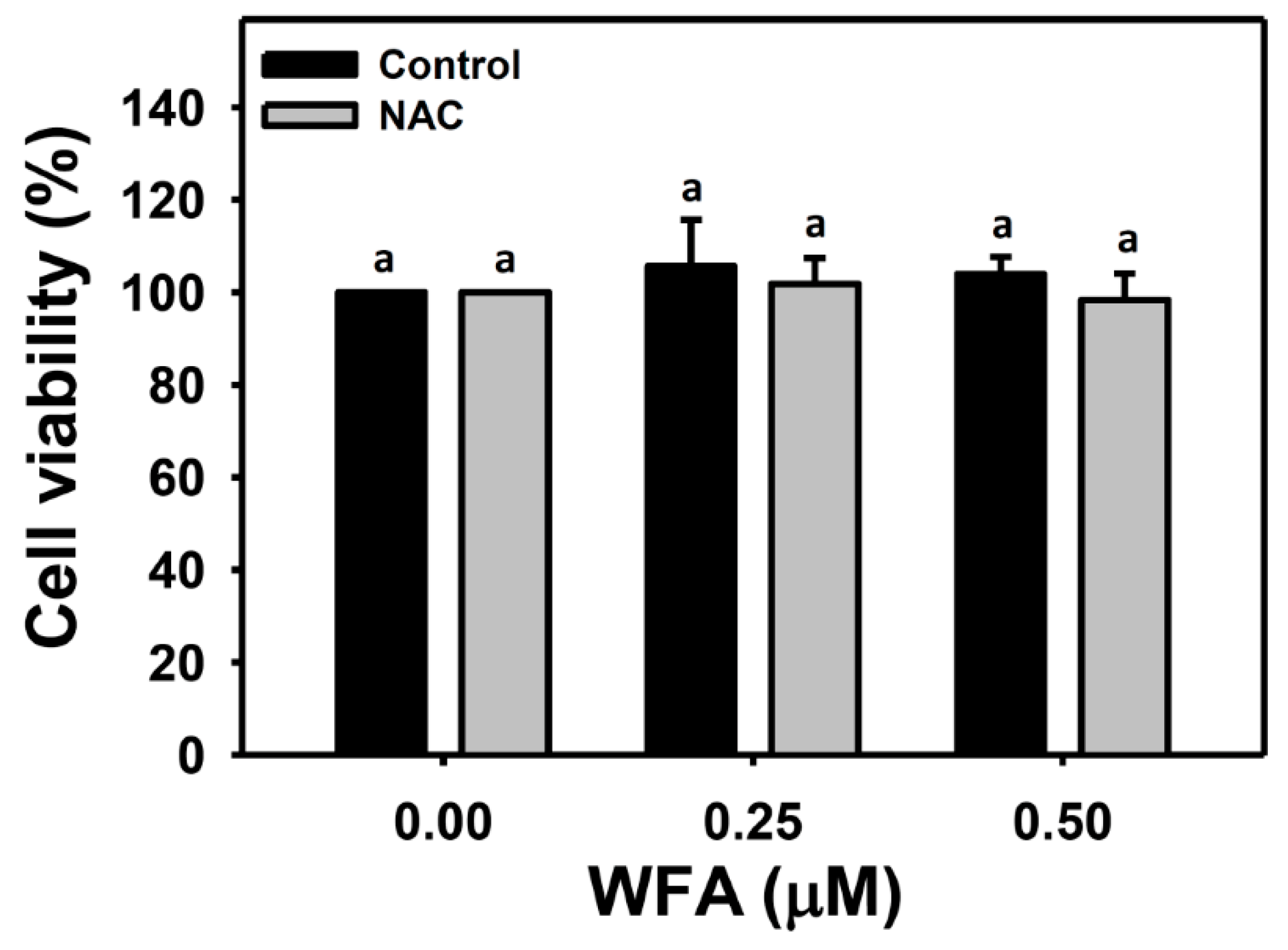
3.1. Identification of the Optimal Concentrations of WFA for Oral Cancer Cell Migration Assay

In the MTS assay (Figure 1), oral cancer cells (Ca9-22) were treated with 0, 0.25, and 0.5 μM of WFA for 24 h with or without NAC pretreatment (2 mM, 1 h). Neither the WFA nor the NAC + WFA (NAC pretreatment and WFA posttreatment) affect the viability of Ca9-22 cells. This result suggests that WFA under 0.5 μM in the single treatment (WFA) or the combined treatment (NAC + WFA) both exhibited no cytotoxic to oral cancer cells (>95% viability). These concentrations were chosen for the following migration related experiments.

3.2. ROS Generation of Oral Cancer Ca9-22 Cells at Low Concentrations of WFA

Figure 2A presented ROS patterns of Ca9-22 cells after NAC and/or WFA treatment. The ROS (+) (%) of Ca9-22 cells after low concentrations of WFA treatments were higher than those of the control, whereas this ROS generation was suppressed by NAC pretreatment (Figure 2B). Therefore, low concentrations of WFA triggered moderate ROS generation in oral cancer Ca9-22 cells.

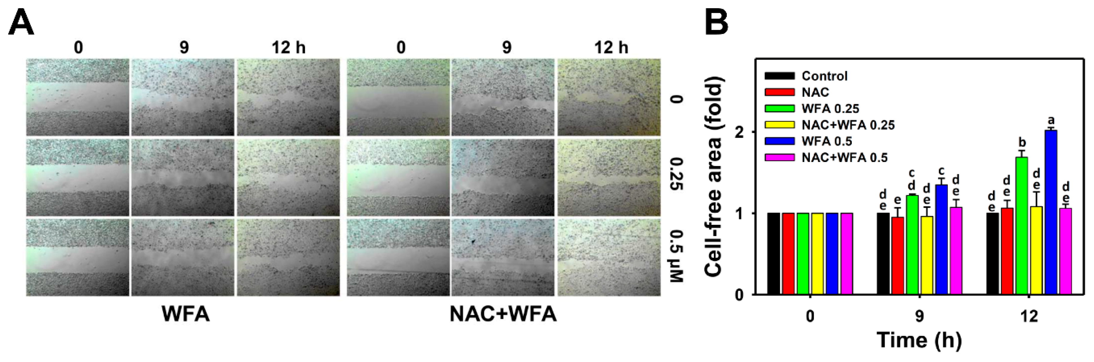
3.3. 2D Migration of Oral Cancer Ca9-22 Cells at Low Concentrations of WFA

Figure 3A demonstrated the wound healing patterns of Ca9-22 cells after NAC and/or WFA treatments. Figure 3B showed that the cell-free area (%) of Ca9-22 cells after low concentrations of WFA treatments was greater than that of the untreated control over time. In contrast, this WFA-induced increase of cell-free area (%) was suppressed by NAC pretreatment. Therefore, low concentrations of WFA triggered 2D migration inhibition in Ca9-22 cells.

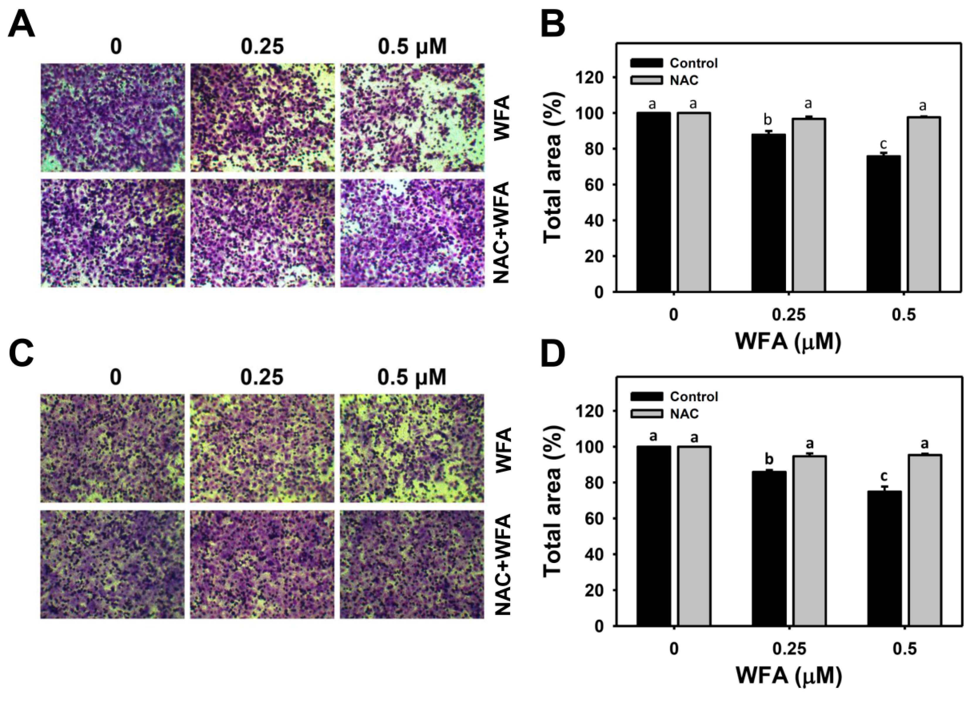
3.4. 3D Migration and Invasion Changes in Oral Cancer Ca9-22 Cells at Low Concentrations of WFA

To further confirm the 2D migration inhibitory effect of WFA, the 3D migration and invasion assays of Ca9-22 cells were performed (Figure 4A,C, respectively). Figure 4B,D showed that low concentrations of WFA suppressed transwell migration and the Matrigel invasion abilities of Ca9-22 cells in a dose-response manner. In contrast, the WFA-induced 3D migration inhibition and invasion were suppressed by NAC pretreatment. Therefore, low concentrations of WFA triggers inhibitory 3D migration and invasion in Ca9-22 cells.

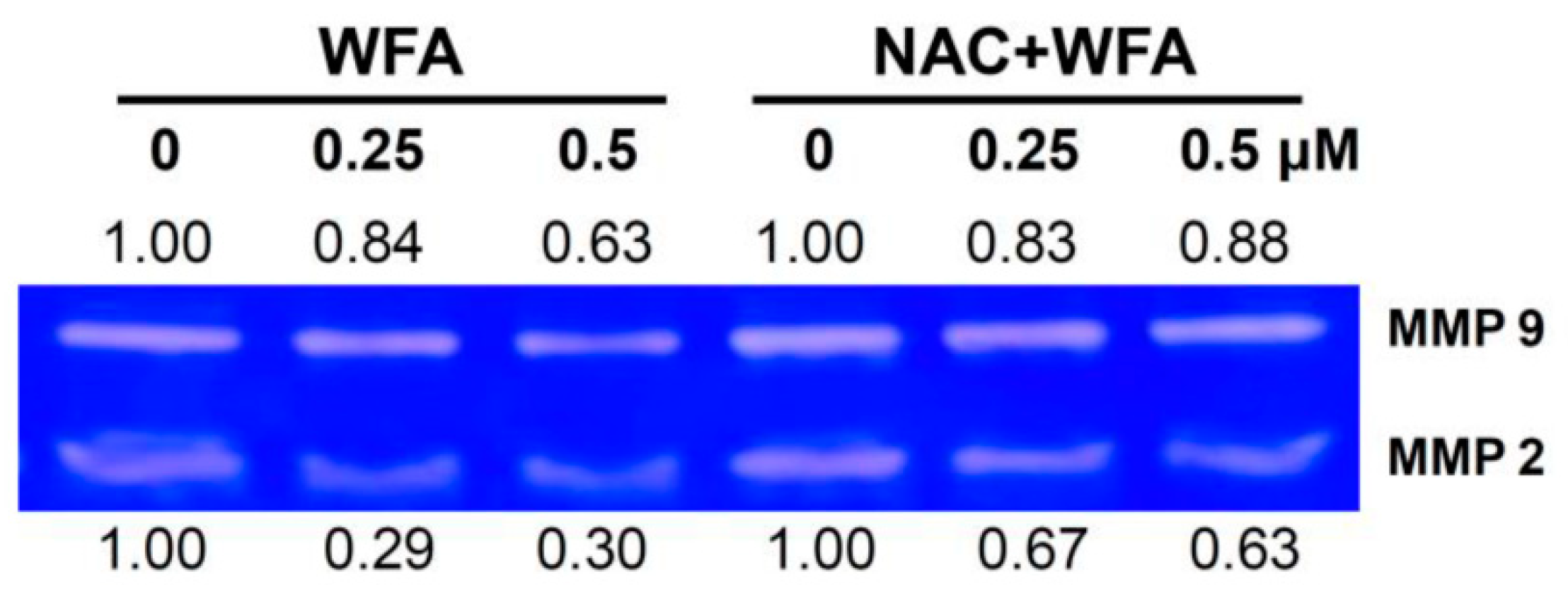
3.5. MMP-2 and MMP-9 Zymography of Oral Cancer Ca9-22 Cells at Low Concentrations of WFA

MMP-2 and MMP-9 activities were proportional to the cell invasion ability [32]. To detect MMP-2 and MMP-9 activities after low concentrations of WFA treatment, a zymography assay was performed. Figure 5 demonstrated the clear zone pattern of MMP-2 and MMP-9 in Ca9-22 cells after NAC and/or WFA treatment. It showed that the MMP-2 and MMP-9 activities of Ca9-22 cells were decreased after WFA treatment. In contrast, these WFA-induced inhibitions of MMP-2 and MMP-9 activities were suppressed by NAC pretreatment. Therefore, low concentrations of WFA triggers inhibition of MMP-2 and MMP-9 activities in Ca9-22 cells.

3.6. Antioxidant Gene Expressions of Oral Cancer Ca9-22 Cells at Low Concentrations of WFA

Under oxidative stress, ROS may activate antioxidant pathways [33,34]. Since moderate ROS is induced by low concentrations of WFA, the mRNA expressions of antioxidant genes [27], including NFE2L2, GSR, GCLC, GPX1, TXN, CAT, SOD1, HMOX1, and NQO1, were examined. Figure 6 showed that low concentrations of WFA significantly induced mRNA expressions of NFE2L2, HMOX1, GSR, and NQO1 genes while expressions of other genes were not significantly affected. Therefore, low concentrations of WFA triggers some antioxidant signaling in Ca9-22 cells.

3.7. Mitogen-Activated Protein Kinase (MAPK) Expressions of Oral Cancer Ca9-22 Cells at Low Concentrations of WFA

To further detect the potential upstream antioxidant signaling in oral cancer cells after low concentrations of WFA treatment, the activation of three members of MAPK, including ERK, JNK, and p38 MAPK was examined. Figure 7 showed that WFA induced phosphorylation of three MAPK members, i.e., p-ERK1/2, p-JNK1/2, and p-p38. In contrast, these WFA-induced MAPK phosphorylations were suppressed by NAC pretreatment. Therefore, low concentrations of WFA triggers MAPK phosphorylations in Ca9-22 cells.

4. Discussion

Previously, we discovered that high cytotoxic concentration of WFA, which was larger than IC50, selectively killed oral cancer cells but rarely damaged normal oral cells [12], i.e., IC50 value of WFA in oral cancer Ca9-22 cells is 3 µM at 24 h MTS assay. In the current study, we focus on the evaluation of the migration regulating effects of low concentration (within 0.5 μM) of WFA in oral cancer Ca9-22 cells, which show 95% viability. This low concentration of WFA inhibits 2D/3D migration, 3D invasion, MMP-2 and MMP-9 activities, whereas it induces ROS generation, antioxidant related gene mRNA expressions and MAPK phosphorylation. The detailed mechanisms for low concentration of WFA inducing inhibition of migration and invasion are discussed below.

4.1. Low Cytotoxic Concentration of Drugs Is Suitable for Migration Study

The standard criteria for studying the migration effect of drugs is based on measurements using low cytotoxic concentrations [35,36,37]. With a high cytotoxic concentration (higher than IC50), WFA had been reported to show migration inhibitory effects against gastric [14] and breast [15] cancer cells, though it may be attributed to apoptosis and cell death. Alternatively, low concentration of WFA with no cytotoxicity avoided side effect of cell death and provided a clear observation for migration response in the current study.

4.2. MMP-2 and MMP-9 Activity Changes in WFA-Treated Oral Cancer Cells

MMP-2 and MMP-9 are important mediators for cell migration, invasion, and metastasis in carcinogenesis [24]. A WFA-derived compound such as 3-azido WFA inhibits MMP-2 activity and migration of prostate PC-3 and cervical HeLa cancer cells [38]. Low concentration of WFA (>95% viability) inhibits MMP-9 activity of cervical Caski and liver SK-Hep-1 cancer cells by downregulating Akt phosphorylation [39].
In agreement with the inhibitory effect on MMP-9 activity [39], we further found that low concentration of WFA (>95% viability) exhibits inhibitory effects on MMP-2 activity in oral cancer Ca9-22 cells. Accordingly, WFA inhibits migration of oral cancer cells by inactivating MMP-2 and MMP-9. Moreover, MMP-2 and MMP-9 are overexpressed in the biopsy specimens of oral squamous cell carcinoma compared to the adjacent normal tissues [40,41]. Therefore, a low concentration of WFA has the potential to inhibit the MMP-2 and MMP-9 activities in order to inhibit migration or metastasis of oral cancer cells.

4.3. ROS Changes in WFA-Treated Oral Cancer Cells

As mentioned above, WFA exhibits a concentration-effect on apoptosis and migration, i.e., high concentration of WFA induces apoptosis while low concentration of WFA inhibits migration. Our previous study [12] demonstrated that the cytotoxic concentrations (>IC50) of WFA induced 90% (+) ROS in oral cancer Ca9-22 cells. In the current study, the low concentration (>95% viability) of WFA induces 70% (+) ROS generation in Ca9-22 cells. It is possible that low concentration of WFA induces a ROS level lower than the redox threshold and leads to cell survival with inhibitory migration. In contrast, high cytotoxic concentration of WFA induces a ROS level higher than the redox threshold and leads to apoptosis and cell death. Accordingly, the differential ROS induction by WFA may lead to distinct fate of oral cancer cells, i.e., migration inhibition or inducible apoptosis.

4.4. Antioxidant Genes Changes in WFA-Treated Oral Cancer Cells

In cancer cells, ROS overproduction is counterbalanced by overexpression of antioxidant activity for redox homeostasis [42]. Moreover, antioxidant genes have the potential to regulate cellular migration. For example, knockdown of HMOX1 and/or NFE2L2 reversed the migration inhibitory effect of semaphorin 6A (SEMA6A) and the SEMA6A-driven downregulation of MMP-9 [43]. Knockdown of NQO1 increases the invasion of human cutaneous squamous cancer SCC12 and SCC13 cells but it is reverted by NQO1 overexpression [44]. Consistently, we found that low concentrations of WFA induced mRNA expressions of NFE2L2, HMOX1, and NQO1 genes, which may lead to inhibitory migration of oral cancer cells.

4.5. MAPK Changes in WFA-Treated Oral Cancer Cells

As mentioned above, both high [12] and low (the current study) concentrations of WFA induced ROS. Moreover, ROS can regulate MAPK signaling [45], which is associated with tumor cell invasion [46]. Cytotoxic concentration of WFA induces apoptosis by phosphorylating p38 and ERK1/2 in leukemic [47] and glioblastomas cells [48], respectively. Similarly, we found that low concentration of WFA induces mild phosphorylation for ERK, JNK, and p38 MAPK.

4.6. The Role of ROS in Low Concentration of WFA Induced Migration Changes and Signaling in Oral Cancer Cells

Under low concentration of WFA, the changes of ROS generation, 2D migration, 3D migration/invasion, MMP-2/MMP-9 activities, antioxidant gene expression, and MAPK phosphorylation are reverted by NAC pretreatment. These results indicate that a low concentration of WFA inhibits migration and induces antioxidant signaling in a ROS-dependent manner in oral cancer cells.

5. Conclusions

Our study focuses on low concentrations of WFA to evaluate its inhibitory effects on migration and invasion in oral cancer Ca9-22 cells. Under low concentrations of WFA, Ca9-22 cells are grown with high viability and retained anti-migration and anti-invasion. Mechanically, this safe treatment of WFA inhibits MMP-2 and MMP-9 activities and induces antioxidant gene expression as well as MAPK activation in oral cancer cells. All these inhibitory migration changes and mechanisms after WFA treatment were suppressed by NAC pretreatment, suggesting that ROS plays an important role in WFA induced inhibitory migration in oral cancer cells. In conclusion, we provide here the first finding that supports low concentration of WFA could be a potent inhibitor for metastasis in oral cancer therapy.

Author Contributions

Conceptualization, L.-C.L. and H.-W.C.; Data curation, J.-Y.T., F.O.-Y., Y.-Y.W. and S.-S.F.Y.; Formal analysis, T.-J.Y.; Investigation, T.-J.Y.; Methodology, J.-Y.T., F.O.-Y., Y.-Y.W. and S.-S.F.Y.; Supervision, L.-C.L. and H.-W.C.; Writing—original draft, T.-J.Y. and H.-W.C.; Writing—review & editing, K.T., L.-C.L. and H.-W.C. All authors have read and agreed to the published version of the manuscript.

Funding

This work was partly supported by funds of the Ministry of Science and Technology (MOST 108-2320-B-037-015-MY3, MOST 108-2314-B-037-020, MOST 108-2314-B-037-080), the National Sun Yat-sen University-KMU Joint Research Project (#NSYSUKMU 109-I002), the Chimei-KMU jointed project (109CM-KMU-007), the Kaohsiung Medical University Research Center (KMU-TC108A04), and the Health and welfare surcharge of tobacco products, the Ministry of Health and Welfare, Taiwan, Republic of China (MOHW109-TDU-B-212-134016).

Conflicts of Interest

The authors declare no conflict of interest.

Abbreviations

WFA: withaferin A; ROS, reactive oxygen species; MMP, matrix metalloproteinase; NFE2L2, nuclear factor, erythroid 2-like 2; HMOX1, heme oxygenase 1; GSR, glutathione-disulfide reductase; NQO1, NAD(P)H quinone dehydrogenase 1; MAPK, mitogen-activated protein kinase; ERK1/2, extracellular signal-regulated kinases 1/2; JNK, c-Jun N-terminal kinase; NAC, N-acetylcysteine; DCFH-DA, 2′,7′-Dichlorodihydrofluorescein diacetate; qRT-PCR, quantitative RT-PCR; NFE2L2, nuclear factor erythroid 2-like 2; GCLC, glutamate-cysteine ligase catalytic subunit; GPX1, glutathione peroxidase 1; TXN, thioredoxin; CAT, catalase; SOD1, superoxide dismutase 1.

References

  1. Bezivin, C.; Tomasi, S.; Lohezic-Le Devehat, F.; Boustie, J. Cytotoxic activity of some lichen extracts on murine and human cancer cell lines. Phytomedicine 2003, 10, 499–503. [Google Scholar] [CrossRef] [PubMed]
  2. Jimenez, L.; Jayakar, S.K.; Ow, T.J.; Segall, J.E. Mechanisms of invasion in head and neck cancer. Arch. Pathol. Lab. Med. 2015, 139, 1334–1348. [Google Scholar] [CrossRef] [Green Version]
  3. Ghate, N.B.; Chaudhuri, D.; Sarkar, R.; Sajem, A.L.; Panja, S.; Rout, J.; Mandal, N. An antioxidant extract of tropical lichen, Parmotrema reticulatum, induces cell cycle arrest and apoptosis in breast carcinoma cell line MCF-7. PLoS ONE 2013, 8, e82293. [Google Scholar] [CrossRef] [PubMed]
  4. Noguti, J.; De Moura, C.F.; De Jesus, G.P.; Da Silva, V.H.; Hossaka, T.A.; Oshima, C.T.; Ribeiro, D.A. Metastasis from oral cancer: An overview. Cancer Genom. Proteom. 2012, 9, 329–335. [Google Scholar]
  5. Malik, F.; Kumar, A.; Bhushan, S.; Khan, S.; Bhatia, A.; Suri, K.A.; Qazi, G.N.; Singh, J. Reactive oxygen species generation and mitochondrial dysfunction in the apoptotic cell death of human myeloid leukemia HL-60 cells by a dietary compound withaferin A with concomitant protection by N-acetyl cysteine. Apoptosis 2007, 12, 2115–2133. [Google Scholar] [CrossRef] [PubMed]
  6. Munagala, R.; Kausar, H.; Munjal, C.; Gupta, R.C. Withaferin A induces p53-dependent apoptosis by repression of HPV oncogenes and upregulation of tumor suppressor proteins in human cervical cancer cells. Carcinogenesis 2011, 32, 1697–1705. [Google Scholar] [CrossRef] [Green Version]
  7. Li, X.; Zhu, F.; Jiang, J.; Sun, C.; Wang, X.; Shen, M.; Tian, R.; Shi, C.; Xu, M.; Peng, F.; et al. Synergistic antitumor activity of withaferin A combined with oxaliplatin triggers reactive oxygen species-mediated inactivation of the PI3K/AKT pathway in human pancreatic cancer cells. Cancer Lett. 2015, 357, 219–230. [Google Scholar] [CrossRef]
  8. Wang, H.C.; Hu, H.H.; Chang, F.R.; Tsai, J.Y.; Kuo, C.Y.; Wu, Y.C.; Wu, C.C. Different effects of 4beta-hydroxywithanolide E and withaferin A, two withanolides from Solanaceae plants, on the Akt signaling pathway in human breast cancer cells. Phytomedicine 2019, 53, 213–222. [Google Scholar] [CrossRef]
  9. Hsu, J.H.; Chang, P.M.; Cheng, T.S.; Kuo, Y.L.; Wu, A.T.; Tran, T.H.; Yang, Y.H.; Chen, J.M.; Tsai, Y.C.; Chu, Y.S.; et al. Identification of withaferin A as a potential candidate for anti-cancer therapy in non-small cell lung cancer. Cancers 2019, 11, 1003. [Google Scholar] [CrossRef] [Green Version]
  10. Xia, S.; Miao, Y.; Liu, S. Withaferin A induces apoptosis by ROS-dependent mitochondrial dysfunction in human colorectal cancer cells. Biochem. Biophys. Res. Commun. 2018, 503, 2363–2369. [Google Scholar] [CrossRef]
  11. Yang, I.-H.; Kim, L.-H.; Shin, J.-A.; Cho, S.-D. Chemotherapeutic effect of withaferin A in human oral cancer cells. J. Cancer Ther. 2015, 6, 735–742. [Google Scholar] [CrossRef] [Green Version]
  12. Chang, H.W.; Li, R.N.; Wang, H.R.; Liu, J.R.; Tang, J.Y.; Huang, H.W.; Chan, Y.H.; Yen, C.Y. Withaferin A induces oxidative stress-mediated apoptosis and DNA damage in oral cancer cells. Front. Physiol. 2017, 8, 634. [Google Scholar] [CrossRef] [PubMed] [Green Version]
  13. Ghosh, K.; De, S.; Mukherjee, S.; Das, S.; Ghosh, A.N.; Sengupta, S.B. Withaferin A induced impaired autophagy and unfolded protein response in human breast cancer cell-lines MCF-7 and MDA-MB-231. Toxicol. In Vitro 2017, 44, 330–338. [Google Scholar] [CrossRef] [PubMed]
  14. Kim, G.; Kim, T.H.; Hwang, E.H.; Chang, K.T.; Hong, J.J.; Park, J.H. Withaferin A inhibits the proliferation of gastric cancer cells by inducing G2/M cell cycle arrest and apoptosis. Oncol. Lett. 2017, 14, 416–422. [Google Scholar] [CrossRef] [Green Version]
  15. Liu, X.; Li, Y.; Ma, Q.; Wang, Y.; Song, A.L. Withaferin-A inhibits growth of drug-resistant breast carcinoma by inducing apoptosis and autophagy, endogenous reactive oxygen species (ROS) production, and inhibition of cell migration and nuclear factor kappa B (Nf-kappaB)/mammalian target of rapamycin (m-TOR) signalling pathway. Med. Sci. Monit. 2019, 25, 6855–6863. [Google Scholar]
  16. Hurd, T.R.; DeGennaro, M.; Lehmann, R. Redox regulation of cell migration and adhesion. Trends Cell Biol. 2012, 22, 107–115. [Google Scholar] [CrossRef] [Green Version]
  17. Chang, Y.T.; Huang, C.Y.; Li, K.T.; Li, R.N.; Liaw, C.C.; Wu, S.H.; Liu, J.R.; Sheu, J.H.; Chang, H.W. Sinuleptolide inhibits proliferation of oral cancer Ca9-22 cells involving apoptosis, oxidative stress, and DNA damage. Arch. Oral Biol. 2016, 66, 147–154. [Google Scholar] [CrossRef]
  18. Huang, C.H.; Yeh, J.M.; Chan, W.H. Hazardous impacts of silver nanoparticles on mouse oocyte maturation and fertilization and fetal development through induction of apoptotic processes. Environ. Toxicol. 2018, 33, 1039–1049. [Google Scholar] [CrossRef]
  19. Wang, T.S.; Lin, C.P.; Chen, Y.P.; Chao, M.R.; Li, C.C.; Liu, K.L. CYP450-mediated mitochondrial ROS production involved in arecoline N-oxide-induced oxidative damage in liver cell lines. Environ. Toxicol. 2018, 33, 1029–1038. [Google Scholar] [CrossRef]
  20. Yen, Y.H.; Farooqi, A.A.; Li, K.T.; Butt, G.; Tang, J.Y.; Wu, C.Y.; Cheng, Y.B.; Hou, M.F.; Chang, H.W. Methanolic extracts of Solieria robusta inhibits proliferation of oral cancer Ca9-22 cells via apoptosis and oxidative stress. Molecules 2014, 19, 18721–18732. [Google Scholar] [CrossRef] [Green Version]
  21. Yen, C.Y.; Chiu, C.C.; Haung, R.W.; Yeh, C.C.; Huang, K.J.; Chang, K.F.; Hseu, Y.C.; Chang, F.R.; Chang, H.W.; Wu, Y.C. Antiproliferative effects of goniothalamin on Ca9-22 oral cancer cells through apoptosis, DNA damage and ROS induction. Mutat. Res. 2012, 747, 253–258. [Google Scholar] [CrossRef] [PubMed]
  22. Chiu, C.C.; Liu, P.L.; Huang, K.J.; Wang, H.M.; Chang, K.F.; Chou, C.K.; Chang, F.R.; Chong, I.W.; Fang, K.; Chen, J.S.; et al. Goniothalamin inhibits growth of human lung cancer cells through DNA damage, apoptosis, and reduced migration ability. J. Agric. Food Chem. 2011, 59, 4288–4293. [Google Scholar] [CrossRef] [PubMed]
  23. Peng, S.Y.; Hsiao, C.C.; Lan, T.H.; Yen, C.Y.; Farooqi, A.A.; Cheng, C.M.; Tang, J.Y.; Yu, T.J.; Yeh, Y.C.; Chuang, Y.T.; et al. Pomegranate extract inhibits migration and invasion of oral cancer cells by downregulating matrix metalloproteinase-2/9 and epithelial-mesenchymal transition. Environ. Toxicol. 2020, 35, 673–682. [Google Scholar] [CrossRef] [PubMed]
  24. Wang, C.Y.; Lin, C.S.; Hua, C.H.; Jou, Y.J.; Liao, C.R.; Chang, Y.S.; Wan, L.; Huang, S.H.; Hour, M.J.; Lin, C.W. Cis-3-O-p-hydroxycinnamoyl ursolic acid induced ROS-dependent p53-mediated mitochondrial apoptosis in oral cancer cells. Biomol. Ther. (Seoul) 2019, 27, 54–62. [Google Scholar] [CrossRef]
  25. Chang, H.W.; Yen, C.Y.; Chen, C.H.; Tsai, J.H.; Tang, J.Y.; Chang, Y.T.; Kao, Y.H.; Wang, Y.Y.; Yuan, S.F.; Lee, S.Y. Evaluation of the mRNA expression levels of integrins alpha3, alpha5, beta1 and beta6 as tumor biomarkers of oral squamous cell carcinoma. Oncol. Lett. 2018, 16, 4773–4781. [Google Scholar]
  26. Yen, C.Y.; Huang, C.Y.; Hou, M.F.; Yang, Y.H.; Chang, C.H.; Huang, H.W.; Chen, C.H.; Chang, H.W. Evaluating the performance of fibronectin 1 (FN1), integrin alpha4beta1 (ITGA4), syndecan-2 (SDC2), and glycoprotein CD44 as the potential biomarkers of oral squamous cell carcinoma (OSCC). Biomarkers 2013, 18, 63–72. [Google Scholar] [CrossRef]
  27. Stagos, D.; Balabanos, D.; Savva, S.; Skaperda, Z.; Priftis, A.; Kerasioti, E.; Mikropoulou, E.V.; Vougogiannopoulou, K.; Mitakou, S.; Halabalaki, M.; et al. Extracts from the mediterranean food plants Carthamus lanatus, Cichorium intybus, and Cichorium spinosum enhanced GSH levels and increased Nrf2 expression in human endothelial cells. Oxid. Med. Cell Longev. 2018, 2018, 6594101. [Google Scholar] [CrossRef] [Green Version]
  28. Livak, K.J.; Schmittgen, T.D. Analysis of relative gene expression data using real-time quantitative PCR and the 2(-Delta Delta C(T)) Method. Methods 2001, 25, 402–408. [Google Scholar] [CrossRef]
  29. Brunn, G.J.; Williams, J.; Sabers, C.; Wiederrecht, G.; Lawrence, J.C., Jr.; Abraham, R.T. Direct inhibition of the signaling functions of the mammalian target of rapamycin by the phosphoinositide 3-kinase inhibitors, wortmannin and LY294002. EMBO J. 1996, 15, 5256–5267. [Google Scholar] [CrossRef]
  30. Fujii, Y.; Yoshihashi, K.; Suzuki, H.; Tsutsumi, S.; Mutoh, H.; Maeda, S.; Yamagata, Y.; Seto, Y.; Aburatani, H.; Hatakeyama, M. CDX1 confers intestinal phenotype on gastric epithelial cells via induction of stemness-associated reprogramming factors SALL4 and KLF5. Proc. Natl. Acad. Sci. USA 2012, 109, 20584–20589. [Google Scholar] [CrossRef] [Green Version]
  31. Baribeau, S.; Chaudhry, P.; Parent, S.; Asselin, E. Resveratrol inhibits cisplatin-induced epithelial-to-mesenchymal transition in ovarian cancer cell lines. PLoS ONE 2014, 9, e86987. [Google Scholar] [CrossRef] [PubMed] [Green Version]
  32. Chien, Y.C.; Liu, L.C.; Ye, H.Y.; Wu, J.Y.; Yu, Y.L. EZH2 promotes migration and invasion of triple-negative breast cancer cells via regulating TIMP2-MMP-2/-9 pathway. Am. J. Cancer Res. 2018, 8, 422–434. [Google Scholar] [PubMed]
  33. Chen, J.; Zhang, Z.; Cai, L. Diabetic cardiomyopathy and its prevention by nrf2: Current status. Diabetes Metab. J. 2014, 38, 337–345. [Google Scholar] [CrossRef] [PubMed] [Green Version]
  34. Hardingham, G.E.; Do, K.Q. Linking early-life NMDAR hypofunction and oxidative stress in schizophrenia pathogenesis. Nat. Rev. Neurosci. 2016, 17, 125–134. [Google Scholar] [CrossRef] [PubMed]
  35. Wang, H.C.; Chu, Y.L.; Hsieh, S.C.; Sheen, L.Y. Diallyl trisulfide inhibits cell migration and invasion of human melanoma a375 cells via inhibiting integrin/facal adhesion kinase pathway. Environ. Toxicol. 2017, 32, 2352–2359. [Google Scholar] [CrossRef] [PubMed]
  36. Shih, Y.L.; Au, M.K.; Liu, K.L.; Yeh, M.Y.; Lee, C.H.; Lee, M.H.; Lu, H.F.; Yang, J.L.; Wu, R.S.; Chung, J.G. Ouabain impairs cell migration, and invasion and alters gene expression of human osteosarcoma U-2 OS cells. Environ. Toxicol. 2017, 32, 2400–2413. [Google Scholar] [CrossRef]
  37. Yeh, C.M.; Hsieh, M.J.; Yang, J.S.; Yang, S.F.; Chuang, Y.T.; Su, S.C.; Liang, M.Y.; Chen, M.K.; Lin, C.W. Geraniin inhibits oral cancer cell migration by suppressing matrix metalloproteinase-2 activation through the FAK/Src and ERK pathways. Environ. Toxicol. 2019, 34, 1085–1093. [Google Scholar] [CrossRef]
  38. Rah, B.; Amin, H.; Yousuf, K.; Khan, S.; Jamwal, G.; Mukherjee, D.; Goswami, A. A novel MMP-2 inhibitor 3-azidowithaferin A (3-azidoWA) abrogates cancer cell invasion and angiogenesis by modulating extracellular Par-4. PLoS ONE 2012, 7, e44039. [Google Scholar] [CrossRef]
  39. Lee, D.H.; Lim, I.H.; Sung, E.G.; Kim, J.Y.; Song, I.H.; Park, Y.K.; Lee, T.J. Withaferin A inhibits matrix metalloproteinase-9 activity by suppressing the Akt signaling pathway. Oncol. Rep. 2013, 30, 933–938. [Google Scholar] [CrossRef] [Green Version]
  40. Tsai, C.H.; Hsieh, Y.S.; Yang, S.F.; Chou, M.Y.; Chang, Y.C. Matrix metalloproteinase 2 and matrix metalloproteinase 9 expression in human oral squamous cell carcinoma and the effect of protein kinase C inhibitors: Preliminary observations. Oral Surg. Oral Med. Oral Pathol. Oral Radiol. Endod. 2003, 95, 710–716. [Google Scholar] [CrossRef]
  41. Patel, B.P.; Shah, P.M.; Rawal, U.M.; Desai, A.A.; Shah, S.V.; Rawal, R.M.; Patel, P.S. Activation of MMP-2 and MMP-9 in patients with oral squamous cell carcinoma. J. Surg. Oncol. 2005, 90, 81–88. [Google Scholar] [CrossRef] [PubMed]
  42. Gorrini, C.; Harris, I.S.; Mak, T.W. Modulation of oxidative stress as an anticancer strategy. Nat. Rev. Drug Discov. 2013, 12, 931–947. [Google Scholar] [CrossRef] [PubMed]
  43. Chen, L.H.; Liao, C.Y.; Lai, L.C.; Tsai, M.H.; Chuang, E.Y. Semaphorin 6A attenuates the migration capability of lung cancer cells via the NRF2/HMOX1 axis. Sci. Rep. 2019, 9, 13302. [Google Scholar] [CrossRef] [PubMed] [Green Version]
  44. Zhang, Q.L.; Li, X.M.; Lian, D.D.; Zhu, M.J.; Yim, S.H.; Lee, J.H.; Jiang, R.H.; Kim, C.D. Tumor suppressive function of NQO1 in cutaneous squamous cell carcinoma (SCC) cells. Biomed. Res. Int. 2019, 2019, 2076579. [Google Scholar] [CrossRef] [PubMed] [Green Version]
  45. Chen, Y.C.; Lu, M.C.; El-Shazly, M.; Lai, K.H.; Wu, T.Y.; Hsu, Y.M.; Lee, Y.L.; Liu, Y.C. Breaking down leukemia walls: Heteronemin, a sesterterpene derivative, induces apoptosis in leukemia Molt4 cells through oxidative stress, mitochondrial dysfunction and induction of talin Expression. Mar. Drugs 2018, 16, 212. [Google Scholar] [CrossRef] [Green Version]
  46. Peng, Q.; Deng, Z.; Pan, H.; Gu, L.; Liu, O.; Tang, Z. Mitogen-activated protein kinase signaling pathway in oral cancer. Oncol. Lett. 2018, 15, 1379–1388. [Google Scholar] [CrossRef] [Green Version]
  47. Mandal, C.; Dutta, A.; Mallick, A.; Chandra, S.; Misra, L.; Sangwan, R.S.; Mandal, C. Withaferin A induces apoptosis by activating p38 mitogen-activated protein kinase signaling cascade in leukemic cells of lymphoid and myeloid origin through mitochondrial death cascade. Apoptosis 2008, 13, 1450–1464. [Google Scholar] [CrossRef]
  48. Grogan, P.T.; Sleder, K.D.; Samadi, A.K.; Zhang, H.; Timmermann, B.N.; Cohen, M.S. Cytotoxicity of withaferin A in glioblastomas involves induction of an oxidative stress-mediated heat shock response while altering Akt/mTOR and MAPK signaling pathways. Investig. New Drugs 2013, 31, 545–557. [Google Scholar] [CrossRef] [Green Version]
Figure 1. Viability of low concentration of Withaferin A (WFA) treatment in oral cancer cells. Oral cancer cells (Ca9-22) were pretreated with or without N-acetylcysteine (NAC) (2 mM, 1 h) and post-treated with different concentrations of WFA for 24 h. For multiple comparison, treatments with the same letter character show nonsignificant difference. Data, mean ± SD (n = 3).
Biomolecules 10 00777 g001
Figure 2. ROS generation effects of low concentrations of WFA in oral cancer cells. (A) ROS patterns of Ca9-22 cells after NAC and/or WFA treatments. Cells were pretreated with or without NAC (2 mM, 1 h) and post-treated with different concentrations of WFA for 24 h, i.e., NAC + WFA vs. WFA. ROS-positive population is marked as ROS (+). (B) Statistics of ROS change in Figure 2A. For multiple comparison, treatments without the same labels (a,b) indicate the significant difference. p < 0.05~0.001. Data, mean ± SD (n = 3).
Biomolecules 10 00777 g002
Figure 3. Two-dimensional anti-migration effects of low concentrations of WFA in oral cancer cells. (A) Two-dimensional migration (wound healing) images of Ca9-22 cells after NAC and/or WFA treatments. Cells were pretreated with or without NAC (2 mM, 1 h) and post-treated with different concentrations of WFA for 0, 9 and 12 h. (B) Statistics of 2D migration change in Figure 3A. For multiple comparison, treatments without the same labels (a–e) indicate the significant difference. p < 0.05~0.0001. Data, mean ± SD (n = 3).
Biomolecules 10 00777 g003
Figure 4. Three-dimensional anti-migration and -invasion effects of low concentrations of WFA in oral cancer cells. (A,C) 3D migration and invasion images of Ca9-22 cells after NAC and/or WFA treatments. Cells were pretreated with or without NAC (2 mM, 1 h) and post-treated with different concentrations of WFA for 21 h. (B,D) Statistics of 3D migration and invasion changes in Figure 4A,B. For multiple comparison, treatments without the same labels (a–c) indicate the significant difference. p < 0.001~0.0001 (B) and p < 0.01~0.001 (D). Data, mean ± SD (n = 3).
Biomolecules 10 00777 g004
Figure 5. MMP-2 and MMP-9 activities of low concentrations of WFA in oral cancer cells. Zymography-detecting MMP-2 and MMP-9 activities in Ca9-22 cells after NAC and/or WFA treatments. Cells were pretreated with or without NAC (2 mM, 1 h) and post-treated with different concentrations of WFA for 48 h. Similar experiments were repeated 3 times.
Biomolecules 10 00777 g005
Figure 6. mRNA expressions of antioxidant genes of low concentrations of WFA in oral cancer cells. Cells were treated with or without 0.5 μM of WFA for 24 h. Treatments (control vs. WFA) without the same labels (a,b) indicate the significant difference. p < 0.05~0.01. Data, mean ± SD (n = 2).
Biomolecules 10 00777 g006
Figure 7. MAPK changes of low concentrations of WFA in oral cancer cells. Ca9-22 cells were pretreated with or without NAC (2 mM, 1 h) and post-treated with different concentrations of WFA for 24 h. ERK1/2, JNK1/2, p38, p-ERK1/2, p-JNK1/2, and p-p38 expressions were detected by Western blotting. The intensity ratio for each p-MAPK expression was adjusted to its matched MAPK and GAPDH intensities. Similar experiments were repeated 3 times.
Biomolecules 10 00777 g007
Table 1. Primer information for antioxidant-associated genes *.
GenesForward Primers (5′→3′)Reverse Primers (5′→3′)Length
TXNGAAGCAGATCGAGAGCAAGACTGGCTCCAGAAAATTCACCCACCT270 bp
GSRGTTCTCCCAGGTCAAGGAGGTTAACCAGCAGCTATTGCAACTGGAGT297 bp
CATATGCAGGACAATCAGGGTGGTCCTCAGTGAAGTTCTTGACCGCT274 bp
SOD1AGGGCATCATCAATTTCGAGC [29]CCCAAGTCTCCAACATGCCTC211 bp
HMOX1CCTTCTTCACCTTCCCCAACATGGCAGAATCTTGCACTTTGTTGC251 bp
NFE2L2GATCTGCCAACTACTCCCAGGTTCTGTAACTCAGGAATGGATAATAGCTCC302 bp
NQO1GAAGGACCCTGCGAACTTTCAGTAGAAAGCACTGCCTTCTTACTCCG258 bp
GCLCACAAGCACCCTCGCTTCAGTACCCTGCAGGCTTGGAATGTCACCT232 bp
GPX1AACCAGTTTGGGCATCAGGAGAGTTCCAGGCAACATCGTTGC256 bp
GAPDHCCTCAACTACATGGTTTACATGTTCC [30]CAAATGAGCCCCAGCCTTCT [31]220 bp
* Primers without reference were designed in this study.

