Article

Placenta, Pericarp, and Seeds of Tabasco Chili Pepper Fruits Show a Contrasting Diversity of Bioactive Metabolites

by
Felipe Cervantes-Hernández
,
Paul Alcalá-González
,
Octavio Martínez
and
José Juan Ordaz-Ortiz
*
Unidad de Genómica Avanzada, Centro de Investigación y de Estudios Avanzados del Instituto Politécnico Nacional (CINVESTAV), Km. 9.6, Libramiento Norte Carretera Irapuato-León, Irapuato, Gto. 36824, Mexico
*
Author to whom correspondence should be addressed.
Metabolites 2019, 9(10), 206; https://doi.org/10.3390/metabo9100206
Submission received: 26 August 2019 / Revised: 21 September 2019 / Accepted: 23 September 2019 / Published: 28 September 2019
(This article belongs to the Special Issue Fruit Metabolism and Metabolomics)

Abstract

:
Chili pepper (Capsicum spp.) is one of the most important horticultural crops worldwide, and its unique organoleptic properties and health benefits have been established for centuries. However, there is little knowledge about how metabolites are distributed throughout fruit parts. This work focuses on the use of liquid chromatography coupled with high resolution mass spectrometry (UHPLC-ESI-HRMS) to estimate the global metabolite profiles of the pericarp, placenta, and seeds of Tabasco pepper fruits (Capsicum frutescens L.) at the red mature stage of ripening. Our main results putatively identified 60 differential compounds between these tissues and seeds. Firstly, we found that pericarp has a higher content of glycosides, showing on average a fold change of 5 and a fold change of 14 for terpenoids when compared with other parts of the fruit. While placenta was the richest tissue in capsaicinoid-related compounds, alkaloids, and tocopherols, with a 35, 3, and 7 fold change, respectively. However, the seeds were richer in fatty acids and saponins with fold changes of 86 and 224, respectively. Therefore, our study demonstrates that a non-targeted metabolomic approach may help to improve our understanding of unexplored areas of plant metabolism and also may be the starting point for a detailed analysis in complex plant parts, such as fruits.

1. Introduction

Chili pepper (Capsicum spp.) is one of the most important crops worldwide. It is used as a main ingredient for many dishes in different cultures, such as Asian, Latin-American, and Mediterranean cultures, due to its organoleptic properties [1]. There are 40 accepted chili species but only five are considered domesticated: C. annuum, C. chinense Jacq, C. frutescens, C. baccatum, and C. pubescens [2]. In 2017, 36 million tons of chili peppers were produced globally, with Mexico being the second largest producer [3]. Wild pepper populations of Tabasco pepper (C. frutescens L.) and C. annuum can be found in some states of Mexico, increasing the relevance for collecting and characterizing these species as resources for the breeding of cultivated peppers [4,5]. Previous studies have extensively described metabolite diversity in C. annuum [6,7] but very little is known on C. chinense and C. frutescens. Most of these studies have undertaken a targeted approach, where the main focus has been to quantitate for specific metabolites, such as capsiate, dihydrocapsiate, capsaicin, dihydrocapsaicin, carotenoids, fatty acids, and amino acids [8,9]. To date, there are no studies with a comprehensive global profiling of tissue specific in C. frutescens and C. chinense.
Capsicum species are known to be rich in compounds such as capsaicinoids, capsinoids, carotenoids, flavonoids, vitamins, essential oils, and other phytochemicals, which provide a unique taste, aromatic properties, and health benefits [10,11,12,13,14]. Capsaicinoids consist mainly of two congeners, capsaicin and dihydrocapsaicin. Capsinoids also have two major analogues, capsiate and dihydrocapsiate. However, there are structural differences: Capsaicinoids are fatty acid amides linked with vanillylamine, whereas capsinoids are fatty acid esters linked to vanillyl alcohol [15,16]. Several compounds identified in the Capsicum species have been studied, because the medicinal potential examples of these compounds are icariside E5, capsosides, and capsianosides [17]; hydroxycinnamic derivatives, O-glycosides of quercetin, luteolin, and chrysoeriol [7]. The reported medicinal benefits are related to areas, such as anti-inflammatory [18], anti-cancer [19], anti-microbial, antioxidant properties [1,20,21], and those with weight-loss properties [22]. In addition, some epidemiological studies of a number of these antioxidants reported that they possess anti-atherosclerotic, antitumor, antimutagenic or anticarcinogenic activity [23,24,25]. It may be that these properties have the ability to help us to address the identification, isolation, and production of nutraceutical compounds or new natural medicinal compounds.
Nevertheless, the location and relative abundance of these metabolites and their precursors in different parts of the chili pepper fruit, such as the pericarp, placenta and seed, remain unclear. It is known that some compounds are synthesized and accumulated into specific tissues in the Capsicum genus [1,26]; for example, capsaicin is synthesized mainly in the placenta [27,28], while anthocyanins are described as being accumulated in pericarp during fruit development [29]. Materska, reported a placental and pericarp comparison in chili pepper fruit, where placenta was the richest in flavonoids, while the pericarp presented a larger diversity in glycosylated compounds. Despite this, little is known about the tissue specific spatial-temporal location of other classes of compounds and products of the secondary metabolism in Capsicum fruits [30]. Investigating the metabolic diversity on fruit tissues is essential in order to gain a comprehensive understanding of the function of specific parts of the fruit at the fundamental level. Consequently, this will enable the possible exploitation of natural products either for pharmaceutical or food products. In the past, these have been done with histochemical methods, by staining tissues sections with various chemical to reveal the presence of specific compounds either by visual or microscope inspection [31].
Metabolomics is defined as the comprehensive analysis of all low molecular weight organic compounds (<1500 Da) in a biological system [32]. Mass spectrometry has become the most widely applied platform for metabolomics, due to the wide range of molecules that can be analyzed on a single run [33]. Global profiling or non-targeted mass spectrometry-based metabolomics have gained importance in the study of crop species and have been applied to investigate potato, tomato, rice, wheat, strawberry, cucumber, and tobacco [34,35,36,37]. In the field of plant metabolomics, liquid chromatography coupled with electrospray ionization high resolution mass spectrometry (UPLC-ESI-HRMS) has emerged as the technique of choice for the putative identification of metabolites in complex matrices. This technique has been widely used, due to its sensitivity, selectivity, and analysis capability [38,39]. Nevertheless, metabolite identification for unknown compounds still remains a big challenge to overcome. In that respect, the recommendations by the Metabolomics Standards Initiative (MSI) recognize five different levels for metabolite confidence annotations. Level 0 requires the full compound 3D structure and stereochemistry information. Levels which are more common include: Level 1 identifications need a confirmation by two orthogonal parameters such as retention time and MS/MS spectrum, normally with match reference standards; and Level 2 requires at least two orthogonal pieces of information, including evidence that excludes all other candidates. Data for Level 2 should describe probable structure and be matched to literature data or databases by diagnostic evidence [40].
Combining existing bioinformatic tools with high resolution mass spectrometry data can reveal unclear relationships of metabolites and their possible function at a spatial-temporal distribution level. As a first attempt to construct the chili fruit metabolome, we produced a hand curated dataset, that contains 60 putative identified metabolites, which include alkaloids and terpenoids that are unreported in Capsicum, with significant differences and relative abundances between three sections (pericarp, placenta, and seed) of Tabasco pepper fruit at the mature red stage, using an UHPLC-QTOF-HRMS platform, combined with the use of Progenesis QI for small molecules, as a tool for the pre-identification of unknown metabolites. Our results underline the global metabolic differences in complexity, mainly based on the secondary metabolism of these fruit parts.

2. Results

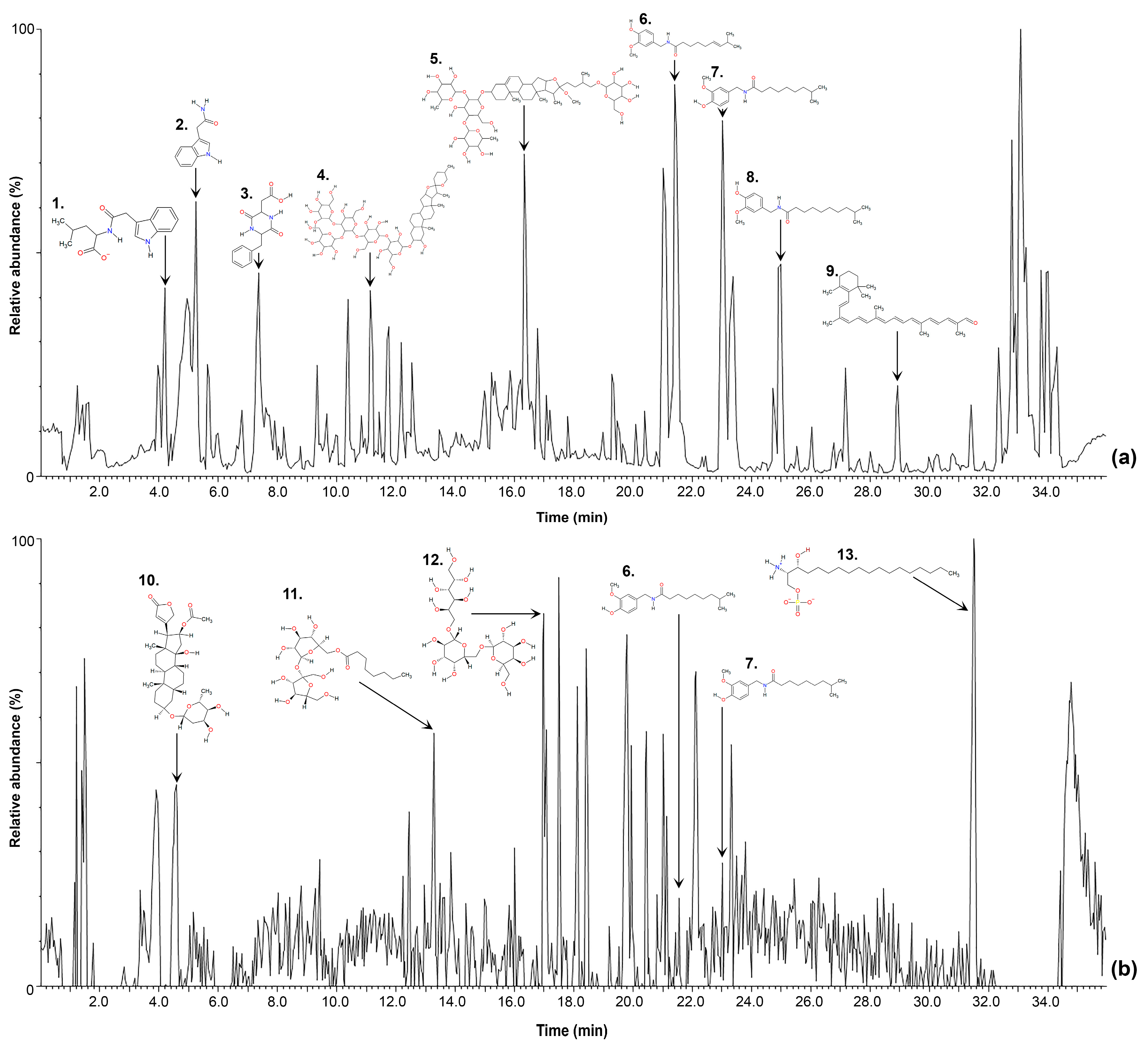
2.1. Non-Targeted Metabolomic Analysis

Our data for two tissues and the seed of Tabasco chili fruit comprised a total of 87 files or chromatograms per ionization mode (positive and negative polarities) as shown in Figure 1 and were uploaded into Progenesis QI. The dataset was first aligned (retention time): Each chromatogram was aligned against each other and automatically compared to a reference profile selected by the software that contained the highest number of features (potential compounds). Then, peak picking was performed by default parameters. A total of 1980 features were detected in the aqueous phase and 1481 were detected in the diethyl ether extract, for both ionization modes. Figure 1 shows a typical chromatographic profile in positive and negative ionization mode of placenta tissue, displaying some representative compounds of chili pepper fruit.
Distribution of the features between parts was compared using principal component analysis (PCA) of loading and score plots describing a significant grouping by part for both extraction phases (Figure 2). In addition, quality controls (QC samples) were also considered, as shown in Figure 2. The QC samples cluster tightly in comparison to the total variance in the projection, suggesting a dataset deemed to be of high quality. Table 1 and Table 2 describe loadings that mostly contribute to principal components for each extraction. Putative organic compounds catalogued as saponins (SPNS), such as Tuberoside J, Matesaponin 5, and Asparagoside B significantly contributed to component 1 in the aqueous phase, while other important features for component 2 belong to flavonoid class (FLV) and SPNS compounds. Furthermore, both components in the organic phase were mainly composed of glycerolipids (GL) and terpenoids (TER), as well as a putative carotenoid, (5cis,5′cis,9cis,11′cis)-1,2,7,7′,8,8′-Hexahydro-1,2-epoxy-ψ, ψ-carotene were important for contribution of component 1 in organic phase.

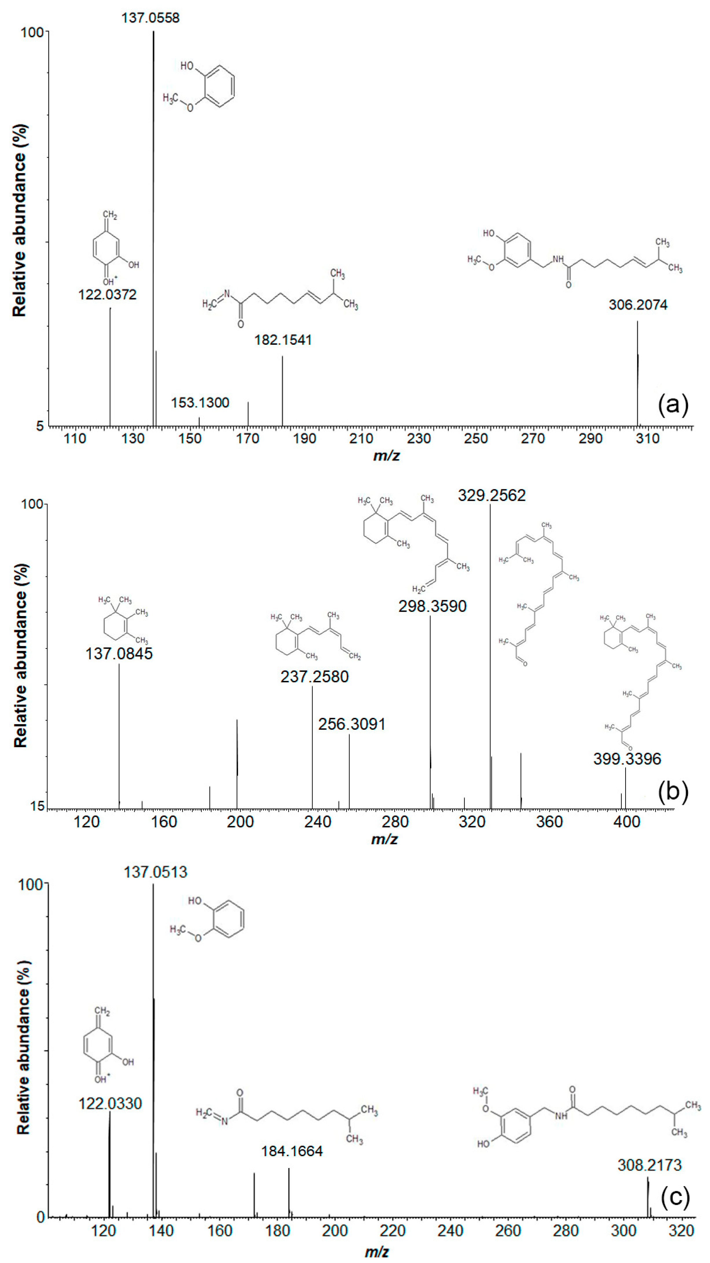
2.2. Level 1 and 2 Metabolomic Identification Analysis

We putatively identified approximately 270 compounds and classified them in 52 compound classes (Table S1). Putative identifications were taken into consideration with a high match score (>90%). Terpenoids, fatty acids, and glycosylated compounds were the most abundant groups. As was predicted, different capsaicinoid compounds were also detected with a high match score. Alkaloids, carotenoids, saponins, and phenolic compounds were also detected in our study.
Capsaicin and dihydrocapsaicin were validated by matching their retention times and MS/MS spectra with those of the analytical standard (Level 1 identification). Furthermore, commonly reported compounds, such as carotenoids and capsaicin related compounds, were detected and putatively identified in the same manner in our samples (Figure 3).

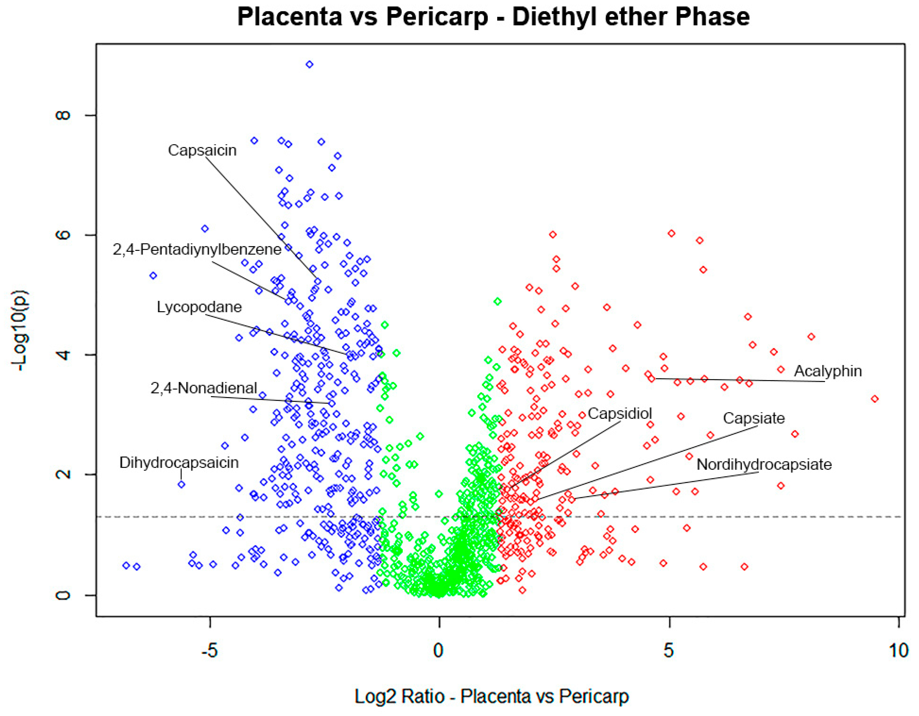
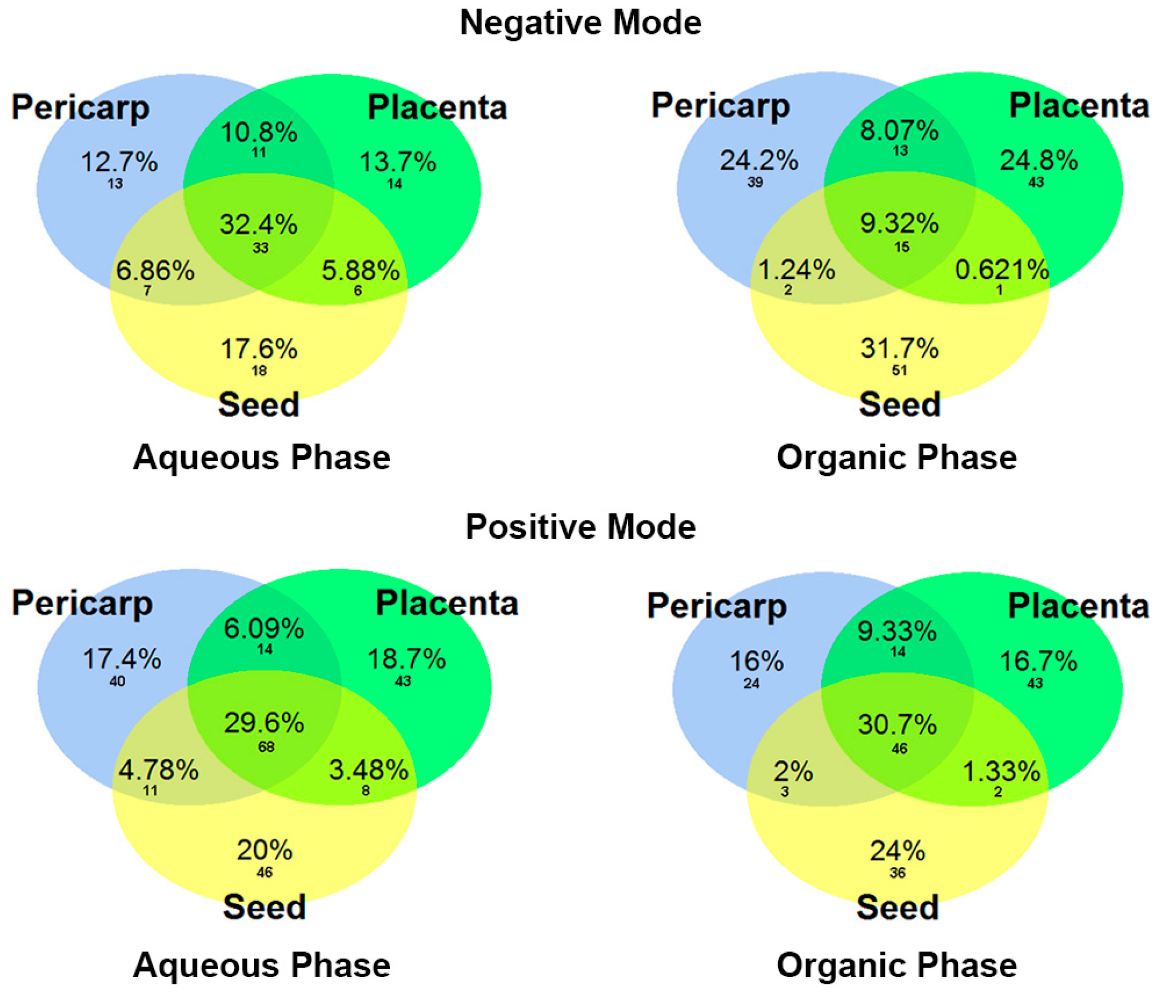
2.3. Different Metabolomic Profiles in Capsicum Sections

Based on results from volcano plot comparisons, we found a total of 60 putative compounds with significant differences between the parts of the Tabasco pepper fruit. Figure 4 shows the distribution of features between placenta and pericarp tissues. As was expected [28], capsaicinoids were more abundant in placenta than pericarp. In contrast, pericarp was richer in glycosylated compounds and terpenoids such as acalyphin, capsiate, and capsidiol. Some significant ions remain unknown that still need to be identified.
After feature screening and putative identification, Venn diagrams were generated (Figure 5) to show similarities and differences between fruit parts, according to the fold change values obtained in Table S1. Noticeably, around 30% of the complete dataset of identification was shared by all three fruit parts in almost all the extraction solvents, the exception being for the organic phase in the negative ionization mode, which only share 9.32% similarities. As can be seen in the Venn diagrams, a greater number of putative metabolites were identified in the positive ionization mode. Those easily detected in the positive ionization mode were the compound classes alkaloids, carotenoids, fatty acids, glycosylated compounds, terpenoids, and saponins; while in the negative ionization mode amino acid-derivate compounds, sphingolipids, and phospholipids were detected.
Listed in Table 3 is the putative identification that presents a differential abundance between fruit parts, including the fragmentation pattern (MS2) and adducts for putative annotation. Fold change values are shown in Table S1.

3. Discussion

The global metabolic comparison between the tissues and seeds of C. frutescens showed several feature differences between the pericarp, placenta, and seed. The Level 1 and 2 confidence metabolite annotations allowed us to assign a putative identification to these ions. Around 30% of metabolites were shared between all three parts. Compounds related to the primary metabolism showed few significant differences, they included amino acid related compounds, fatty acids, and phospholipids. As shown in the Venn diagram (Figure 5) and Table S1, placenta and pericarp have the biggest compound class diversity. Significantly, the seeds presented a higher number of putative identifications, and these were primarily saponins, terpenes, and fatty acids.
Pericarp compound classes were mainly composed of glycosylated compounds and terpenoids. Complementary to these findings, Materska demonstrated that chili pepper pericarp is abundant in glycosylated compounds [30]. Likewise, terpenoids were distributed in the whole fruit but pericarp showed a slightly higher proportion of them. These compounds are highly abundant in spices and herbs and give a wide range on the aroma and flavor spectrum [1]. Similarly, terpenoids have been described as showing antibiotic properties [41] and have been used in fragrances [42].
Placental tissue showed a large number of previously reported compounds with bioactivity, mainly capsaicin- and capsinoids-related compounds. In addition, alkaloids and tocopherols were present, a fact that is in agreement with current literature [28,30]. Found to be abundant in this fruit compartment were 6-O-acetylaustroinulin (terpenoid) and Myrciacitrin V (flavonoid) and they have not been reported in the Capsicum fruit.
The compounds found in chili seeds were predominantly fatty acids (3,16-Dihydroxypalmitate, Sterculynic acid) and saponins (Capsicoside A, Eleutheroside L and Tragopogonsaponin F) where they function as reserve nutrients for embryo development and propagation [43]. Moreover, seeds showed the presence of terpenes (Ursolic acid 3-[glucosyl-(1->4)-xyloside]) which are known to function as a natural promoter of predation and, as a consequence, a seed disperser [44]. Ritota et al. (2010) reported an abundance of fatty acids in sweet pepper species by nuclear magnetic resonance spectroscopy, in which polyunsaturated fatty acids were easily detected and pre-identified [6].
Our results were consistent with previously reported findings regarding the large diversity of secondary metabolites in fruits of Capsicum species and the non-targeted metabolomics profiling of Solanaceae [10,26,39,45,46,47,48]. Furthermore, new compounds, such as Myrciacitrin V, Feruloyl-β-sitosterol, 6-O-acetylaustroinulin and others, were putatively annotated as statistically significant in specific fruit parts.
Capsaicin, Dihydrocapsaicin, and capsaicinoids derivatives mainly accumulate in the placenta, as previously reported [27,49]. This class of compounds represents the most described and abundant metabolite in this genus and are predominantly known as being responsible for the pungency. Different bioactivity assays have been developed, demonstrating properties of capsaicinoids over different cell lines and metabolism, including as an analgesic and for weight-loss [22,50]. Large abundance of capsaicinoids in the chili fruit placenta was proposed by Tewksbury and Nabhan (2001), who suggest that capsaicin selectively discourages vertebrate predators (capsaicin has been found to repel or poison mammals) without deterring more effective and important seed dispersers, such as birds. [51].
A variety of new compounds in Capsicum genus, also reported in different species, were detected in pericarp, including isopimaric acid [41], which is a terpenoid with bioactive properties. Additionally, other compounds such as Pedalitin [52], Xi-8-acetonyldihydrosanguinarine [53], Pratenol B [54], Uralenneoside [55] were detected in pericarp and have been previously reported as bioactive compounds. Quercetin 3-(6″-malonyl-glucoside) is an anthocyanin-related compound that has not been reported in pepper fruit, but this compound class is well known to be localized mainly in pericarp [56,57], due to its involvement as a protection system against solar damage in plants and to attract potential pollinators [29].
New putative compounds in placental tissue, such as Lycopodane [58], 2,4-Pentadiynylbenzene [59], Myrciacitrin V [60], Cinncassiol C [61], and 6-O-acetylaustroinulin [62] have been reported as bioactive compounds, supporting the nutraceutical properties of chili pepper against metabolic disorders.
The existence of terpenes in seeds may result in different aromas that have been shown to firstly attract birds to mature fruits during the day [63] and secondly, to promote the dispersal of seeds. This function supports the ecological relationship between birds and chili pepper fruits, attracting the most beneficial vertebrate predators [51].
In summary, the non-targeted LC-MS metabolomics method that was developed in this study is shown to be a powerful tool for the putative identification of tissue-specific secondary metabolites at the red mature stage of chili pepper fruit. The use of databases available online gave rise to a faster comprehensive elucidation of global characteristics of a complex matrix than more traditional phytochemical studies. Nutraceutical, aroma, flavor, and new compounds that have not been reported before were putatively identified and related to pericarp, placenta or seeds of C. frutescens. As presented here, some of these compounds have been reported with bioactivity properties, supporting empirical properties of pepper fruit that have been known for centuries. The procedure developed here will be utilized for further studies in our laboratory, including to enable the exploration of comparisons between wild cultivars of chili pepper fruit with their cultivated counterparts and for the further understanding of secondary metabolism in this crop. We recommend that complementary analysis should be carried out to confirm structural elucidation. In addition, compound isolation and bioactivity properties should be considered in future studies.

4. Materials and Methods

4.1. Plant Material and Dissection of Tissues and Seed

Seeds of Tabasco pepper (C. frutescens L.) were treated with 3% hypochlorite solution. Plants were grown in optimum conditions (30–32 °C), at greenhouse facilities between June and September of 2016. Fruits from different plants were collected at 60 DPA (red ripe stage), washed with deionized water and immediately frozen with liquid nitrogen and stored at −80 °C until dissection and analysis.
Five biological replicates (plants) were considered for the experiment and three fruits per plant were collected. Each fruit was first placed into dry ice to facilitate hand dissection into pericarp, placenta, and seed using a sterile scalpel. All fruit parts were ground using a ball mill (Retsch MM301) under cold conditions and applying liquid nitrogen.

4.2. Chemicals, Reagents, and Standards

All chemicals and reagents were purchased from AccesoLab S.A. de C.V. (Mexico, Mexico). Capsaicin and dihydrocapsaicin analytical standards, formic acid, methanol, acetonitrile were HPLC grade and purchased from Sigma–Aldrich (Mexico, Mexico).

4.3. Sample Extraction and UHPLC-MS Analysis

For metabolite extraction, the method employed was adapted from Matyash [64] as follows: Methanol, 1.5 mL, was added to 100 mg of sample in a test tube and vortexed for 1 min, then, 5 mL of diethyl ether was added. The mixture was incubated with gentle stirring for one hour at room temperature. Next, 1.5 mL of ultra-pure water (18 Ω, milli-Q system) was added and mixed vigorously for a further minute then kept at room temperature for 10 min to allow phase separation. After that, the sample was centrifuged at 1000 × g for 10 min. Aqueous and organic layers were recovered separately and vacuum dried (miVac®, Genevac) at 30 °C for 30 min and finally kept at −80 °C until further analysis.
Three quality control (QC) samples were prepared to account for instrument drift and system calibration during analysis in UHPLC-QTOF-HRMS; each QC sample was prepared by mixing homogenously all sample extracts into a new single vial, in both separated phases containing polar and non-polar compounds. QC samples were distributed at the beginning, middle, and end of the injection run list. Analytical standards of capsaicin and dihydrocapsaicin were injected under the same conditions as samples. Extraction blanks were also considered during the experiment.
For LC-MS analysis, all samples (including QC, analytical standards and blank extraction) were resuspended in 1 mL of acetonitrile/ultra-pure water 50:50 (v/v) and filtered through a membrane of 0.2 µm (PTFE, Agilent Technologies, Santa Clara, USA). Samples were injected according to a randomized list order on an UPLC® (Acquity class I, Waters, Milford, CA, USA) coupled with an orthogonal QTOF (SYNAPT G1 HDMS, Waters, Milford, CA, USA) mass spectrometer. Chromatographic separation was achieved on a reversed phase CSH C18 column (2.1 mm × 150 mm, 1.7 µm, Waters, Milford, USA) maintained at 30 °C during chromatographic separation. Auto-sampling of 10 µL per sample was injected. Compounds were eluted using ultra-pure water with 0.1% (v/v) formic acid (solvent A) and acetonitrile with 0.1% (v/v) formic acid (solvent B) with a flow rate of 0.3 mL/min with the following gradient program: From 0.5 to 30 min, 1–75% B; 30 to 31 min, 75% B; 31 to 31.5 min, 75–100% B; 31.5 to 34.5, 100% B; 34.5 to 34.6, 100-1% B; 34.6 to 36 min, 1% B. The mass spectrometer mass range was set from 50 to 1500 Da. Both ionization modes were injected separated. For negative electrospray ionization (ESI) mode, the conditions were set as follows: Capillary voltage 2 kV; cone voltage 40 V; source temperature 150 °C; cone gas flow 20 L/h; desolvation temperature 350 °C; desolvation gas flow 600 L/h. For the positive ESI mode: Capillary voltage 3 kV; cone voltage 40 V; source temperature 130 °C; desolvation temperature 350 °C; desolvation gas flow 700 L/h. Leucine-Enkephalin (2 ng/mL) was infused as LockSpray reference internal mass calibrant at a flow rate of 5 µL/min and its signal was monitored every 10 s. The data format was collected in a continuum mode with a MS scan time of 1.5 s. In both the positive and negative ionization mode, data were acquired in MSE experiments; using Argon as the collision gas with a collision energy in the trap region of 6 eV (Function 1, low energy) and ranged from 20–40 eV (Function 2, high voltage).

4.4. Data Analysis

Raw data was imported to Progenesis QI for small molecules software (Non-Linear Dynamics, Waters, Milford, MA, USA) for automatic alignment, normalization, deconvolution, and compound pre-identification over all samples separating the aqueous and organic phases. The RT range was limited from 0.5 to 35 min for pre-identification method. Pre-identification was performed using Chemspider Databases (PlantCyc, Plant Metabolic Network, KEGG, HMDB and ChEBI) and with an in-house database with a minimum match of 90% for precursor ions, MS/MS data and isotope distribution was included for increasing match score values. Statistics and graphics were performed using EZinfo 3.0 (Waters, Milford, MA, USA) and R (3.3.3v, Vienna, Austria) [65] software. Compounds were grouped according to their compound classes. The resulting data was first mean centered and scaled to Pareto and then submitted to a principal component analysis (PCA) using the first three components. Results were analyzed using one-way ANOVA and q-values were established using the false discovery rate (FDR < 0.01) to correct multiple comparisons by the Benjamini–Hochberg procedure [66].

Supplementary Materials

The following are available online at https://www.mdpi.com/2218-1989/9/10/206/s1, Table S1: Putative identification.xlsx.

Author Contributions

Conceptualization, J.J.O.-O.; data curation, F.C.-H. and P.A-G.; formal analysis, F.C.-H. and P.A.-G.; funding acquisition, J.J.O.-O.; methodology, F.C.-H. and P.A.-G.; supervision, O.M. and J.J.O.-O.; writing—original draft, F.C.-H.; writing—review and editing, O.M. and J.J.O.-O.

Funding

This work was supported by Consejo Nacional de Ciencia y Tecnología (CONACYT Mexico) for grant number 264354 and scholarship: 587878.

Acknowledgments

We thank Fernando Hernández-Godinez (Computational Biology Lab), María Esperanza Anaya-Gil (Metabolomics Lab), and Neftalí Ochoa-Alejo for their assistance, support, and suggestions during the experiment.

Conflicts of Interest

The funders had no role in the design of the study; in the collection, analyses, or interpretation of data; in the writing of the manuscript, or in the decision to publish the result.

References

  1. Guzman, I.; Bosland, P.W.; O’Connell, M.A. Heat, Color, and Flavor Compounds in Capsicum Fruit. In The Biological Activity of Phytochemicals; Springer: New York, NY, USA, 2011; pp. 109–126. [Google Scholar] [CrossRef]
  2. Bebeli, P.J.; Mazzucato, A. The Solanaceae—A Review of Recent Research on Genetic Resources and Advances in the Breeding of Tomato, Pepper and Eggplant. Eur. J. Plant Sci. Biotechnol. 2008, 2, 3–30. [Google Scholar]
  3. FAOSTAT. Available online: http://www.fao.org/faostat/en/#data/QC (accessed on 2 November 2018).
  4. Velázquez-Ventura, J.C.; de la Cruz-Lázaro, E.; Osorio-Osorio, R.; Preciado-Rangel, P. Morphological Variation of Wild Peppers (Capsicum spp.) from the State of Tabasco, Mexico. Emir. J. Food Agric. 2018, 30, 115–121. [Google Scholar] [CrossRef]
  5. Hayano-Kanashiro, C.; Gámez-Meza, N.; Medina-Juárez, L.Á. Wild Pepper Capsicum annuum L. var. glabriusculum: Taxonomy, Plant Morphology, Distribution, Genetic Diversity, Genome Sequencing, and Phytochemical Compounds. Crop Sci. 2016, 56, 1–11. [Google Scholar] [CrossRef]
  6. Ritota, M.; Marini, F.; Sequi, P.; Valentini, M. Metabolomic Characterization of Italian Sweet Pepper (Capsicum annum L.) by Means of HRMAS-NMR Spectroscopy and Multivariate Analysis. J. Agric. Food Chem. 2010, 58, 9675–9684. [Google Scholar] [CrossRef] [PubMed]
  7. Marín, A.; Ferreres, F.; Tomás-Barberán, F.A.; Gil, M.I. Characterization and Quantitation of Antioxidant Constituents of Sweet Pepper (Capsicum annuum L.). J. Agric. Food Chem. 2004, 52, 3861–3869. [Google Scholar] [CrossRef]
  8. Fayos, O.; Savirón, M.; Orduna, J.; Barbero, G.F.; Mallor, C.; Garcés-Claver, A. Quantitation of Capsiate and Dihydrocapsiate and Tentative Identification of Minor Capsinoids in Pepper Fruits (Capsicum spp.) by HPLC-ESI-MS/MS(QTOF). Food Chem. 2019, 270, 264–272. [Google Scholar] [CrossRef] [PubMed]
  9. Ananthan, R.; Subhash, K.; Longvah, T. Capsaicinoids, Amino Acid and Fatty Acid Profiles in Different Fruit Components of the World Hottest Naga King Chilli (Capsicum chinense Jacq). Food Chem. 2018, 238, 51–57. [Google Scholar] [CrossRef] [PubMed]
  10. Aranha, B.C.; Hoffmann, J.F.; Barbieri, R.L.; Rombaldi, C.V.; Chaves, F.C. Untargeted Metabolomic Analysis of Capsicum spp. by GC-MS. Phytochem. Anal. 2017, 28, 439–447. [Google Scholar] [CrossRef] [PubMed]
  11. Fayos, O.; De Aguiar, A.C.; Jiménez-Cantizano, A.; Ferreiro-González, M.; Garcés-Claver, A.; Martínez, J.; Mallor, C.; Ruiz-Rodríguez, A.; Palma, M.; Barroso, C.G.; et al. Ontogenetic Variation of Individual and Total Capsaicinoids in Malagueta Peppers (Capsicum frutescens) during Fruit Maturation. Molecules 2017, 22, 736. [Google Scholar] [CrossRef]
  12. Kumar, O.A.; Tata, S.S. Ascorbic Acid Contents in Chili Peppers (Capsicum L.). Not. Sci. Biol. 2009, 1, 50–52. [Google Scholar] [CrossRef]
  13. Loizzo, M.R.; Pugliese, A.; Bonesi, M.; Menichini, F.; Tundis, R. Evaluation of Chemical Profile and Antioxidant Activity of Twenty Cultivars from Capsicum annuum, Capsicum baccatum, Capsicum chacoense and Capsicum chinense: A Comparison between Fresh and Processed Peppers. LWT-Food Sci. Technol. 2015, 64, 623–631. [Google Scholar] [CrossRef]
  14. Nagy, Z.; Daood, H.; Koncsek, A.; Molnár, H.; Helyes, L. The Simultaneous Determination of Capsaicinoids, Tocopherols, and Carotenoids in Pungent Pepper Powder. J. Liq. Chromatogr. Relat. Technol. 2017, 40, 199–209. [Google Scholar] [CrossRef]
  15. Kobata, K.; Tate, H.; Iwasaki, Y.; Tanaka, Y.; Ohtsu, K.; Yazawa, S.; Watanabe, T. Isolation of Coniferyl Esters from Capsicum baccatum L., and Their Enzymatic Preparation and Agonist Activity for TRPV1. Phytochemistry 2008, 69, 1179–1184. [Google Scholar] [CrossRef]
  16. Huang, W.; Cheang, W.S.; Wang, X.; Lei, L.; Liu, Y.; Ma, K.Y.; Zheng, F.; Huang, Y.; Chen, Z.-Y. Capsaicinoids but Not Their Analogue Capsinoids Lower Plasma Cholesterol and Possess Beneficial Vascular Activity. J. Agric. Food Chem. 2014, 62. [Google Scholar] [CrossRef] [PubMed]
  17. Iorizzi, M.; Lanzotti, V.; De Marino, S.; Zollo, F.; Blanco-Molina, M.; Macho, A.; Muñoz, E. New Glycosides from Capsicum annuum L. Var. acuminatum. Isolation, Structure Determination, and Biological Activity. J. Agric. Food Chem. 2001, 49, 2022–2029. [Google Scholar] [CrossRef] [PubMed]
  18. Spiller, F.; Alves, M.K.; Vieira, S.M.; Carvalho, T.A.; Leite, C.E.; Lunardelli, A.; Poloni, J.A.; Cunha, F.Q.; de Oliveira, J.R. Anti-Inflammatory Effects of Red Pepper (Capsicum baccatum) on Carrageenan- and Antigen-Induced Inflammation. J. Pharm. Pharmacol. 2008, 60, 473–478. [Google Scholar] [CrossRef]
  19. Anandakumar, P.; Kamaraj, S.; Jagan, S.; Ramakrishnan, G.; Devaki, T. Capsaicin Provokes Apoptosis and Restricts Benzo(a)Pyrene Induced Lung Tumorigenesis in Swiss Albino Mice. Int. Immunopharmacol. 2013, 17, 254–259. [Google Scholar] [CrossRef] [PubMed]
  20. Jolayemi, A.; Ojewole, J. Comparative Anti-Inflammatory Properties of Capsaicin and Ethyl Acetate Extract of Capsicum frutescens Linn [Solanaceae] in Rats. Afr. Health Sci. 2013, 13, 357–361. [Google Scholar] [CrossRef]
  21. M, S.; Gaur, R.; Sharma, V.; Chhapekar, S.S.; Das, J.; Kumar, A.; Yadava, S.K.; Nitin, M.; Brahma, V.; Abraham, S.K.; et al. Comparative Analysis of Fruit Metabolites and Pungency Candidate Genes Expression between Bhut Jolokia and Other Capsicum Species. PLoS ONE 2016, 11, e0167791. [Google Scholar] [CrossRef]
  22. Varghese, S.; Kubatka, P.; Rodrigo, L.; Gazdikova, K.; Caprnda, M.; Fedotova, J.; Zulli, A.; Kruzliak, P.; Büsselberg, D. Chili Pepper as a Body Weight-Loss Food. Int. J. Food Sci. Nutr. 2017, 68, 392–401. [Google Scholar] [CrossRef]
  23. Cai, Y.; Luo, Q.; Sun, M.; Corke, H. Antioxidant Activity and Phenolic Compounds of 112 Traditional Chinese Medicinal Plants Associated with Anticancer. Life Sci. 2004, 74, 2157–2184. [Google Scholar] [CrossRef] [PubMed]
  24. Jackson, J.K.; Higo, T.; Hunter, W.L.; Burt, H.M. The Antioxidants Curcumin and Quercetin Inhibit Inflammatory Processes Associated with Arthritis. Inflamm. Res. 2006, 55, 168–175. [Google Scholar] [CrossRef]
  25. Liu, R.H. Potential Synergy of Phytochemicals in Cancer Prevention: Mechanism of Action. J. Nutr. 2004, 134, 3479S–3485S. [Google Scholar] [CrossRef] [PubMed] [Green Version]
  26. Wahyuni, Y.; Ballester, A.-R.; Sudarmonowati, E.; Bino, R.J.; Bovy, A.G. Metabolite Biodiversity in Pepper (Capsicum) Fruits of Thirty-Two Diverse Accessions: Variation in Health-Related Compounds and Implications for Breeding. Phytochemistry 2011, 72, 1358–1370. [Google Scholar] [CrossRef] [PubMed]
  27. Gamboa-Becerra, R.; Ramírez-Chávez, E.; Molina-Torres, J.; Winkler, R. MSI.R Scripts Reveal Volatile and Semi-Volatile Features in Low-Temperature Plasma Mass Spectrometry Imaging (LTP-MSI) of Chilli (Capsicum annuum). Anal. Bioanal. Chem. 2015, 407, 5673–5684. [Google Scholar] [CrossRef] [PubMed]
  28. Nugroho, L.H. Red Pepper (Capsicum spp.) Fruit: A Model for the Study of Secondary Metabolite Product Distribution and Its Management. AIP Conf. Proc. 2016, 1744, 020034. [Google Scholar] [CrossRef]
  29. Aza-González, C.; Herrera-Isidrón, L.; Núñez-Palenius, H.G.; Martínez De La Vega, O.; Ochoa-Alejo, N. Anthocyanin Accumulation and Expression Analysis of Biosynthesis-Related Genes during Chili Pepper Fruit Development. Biol. Plant. 2013, 57, 49–55. [Google Scholar] [CrossRef]
  30. Materska, M. Bioactive Phenolics of Fresh and Freeze-Dried Sweet and Semi-Spicy Pepper Fruits (Capsicum annuum L.). J. Funct. Foods 2014, 7, 269–277. [Google Scholar] [CrossRef]
  31. Li, B.; Bhandari, D.R.; Janfelt, C.; Römpp, A.; Spengler, B. Natural products in Glycyrrhiza glabra (licorice) rhizome imaged at the cellular level by atmospheric pressure matrix-assisted laser desorption/ionization tandem mass spectrometry imaging. Plant J. 2014, 80, 161–171. [Google Scholar] [CrossRef]
  32. Viant, M.R.; Kurland, I.J.; Jones, M.R.; Dunn, W.B. How Close Are We to Complete Annotation of Metabolomes? Curr. Opin. Chem. Biol. 2017, 36, 64–69. [Google Scholar] [CrossRef]
  33. Kamphorst, J.J.; Lewis, I.A. Editorial Overview: Recent Innovations in the Metabolomics Revolution. Curr. Opin. Biotechnol. 2017, 43, 4–7. [Google Scholar] [CrossRef] [PubMed]
  34. Frank, T.; Engel, K.-H. 8-Metabolomic Analysis of Plants and Crops. In Metabolomics in Food and Nutrition; Woodhead Publishing Limited: Sawston, UK, 2013; pp. 148–191. [Google Scholar] [CrossRef]
  35. Schauer, N.; Fernie, A.R. Plant Metabolomics: Towards Biological Function and Mechanism. Trends Plant Sci. 2006, 11, 508–516. [Google Scholar] [CrossRef] [PubMed]
  36. Christ, B.; Pluskal, T.; Aubry, S.; Weng, J.-K. Contribution of Untargeted Metabolomics for Future Assessment of Biotech Crops. Trends Plant Sci. 2018, 23, 1047–1056. [Google Scholar] [CrossRef] [PubMed]
  37. Fang, C.; Fernie, A.R.; Luo, J. Exploring the Diversity of Plant Metabolism. Trends Plant Sci. 2019, 24, 83–98. [Google Scholar] [CrossRef] [PubMed]
  38. Bijttebier, S.; Van der Auwera, A.; Foubert, K.; Voorspoels, S.; Pieters, L.; Apers, S. Bridging the Gap between Comprehensive Extraction Protocols in Plant Metabolomics Studies and Method Validation. Anal. Chim. Acta 2016, 935, 136–150. [Google Scholar] [CrossRef]
  39. Matsuda, F.; Yonekura-Sakakibara, K.; Niida, R.; Kuromori, T.; Shinozaki, K.; Saito, K. MS/MS Spectral Tag-Based Annotation of Non-Targeted Profile of Plant Secondary Metabolites. Plant J. 2009, 57, 555–577. [Google Scholar] [CrossRef]
  40. Blaženović, I.; Kind, T.; Ji, J.; Fiehn, O. Software Tools and Approaches for Compound Identification of LC-MS/MS Data in Metabolomics. Metabolites 2018, 8, 31. [Google Scholar] [CrossRef]
  41. Smith, E.; Williamson, E.; Zloh, M.; Gibbons, S. Isopimaric Acid from Pinus nigra Shows Activity against Multidrug-Resistant and EMRSA Strains of Staphylococcus aureus. Phyther. Res. 2005, 19, 538–542. [Google Scholar] [CrossRef]
  42. Bakkali, F.; Averbeck, S.; Averbeck, D.; Idaomar, M. Biological Effects of Essential Oils—A Review. Food Chem. Toxicol. 2008, 46, 446–475. [Google Scholar] [CrossRef]
  43. Baud, S.; Dubreucq, B.; Miquel, M.; Rochat, C.; Lepiniec, L. Storage Reserve Accumulation in Arabidopsis: Metabolic and Developmental Control of Seed Filling. Arab. B. 2008, e0113. [Google Scholar] [CrossRef] [PubMed]
  44. Rodríguez, A.; Alquézar, B.; Peña, L. Fruit Aromas in Mature Fleshy Fruits as Signals of Readiness for Predation and Seed Dispersal. New Phytol. 2013, 197, 36–48. [Google Scholar] [CrossRef] [PubMed]
  45. Rabara, R.C.; Tripathi, P.; Rushton, P.J. Comparative Metabolome Profile between Tobacco and Soybean Grown under Water-Stressed Conditions. BioMed Res. Int. 2017, 2017, 3065251. [Google Scholar] [CrossRef] [PubMed]
  46. Yasir, M.; Sultana, B.; Anwar, F. LC—ESI—MS/MS Based Characterization of Phenolic Components in Fruits of Two Species of Solanaceae. J. Food Sci. Technol. 2017, 55, 2370–2376. [Google Scholar] [CrossRef] [PubMed]
  47. López-Gresa, M.P.; Lisón, P.; Campos, L.; Rodrigo, I.; Rambla, J.L.; Granell, A.; Conejero, V.; Bellés, J.M. A Non-Targeted Metabolomics Approach Unravels the VOCs Associated with the Tomato Immune Response against Pseudomonas Syringae. Front. Plant Sci. 2017, 8, 01188. [Google Scholar] [CrossRef] [PubMed]
  48. Heinig, U.; Aharoni, A. Analysis of Steroidal Alkaloids and Saponins in Solanaceae Plant Extracts Using UPLC-QTOF Mass Spectrometry. In Methods in Molecular Biology; Rodríguez-Concepción, M., Ed.; Springer: New York, NY, USA, 2014; Volume 1153, pp. 171–185. [Google Scholar] [CrossRef]
  49. Aza-González, C.; Núñez-Palenius, H.G.; Ochoa-Alejo, N. Molecular Biology of Capsaicinoid Biosynthesis in Chili Pepper (Capsicum spp.). Plant Cell Rep. 2011, 30, 695–706. [Google Scholar] [CrossRef] [PubMed]
  50. Reyes-Escogido, D.M.; Gonzalez-Mondragon, E.G.; Vazquez-Tzompantzi, E. Chemical and Pharmacological Aspects of Capsaicin. Molecules 2011, 16, 1253–1270. [Google Scholar] [CrossRef] [PubMed] [Green Version]
  51. Tewksbury, J.J.; Nabhan, G.P. Seed Dispersal: Directed Deterrence by Capsaicin in Chillies. Nature 2001, 412, 403–404. [Google Scholar] [CrossRef]
  52. Yamazaki, K.; Iwashina, T.; Kitajima, J.; Gamou, Y.; Yoshida, A.; Tannowa, T. External and Internal Flavonoids from Madagascarian Uncarina Species (Pedaliaceae). Biochem. Syst. Ecol. 2007, 35, 743–749. [Google Scholar] [CrossRef]
  53. Furuya, T.; Ikuta, A.; Syōno, K. Alkaloids from Callus Tissue of Papaver somniferum. Phytochemistry 1972, 11, 3041–3044. [Google Scholar] [CrossRef]
  54. Yoshihara, T.; Sakuma, T.; Ichihara, A. Yellow Fluorescent Stress Compounds, Pratenols A and B, from Red Clover (Trifolium pratense) Infected by Kahatiella Caulivora. Biosci. Biotechnol. Biochem. 1992, 56, 1955–1958. [Google Scholar] [CrossRef]
  55. Jia, S.S.; Ma, C.M.; Li, Y.H.; Hao, J.H. Glycosides of Phenolic Acid and Flavonoids from the Leaves of Glycyrrhiza uralensis Ficsh. Yao Xue Xue Bao 1992, 27, 441–444. [Google Scholar] [PubMed]
  56. Li, Q.; Somavat, P.; Singh, V.; Chatham, L.; Gonzalez de Mejia, E. A Comparative Study of Anthocyanin Distribution in Purple and Blue Corn Coproducts from Three Conventional Fractionation Processes. Food Chem. 2017, 231, 332–339. [Google Scholar] [CrossRef] [PubMed]
  57. Wahyuni, Y.; Stahl-Hermes, V.; Ballester, A.R.; de Vos, R.C.H.; Voorrips, R.E.; Maharijaya, A.; Molthoff, J.; Zamora, M.V.; Sudarmonowati, E.; Arisi, A.C.M.; et al. Genetic Mapping of Semi-Polar Metabolites in Pepper Fruits (Capsicum sp.): Towards Unravelling the Molecular Regulation of Flavonoid Quantitative Trait Loci. Mol. Breed. 2014, 33, 503–518. [Google Scholar] [CrossRef] [PubMed]
  58. Takayama, H.; Katakawa, K.; Kitajima, M.; Yamaguchi, K.; Aimi, N. Ten New Lycopodium Alkaloids Having the Lycopodane Skeleton Isolated from Lycopodium Serratum THUNB. Chem. Pharm. Bull. Tokyo 2003, 51, 1163–1169. [Google Scholar] [CrossRef] [PubMed]
  59. Pappas, R.S.; Sturtz, G. Unusual Alkynes Found in the Essential Oil of Artemisia dracunculus L. Var. dracunculus from the Pacific Northwest. J. Essent. Oil Res. 2001, 13, 187–188. [Google Scholar] [CrossRef]
  60. Bahmani, M.; Golshahi, H.; Saki, K.; Rafieian-Kopaei, M.; Delfan, B.; Mohammadi, T. Medicinal Plants and Secondary Metabolites for Diabetes Mellitus Control. Asian Pac. J. Trop. Dis. 2014, 4, S687–S692. [Google Scholar] [CrossRef]
  61. Nohara, T.; Nishioka, I.; Tokubuchi, N.; Miyahara, K.; Kawasaki, T. Cinncassiol C1, a Novel Type of Diterpene from Cinnamomi Cortex. Chem. Pharm. Bull. Tokyo 1980, 28, 1969–1970. [Google Scholar] [CrossRef]
  62. Novillo, F.; Velasco, E.; Delgado, G. Tri- and Diterpenoids from Some Species of Euphorbiaceae. Evaluation of Their Antiinflammatory and Cytotoxic Properties. Planta Med. 2015, 81, PM_106. [Google Scholar] [CrossRef]
  63. Borges, R.M.; Ranganathan, Y.; Krishnan, A.; Ghara, M.; Pramanik, G. When Should Fig Fruit Produce Volatiles? Pattern in a Ripening Process. Acta Oecol. 2011, 37, 611–618. [Google Scholar] [CrossRef]
  64. Matyash, V.; Liebisch, G.; Kurzchalia, T.V.; Shevchenko, A.; Schwudke, D. Lipid Extraction by Methyl-Tert-Butyl Ether for High-Throughput Lipidomics. J. Lipid Res. 2008, 49, 1137–1146. [Google Scholar] [CrossRef] [PubMed]
  65. R Core Team. R: A Language and Environment for Statistical Computing; R Foundation for Statistical Computing: Vienna, Austria, 2018. Available online: https://www.R-project.org/ (accessed on 3 November 2018).
  66. Benjamini, Y.; Yekutieli, D. The control of the false discovery rate in multiple testing under dependency. Ann. Stat. 2001, 29, 1165–1188. [Google Scholar]
Figure 1. (a) Base peak intensity chromatographic profile of placenta tissue from chili pepper fruit on a Charged Surface Hybrid (CSH) C18 column obtained with Electrospray Ionization (ESI) positive mode on a mass range from 100 to 1500. 1. 2-[(1H-Indol-3-ylacetyl) amino]-4-methylpentanoate; 2. Indole-3-acetamide; 3. L-cis-Cyclo (aspartylphenylalanyl); 4. Capsicosin; 5. Yamogenintetroside B; 6. Capsaicin; 7. Dihydrocapsaicin; 8. Homodihydrocapsaicin; 9. β-Carotinal. (b) Base peak intensity chromatographic profile of placenta tissue from chili pepper fruit on a CSH C18 column obtained with ESI negative mode. 10. Oleandrigenin monodigitoxoside; 11. β-d-fructofuranosyl 6-O-octanoyl-α-d-glucopyranoside; 12. β-(1->6)-galactotriitol; 13. (2S,3R)-2-Azaniumyl-3-hydroxyoctadecyl phosphate.
Metabolites 09 00206 g001
Figure 2. (a) Principal Component Analysis (PCA) Bi-plot of loadings (features: crosses) obtained in ESI positive mode and scores (samples: colored circles) extracted with methanol:water phase (component 1:33.10%; component 2: 15.72%; loadings = 1294 features; n = 26); (b) PCA Bi-plot of loadings (features: crosses) and scores (samples: colored circles) extracted with diethyl ether phase (component 1:37.15%; component 2: 15.14%; loadings = 1391 features; n = 24).
Metabolites 09 00206 g002
Figure 3. Mass spectrum of most common compounds in Tabasco chili pepper using Ultra High Pressure Liquid Chromatography MSMS Quadrupole Time of Flight (UHPLC-MS2 Q-TOF; collision energy ramp: 20–40 eV) ESI positive ionization mode of placenta tissue, as putatively identified by Progenesis QI for small molecules. (a) Capsaicin; (b) β-carotinal; and (c) dihydrocapsaicin.
Metabolites 09 00206 g003
Figure 4. Volcano plot comparison of relative abundance between tissues of 1394 features in ESI positive ionization mode: Placenta (left), and pericarp (right), unchanged (green); one-way ANOVA p = 0.05 (dotted line) Y axis: p value, X axis: fold change.
Metabolites 09 00206 g004
Figure 5. Venn diagrams of the complete dataset of putative metabolites in different fruit parts at the red mature stage of Tabasco chili pepper (C. frutescens); labels are in percent and number of metabolites.
Metabolites 09 00206 g005
Table 1. Loadings most contributing to principal components for the aqueous phase.
Putative IdentificationClassPC1
Tuberoside J SPNS0.2012
Asparagoside BSPNS0.1939
Matesaponin 5SPNS0.1871
Oleanolic acid 3-O-[O-β-d-glucopyranosyl-(1->4)-O-β-d-glucopyranosyl-(1->3)-O-α-l-rhamnopyranosyl- (1->2)-α -l-arabinopyranoside]SPNS0.1822
CapsicosinSPNS0.1650
PC2
(3″-Apiosyl-6″-malonyl) astragalinFLV0.1401
Pratenol BBZD0.0881
Asparagoside BSPNS0.0750
Matesaponin 5SPNS0.0673
Kaempferol 3-xylosylglucosideFLV0.0658
PC1: Principal Component 1; PC2: Principal Component 2; BZD: benzoyl derivate; FLV: flavonoid; SPNS: saponin.
Table 2. Loading most contributing to principal components for the organic phase.
Putative IdentificationClassPC1
AbietaneTER0.1450
(5cis,5′cis,9cis,11′cis)-1,2,7,7′,8,8′-Hexahydro-1,2-epoxy-ψ, ψ-caroteneCARO0.1429
Lycoperoside DSPNS0.1324
PhyllohydroquinoneTER0.1302
2-CaprylooleomyristinGL−0.0796
PC2
α,α′-Trehalose 6-mycolateGL0.1505
2-CaprylooleomyristinGL0.1226
MG(14:0/0:0/0:0)GL−0.1284
UralenneosideBZD−0.0757
AbietaneTER0.0719
PC1: Principal Component 1; PC2: principal Component 2; BZD: benzoyl derivate; Caro: carotenoid; GL: Glycerolipids; SPNS: saponin; TER: terpenoid.
Table 3. Differential putative identifications in parts of Tabasco pepper fruit by UHPLC-MS2 in both ESI modes.
Compound NameFormulaClassAdductPrecursor (m/z)Fragments (m/z)
α-campholenaldehydeC10H16OTER[M + H − H2O] +135.1180109.1021(3.7)
JasmoloneC11H16O2JASM[M + H − 2H2O] +145.1027133.1026(4.0), 121.1024(3.3), 107.0864(3.5)
2,4-PentadiynylbenzeneC11H8BZD[2M + NH4] +298.1669177.0684(2.8), 145.0399(1.6), 117.0428(1.9)
UralenneosideC12H14O8BZD[M + H] +287.0755287.0741(63.6), 285.0609(0.4), 257.0637(2.6), 203.0493(0.5), 153.0300(1.45), 135.0542(0.1)
Synephrine acetonideC12H17NO2BZD[2M + FA − H] -459.2565208.2805(8.9)
CuscohygrineC13H24N2OAK[M + H] +225.1977197.1340(5.5), 183.1184 (5.6)
AcalyphinC14H20N2O9GC[M + Na] +383.1044325.0952(1.3), 299.0774(4.6), 165.0311(0.3)
Pratenol BC15H12O7BZD[M + H − H2O] +287.0546153.0195(2.8), 131.0512 (2.3)
LycopodaneC15H25NAK[M + H − 2H2O] +220.3782184.1841(5.8)
PedalitinC16H12O7FLV[M + H − H2O] +299.0570299.0568(7.3), 165.0197(0.5)
NordihydrocapsaicinC17H27NO3CAPS[M+H] +294.2055285.2240(3.6), 257.2282(2.8), 189.1653(3.9)
Nerolidyl acetateC17H28O2TER[M + H − 2H2O] +229.1966161.134 (12.7)
CapsaicinC18H27NO3CAPS[M + H] +306.2075182.1559(0.2), 137.0605(15.4), 122.0371(5.8)
DihydrocapsaicinC18H29NO3CAPS[M + H] +308.22409137.061 (5.5)
ArtocarbeneC19H18O4PPN[M + H] +311.1301175.0771(2.5), 169.0756(3.5), 163.0764(0.9), 160.0537(0.7), 137.0614(2.2), 131.0511(2.0)
1-(4-hydroxyphenyl)-7-phenyl-(6E)-6-hepten-3-olC19H22O2PPN[M + Cl] −317.1345131.0808 (0.3)
Sterculynic acidC19H30O2FAT[M + H − H2O] +273.2235273.2220(60.9), 255.2121(53.2), 173.1339(6.3), 163.0616(4.3), 161.1336(28.8), 147.1183(8.0)
Kaempferol 3-O-arabinosideC20H18O10FLV[M + H − 2H2O] +383.0783325.0730(4.0), 299.0568(7.3), 165.0197(0.5)
all-trans-3,4-DidehydroretinoateC20H26O2PRN[M + H] +281.1929181.1024(21.3), 165.0731(19.1), 157.1027(23.0), 155.0870(37.3), 145.1027(27.6), 128.0636(66.9)
Cinncassiol CC20H28O7TER[M + H − H2O] +363.1781332.1368(0.6), 314.1253(0.5), 222.1141(0.6), 136.0677(2.1), 135.0456(0.2), 119.0495(0.8)
Isopimaric acidC20H30O2TER[M + H − 2H2O] +285.2239284.2974(0.7), 257.2282(2.8)
2′-HydroxyisoorientinC21H20O12FLV[M + H] +465.1051303.0512 (7.4)
5,7,3′-trihydroxy-3,5′-dimethoxy-2′-(3′-methylbut-2-enyl)flavoneC22H22O7FLV[M + H] +399.1472381.1379 (0.9)
Vestitone 7-glucosideC22H26O9PPN[M + ACN + H] +417.1577221.0831 (2.6)
6-O-AcetylaustroinulinC22H36O4TER[M + ACN + Na] +787.5307733.4879 (7.0)
xi-8-AcetonyldihydrosanguinarineC23H19NO5AK[M + H − H2O] +372.1245344.1288(0.8), 149.0352(0.3)
Quercetin 3-(6″-malonyl-glucoside)C24H22O15FLV[M + H] +551.1061303.0514 (23.3)
12′-apo-β-carotenalC25H34OTER[2M + FA − H] −745.5259685.5227(231.2), 539.4294(47.0)
Kaempferol 3-xylosylglucosideC26H28O15FLV[M + H] +581.1525341.2486(1.6), 287.0557(49.5), 153.0195(2.8), 131.0512(2.3)
11′-Carboxy-α-tocopherolC26H42O4TPHE[M + H] +419.3222177.1023 (0.5)
β-tocopherolC28H48O2TPHE[2M − H] -831.7267417.6959(90.2)
AmarogentinC29H30O13GC[M + NH4] +604.2051325.073(4.0), 299.0568(7.3), 165.0197(0.5)
Rhamnazin 3-rutinosideC29H34O16FLV[M + 2Na − H] +683.1485303.0514 (23.3)
Myrciacitrin VC30H30O13FLV[M + ACN + H] +640.2098151.0407 (5.4)
Bryononic acidC30H46O3CBN[M + ACN + Na] +518.3645358.1972(0.5), 342.2300(1.5), 320.2464(1.8), 222.1338(0.3), 196.1848(0.3)
3,7-Dihydroxy-25-methoxycucurbita-5,23-dien-19-alC31H50O4STR[M + Cl] −521.3404485.7277(21.0)
Capsianoside IC32H52O14TER[M + Na] +683.3298683.3291(4.4), 365.1088(1.6), 363.0929(1.6), 271.2444(7.5)
Diosgenin 3-O-beta-d-glucosideC33H52O8TER[M + H] +577.3759468.2101(67.0), 441.1756(9.8), 415.3230(23.2), 397.3135(4.5), 397.1857(3.8), 271.0622(4.8)
Kidjoranin-3-O-β-digitoxopyranosideC36H48O10SPNS[M − H2O − H] −621.3029621.3011(167.6), 579.2889(8.2), 285.1144(46.0), 255.0975(5.6)
Feruloyl-β-sitosterolC39H58O4TER[2M + Hac − H] −1239.90121239.893(28.0), 887.5754(11.0)
Ubiquinol-6C39H60O4PRN[M + Na] +615.4544394.3743(20.0), 322.2779(2.0), 310.3341(3.8), 134.1078(2.0)
Fistuloside AC39H62O13SPNS[M + H] +739.4309577.3766(32.5), 468.2101(67.0), 441.1756(9.8), 415.3230(23.2), 397.3135 (4.5),271.0622(4.8)
NigroxanthinC40H54O2TER[M + 2Na − H] +611.3841467.2684(1.0), 449.3285(2.3), 305.2134(4.5), 287.2032(2.6), 269.1927(1.9)
Ursolic acid 3-[glucosyl-(1->4)-xyloside]C41H66O12TER[M + Na] +773.4399773.4389(4.5), 686.3793(2.1), 611.3844(5.0), 449.3285(2.3), 305.2134(4.5), 287.2032(2.6)
Melilotoside BC41H68O12TER[M + H] +753.4203267.1773 (0.4)
Licoricesaponin C2C42H62O15TER[M + Na] +829.3952829.3948(32.4), 723.3561(3.4),624.3786(4.6),310.1940(1.3), 250.1564(8.8), 146.0618(1.3)
Tuberoside LC51H84O23SPNS[M + H] +1065.5627670.3848(1.4), 611.3847(1.6), 449.3291(1.6), 432.3226(0.5)
Yamogenintetroside BC52H86O22SPNS[M + 2Na − H] +1107.5319854.4602(7.0), 762.4256(11.1), 559.4917(1.7), 541.4820(2.5), 426.3396(5.1), 309.1197(1.9)
Oleanolic acid 3-O-[O-β-d-glucopyranosyl-(1->4)-O-β-d-glucopyranosyl-(1->3)-O-α-l-rhamnopyranosyl-(1->2)-α-l-arabinopyranoside]C53H86O21TER[M + Na] +1081.5464773.4401(1.3), 611.3846(3.4), 449.3298(3.2), 153.0195(6.5)
Tragopogonsaponin FC56H80O21SPNS[M + CH3OH + H] +1121.5515786.4322 (43.4)
Trigofoenoside GC56H92O27SPNS[M + H] +1197.59951197.5994(31.3), 829.3948(32.4), 723.3561(3.4), 624.3786(4.6), 338.1889(1.5), 250.1564(8.8)
Hovenoside DC57H92O26TER[M + CH3OH + H] +1225.60281210.6307(124.9), 1064.5708(55.2), 870.4542(25.2), 442.3347(11.2), 325.1173(14.4), 301.0726(19.0)
CapsicosinC57H94O29TER[M + H] +1243.6144595.3883(18.9), 433.3333(18.6), 415.3237(9.2), 289.2185(18.0), 271.2091(10.8), 161.1340(12.7)
Eleutheroside LC59H96O25SPNS[M + Na] +1227.6129932.493(12.4), 399.3288(8.4), 285.2599(2.2)
β-l-arabinose 1-phosphate(2-)C5H9O8P-2GC[M + Cl] −262.9693262.9688(2.6), 218.9505(4.6)
Capsicoside AC63H106O35SPNS[M + H − 2H2O] +1387.6618901.4882(5.1), 739.4320(24.5), 577.3766(32.5), 468.2101(67.0), 441.1756(9.8), 415.3230(23.2)
Matesaponin 5C65H106O31SPNS[M + Na] +1405.6713757.4389(195.6), 595.3839(200.6), 451.2716(56.9), 289.2162(151.2), 271.2075(66.5), 253.1970(56.8)
PyridoxamineC8H12N2O2PYR[M+H−H2O] +151.0872135.0247 (0.3)
3-[3,4-Dihydroxy-2-(hydroxymethyl)-1-pyrrolidinyl]propanamideC8H16N2O4AK[M + H − H2O] +187.1093175.1117(1.3), 155.0443(0.2), 116.0711(1.3), 112.0767(0.5), 109.0296(0.9)
2,4-NonadienalC9H14OCBN[M + K] +177.0683169.1144(11.7), 157.1133(15.3), 155.0982(17.7), 153.0822(13.1), 142.0889(25.6), 128.0724(24.6)
AK: Alkaloids; BZD: benzoyl derivate; CAPS: capsaicinoids; CBN: carbonyl derivate; FAT: fatty acids; FLV: flavonoids; GC: glycoside compounds; Jasm: jasmones; PPN: phenylpropanoids; PYR: pyrazines; SPNS: saponins; TER: terpenoids; TPHE: tocopherols. Relative abundance values of fragments ions are in brackets.

