HPLC-qTOF-MS/MS-Based Profiling of Flavan-3-ols and Dimeric Proanthocyanidins in Berries of Two Muscadine Grape Hybrids FLH 13-11 and FLH 17-66


Abstract
:1. Introduction
2. Results
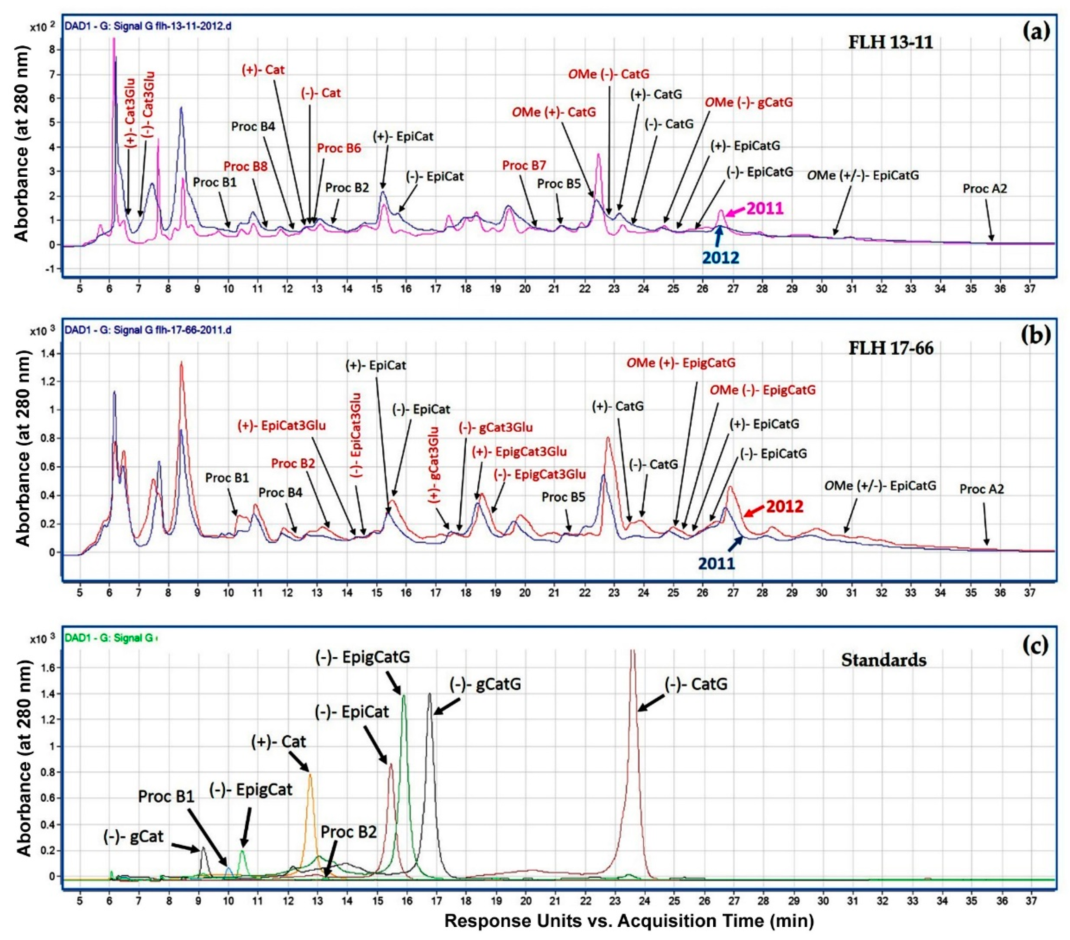
2.1. Metabolite Peak Profile Comparison among FLH 13-11, FLH 17-66, and Standards
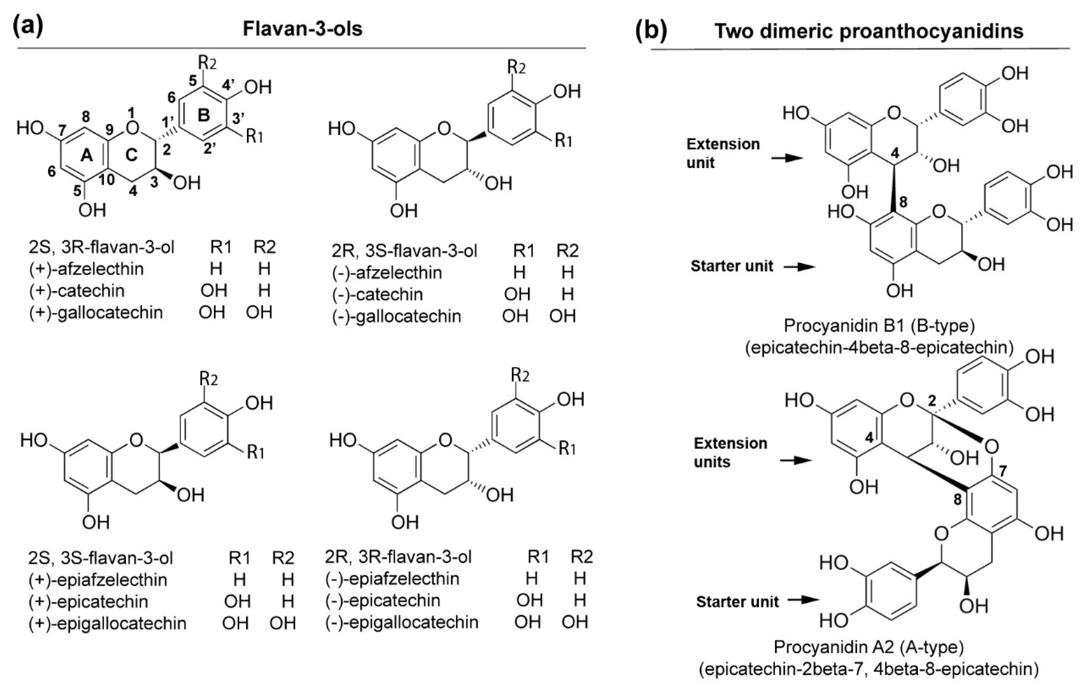
2.2. HPLC-qTOF-MS/MS Based Characterization of Flavan-3-ol Aglycones, Conjugates, and Dimeric PAs Standards
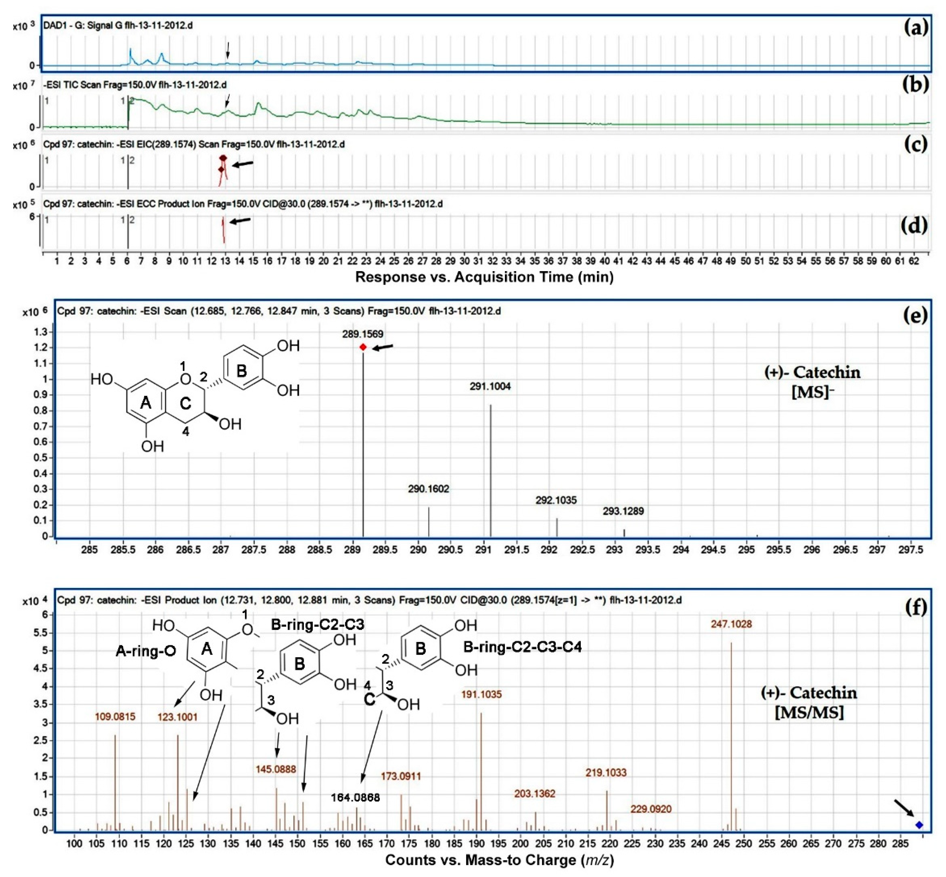
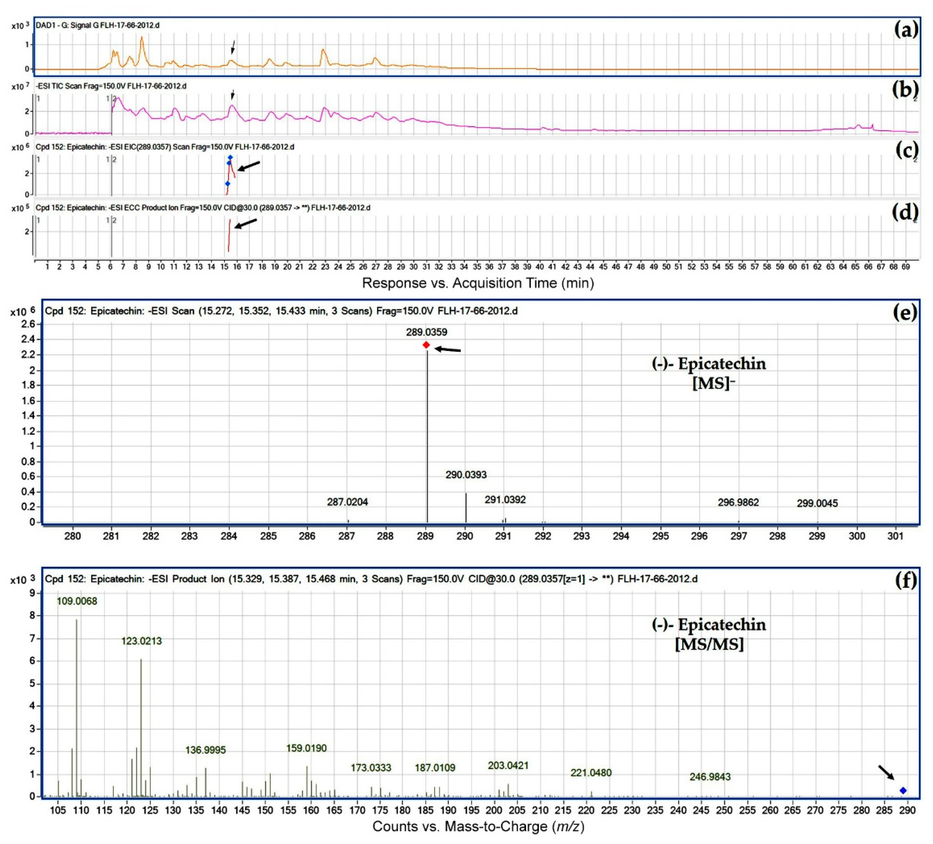
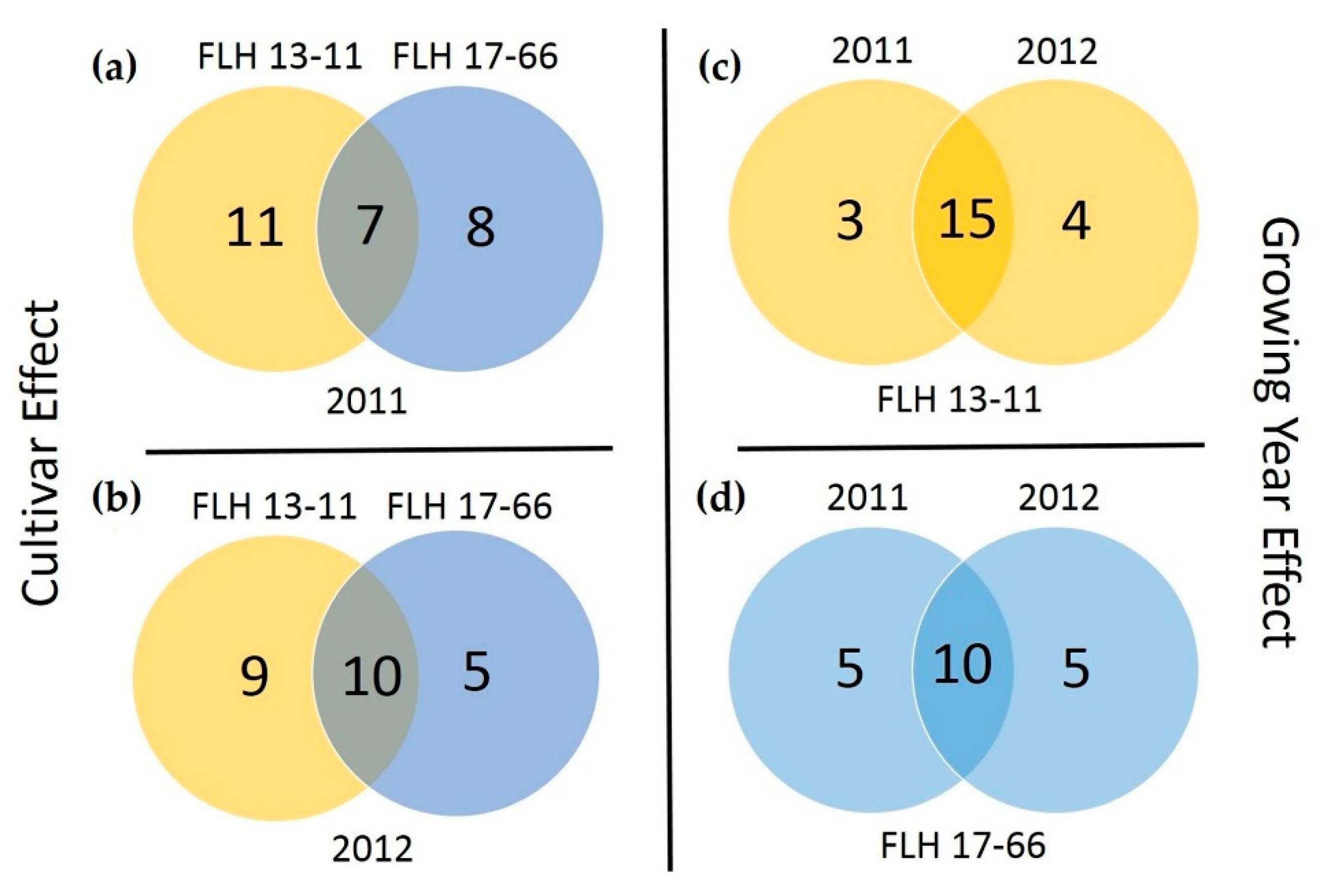
2.3. Flavan-3-ol Aglycone Profiles in Extracts
2.4. Flavan-3-ol Conjugate Profiles in Extracts
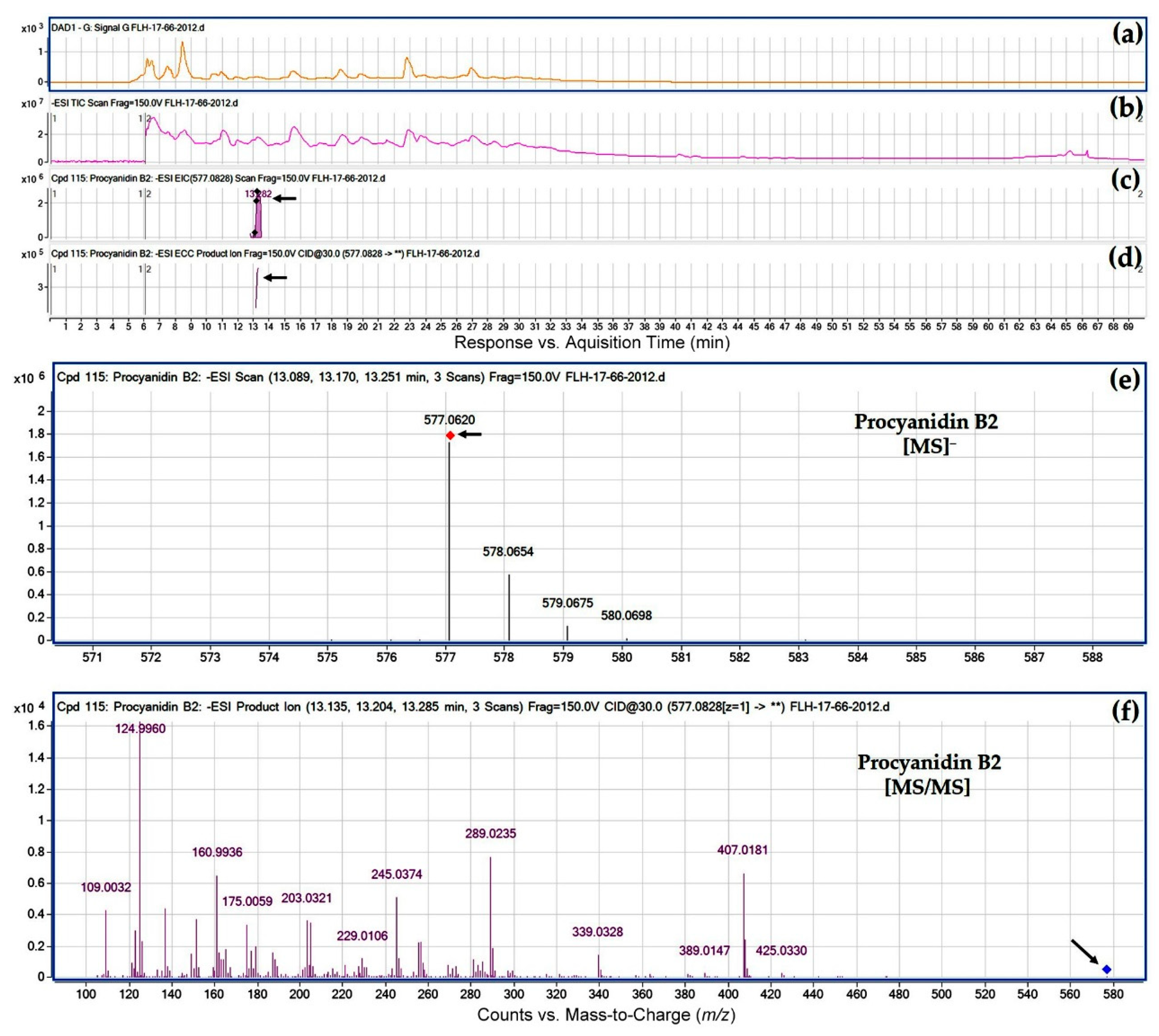
2.5. Dimeric Proanthocyanidin Profiles in Extracts
3. Discussion
4. Materials and Methods
4.1. Chemical Agents and Standards
4.2. Plant Material
4.3. Extraction of Flavan-3-ols
4.4. High Performance Liquid Chromatograph-Quadrupole Time-of-Flight-Tandem Mass Spectrometer (HPLC-qTOF-MS/MS) Analysis
4.5. Structure Annotation
5. Conclusions
Supplementary Materials
Author Contributions
Funding
Acknowledgments
Conflicts of Interest
References
- Xie, D.-Y.; Dixon, R.A. Proanthocyanidin biosynthesis—Still more questions than answers? Phytochemistry 2005, 66, 2127–2144. [Google Scholar] [CrossRef] [PubMed]
- Zhang, S.T.; Li, L.X.; Cui, Y.; Luo, L.X.; Li, Y.Y.; Zhou, P.Y.; Sun, B.S. Preparative high-speed counter-current chromatography separation of grape seed proanthocyanidins according to degree of polymerization. Food Chem. 2017, 219, 399–407. [Google Scholar] [CrossRef] [PubMed]
- Spranger, I.; Sun, B.; Mateus, A.M.; de Freitas, V.; Ricardo-Da-Silva, J.M. Chemical characterization and antioxidant activities of oligomeric and polymeric procyanidin fractions from grape seeds. Food Chem. 2008, 108, 519–532. [Google Scholar] [CrossRef] [PubMed]
- Jonker, A.; Yu, P. The Role of Proanthocyanidins Complex in Structure and Nutrition Interaction in Alfalfa Forage. Int. J. Mol. Sci. 2016, 17, 793. [Google Scholar] [CrossRef] [PubMed]
- Blade, C.; Aragones, G.; Arola-Arnal, A.; Muguerza, B.; Bravo, F.I.; Salvado, M.J.; Arola, L.; Suarez, M. Proanthocyanidins in health and disease. Biofactors 2016, 42, 5–12. [Google Scholar] [PubMed]
- Jonker, A.; Yu, P.Q. The Occurrence, Biosynthesis, and Molecular Structure of Proanthocyanidins and Their Effects on Legume Forage Protein Precipitation, Digestion and Absorption in the Ruminant Digestive Tract. Int. J. Mol. Sci. 2017, 18, 1105. [Google Scholar] [CrossRef] [PubMed]
- Gourineni, V.; Shay, N.F.; Chung, S.; Sandhu, A.K.; Gu, L.W. Muscadine Grape (Vitis rotundifolia) and Wine Phytochemicals Prevented Obesity-Associated Metabolic Complications in C57BL/6J Mice. J. Agric. Food Chem. 2012, 60, 7674–7681. [Google Scholar] [CrossRef] [PubMed]
- Brown, W.L. The anthocyanin pigment of the hunt muscadine grape. J. Am. Chem. Soc. 1940, 62, 2808–2810. [Google Scholar] [CrossRef]
- Olien, W.C. Introduction to the Muscadines. In Muacadine Grapes; Basiouny, F.M., Himelrick, D.G., Eds.; ASHA Press: Alexandria, VA, USA, 2001; pp. 1–13. [Google Scholar]
- Sandhu, A.K.; Gu, L. Antioxidant capacity, phenolic content, and profiling of phenolic compounds in the seeds, skin, and pulp of Vitis rotundifolia (Muscadine Grapes) As determined by HPLC-DAD-ESI-MS(n). J. Agric. Food Chem. 2010, 58, 4681–4692. [Google Scholar] [CrossRef] [PubMed]
- Pastrana-Bonilla, E.; Akoh, C.C.; Sellappan, S.; Krewer, G. Phenolic content and antioxidant capacity of muscadine grapes. J. Agric. Food Chem. 2003, 51, 5497–5503. [Google Scholar] [CrossRef] [PubMed]
- You, Q.; Chen, F.; Wang, X.; Sharp, J.L.; You, Y. Analysis of phenolic composition of Noble muscadine (Vitis rotundifolia) by HPLC-MS and the relationship to its antioxidant capacity. J. Food Sci. 2012, 77, C1115–C1123. [Google Scholar] [CrossRef] [PubMed]
- Plundrich, N.; Grace, M.H.; Raskin, I.; Ann Lila, M. Bioactive polyphenols from muscadine grape and blackcurrant stably concentrated onto protein-rich matrices for topical applications. Int. J. Cosmet. Sci. 2013, 35, 394–401. [Google Scholar] [CrossRef] [PubMed]
- Wei, Z.; Luo, J.; Huang, Y.; Guo, W.; Zhang, Y.; Guan, H.; Xu, C.; Lu, J. Profile of Polyphenol Compounds of Five Muscadine Grapes Cultivated in the United States and in Newly Adapted Locations in China. Int. J. Mol. Sci. 2017, 18, 631. [Google Scholar] [CrossRef] [PubMed]
- Xu, C.; Yagiz, Y.; Hsu, W.Y.; Simonne, A.; Lu, J.; Marshall, M.R. Antioxidant, antibacterial, and antibiofilm properties of polyphenols from muscadine grape (Vitis rotundifolia Michx.) pomace against selected foodborne pathogens. J. Agric. Food Chem. 2014, 62, 6640–6649. [Google Scholar] [CrossRef] [PubMed]
- Talcott, S.T.; Lee, J.H. Ellagic acid and flavonoid antioxidant content of muscadine wine and juice. J. Agric. Food Chem. 2002, 50, 3186–3192. [Google Scholar] [CrossRef] [PubMed]
- Mortensen, J.A. Cultivars. In Muscadine Grapes; Basiounny, F.M., Himelrick, D.G., Eds.; ASHS Press: Alexandria, VA, USA, 2001; pp. 91–105. [Google Scholar]
- You, Q.; Chen, F.; Sharp, J.L.; Wang, X.; You, Y.R.; Zhang, C.J. High-performance liquid chromatography-mass spectrometry and evaporative light-scattering detector to compare phenolic profiles of muscadine grapes. J. Chromatogr. A 2012, 1240, 96–103. [Google Scholar] [CrossRef] [PubMed]
- Quideau, S.; Deffieux, D.; Douat-Casassus, C.; Pouysegu, L. Plant Polyphenols: Chemical Properties, Biological Activities, and Synthesis. Angew. Chem. Int. Ed. 2011, 50, 586–621. [Google Scholar] [CrossRef] [PubMed]
- Li, H.J.; Deinzer, M.L. Tandem mass spectrometry for sequencing proanthocyanidins. Anal. Chem. 2007, 79, 1739–1748. [Google Scholar] [CrossRef] [PubMed]
- Callemien, D.; Collin, S. Use of RP-HPLC-ESI(−)-MS/MS to differentiate various proanthocyanidin isomers in lager beer extracts. J. Am. Soc. Brew. Chem. 2008, 66, 109–115. [Google Scholar] [CrossRef]
- Delcambre, A.; Saucier, C. Identification of new flavan-3-ol monoglycosides by UHPLC-ESI-Q-TOF in grapes and wine. J. Mass Spectrom. 2012, 47, 727–736. [Google Scholar] [CrossRef] [PubMed]
- Gu, L.W.; Kelm, M.A.; Hammerstone, J.F.; Zhang, Z.; Beecher, G.; Holden, J.; Haytowitz, D.; Prior, R.L. Liquid chromatographic/electrospray ionization mass spectrometric studies of proanthocyanidins in foods. J. Mass Spectrom. 2003, 38, 1272–1280. [Google Scholar] [CrossRef] [PubMed]
- Wang, X.; Tong, H.R.; Chen, F.; Gangemi, J.D. Chemical characterization and antioxidant evaluation of muscadine grape pomace extract. Food Chem. 2010, 123, 1156–1162. [Google Scholar] [CrossRef]
- Martino, K.G.; Paul, M.S.; Pegg, R.B.; Kerr, W.L. Effect of time-temperature conditions and clarification on the total phenolics and antioxidant constituents of muscadine grape juice. LWT-Food Sci. Technol. 2013, 53, 327–330. [Google Scholar] [CrossRef]
- Zhang, Y.; Chang, S.K.C.; Stringer, S.J. Characterization of titratable acids, phenolic compounds, and antioxidant activities of wines made from eight mississippi-grown muscadine varieties during fermentation. LWT-Food Sci. Technol. 2017, 86, 302–311. [Google Scholar] [CrossRef]
- Gerald, T.M.; Boyd, L.C.; Delauder, S.F.; Lewis, W.E. Procyanidins from muscadine seeds analysis of antioxidant capacity. Agro Food Ind. Hi-Tech 2011, 22, 19–22. [Google Scholar]


| Standards | MW (g/mol) | RT (min) | [M − H]⁻ (m/z) | [MS/MS] (m/z) |
|---|---|---|---|---|
| (+)-Catechin | 290.26 | 12.761 | 289.0558 | 109.0140- 121.0136- 123.0289- 125.0089- 137.0078- 145.088- 151.0230- 159.0238- 187.0227- 203.0529- 212.0308- 221.0634- 245.0640 |
| (−)-Epicatechin | 290.27 | 15.619 | 289.0558 | 109.0137- 121.0131- 123.0286- 125.0089- 137.0079- 151.0218- 159.0276- 187.0225- 203.0532- 212.0306- 221.0627- 245.0619 |
| (−)-Gallocatechin | 306.27 | 9.166 | 305.0495 | 109.0178- 121.0176- 123.0291- 125.0117- 137.0116- 151.0274- 159.0318- 167.0204- 175.0250- 179.0199- 186.0174- 203.0183- 219.0500 |
| (−)-Epigallocatechin | 306.27 | 10.338 | 305.0518 | 109.0152- 121.0157- 123.0273- 125.0097- 137.0096- 151.0257- 159.0288- 167.0191- 175.0235- 179.0157- 189.0375- 203.0180- 219.0513 |
| (−)-Catechin gallate | 442.37 | 23.590 | 441.0686 | 109.0153- 121.0145- 123.0298- 125.0095- 137.0092- 151.0245- 159.0293- 168.9982- 175.0565- 179.0196- 189.0382- 203.0543- 221.0658- 245.0653- 271.0450- 289.0552 |
| (−)-Epigallocatechin gallate | 458.00 | 16.149 | 457.0565 | 109.0146- 121.0144- 122.9944- 125.0088- 137.0081- 151.0229- 159.0277- 168.9973- 179.0193- 192.9972- 204.0225- 221.0277- 244.0435- 269.0319- 287.0353- 305.0463 |
| (−)-Gallocatechin gallate | 458.00 | 15.872 | 457.0577 | 109.0152- 121.0145- 122.9962- 125.0097- 137.0087- 151.0233- 159.0286- 168.9982- 179.0194- 189.0340- 203.0197- 221.0312- 245.0261- 269.0258- 287.0393- 305.0486 |
| Procyanidin B1 | 578.52 | 10.441 | 577.1225 | 109.0138- 121.0139- 123.0284- 125.0081- 137.0077- 151.0232- 159.0269- 161.0097- 163.0197- 179.0178- 189.0348- 203.0529- 221.0645- 245.0625- 271.0448- 287.0327- 289.0541- 299.0364- 315.0697- 321.0582- 339.0693- 407.0601- 425.0709 |
| Procyanidin B2 | 578.52 | 13.674 | 577.1224 | 109.0132- 121.0146- 123.0283- 125.0082- 137.0086- 151.0221- 159.0269- 161.0093- 163.0232- 179.0196- 189.0369- 203.0519- 221.0643- 245.0621- 271.0435- 286.0298- 289.0535- 297.0203- 315.0647- 339.0721- 407.0602- 425.0712 |
| Compound | MW (g/mol) | Cultivar | Year of Harvest | Ret. Tim (min) | [M − H]− (m/z) | [MS/MS] (m/z) Profiles | Figure # | |
|---|---|---|---|---|---|---|---|---|
| 2011 | 2012 | |||||||
| (+)-Catechin | 290.26 | FLH 13-11 | √ | √ | 12.806 | 289.1574 | 109.0815- 123.1001- 137.0823- 145.0888- 151.1007- 163.1031- 173.0911- 191.1035- 203.1362- 219.1033- 247.1028 | Figure 3 |
| FLH 17-66 | – | – | – | – | ||||
| (−)-Catechin | 290.26 | FLH 13-11 | √ | √ | 12.872 | 289.1586 | 109.0824- 123.1012- 137.0835- 145.0903- 151.1025- 163.1047- 173.0926- 191.1046- 203.1395- 219.1048- 247.1045 | Figure S1 |
| FLH 17-66 | – | – | – | – | ||||
| (+)-Epicatechin | 290.27 | FLH 13-11 | √ | √ | 15.029 | 289.1574 | 109.0817- 123.1005- 137.0835- 151.1017- 161.1221- 175.1292- 188.1158- 203.1425- 212.1182- 221.1558- 247.1014 | Figure S2 |
| FLH 17-66 | √ | – | 15.277 | 289.0377 | 109.0083- 123.0218- 137.0031- 148.9987- 159.0178- 173.0298- 191.0071- 202.0256- 221.0492- 245.0482 | |||
| (−)-Epicatechin | 290.27 | FLH 13-11 | √ | √ | 15.629 | 289.1574 | 109.0819- 123.1007- 137.0831- 151.1016- 159.1082- 175.1354- 187.1087- 203.1421- 212.1219- 221.1560- 245.1602- 271.1448 | Figure 4 |
| FLH 17-66 | √ | √ | 15.398 | 289.0357 | 109.0068- 123.0213- 136.9995- 151.0132- 159.0190- 173.0333- 187.0109- 203.0421- 221.0480- 246.9843 | |||
| Compound | MW (g/mol) | Cultivar | Year of Harvest | Ret. Tim (min) | [MS]⁻ (m/z) | [MS/MS] (m/z) Profiles | Figure # | |
|---|---|---|---|---|---|---|---|---|
| 2011 | 2012 | |||||||
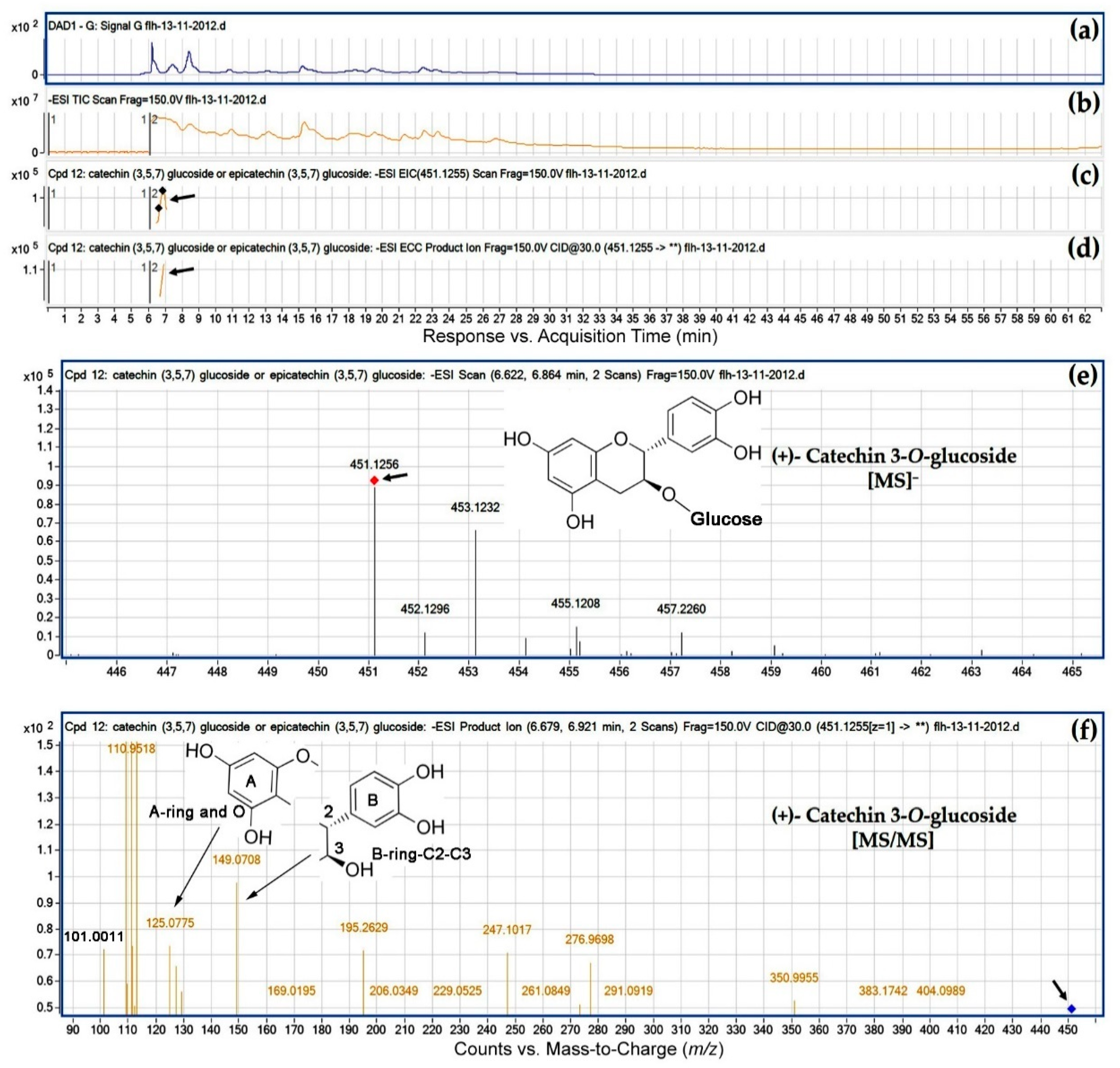
| (+)-Catechin 3-O-glucoside | 452.41 | FLH 13-11 | – | √ | 6.8 | 451.1255 | 101.0781- 108.9543- 109.0471- 110.9518- 112.9492- 125.0775- 149.0708- 169.0195- 195.2629- 206.0349- 229.0525- 247.1017- 276.9698- 350.9955- 404.0989 | Figure 5 |
| FLH 17-66 | – | – | – | – | ||||
| (−)-Catechin 3-O-glucoside | 452.41 | FLH 13-11 | – | √ | 6.991 | 451.1255 | 101.0752- 108.9545- 109.9014- 110.9520- 112.9487-125.0556- 132.8862- 149.0722- 201.1385- 215.1058- 245.1000- 263.1016- 273.0970- 301.7091- 337.1560- 350.9861- 393.1542- 408.0436 | Figure S3 |
| FLH 17-66 | – | – | – | – | ||||
| (+)-Epicatechin 3-O-glucoside | 452.41 | FLH 13-11 | – | – | – | – | Figure S4 | |
| FLH 17-66 | √ | √ | 14.185 | 451.0948 | 100.9949- 112.9969- 124.9885- 136.9909- 152.0334- 168.9777- 188.9751- 216.9631- 229.9739- 246.9749- 258.9704- 272.9450- 287.9723- 300.9451- 343.9958- 391.0083 | |||
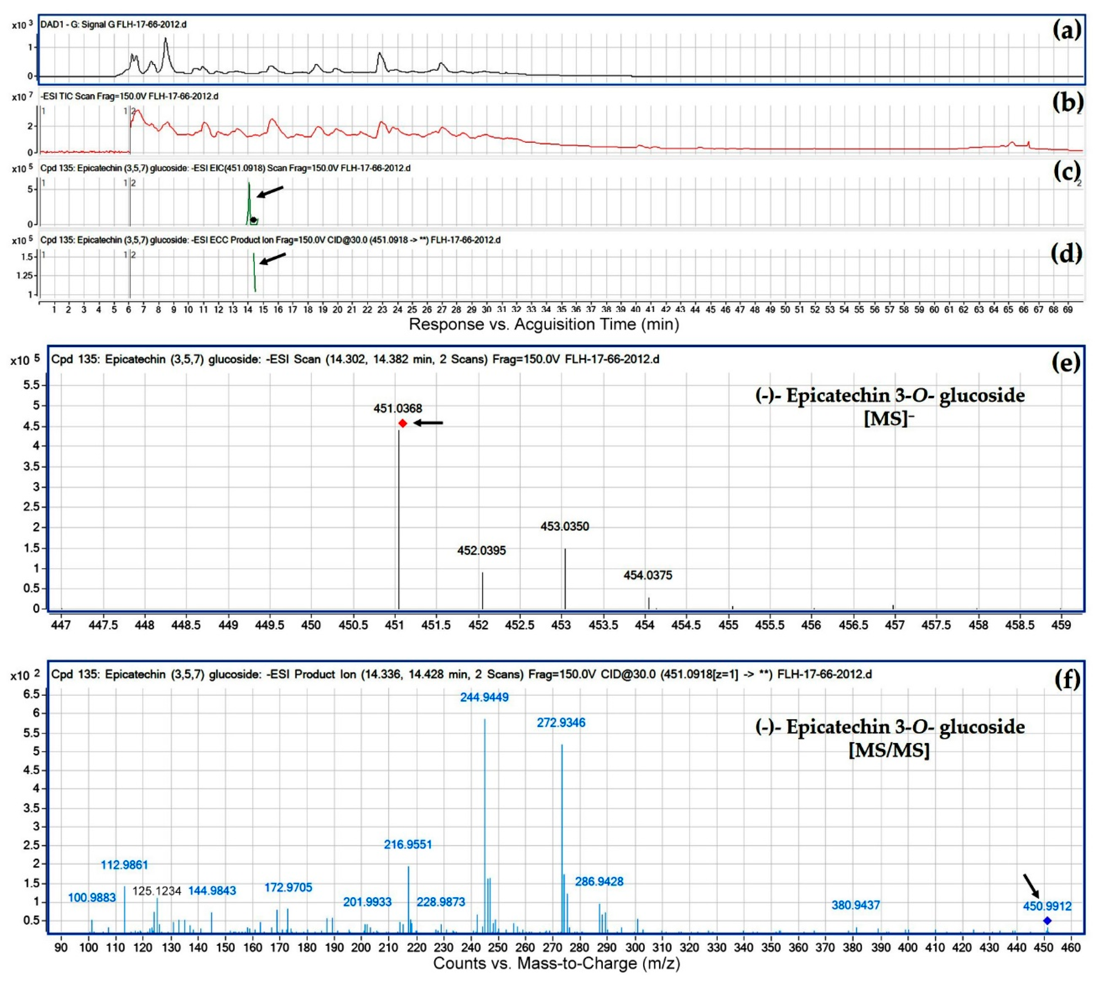
| (−)-Epicatechin 3-O-glucoside | 452.41 | FLH 13-11 | – | – | – | – | Figure 6 | |
| FLH 17-66 | – | √ | 14.382 | 451.0918 | 100.9883- 112.9861- 124.9861- 144.9843- 162.9523- 172.9705- 188.9668- 201.9933- 216.9551- 228.9873- 244.9449- 255.4556- 272.9346- 286.9428- 300.9102- 380.9437 | |||
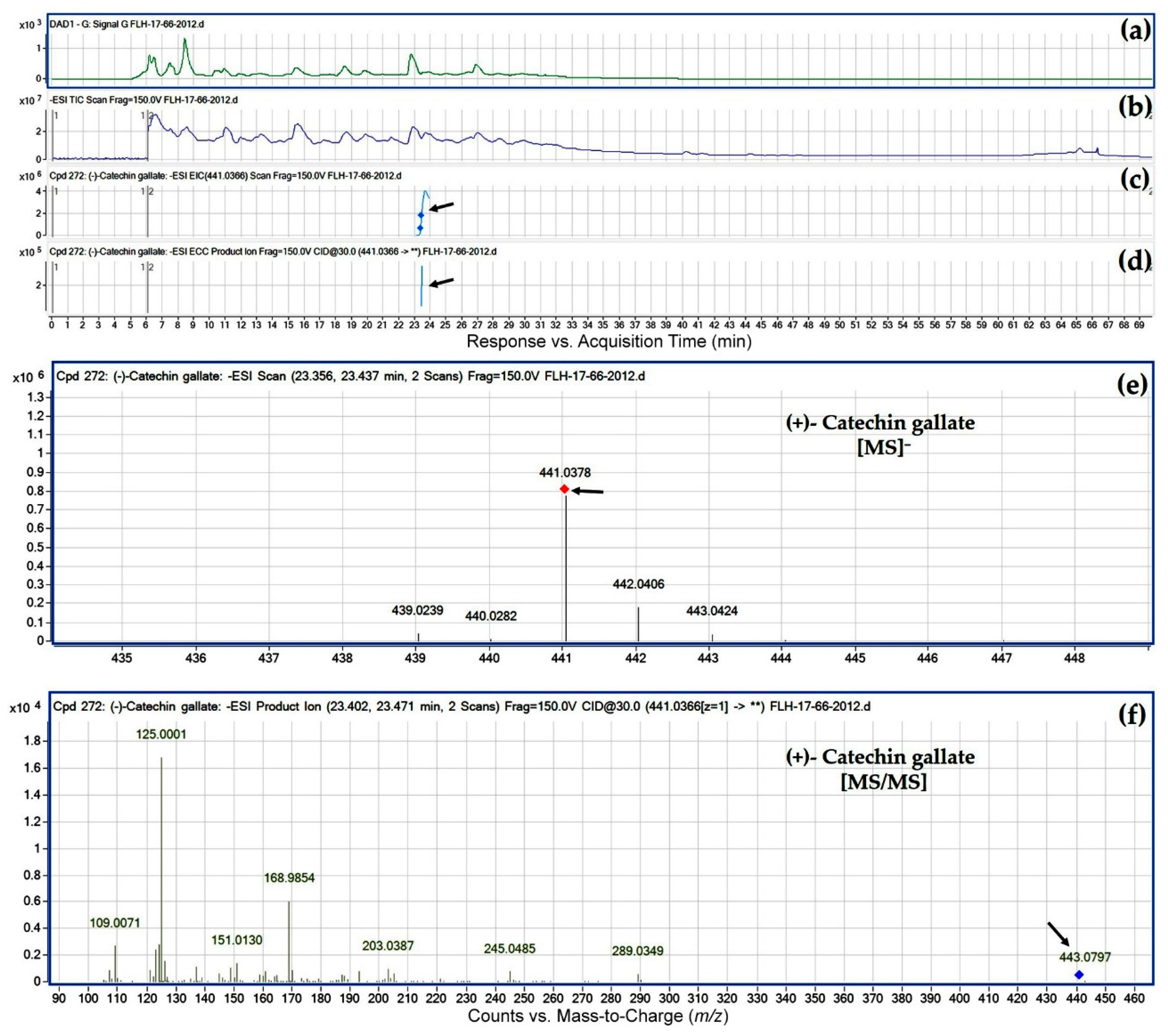
| (+)-Catechin gallate | 442.37 | FLH 13-11 | √ | √ | 23.159 | 441.1883 | 109.0819- 125.0805- 137.0829- 151.1012- 161.1235- 169.0794- 179.1017- 193.0835- 203.1424- 221.1560- 245.1601- 259.1415- 271.1424- 289.1571- 303.1370- 331.1358- | Figure 7 |
| FLH 17-66 | √ | √ | 23.436 | 441.0366 | 109.0071- 125.0001- 136.9989- 151.0130- 161.0310- 168.9854- 179.0047- 192.9813- 203.0387- 211.0086- 221.0537- 245.0485- 256.0208- 289.0349 | |||
| (−)-Catechin gallate | 442.37 | FLH 13-11 | √ | √ | 23.795 | 441.1883 | 109.0820- 125.0805- 137.0833- 151.1014- 169.0795- 179.1017- 193.0838- 203.1422- 221.1563- 245.1603- 271.1434- 289.1570- 303.1388- 331.1365 | Figure S5 |
| FLH 17-66 | √ | √ | 23.84 | 441.04 | 109.0075- 125.0004- 136.9990- 151.0140- 161.0307- 168.9855- 187.0088- 203.0416- 221.0477- 245.0471- 259.0269- 271.0268- 289.0332 | |||
| (+)-Epicatechin gallate | 442.37 | FLH 13-11 | – | √ | 25.094 | 441.1883 | 109.0821- 125.0805- 137.0831- 151.1013- 169.0792- 179.1019- 188.1141- 193.0832- 203.1421- 209.1312- 221.1577- 245.1607- 259.1414- 271.1441- 289.1568- 303.1366 | Figure S6 |
| FLH 17-66 | √ | √ | 25.665 | 441.0366 | 109.0070- 125.0005- 137.0002- 146.0097- 151.0127- 163.0083- 188.0163- 203.0384- 221.0529- 235.1838- 245.0512- 265.3904- 289.0262- 342.0549- 379.8921 | |||
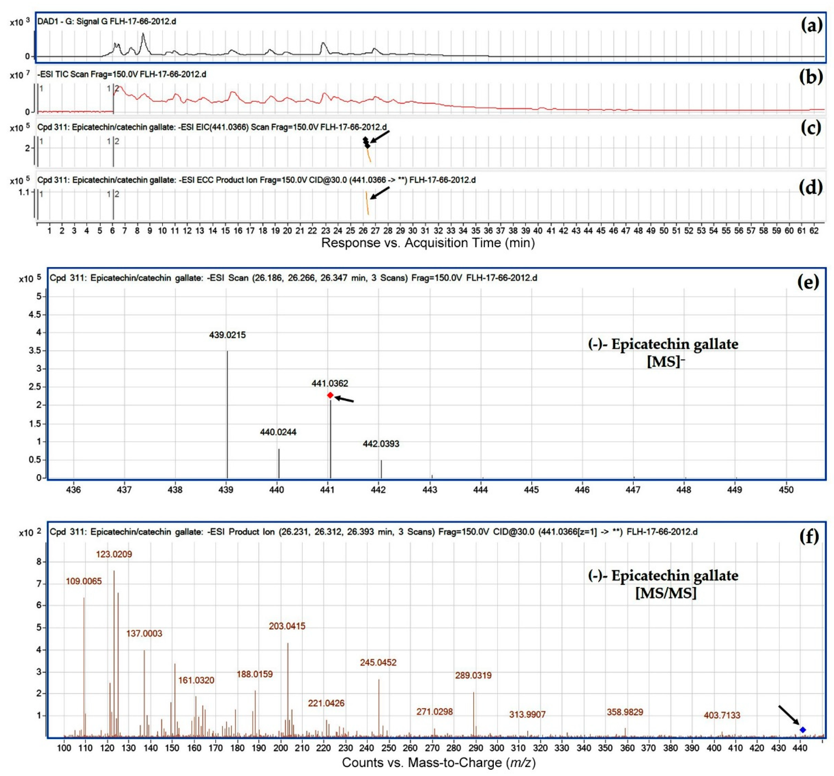
| (−)-Epicatechin gallate | 442.37 | FLH 13-11 | √ | √ | 25.818 | 441.1892 | 109.0836- 125.0810- 137.0830- 151.1038- 164.0731- 179.1025- 187.1074- 195.0042- 203.1427- 221.1591- 245.1588- 254.1340- 275.1032- 289.1590- 301.1176- 315.1329- | Figure 8 |
| FLH 17-66 | √ | √ | 26.312 | 441.0366 | 109.0065- 123.0209- 137.0003- 145.0019- 151.0112- 161.0320- 179.0064- 188.0159- 203.0415- 221.0426- 235.0150- 245.0452- 258.9808- 271.0298- 289.0319- 313.9907- 331.9910- 358.9829- 403.7133 | |||
| (+)-Gallocatechin 3-O-glucoside | 468.00 | FLH 13-11 | – | – | – | – | Figure S7 | |
| FLH 17-66 | √ | – | 17.476 | 467.9999 | 106.9921- 125.0015- 133.9739- 157.0012- 168.9887- 179.0102- 200.9870- 228.9835- 246.9902- 274.9884- 300.9626- 317.0175- 346.9813- 367.9021- 432.2409- 465.6268 | |||
| (−)-Gallocatechin 3-O-glucoside | 468.00 | FLH 13-11 | – | – | – | – | Figure S8 | |
| FLH 17-66 | √ | – | 17.656 | 467.9999 | 106.9969- 125.0027- 135.0205- 168.9884- 184.9934- 210.9862- 228.9798- 250.0131- 274.9883- 283.9547- 300.9604- 315.9707- 338.4035- 367.0427- | |||
| (+)-Epigallocatechin 3-O-glucoside | 468.00 | FLH 13-11 | – | – | – | – | Figure S9 | |
| FLH 17-66 | √ | √ | 18.234 | 467.9974 | 106.9919- 124.9998- 145.0057- 156.9997- 168.1867- 184.9947- 200.9880- 228.9807- 244.9756- 256.9695- 274.9827- 290.9684- 300.9618- 313.0098 | |||
| (−)-Epigallocatechin 3-O-glucoside | 468.00 | FLH 13-11 | – | – | – | – | Figure S10 | |
| FLH 17-66 | √ | √ | 18.875 | 467.9974 | 106.9919- 125.0000- 145.0007- 159.0135- 168.9849- 184.9949- 200.9831- 228.9837- 256.9720- 274.9860- 300.9609- 313.0135- 340.0573- 465.0236 | |||
| O-Methylated (+)-Catechin gallate | 456.00 | FLH 13-11 | √ | √ | 22.362 | 455.3199 | 101.0748- 113.0764- 125.0799- 131.0919- 143.0946- 161.1074- 169.0767- 189.0931- 217.0827- 245.1269- 263.2316- 274.0943- 283.1247- 291.0934- 301.0835- 311.6865- 329.1163- 340.0422- 355.0671- 399.0656 | Figure S11 |
| FLH 17-66 | – | – | – | – | ||||
| O-Methylated (−)-Catechin gallate | 456.00 | FLH 13-11 | – | √ | 22.928 | 455.3199 | 101.0749- 113.0780- 125.0801- 132.0988- 143.0908- 161.1085- 173.0920- 191.0647- 217.0746- 247.0945- 263.2333- 295.0561- 311.1188- 340.0548- 355.0660- 375.2308- 399.0789 | Figure S12 |
| FLH 17-66 | – | – | – | – | ||||
| O-Methylated (+/−)-Epicatechin gallate | 456.00 | FLH 13-11 | √ | √ | 30.36 | 455.2054 | 107.0648- 11.0601- 125.0799- 139.1013- 149.0834- 169.0778- 173.0796- 185.1217- 191.1028- 202.1402- 217.0770- 226.1377- 235.1698- 259.1825- 270.1331- 285.1589- 303.1738- 315.0628- 335.0702- 361.2071 | Figure S13 |
| 456.00 | FLH 17-66 | – | √ | 30.839 | 455.0504 | 106.9945- 124.9977- 136.9965- 148.9984- 168.9866- 177.0170- 183.0117- 196.8803- 202.0292- 217.0550- 220.0302- 228.0043- 241.0177- 253.9952- 269.0249- 274.9825- 285.0435- 303.0430- 310.9758- 348.2291- 387.2122- 446.0391 | ||
| O-Methylated (−)-Gallocatechin gallate | 472.40 | FLH 13-11 | √ | – | 24.923 | 471.2039 | 107.0656- 109.0812- 125.0809- 137.0832- 145.0904- 151.1014- 161.0884- 169.0798- 183.1141- 201.1258- 213.1294- 225.1304- 243.1426- 257.1262- 269.1280- 287.1421- 303.1401- 313.1232 | Figure S14 |
| FLH 17-66 | – | – | – | – | ||||
| O-Methylated (+)-Epigallocatechin gallate | 472.40 | FLH 13-11 | – | – | – | – | Figure S15 | |
| FLH 17-66 | √ | – | 25.018 | 471.0486 | 106.9918- 125.0014- 151.0132- 160.9981- 168.9872- 183.0166- 201.0280- 213.0195- 225.0247- 243.0342- 257.0042- 269.0087- 288.0210- 303.0141 | |||
| O-Methylated (−)-Epigallocatechin gallate | 472.40 | FLH 13-11 | – | – | – | – | Figure S16 | |
| FLH 17-66 | √ | – | 25.347 | 471.0486 | 106.9950- 124.9992- 133.0056- 151.0090- 160.9945- 164.9891- 168.9837- 173.0359- 178.9967- 183.0169- 188.0060- 199.0158- 213.0245- 241.0151- 269.0020- 297.9831- 313.9682- 337.9594 | |||
| Compounds | MW (g/mol) | Cultivar | Year of Harvest | Ret. Tim (min) | MS⁻ (m/z) | [MS/MS] (m/z) Profiles | Figure # | |
|---|---|---|---|---|---|---|---|---|
| 2011 | 2012 | |||||||
| Procyanidin A2 | 576.51 | FLH 13-11 | √ | √ | 35.193 | 575.2385 | 125.0804- 137.0830- 151.0966- 161.0901- 169.0802- 175.1030- 191.0990- 201.1213- 217.1222- 229.1250- 243.1107- 257.1236- 271.1085- 287.1407- 351.0316- 394.1682- 407.1760- 449.1959 | Figure 9 |
| FLH 17-66 | √ | √ | 35.684 | 575.0652 | 109.0070- 124.9985- 136.9988- 152.9935- 160.9962- 168.9837- 175.0088- 187.0083- 199.0077- 215.0015- 227.000- 230.9939- 242.9965- 257.0099- 270.9877- 285.0011- 296.9985- 312.9930- 327.0119- 378.0426- 394.0270- 407.0324- 425.0363- 449.0414 | |||
| Procyanidin B1 [epicatechin-(4β→8)-catechin] | 578.52 | FLH 13-11 | √ | – | 10.008 | 577.2579 | 109.0823- 125.0812- 137.0845- 151.0997- 161.0911- 179.1013- 187.1092- 205.1215- 217.1259- 229.1257- 245.1596- 256.1226- 273.1247- 281.1328- 289.1590- 297.1640- 339.1805- 381.1925- 393.1828- 407.1804- 425.1916- 451.2157 | Figure S17 |
| FLH 17-66 | – | √ | 10.068 | 577.0828 | 108.9944- 124.9868- 136.9845- 152.9751- 160.9802- 174.9912- 203.0163- 229.9978- 245.0179- 254.9655- 272.9779- 289.0023- 297.9319- 312.9674- 339.0092- 406.9878 | |||
| Procyanidin B2 [epicatechin-(4β→8)-epicatechin] | 578.52 | FLH 13-11 | √ | √ | 13.528 | 577.2551 | 109.0809- 125.0801- 137.0810- 149.0838- 161.0938- 165.0838- 175.1065- 191.1055- 205.1202- 229.1258- 245.1590- 269.1261- 289.1558- 329.1543- 367.2092- 393.1943- 407.1765- 439.2095- 533.2681 | Figure 10 |
| FLH 17-66 | √ | √ | 13.21 | 577.0828 | 109.0032- 124.9960- 136.9946- 151.0073- 160.9936- 175.0059- 187.0037- 203.0321- 221.0419- 229.0106- 245.0374- 254.9878- 268.9982- 280.9984- 289.0235- 339.0328- 389.0147- 407.0181- 425.0330 | |||
| Procyanidin B4 [catechin-(4β→8)-epicatechin] | 578.52 | FLH 13-11 | √ | √ | 12.24 | 577.2551 | 109.0814- 125.0805- 137.0823- 149.0852- 165.0824- 179.1032- 191.1036- 201.1267- 205.1212- 227.1137- 247.1403- 269.1262- 289.1557- 329.1593- 353.1942- 367.2122- 393.1957- 407.1776- 425.1887- 439.2070 | Figure S18 |
| FLH 17-66 | – | √ | 12.407 | 577.0828 | 109.0231- 125.0156- 137.0162- 149.0119- 163.0261- 165.0054- 177.0335- 191.0167- 201.0358- 215.0238- 227.0187- 241.0249- 269.0177- 289.0338- 367.0710- 404.0440- 439.0388- 541.0047 | |||
| Procyanidin B5 [epicatechin-(4β→6)-epicatechin] | 578.52 | FLH 13-11 | √ | √ | 21.369 | 577.2193 | 109.0808- 125.0802- 137.0823- 151.1006- 161.0891- 165.0832- 179.0649- 187.1050- 205.1192- 229.1270- 245.1575- 271.1073- 289.1563- 316.1109- 329.1598- 339.1809- 359.1454- 381.1959- 407.1782- 425.1849- 439.2071- 451.2220- 463.1951 | Figure S19 |
| FLH 17-66 | – | √ | 21.467 | 577.0828 | 109.0011- 124.9934- 136.9912- 151.0032- 160.9909- 175.0013- 187.0008- 203.0265- 214.9922- 227.0197- 245.0321- 255.9888- 270.9718- 280.9948- 289.0166- 315.9665- 339.0265- 407.0129- 463.0166 | |||
| Procyanidin B6 [catechin-(4β→6)-catechin] | 578.52 | FLH 13-11 | √ | √ | 12.886 | 577.2551 | 109.0813- 125.0800- 137.0824- 151.1011- 161.0883- 165.0837- 179.1019- 187.1074- 205.1203- 229.1273- 245.1579- 273.1228- 289.1562- 329.1583- 339.1792- 357.1931- 407.1774- 425.1894-439.2095- 451.2070 | Figure S20 |
| FLH 17-66 | – | – | – | – | ||||
| Procyanidin B7 [epicatechin-(4β→6)-catechin] | 578.52 | FLH 13-11 | √ | √ | 20.289 | 577.2551 | 109.0793- 125.0802- 137.0822- 149.0840- 165.0846- 179.1029- 189.1219- 207.1402- 229.1203- 243.1446- 271.1450- 289.1552- 301.1616- 329.1578- 353.1956- 377.1962- 407.1900- 425.1968- 439.2060- 449.2324- 475.2127- 509.2548- 533.2597- 559.2447 | Figure S21 |
| FLH 17-66 | – | – | – | – | ||||
| Procyanidin B8 [catechin-(4β→6)-epicatechin] | 578.52 | FLH 13-11 | √ | – | 11.255 | 577.2579 | 109.0822- 125.0815- 137.0833- 151.1016- 161.0898- 165.0855- 175.1072- 187.1098- 205.1217- 229.1266- 245.1602- 256.1230- 273.1279- 289.1577- 299.1461- 329.1573- 339.1795- 381.2008- 407.1800- 425.1916- 439.2084- 451.2077 | Figure S22 |
| FLH 17-66 | – | – | – | – | ||||
© 2018 by the authors. Licensee MDPI, Basel, Switzerland. This article is an open access article distributed under the terms and conditions of the Creative Commons Attribution (CC BY) license (http://creativecommons.org/licenses/by/4.0/).
Share and Cite
Yuzuak, S.; Ballington, J.; Xie, D.-Y. HPLC-qTOF-MS/MS-Based Profiling of Flavan-3-ols and Dimeric Proanthocyanidins in Berries of Two Muscadine Grape Hybrids FLH 13-11 and FLH 17-66. Metabolites 2018, 8, 57. https://doi.org/10.3390/metabo8040057
Yuzuak S, Ballington J, Xie D-Y. HPLC-qTOF-MS/MS-Based Profiling of Flavan-3-ols and Dimeric Proanthocyanidins in Berries of Two Muscadine Grape Hybrids FLH 13-11 and FLH 17-66. Metabolites. 2018; 8(4):57. https://doi.org/10.3390/metabo8040057
Chicago/Turabian StyleYuzuak, Seyit, James Ballington, and De-Yu Xie. 2018. "HPLC-qTOF-MS/MS-Based Profiling of Flavan-3-ols and Dimeric Proanthocyanidins in Berries of Two Muscadine Grape Hybrids FLH 13-11 and FLH 17-66" Metabolites 8, no. 4: 57. https://doi.org/10.3390/metabo8040057
APA StyleYuzuak, S., Ballington, J., & Xie, D.-Y. (2018). HPLC-qTOF-MS/MS-Based Profiling of Flavan-3-ols and Dimeric Proanthocyanidins in Berries of Two Muscadine Grape Hybrids FLH 13-11 and FLH 17-66. Metabolites, 8(4), 57. https://doi.org/10.3390/metabo8040057