
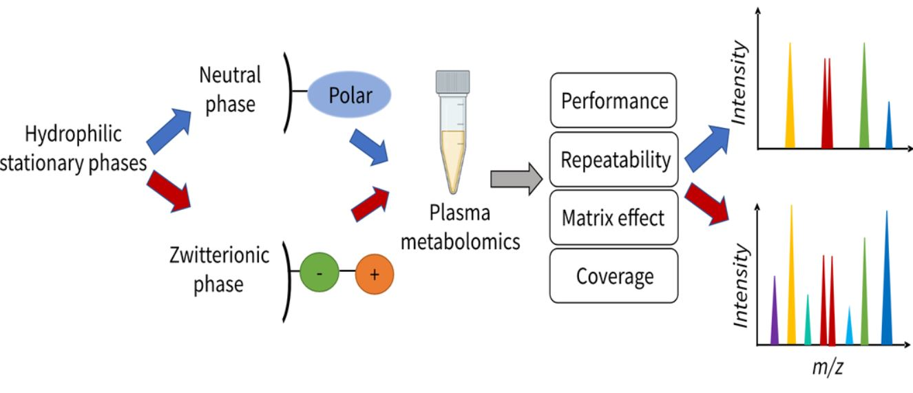
Systematic Evaluation of HILIC Stationary Phases for Global Metabolomics of Human Plasma

 , , and
Abstract
: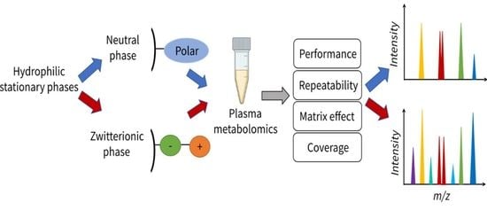
1. Introduction
2. Results and Discussion
2.1. Column Selectivity and Performance
2.2. Retention Mechanism
2.3. Matrix Effect
2.4. Repeatability Evaluation
2.4.1. Intra-Batch Repeatability
2.4.2. Inter-Batch Repeatability
2.5. Untargeted Metabolomics Analysis for Human Plasma
3. Materials and Methods
3.1. Chemicals and Materials
3.2. Standard Solutions
3.3. Sample Preparation
3.4. Instrumentation and LC-MS Acquisitions
3.5. Matrix Effect
3.6. Repeatability
3.7. Data Analysis
4. Conclusions
Supplementary Materials
Author Contributions
Funding
Institutional Review Board Statement
Informed Consent Statement
Data Availability Statement
Acknowledgments
Conflicts of Interest
References
- Rosato, A.; Tenori, L.; Cascante, M.; Ramon, P.; Carulla, D.A.; Martins, V.A.P.; Saccenti, E. From correlation to causation: Analysis of metabolomics data using systems biology approaches. Metabolomics 2018, 14, 1–20. [Google Scholar] [CrossRef] [PubMed] [Green Version]
- Johnson, C.H.; Ivanisevic, J.; Siuzdak, G. Metabolomics: Beyond biomarkers and towards mechanisms. Nat. Publ. Gr. 2016, 17, 451–459. [Google Scholar] [CrossRef] [PubMed] [Green Version]
- Coras, R.; Murillo-Saich, J.; Guma, M. Circulating Pro- and Anti-Inflammatory Metabolites and Its Potential Role in Rheumatoid Arthritis Pathogenesis. Cells 2020, 9, 827. [Google Scholar] [CrossRef] [Green Version]
- Gallart-Ayala, H.; Konz, I.; Mehl, F.; Teav, T.; Oikonomidi, A.; Peyratout, G.; van der Velpen, V.; Popp, J.; Ivanisevic, J. A global HILIC-MS approach to measure polar human cerebrospinal fluid metabolome: Exploring gender-associated variation in a cohort of elderly cognitively healthy subjects. Anal. Chim. Acta 2018, 1037, 327–337. [Google Scholar] [CrossRef] [PubMed]
- Tang, D.Q.; Zou, L.; Yin, X.X.; Ong, C.N. HILIC-MS for metabolomics: An attractive and complementary approach to RPLC-MS. Mass Spectrom. Rev. 2014, 35, 574–600. [Google Scholar] [CrossRef] [PubMed]
- Medvedovici, A.; Bacalum, E.; David, V. Sample preparation for large-scale bioanalytical studies based on liquid chromatographic techniques. Biomed. Chromatogr. 2018, 32, e4137. [Google Scholar] [CrossRef] [PubMed]
- Buszewski, B.; Noga, S. Hydrophilic interaction liquid chromatography (HILIC)-a powerful separation technique. Anal. Bioanal. Chem. 2012, 402, 231–247. [Google Scholar] [CrossRef] [PubMed] [Green Version]
- McCalley, D.V. Study of retention and peak shape in hydrophilic interaction chromatography over a wide pH range. J. Chromatogr. A 2015, 1411, 41–49. [Google Scholar] [CrossRef] [PubMed]
- Li, J.; Wang, Q.L.; Liu, Y.; Ke, Y.; Fan, Q.Q.; Zhou, P.; An, M.C.; Liu, H.M. Simultaneous determination of 24 free amino acids in MGC803 cells by hydrophilic interaction liquid chromatography with tandem mass spectrometry. J. Chromatogr. B Anal. Technol. Biomed. Life Sci. 2019, 1132, 121792. [Google Scholar] [CrossRef] [PubMed]
- Virgiliou, C.; Gika, H.G.; Theodoridis, G.A. HILIC-MS-MS Multi-Targeted Method for Metabolomics. Metab. Profiling Methods Protoc. 2018, 1738, 133–147. [Google Scholar] [CrossRef]
- Arase, S.; Kimura, S.; Ikegami, T. Method optimization of hydrophilic interaction chromatography separation of nucleotides using design of experiment approaches I: Comparison of several zwitterionic columns. J. Pharm. Biomed. Anal. 2018, 158, 307–316. [Google Scholar] [CrossRef] [PubMed]
- Sriboonvorakul, N.; Leepipatpiboon, N.; Dondorp, A.M.; Pouplin, T.; White, N.J.; Tarning, J.; Lindegardh, N. Liquid chromatographic-mass spectrometric method for simultaneous determination of small organic acids potentially contributing to acidosis in severe malaria. J. Chromatogr. B Anal. Technol. Biomed. Life Sci. 2013, 941, 116–122. [Google Scholar] [CrossRef] [PubMed] [Green Version]
- Guo, Y. Recent progress in the fundamental understanding of hydrophilic interaction chromatography (HILIC). Analyst 2015, 140, 6452–6466. [Google Scholar] [CrossRef] [PubMed]
- Contrepois, K.; Jiang, L.; Snyder, M. Optimized analytical procedures for the untargeted metabolomic profiling of human urine and plasma by combining hydrophilic interaction (HILIC) and reverse-phase liquid chromatography (RPLC)-mass spectrometry. Mol. Cell. Proteom. 2015, 14, 1684–1695. [Google Scholar] [CrossRef] [PubMed] [Green Version]
- Sillner, N.; Walker, A.; Harrieder, E.M.; Schmitt-Kopplin, P.; Witting, M. Development and application of a HILIC UHPLC-MS method for polar fecal metabolome profiling. J. Chromatogr. B Anal. Technol. Biomed. Life Sci. 2019, 1109, 142–148. [Google Scholar] [CrossRef] [PubMed]
- Erngren, I.; Haglöf, J.; Engskog, M.K.R.; Nestor, M.; Hedeland, M.; Arvidsson, T.; Pettersson, C. Adduct formation in electrospray ionisation-mass spectrometry with hydrophilic interaction liquid chromatography is strongly affected by the inorganic ion concentration of the samples. J. Chromatogr. A 2019, 1600, 174–182. [Google Scholar] [CrossRef] [PubMed]
- Elmsjö, A.; Haglöf, J.; Engskog, M.K.R.; Erngren, I.; Nestor, M.; Arvidsson, T.; Pettersson, C. Method selectivity evaluation using the co-feature ratio in LC/MS metabolomics: Comparison of HILIC stationary phase performance for the analysis of plasma, urine and cell extracts. J. Chromatogr. A 2018, 1568, 49–56. [Google Scholar] [CrossRef] [PubMed]
- West, C.; Auroux, E. Deconvoluting the effects of buffer salt concentration in hydrophilic interaction chromatography on a zwitterionic stationary phase. J. Chromatogr. A 2016, 1461, 92–97. [Google Scholar] [CrossRef]
- Alpert, A.J. Effect of salts on retention in hydrophilic interaction chromatography. J. Chromatogr. A 2018, 1538, 45–53. [Google Scholar] [CrossRef] [PubMed]
- Ahn, J.; Bones, J.; Yu, Y.Q.; Rudd, P.M.; Gilar, M. Separation of 2-aminobenzamide labeled glycans using hydrophilic interaction chromatography columns packed with 1.7 μm sorbent. J. Chromatogr. B 2010, 878, 403–408. [Google Scholar] [CrossRef]
- Bajad, S.U.; Lu, W.; Kimball, E.H.; Yuan, J.; Peterson, C.; Rabinowitz, J.D. Separation and quantitation of water soluble cellular metabolites by hydrophilic interaction chromatography-tandem mass spectrometry. J. Chromatogr. A 2006, 1125, 76–88. [Google Scholar] [CrossRef] [PubMed]
- Tufi, S.; Lamoree, M.; de Boer, J.; Leonards, P. Simultaneous analysis of multiple neurotransmitters by hydrophilic interaction liquid chromatography coupled to tandem mass spectrometry. J. Chromatogr. A 2015, 1395, 79–87. [Google Scholar] [CrossRef] [PubMed]
- Greco, G.; Letzel, T. Main interactions and influences of the chromatographic parameters in HILIC separations. J. Chromatogr. Sci. 2013, 51, 684–693. [Google Scholar] [CrossRef] [PubMed]
- Wang, J.; Guo, Z.; Shen, A.; Yu, L.; Xiao, Y.; Xue, X.; Zhang, X.; Liang, X. Hydrophilic-subtraction model for the characterization and comparison of hydrophilic interaction liquid chromatography columns. J. Chromatogr. A 2015, 1398, 29–46. [Google Scholar] [CrossRef] [PubMed]
- Ekdahl, A.; Johansson, M.C.; Ahnoff, M. Tracing and separating plasma components causing matrix effects in hydrophilic interaction chromatography-electrospray ionization mass spectrometry. J. Chromatogr. B Anal. Technol. Biomed. Life Sci. 2013, 923–924, 83–91. [Google Scholar] [CrossRef] [PubMed]
- Takayama, N.; Lim, L.W.; Takeuchi, T. Retention behavior of inorganic anions in hydrophilic interaction chromatography. Anal. Sci. 2017, 33, 619–625. [Google Scholar] [CrossRef] [PubMed] [Green Version]
- Iturrospe, E.; Da Silva, K.M.; Talavera Andújar, B.; Cuykx, M.; Boeckmans, J.; Vanhaecke, T.; Covaci, A.; van Nuijs, A.L.N. An exploratory approach for an oriented development of an untargeted hydrophilic interaction liquid chromatography-mass spectrometry platform for polar metabolites in biological matrices. J. Chromatogr. A 2021, 1637, 461807. [Google Scholar] [CrossRef] [PubMed]
- Smith, C.A.; Want, E.J.; O’Maille, G.; Abagyan, R.; Siuzdak, G. XCMS: Processing mass spectrometry data for metabolite profiling using nonlinear peak alignment, matching, and identification. Anal. Chem. 2006, 78, 779–787. [Google Scholar] [CrossRef] [PubMed]
- DeFelice, B.C.; Mehta, S.S.; Samra, S.; Čajka, T.; Wancewicz, B.; Fahrmann, J.F.; Fiehn, O. Mass Spectral Feature List Optimizer (MS-FLO): A Tool To Minimize False Positive Peak Reports in Untargeted Liquid Chromatography-Mass Spectroscopy (LC-MS) Data Processing. Anal. Chem. 2017, 89, 3250–3255. [Google Scholar] [CrossRef] [PubMed]
- Pang, Z.; Chong, J.; Zhou, G.; De Lima Morais, D.A.; Chang, L.; Barrette, M.; Gauthier, C.; Jacques, P.É.; Li, S.; Xia, J. MetaboAnalyst 5.0: Narrowing the gap between raw spectra and functional insights. Nucleic Acids Res. 2021, 49, W388–W396. [Google Scholar] [CrossRef]
- Haug, K.; Cochrane, K.; Nainala, V.C.; Williams, M.; Chang, J.; Jayaseelan, K.V.; O’Donovan, C. MetaboLights: A resource evolving in response to the needs of its scientific community. Nucleic Acids Res. 2020, 48, D440–D444. [Google Scholar] [CrossRef] [PubMed] [Green Version]


| ZIC-c (51 Metabolites Detected) | BEH (47 Metabolites Detected) | |||
|---|---|---|---|---|
| Metabolite Classes | Matrix Effect | Salt Effect | Matrix Effect | Salt Effect |
| Amino acids | 11 (16 *) | 11 (16) | 14 (17) | 4 (17) |
| Amines | 2 (4) | 3 (4) | 2 (4) | 1 (4) |
| Sugar and Sugar phosphate | 5 (5) | 4 (5) | 4 (4) | 2 (4) |
| Nucleoside and Nucleotide | 10 (13) | 10 (13) | 8 (11) | 7 (11) |
| Acylcarnitines | 1 (2) | 0 (2) | 0 (2) | 1 (2) |
| Organic acids | 8 (11) | 8 (11) | 6 (9) | 6 (9) |
Publisher’s Note: MDPI stays neutral with regard to jurisdictional claims in published maps and institutional affiliations. |
© 2022 by the authors. Licensee MDPI, Basel, Switzerland. This article is an open access article distributed under the terms and conditions of the Creative Commons Attribution (CC BY) license (https://creativecommons.org/licenses/by/4.0/).
Share and Cite
Hosseinkhani, F.; Huang, L.; Dubbelman, A.-C.; Guled, F.; Harms, A.C.; Hankemeier, T. Systematic Evaluation of HILIC Stationary Phases for Global Metabolomics of Human Plasma. Metabolites 2022, 12, 165. https://doi.org/10.3390/metabo12020165
Hosseinkhani F, Huang L, Dubbelman A-C, Guled F, Harms AC, Hankemeier T. Systematic Evaluation of HILIC Stationary Phases for Global Metabolomics of Human Plasma. Metabolites. 2022; 12(2):165. https://doi.org/10.3390/metabo12020165
Chicago/Turabian StyleHosseinkhani, Farideh, Luojiao Huang, Anne-Charlotte Dubbelman, Faisa Guled, Amy C. Harms, and Thomas Hankemeier. 2022. "Systematic Evaluation of HILIC Stationary Phases for Global Metabolomics of Human Plasma" Metabolites 12, no. 2: 165. https://doi.org/10.3390/metabo12020165