An Unusual Presentation of Pectoralis Major Pyomyositis Presenting as Septic Arthritis of the Shoulder: A Case Report and Review of the Literature


Abstract
:1. Introduction
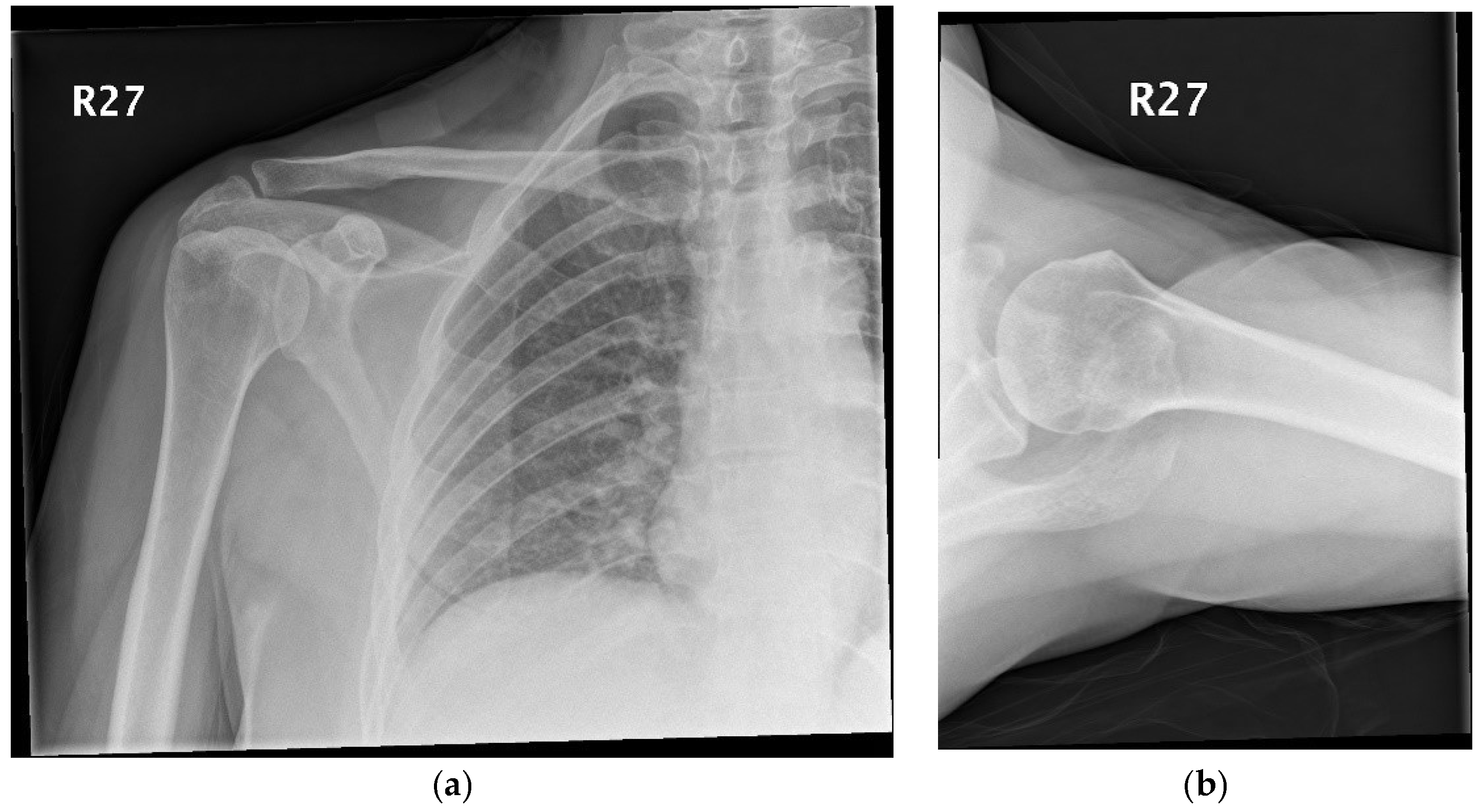
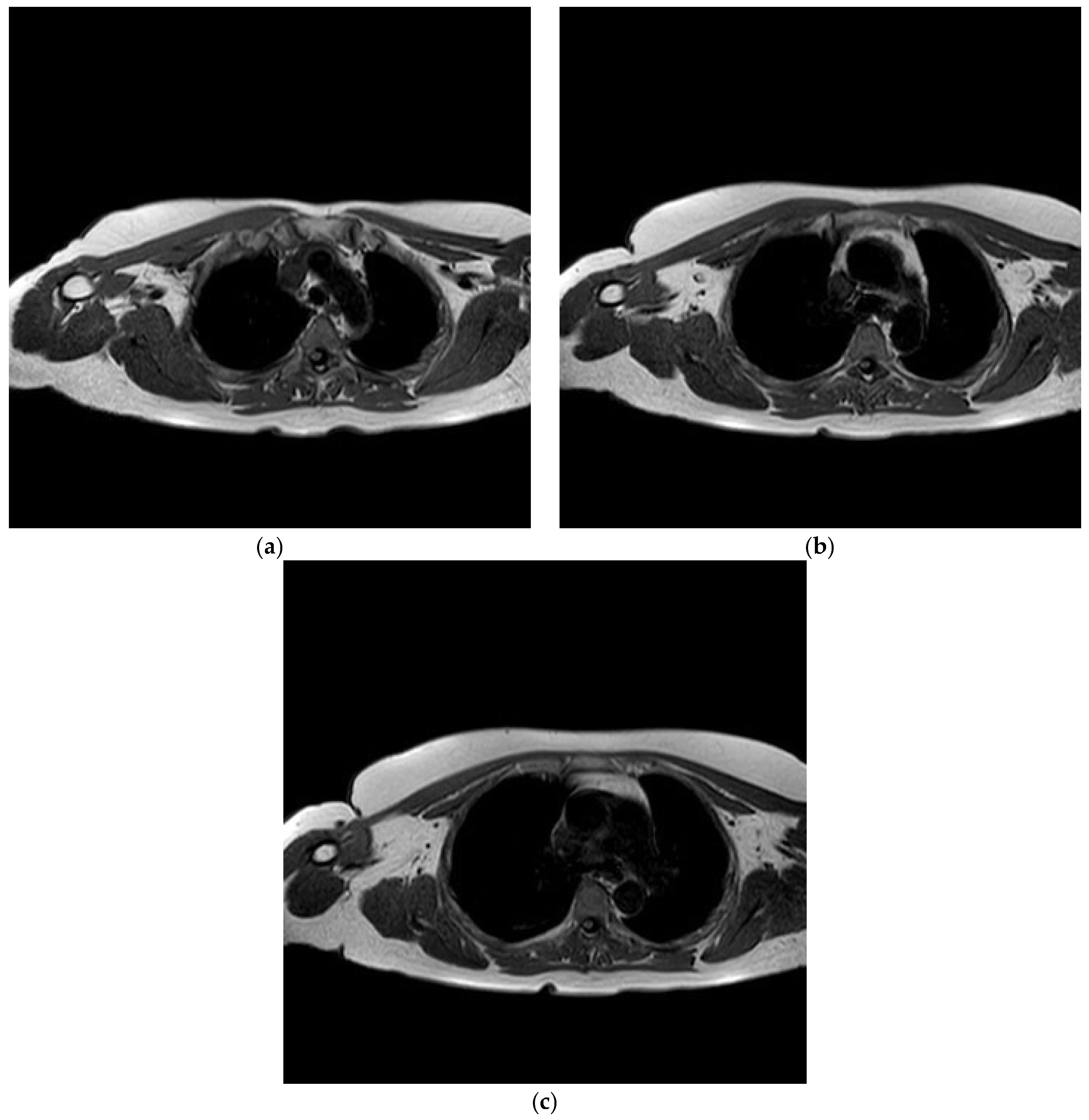
2. Case Report
3. Discussion
4. Conclusions
Funding
Conflicts of Interest
References
- Chiedozi, L.C. Pyomyositis: review of 205 cases in 112 patients. Am. J. Surg. 1979, 137, 255–259. [Google Scholar] [CrossRef]
- Comegna, L.; Guidone, P.I.; Prezioso, G.; Franchini, S.; Petrosino, M.I.; Di Filippo, P.; Chiarelli, F.; Mohn, A.; Rossi, N. Pyomyositis is not only a tropical pathology: A case series. J. Med. Case Reports 2016, 10, 372. [Google Scholar] [CrossRef] [PubMed]
- Glynne Andrew, J.; Mintowt Czyz, W. Pyomyositis presenting as septic arthritis: A report of 2 cases. Acta Orthopaedica Scandinavica 1988, 59, 587–588. [Google Scholar] [CrossRef] [Green Version]
- King, R.J.; Laugharne, D.; Kerslake, R.W.; Holdsworth, B.J. Case report: Primary obturator pyomyositis: A diagnostic challenge. J. Bone Joint Surg. B. R. 2003, 85, 895–898. [Google Scholar] [CrossRef]
- Papadopoulos, M.; Chugh, S.; Fitzgerald, R.; Thomas, R.J. Obturator internus pyomyositis. Orthopedics 2000, 23, 383–384. [Google Scholar] [PubMed]
- Oliver, C.; Syed, Z.N.; Sean, H.; John, A.S. Extensive pyogenic myositis of the hip in an immuno-competent patient. JRSM Short Rep. 2011, 2, 90. [Google Scholar]
- Kattimani, R.; McConnell, J.; Waite, J. Pyomyositis of gluteus medius: A case report and review of the literature. J. Orthop. Case Rep. 2017, 7, 48. [Google Scholar] [PubMed]
- Jayoussi, R.; Bialik, V.; Eyal, A.; Shehadeh, N.; Etzioni, A. Pyomyositis caused by vigorous exercise in a boy. Acta Paediat. 1995, 84, 226–227. [Google Scholar] [CrossRef]
- Ross, J.J.; Shamsuddin, H. Sternoclavicular septic arthritis: review of 180 cases. Medicine 2004, 83, 139–148. [Google Scholar] [CrossRef] [PubMed]
- Shepherd, J.J. Tropical myositis, is it an entity and what is its causes? Lancet 1983, 1240–1242. [Google Scholar] [CrossRef]
- Mazur, J.M.; Ross, G.; Cummings, J.; Hahn, J.G.; McCluskey, W.P. Usefulness of magnetic resonance imaging for the diagnosis of acute musculoskeletal infections in children. J. Pediat. Orthop. 1995, 15, 144–147. [Google Scholar] [CrossRef]
- Joseph, F.W.; James, O.P.; Dellinger, E.P. 51-Pyomyositis Tropicans. Available online: https://doi.org/10.1016/B978-1-4377-0126-5.00051-3 (accessed on 21 March 2012).
- Bickels, J.; Ben-Sira, L.; Kessler, A.; Wientroub, S. Primary pyomyositis. JBJS 2002, 84, 2277–2286. [Google Scholar] [CrossRef]


© 2018 by the authors. Licensee MDPI, Basel, Switzerland. This article is an open access article distributed under the terms and conditions of the Creative Commons Attribution (CC BY) license (http://creativecommons.org/licenses/by/4.0/).
Share and Cite
Saad, A.; Shahban, S.; Elgamal, T. An Unusual Presentation of Pectoralis Major Pyomyositis Presenting as Septic Arthritis of the Shoulder: A Case Report and Review of the Literature. Diseases 2018, 6, 100. https://doi.org/10.3390/diseases6040100
Saad A, Shahban S, Elgamal T. An Unusual Presentation of Pectoralis Major Pyomyositis Presenting as Septic Arthritis of the Shoulder: A Case Report and Review of the Literature. Diseases. 2018; 6(4):100. https://doi.org/10.3390/diseases6040100
Chicago/Turabian StyleSaad, Ahmed, Shafiq Shahban, and Tarek Elgamal. 2018. "An Unusual Presentation of Pectoralis Major Pyomyositis Presenting as Septic Arthritis of the Shoulder: A Case Report and Review of the Literature" Diseases 6, no. 4: 100. https://doi.org/10.3390/diseases6040100
APA StyleSaad, A., Shahban, S., & Elgamal, T. (2018). An Unusual Presentation of Pectoralis Major Pyomyositis Presenting as Septic Arthritis of the Shoulder: A Case Report and Review of the Literature. Diseases, 6(4), 100. https://doi.org/10.3390/diseases6040100