Article

Microsyntenic Clusters Reveal Conservation of lncRNAs in Chordates Despite Absence of Sequence Conservation

by
Carlos Herrera-Úbeda
1,
Marta Marín-Barba
2,
Enrique Navas-Pérez
1,
Jan Gravemeyer
3,
Beatriz Albuixech-Crespo
1,
Grant N. Wheeler
2 and
Jordi Garcia-Fernàndez
1,*
1
Department of Genetics, Microbiology and Statistics, Faculty of Biology, University of Barcelona, 08028 Barcelona, Spain
2
School of Biological Sciences, University of East Anglia, Norwich Research Park, Norwich NR4 7TU, UK
3
German Cancer Research Center, 69120 Heidelberg, Germany
*
Author to whom correspondence should be addressed.
Biology 2019, 8(3), 61; https://doi.org/10.3390/biology8030061
Submission received: 23 July 2019 / Revised: 19 August 2019 / Accepted: 21 August 2019 / Published: 24 August 2019
(This article belongs to the Section Evolutionary Biology)

Abstract

:
Homologous long non-coding RNAs (lncRNAs) are elusive to identify by sequence similarity due to their fast-evolutionary rate. Here we develop LincOFinder, a pipeline that finds conserved intergenic lncRNAs (lincRNAs) between distant related species by means of microsynteny analyses. Using this tool, we have identified 16 bona fide homologous lincRNAs between the amphioxus and human genomes. We characterized and compared in amphioxus and Xenopus the expression domain of one of them, Hotairm1, located in the anterior part of the Hox cluster. In addition, we analyzed the function of this lincRNA in Xenopus, showing that its disruption produces a severe headless phenotype, most probably by interfering with the regulation of the Hox cluster. Our results strongly suggest that this lincRNA has probably been regulating the Hox cluster since the early origin of chordates. Our work pioneers the use of syntenic searches to identify non-coding genes over long evolutionary distances and helps to further understand lncRNA evolution.

1. Introduction

Identifying and understanding the factors that underlie the evolution of morphological complexity is one of the central issues in the field of evolutionary developmental biology (or evo-devo). From the initial claims that gene duplication and neofunctionalization were at the core of phenotypic change [1], the current view also takes into account the fine-tuning of gene regulation [2] and increasing the proteome and interactome complexity through additional processes. In this regard, molecular mechanisms such as alternative splicing or RNA-editing, and the RNA world, with molecules like small miRNA or long non coding RNAs (lncRNAs), allow deeper and multifaceted levels of gene regulation [3]. LncRNA-mediated regulation stands out as a quick and efficient mechanism to modulate gene expression, as these molecules are function-ready almost immediately after or even during transcription and can be rapidly degraded by the cellular machinery [3,4,5]. These characteristics make lncRNAs sharp regulators of the myriad biological processes in which they are involved, such as chromatin remodeling, protein scaffolding or gene expression regulation through direct binding to genomic enhancers [6,7].
The study of lncRNAs from an evolutionary perspective has been hindered by their lack of strong primary sequence conservation [8,9], their apparent lack of secondary structure conservation [10], and their massive genomic generation and decay rate [11]. The cephalochordate Branchiostoma lanceolatum represents the earliest branching chordate lineage and holds a genome that seems to have retained many of the features of the ancestral pre-duplicative vertebrate [12,13]. Searches of lncRNAs conserved between amphioxus and vertebrates based on sequence similarity have been unsuccessful [14,15], probably due to the long evolutionary distance that separates these lineages [16]. Recently, however, a strategy to identify conserved intergenic lncRNAs (lincRNAs) by means of syntenic analyses has been successfully attempted, but limited to closely related species [17,18].
Here, we have developed a pipeline called LincOFinder that finds conserved clusters of microsynteny between two distant organisms surrounding an intergenic lncRNA. Furthermore, we use this tool to study the conservation and evolution of the lincRNA repertoire in the chordate lineage, finding up to 16 lincRNAs putatively conserved between amphioxus and human. Finally, we further study the case of Hotairm1, assessing its developmental expression in amphioxus and Xenopus and showing that its inhibition during X. tropicalis development produces a severe headless phenotype, probably by disrupting the chromatin dynamics of the anterior Hox cluster. Overall, our work pioneers the use of syntenic searches to identify non-coding genes over long evolutionary distances and helps to further understand lincRNA evolution in the frame of the invertebrate-vertebrate transition.

2. Materials and Methods

2.1. Amphioxus and Human Coding and lincRNA Datasets

We used the intergenic and bidirectional fractions from the lncRNAs dataset provided by Marlétaz et al. [15] to obtain an amphioxus lincRNA fraction (1318 genes), and their protein-coding genes supported by orthology as the coding fraction (10,832 genes). The human coding genes were obtained from the Ensembl annotation of the Ch38.96 genome assembly [19]. Finally, the orthologous gene families described in Marlétaz et al. [15] were used to assess the amphioxus/human gene orthologies.

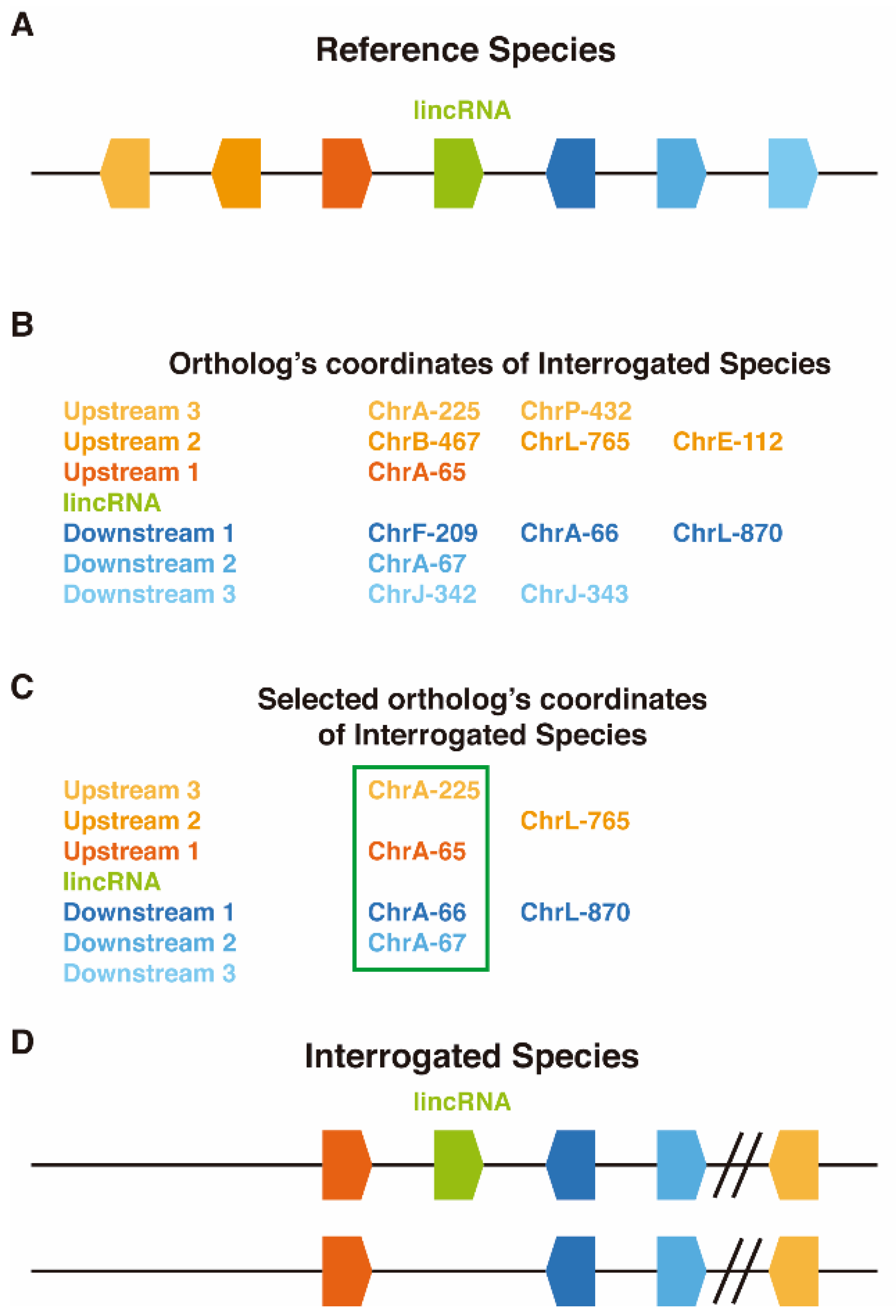
2.2. LincOFinder (lincRNA Orthology Finder)

LincOFinder (https://github.com/cherrera1990/LincOFinder) is a program designed to identify shared microsyntenic clusters surrounding lincRNAs between two species. These are named the “reference” (Ref) and the “interrogated” species (Int). In the first, nonautomated step, the genes of both species (only the coding genes for Int) need to be arranged according to their position within the corresponding chromosome or scaffold, then a virtual coordinate according to their position is stablished (e.g., the first gene in chromosome A will be chrA-1, the second chrA-2, etc.). Furthermore, the orthologies between the genes of both species are established, using sets of known orthologous families or with the help of programs like Orthofinder [20]. Once the data is properly formatted as indicated in the ReadMe.md of LincOFinder, each annotated lincRNA from Ref is used as a reference point. The three upstream and three downstream genes neighboring the lincRNA are selected, and the orthology coordinates from Int are parsed into a distance matrix (Figure 1). The reasoning behind selecting only the three upstream and three downstream genes is to try to be astringent enough to comply with the definition of microsynteny [21] but at the same time allowing insertions and deletions up to a certain degree and the discovering, in case they exist, of larger clusters. Only genes present in the same chromosome are taken into account for distance assessment, and comparisons between paralogs of the same Ref gene are avoided. Then, a UPGMA hierarchical clustering algorithm [22] is used to create viable distance clusters (the ones that comply with the previously stated restrictions), and the cluster with the minimum distance between two neighboring genes is selected, thus identifying possible microsynteny clusters. If several possible clusters are formed, then they are displayed separately. These microsynteny clusters should be further filtered by selecting only those that harbor adjacent genes. Finally, candidates are manually curated by looking for the presence of lincRNAs in the microsyntenic region of Int (the algorithm is blind to Int ncRNAs due to the possibility of missing unannotated syntenic lincRNAs that could be, for example, present in the form of ESTs). This step can be done using a genome browser such us UCSC [23] (Figure 1).

2.3. Xenopus Embryos and MO Injections

All experiments were performed in compliance with the relevant laws and institutional guidelines of the University of East Anglia. The research has been approved by the local ethical review committee according to UK Home Office under Project License PPL 70/8876. X. tropicalis females were primed 24 h before the eggs were required and induced 5 h prior the experiment both with Chorulon (human Chorionic Gonadotropin). X. tropicalis males were also primed with Chorulon. Eggs were naturally obtained and fertilized with a sperm solution in Leibovitz’s L-15 Medium supplemented with 10% calf serum and left at room temperature for 5 min. After that, embryos were immersed in 0.05× MMR (Marc’s Modified Ringer’s) (50 mM NaCl, 1 mM KCl, 0.5 mM MgCl2, 1 mM CaCl2, 2.5 mM HEPES pH 7.5) for 20 min at room temperature, then washed in 2% l-cysteine pH = 8 for 7 min and rinsed several times with 0.05× MMR. Embryos were incubated in a BSA (bovine serum albumin) coated Petri dish in 0.05× MMR at 26 °C.
Morpholino oligos (MO) were designed and provided by Gene Tools. Morpholino sequences are: Standard control CCTCTTACCTCAGTTACAATTTATA, Hotairm1 AATCACTATTTGCTCCTTACCGGGT. Microinjections were carried out in 3% Ficoll PM400 (Sigma St. Louis, MO, USA) at 2-cell stage in both cells, and then embryos were incubated at 26 °C. Once Xenopus embryos reached the appropriate stage, they were snap frozen in liquid nitrogen for RNA extraction or fixed for whole mount in situ hybridization using MEMFA (3.7% formaldehyde, 1× MEM salts), then washed in PBST (PBS, 0.1% Tween), dehydrated in a serial dilution of ethanol and kept at 4 °C or −20 °C.

2.4. Xenopus RNA Extraction, cDNA Synthesis, PCR and Quantitative PCR

RNA was extracted using High Pure RNA isolation kit (Roche Basel, Switzerland) and 1 μg of RNA was taken to synthesize cDNA using Maxima First Strand cDNA synthesis kit (Thermofisher Waltham, MA, USA). RT-PCR was performed using SYBR Green detection method. Primers were designed using Primer3plus; gapdh was used as a control housekeeper gene. Primer sequences are indicated in 5′–3′ direction.
hoxa1—F: AGAAGTTTGCCGGTCTCCTT, R: AAGCCATATTCCCCAGCTTT
hoxa4—F: CAGTATCCACCCCGAAAAGA, R: GGGTTCCCCTCCACTGTAAT
hoxa5—F: GTCAGTGCAACCCCAAATCT, R: TTTCCTTCTGGCCCTCCTAT
hoxa6—F: GGAAGTACAGCAGCCCTGTC, R: GTAGGTCTGCCTCCCTCTCC
hoxa7—F: GACTCCCATTTCCGCATCTA, R: GGTAACGGGTGTAGGTCTGG
gapdh—F: ACTACCGTCCATGCCTTCAC, R: TCAGGGATGACTTTCCCAAC
For RT-PCR, ~100 ng of cDNA was used for amplification and the total PCR product was loaded to a 10 mg/mL agarose gel.
P300—F: GATTGCTACACCACCTTCTC, R: CCATGGGAGTCTTGACAATC
hotairm1—F1: CACAGTGCAGATGTCAGTGC, F2: CTACGGAGAGATACTTGCAC, R1: ATGCACGGTGTGATCAGTCG, R2: AAGCAATAACCGAGGCCTCT

2.5. Xenopus Whole-Mount In Situ Hybridization

In situ hybridization was carried out as previously described [24]. Probes were synthetized for X. tropicalis hoxa1 and hotairm1 using the following primers (sequences 5′–3′) hoxa1F: GATCGTTTTGTGGTCGGACG, hoxa1R: GCAGCAATTTCTACCCTGCG, hotairm1F: CTACGGAGAGATACTTGCAC, hotairm1R: AAGCAATAACCGAGGCCTCT.
Otx2 and engrailed vectors for probe synthesis were kindly provided by Professor N. Papalopulu (Manchester).

2.6. Amphioxus Embryo Collection and Whole-Mount In Situ Hybridization

Ripe adult amphioxus specimens were collected in Banyuls-sur-mer, France. Spawning was induced as previously described [25] in a dry laboratory. After in vitro fertilization, embryos were cultured at 18 °C until they reach the desired stage and fixed with 4% PFA in MOPS buffer overnight at 4 °C.
The hybridization chain reaction (HCR) [26] in situ v3.0 kit by Molecular Instruments (Los Angeles, CA, USA) was used following the protocol provided by the manufacturer for zebrafish embryos, with some adjustments to the probe and hairpins concentration (2pMol and 18pMol respectively) and using nests with a 0.4 µm mesh. The sequence provided for the probe synthesis for Hotairm1 was the following (5′–3′) AAGGAGAGACGAAAGTCACCGGGACAAACCGGAGGATGTCTCGGAGGACCCTACCACCGCTCCCGCCTGTGCTCTACAGGTCACCAGGTGGGGATAGCACAACATGTCCTCTCTAGACATCTCTACTACACGCAGCTTGCTACCTGAAAGTTATCATATCTAGAATGTATATCTGCTTCAGTGTAAGCAACG.

3. Results and Discussion

3.1. Conserved Microsynteny Clusters

In order to identify conserved lincRNAs across chordates, we developed a pipeline called LincOFinder and used previously described Branchiostoma lanceolatum-Homo sapiens orthology families to detect conserved microsynteny clusters around specific amphioxus lincRNAs [15,23] (see Methods). Here we present the most reliable set of homologous lincRNAs that we were able to produce. Although in our pipeline three upstream and three downstream genes are considered, the output must be trimmed to extract the bona fide orthologous lincRNAs. In this case, we decided to restrict the distance between coding genes to one, and to consider only the clusters formed by one upstream gene, the lincRNA and one downstream gene. From the 32 clusters, only the 16 presented in Table 1 were considered to have a bona fide orthologous lincRNA. We also analyzed under these restrictions the clusters formed by two upstream genes and the lincRNA and by the lincRNA and two downstream genes (Table S1, Table S2). The rate of orthologous lincRNA finding was around 45% in the aforementioned analyses and the whole raw output is available in the supplementary info (File S1). Using this approach, we were able to obtain a list of 16 lincRNAs putatively conserved between human and amphioxus (Table 1). To our knowledge, this list represents the best set of highly curated lincRNAs with the deepest evolutionary conservation reported to date [27]. The main advantage of LincOFinder over other methods based on lincRNA sequence conservation is that it relies on microsyntenic conservation and a proper establishment of interspecific orthology relationships, which are more evolutionary constrained than the highly mutable nucleotide sequences of lincRNAs. In conclusion, LincOFinder can help to uncover conserved lincRNAs over deep evolutionary distances, in any species for which proper gene annotation data is available.

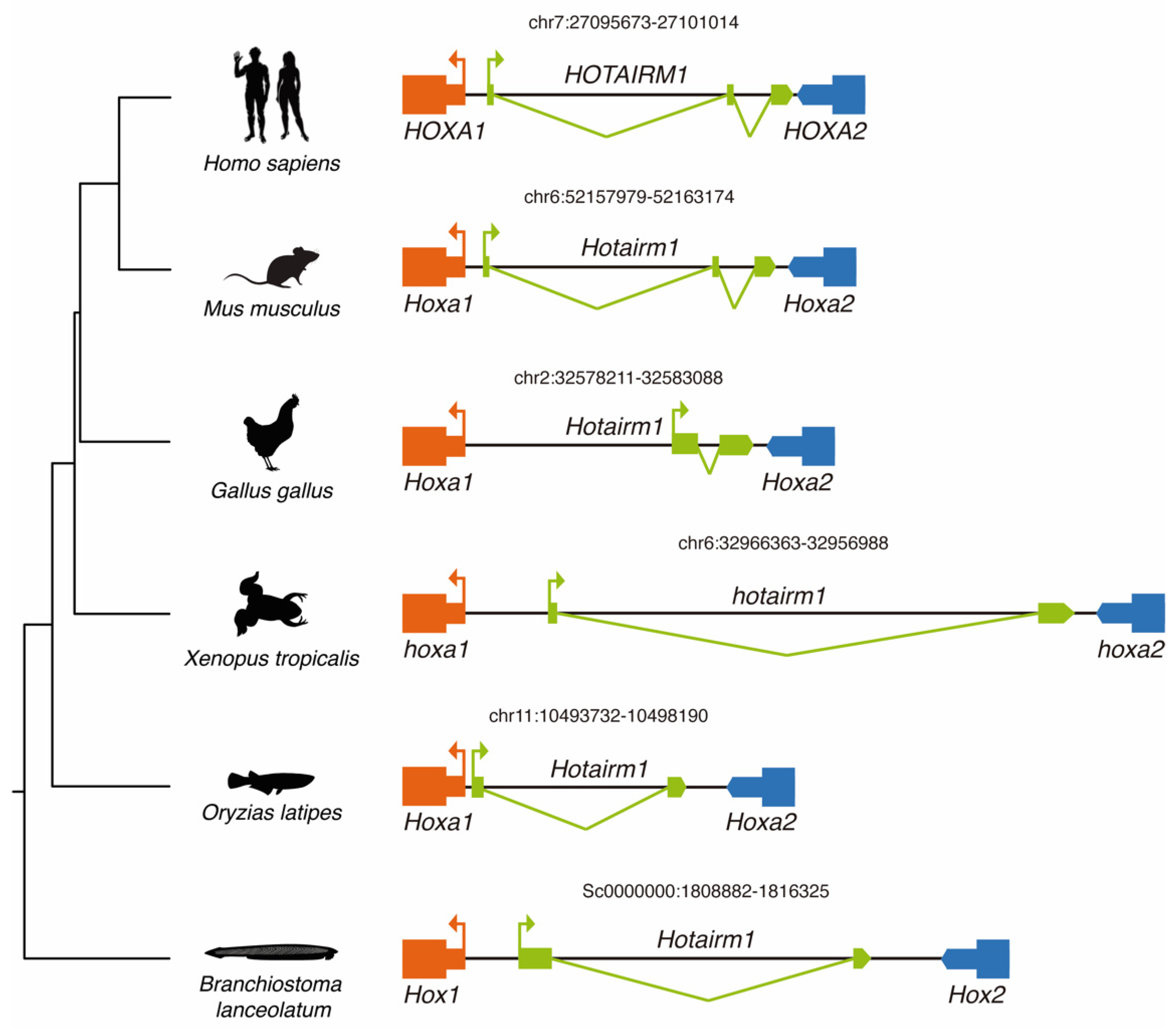
3.2. Conservation of HOTAIRM1 across Chordates

HOTAIRM1 was selected for further study because its conservation across several vertebrate lineages has been previously underscored [28,29], and its mechanism of action has been thoroughly studied [30]. HOTAIRM1 was identified for the first time in myelopoietic human cells [31], during a screen for transcriptionally active intergenic elements within the HoxA cluster. In amphioxus it is situated in the Hox cluster between Hox1 and Hox2, and between Hoxa1 and Hoxa2 in vertebrates (Figure 2). According to our microsynteny-based analysis, Hotairm1 is conserved in most of the chordate species analyzed, with the notable exception of zebrafish (Figure 2). Nonetheless, Hotairm1 appears to be present in other actinopterigians like spotted gar (data not shown), as well as in teleosts like medaka (Figure 2). Given that the Hox cluster is disintegrated in tunicates [32] and due to the absence of an antisense transcript 5′ of Ciona intestinallis Hoxa1, we were unable to confirm the presence of Hotairm1 in this chordate subphylum. Finally, we could not find any trace of this lincRNA in the genomes of the cyclostomes Eptapretus burger and Petromyzon marinus possibly because the microsynteny is lost in this region, due to a lineage-specific loss or alternatively because the lncRNA annotation was deficient [33].
Our results add up to previous studies that have described the presence of Hotairm1 in mammals [28], birds and reptiles [29], strongly suggesting that Hotairm1 was retained within the HoxA cluster after the vertebrate-specific rounds of genome duplication [34], and that its origin predates, at least, the appearance of extant chordate lineages more than 500 million years ago.

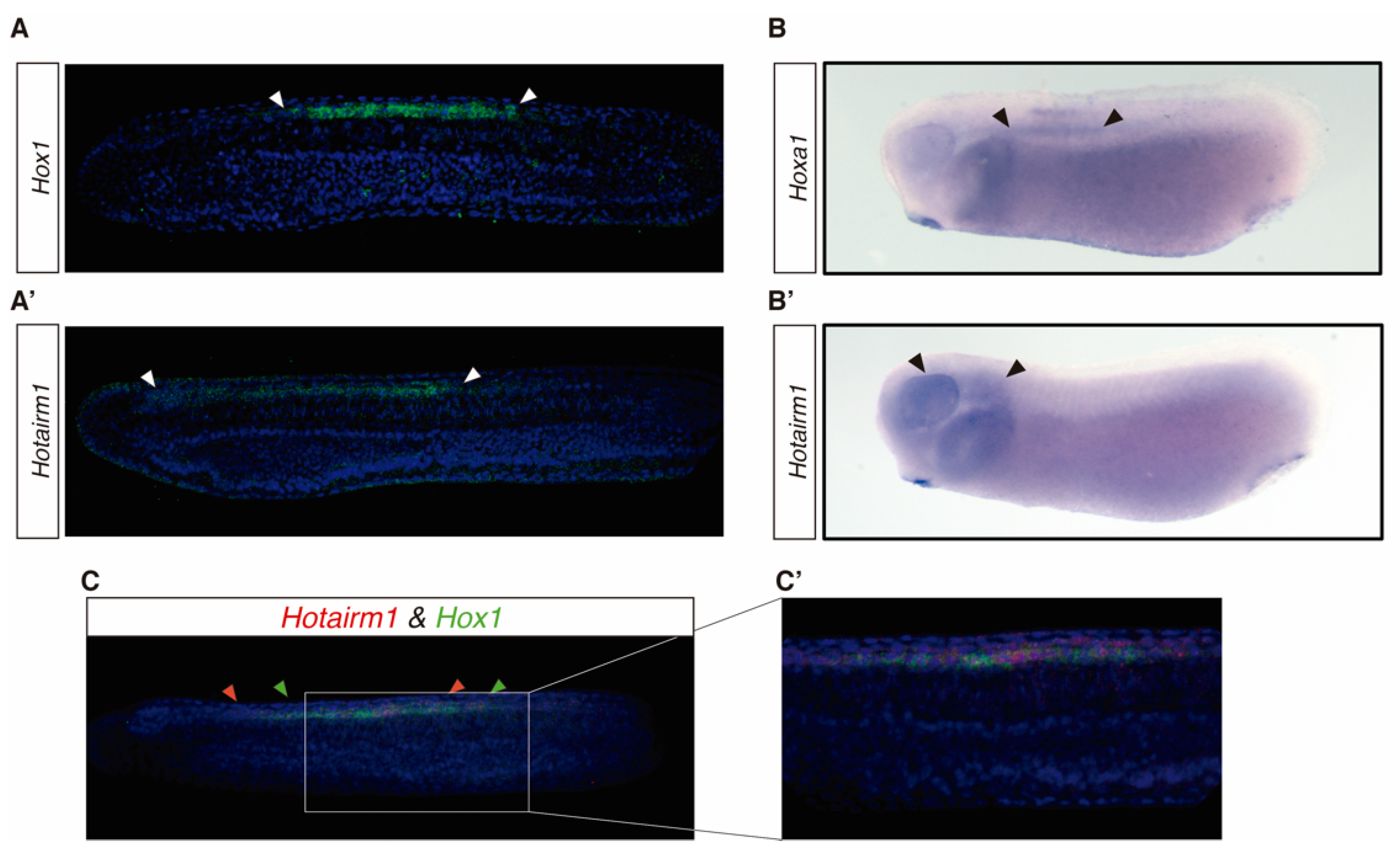
3.3. HOTAIRM1 Expression Patterns in Amphioxus and Xenopus

The expression domain of Hotairm1 is mostly unknown, although it has been observed to be significantly increased or decreased in several types of cancer [35,36,37]. Furthermore, its expression is dynamically regulated during neuronal differentiation, showing a sharp increase in early differentiating neurons [38]. According to available RNA-seq data [15], the expression of Hotairm1 during B. lanceolatum development peaks at 27 h post fertilization (hpf) (File S1). To investigate Hotairm1 expression during amphioxus development, we performed fluorescent in situ hybridization in embryos from 18 hpf to 48 hpf. At 18 hpf we couldn’t detect any signal, while at 21 hpf the expression of Hotairm1 appeared in scattered cells in the presomitic mesoderm and in the neural plate partly overlapping Hox1 expression domain (data not shown) [39]. At 30 hpf, 36 hpf and 48 hpf Hotairm1 expression is restricted to the neural tube from the 5th somite towards the anterior developing neural tube, probably reaching the Di-Mesencephalic primordium (DiMes) [39,40] (Figure 3A’). Relevantly, the expression domain of Hotairm1 in this developmental stage overlaps with Hox1 which is also expressed in the developing neural tube and localized in the hindbrain (Figure 3C,C’) [41,42].
In the vertebrate X. tropicalis, the expression of hotairm1 was detected in the midbrain, hindbrain and the pharyngeal arch (Figure 3B’). At the same time, hoxa1 in this species is expressed in the pharyngeal arch and the anterior developing neural tube (Figure 3B) [43].
These results suggest that the expression domain of hotairm1 is conserved between B. lanceolatum and X. tropicalis, being expressed in the anterior half of the developing neural tube and partly overlapping with Hox1 and hoxa1 expression, respectively (Figure 3).

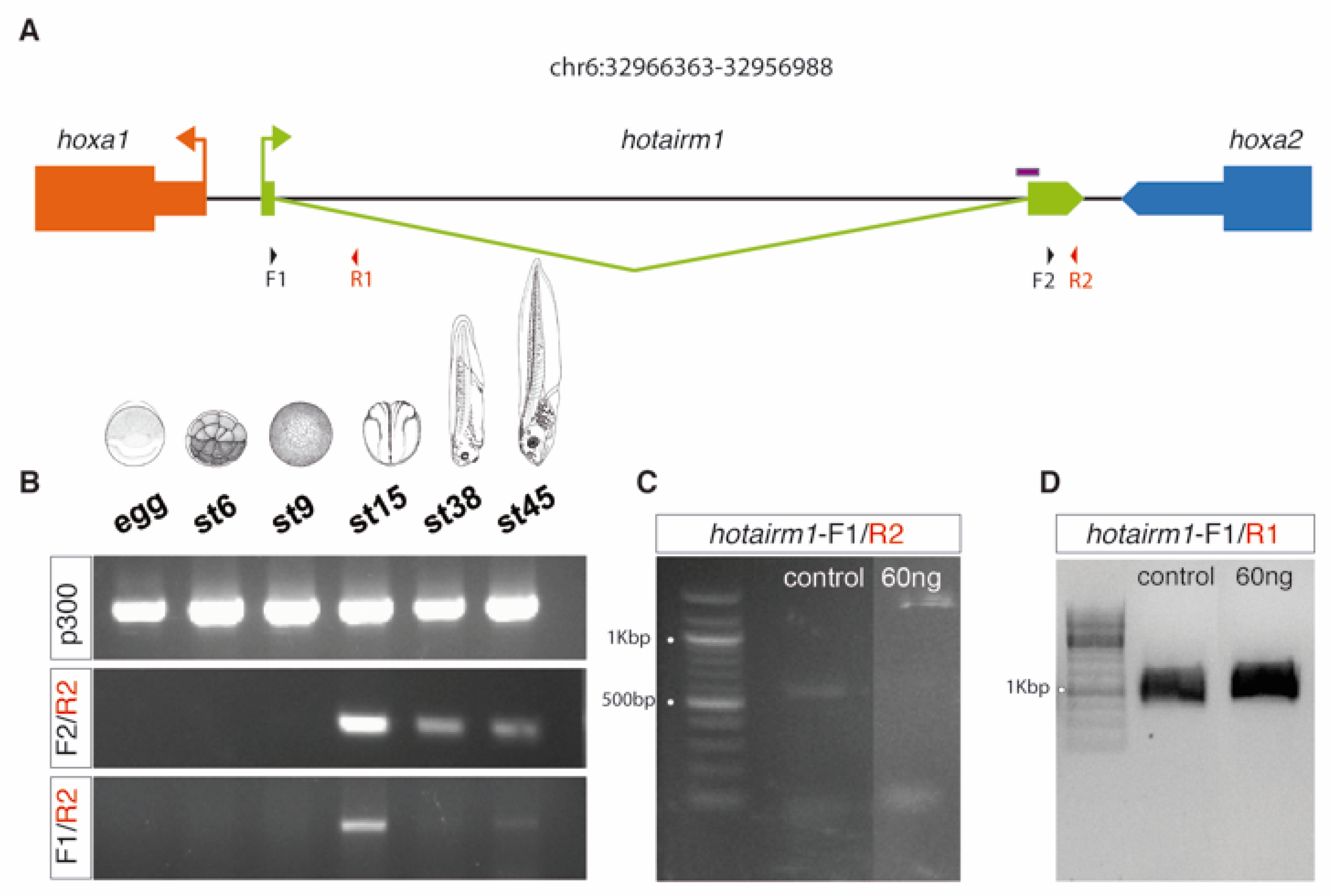
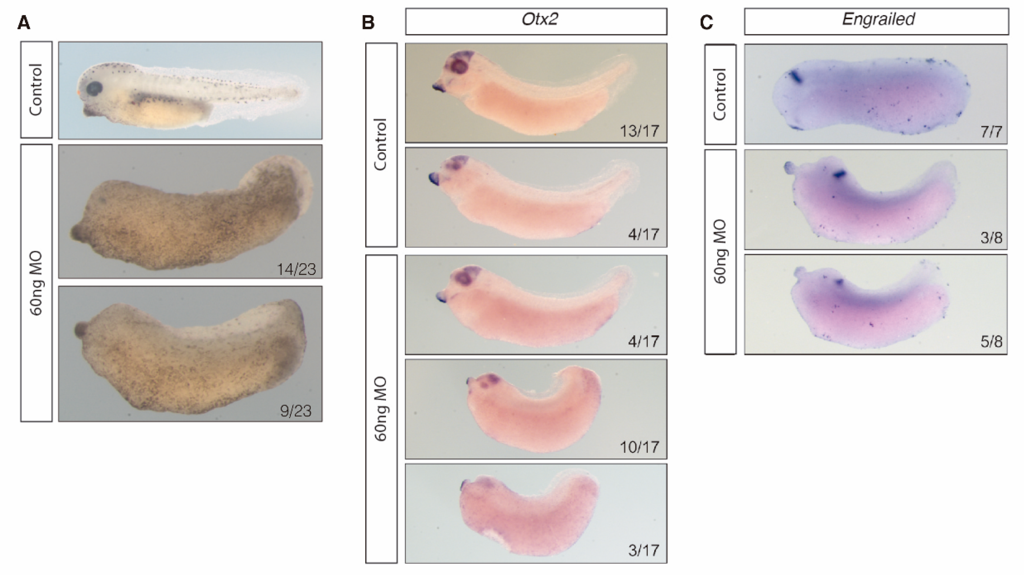
3.4. HOTAIRM1 Function and Expression Conservation

Remarkably, HOTAIRM1 has been described to act as a regulator of the chromatin state within the Hox cluster in human cells. Wang & Dostle [30] found that two HOTAIRM1 isoforms, one spliced and one unspliced, play diverging roles in the regulation of the HoxA cluster chromatin state in presence of retinoic acid. Their findings indicate that the spliced isoform binds to the polycomb repressive complex 2 (PRC2) and changes the chromatin state repressing the medial HoxA genes (HOXA4, HOXA5, HOXA6). The unspliced isoform, on the other hand, binds to the UTX/MLL complex and promotes the expression of the proximal HoxA genes (HOXA1 and HOXA2).
To gain insight into the function of hotairm1 we tried to alter the expression balance of its isoforms during X. tropicalis development. In order to achieve this, we used a morpholino oligonucleotide targeting the 3′ splice junction, thus forcing an isoform switch towards the unspliced state (Figure 4).
Strikingly, the morpholino treatment resulted in a headless tadpole-stage embryo (Figure 5A). This phenotype is characterized by the decrease of expression of brain markers such as otx2 (forebrain-midbrain boundary marker) and engrailed (midbrain-hindbrain boundary marker) (Figure 5B,C). These results suggest that alterations in the balance of Hotairm1 isoforms produce a severe disruption in the development of the anterior part of the central neural system (Figure 5).
Finally, in order to check whether the expression of HoxA genes was altered in MO-treated Xenopus embryos, we performed Real Time quantitative PCR at stage 18, when neurulation is taking place. Our results show a significant upregulation of medial Hox genes hoxa5 and hoxa6, and a downregulation of hoxa4, compared with control embryos. Remarkably, no significant change in the expression of the proximal HoxA gene, hoxa1, was observed. These results suggest that HOTAIRM1 function is partially conserved between Xenopus and human (Figure 6).

4. Conclusions

We have developed a novel pipeline called lincOFinder that establishes bona fide microsyntenic clusters to detect deeply conserved lincRNAs. Applying this tool to investigate the invertebrate-vertebrate transition, we have managed to identify 16 lincRNA putatively conserved between amphioxus and humans. To our knowledge, this represents the first successful identification of homologous lincRNAs over very long evolutionary distances. We show that one of these conserved lincRNAs, Hotairm1, is expressed along the anterior half of the neural tube during amphioxus and Xenopus development. The injection of MO targeting the 3′ splice junction triggers an imbalance between the spliced and unspliced form resulting in the disruption of the proximal and medial hoxa genes. This change in hoxa expression produces in a tadpole a patterning defect in the anterior neural system leading to a headless phenotype. However, further work needs to be done to elucidate the molecular mechanism underlying this severe phenotype. This nonetheless, is a reliable indicative that this lincRNA is at least to some degree conserved in amphioxus, Xenopus and human, allowing us to infer that it is conserved in the phylum Chordata and that regulation of the Hox cluster by lincRNAs may be traced back to the origin of chordates.

Supplementary Materials

The following are available online at https://www.mdpi.com/2079-7737/8/3/61/s1, Table S1: Putatively conserved lincRNAs between Homo sapiens and Branchiostoma lanceolatum. Analysis of the genes surrounding the lincRNA focusing on three genes (upstream2, upstream1 and lincRNA); Table S2: Putatively conserved lincRNAs between Homo sapiens and Branchiostoma lanceolatum. Analysis of the genes surrounding the lincRNA focusing on three genes (lincRNA, downstream1 and downstream2); File S1: Homology_Expression_RawOut.xlsx.

Author Contributions

Conceptualization, C.H.-U., E.N.-P. and M.M.-B.; methodology, C.H.-U. and M.M.-B.; software, C.H.-U. and J.G.; validation, C.H.-U., and M.M.-B.; formal analysis, C.H.-U. and J.G.; investigation, C.H.-U., B.A.-C. and M.M.-B.; resources, J.G.-F., G.N.W.; data curation, C.H.-U. and J.G.; writing—original draft preparation, C.H.-U., M.M.-B. and E.N.-P.; writing—review and editing, C.H.-U., M.M.-B., J.G.-F., G.N.W. and E.N.-P.; visualization, C.H.-U. and M.M.-B.; supervision, J.G.-F. and G.N.W.; project administration, J.G.-F.; funding acquisition, J.G.-F. and G.N.W.

Funding

This research was funded by grant BFU2017-86152-P (Ministerio de Ciencia, Innovación y Universidades, Spanish Government) to J.G.-F., C.H.-U. holds a predoctoral FPI contract (Ministerio de Ciencia, Innovación y Universidades, Spanish Government) and People Program (Marie Curie Actions) of the European Union’s Seventh Framework Program FP7 under REA Grant agreement number 607142 (DevCom) to GW.

Acknowledgments

We wish to thank Mauel Irimia for help, advice and support, and Tom Lewin for the modifications to the HCR protocol for ish.

Conflicts of Interest

The authors declare no conflict of interest. The funders had no role in the design of the study; in the collection, analyses, or interpretation of data; in the writing of the manuscript, or in the decision to publish the results.

References

  1. Holland, P.W.H.; Garcia-Fernàndez, J.; Williams, N.A.; Sidow, A. Gene duplications and the origins of vertebrate development. Development 1994, 1994, 125–133. [Google Scholar]
  2. Schmitz, J.F.; Zimmer, F.; Bornberg-Bauer, E. Mechanisms of transcription factor evolution in Metazoa. Nucleic Acids Res. 2016, 44, 6287–6297. [Google Scholar] [CrossRef] [PubMed]
  3. Morris, K.V.; Mattick, J.S. The rise of regulatory RNA. Nat. Rev. Genet. 2014, 15, 423–437. [Google Scholar] [CrossRef] [PubMed] [Green Version]
  4. Zampetaki, A.; Albrecht, A.; Steinhofel, K. Long Non-coding RNA Structure and Function: Is There a Link? Front. Physiol. 2018, 9, 1201. [Google Scholar] [CrossRef] [PubMed]
  5. Wan, Y.; Kertesz, M.; Spitale, R.C.; Segal, E.; Chang, H.Y. Understanding the transcriptome through RNA structure. Nat. Rev. Genet. 2011, 12, 641–655. [Google Scholar] [CrossRef] [PubMed] [Green Version]
  6. Ponting, C.P.; Oliver, P.L.; Reik, W. Evolution and functions of long noncoding RNAs. Cell 2009, 136, 629–641. [Google Scholar] [CrossRef]
  7. Fico, A.; Fiorenzano, A.; Pascale, E.; Patriarca, E.J.; Minchiotti, G. Long non-coding RNA in stem cell pluripotency and lineage commitment: functions and evolutionary conservation. Cell. Mol. Life Sci. 2019, 76, 1459–1471. [Google Scholar] [CrossRef]
  8. Diederichs, S. The four dimensions of noncoding RNA conservation. Trends Genet. 2014, 30, 121–123. [Google Scholar] [CrossRef]
  9. Jathar, S.; Kumar, V.; Srivastava, J.; Tripathi, V. Technological developments in lncRNA biology. In Advances in Experimental Medicine and Biology; Springer: Singapore, 2017; Vol. 1008, pp. 283–323. [Google Scholar]
  10. Rivas, E.; Clements, J.; Eddy, S.R. A statistical test for conserved RNA structure shows lack of evidence for structure in lncRNAs. Nat. Methods 2017, 14, 45–48. [Google Scholar] [CrossRef]
  11. Neme, R.; Tautz, D. Fast turnover of genome transcription across evolutionary time exposes entire non-coding DNA to de novo gene emergence. Elife 2016, 5. [Google Scholar] [CrossRef]
  12. Garcia-Fernandez, J.; Benito-Gutierrez, E. It’s a long way from amphioxus: descendants of the earliest chordate. Bioessays 2009, 31, 665–675. [Google Scholar] [CrossRef]
  13. Paps, J.; Holland, P.W.H.; Shimeld, S.M. A genome-wide view of transcription factor gene diversity in chordate evolution: less gene loss in amphioxus? Brief. Funct. Genomics 2012, 11, 177–186. [Google Scholar] [CrossRef] [Green Version]
  14. Putnam, N.H.; Butts, T.; Ferrier, D.E.K.; Furlong, R.F.; Hellsten, U.; Kawashima, T.; Robinson-Rechavi, M.; Shoguchi, E.; Terry, A.; Yu, J.-K.; et al. The amphioxus genome and the evolution of the chordate karyotype. Nature 2008, 453, 1064–1071. [Google Scholar] [CrossRef] [Green Version]
  15. Marlétaz, F.; Firbas, P.N.; Maeso, I.; Tena, J.J.; Bogdanovic, O.; Perry, M.; Wyatt, C.D.R.; de la Calle-Mustienes, E.; Bertrand, S.; Burguera, D.; et al. Amphioxus functional genomics and the origins of vertebrate gene regulation. Nature 2018, 564, 64–70. [Google Scholar] [CrossRef] [Green Version]
  16. Bertrand, S.; Escriva, H.; Williams, N.A.; Holland, N.D.; Holland, L.Z. Evolutionary crossroads in developmental biology: amphioxus. Development 2011, 138, 4819–4830. [Google Scholar] [CrossRef] [Green Version]
  17. Pegueroles, C.; Iraola-Guzmán, S.; Chorostecki, U.; Ksiezopolska, E.; Saus, E.; Gabaldón, T. Transcriptomic analyses reveal groups of co-expressed, syntenic lncRNAs in four species of the genus Caenorhabditis. RNA Biol. 2019, 16, 320–329. [Google Scholar] [CrossRef] [Green Version]
  18. Bush, S.J.; Muriuki, C.; McCulloch, M.E.B.; Farquhar, I.L.; Clark, E.L.; Hume, D.A. Cross-species inference of long non-coding RNAs greatly expands the ruminant transcriptome. Genet. Sel. Evol. 2018, 50, 20. [Google Scholar] [CrossRef] [Green Version]
  19. Zerbino, D.R.; Achuthan, P.; Akanni, W.; Amode, M.R.; Barrell, D.; Bhai, J.; Billis, K.; Cummins, C.; Gall, A.; Girón, C.G.; et al. Ensembl 2018. Nucleic Acids Res. 2018, 46, D754–D761. [Google Scholar] [CrossRef]
  20. Emms, D.M.; Kelly, S. OrthoFinder: solving fundamental biases in whole genome comparisons dramatically improves orthogroup inference accuracy. Genome Biol. 2015, 16, 157. [Google Scholar] [CrossRef]
  21. Bogdanovic, O.; Alexis, M.S.; Tena, J.J.; Maeso, I.; Fernandez-Minan, A.; Fraser, H.B.; Gomez-Skarmeta, J.L.; Roy, S.W.; Irimia, M.; de la Calle-Mustienes, E. Extensive conservation of ancient microsynteny across metazoans due to cis-regulatory constraints. Genome Res. 2012, 22, 2356–2367. [Google Scholar]
  22. Sokal, R.R. A statistical method for evaluating systematic relationships. Univ. Kansas, Sci. Bull. 1958, 38, 1409–1438. [Google Scholar]
  23. Kent, W.J.; Sugnet, C.W.; Furey, T.S.; Roskin, K.M.; Pringle, T.H.; Zahler, A.M.; Haussler, D. The human genome browser at UCSC. Genome Res. 2002, 12, 996–1006. [Google Scholar] [CrossRef]
  24. Monsoro-Burq, A.H. A Rapid Protocol for Whole-Mount In Situ Hybridization on Xenopus Embryos. Cold Spring Harb. Protoc. 2007, 2007. [Google Scholar] [CrossRef]
  25. Fuentes, M.; Benito, E.; Bertrand, S.; Paris, M.; Mignardot, A.; Godoy, L.; Jimenez-Delgado, S.; Oliveri, D.; Candiani, S.; Hirsinger, E.; et al. Insights into spawning behavior and development of the european amphioxus (Branchiostoma lanceolatum). J. Exp. Zool. Part B Mol. Dev. Evol. 2007, 308B, 484–493. [Google Scholar] [CrossRef]
  26. Choi, H.M.T.; Schwarzkopf, M.; Fornace, M.E.; Acharya, A.; Artavanis, G.; Stegmaier, J.; Cunha, A.; Pierce, N.A. Third-generation in situ hybridization chain reaction: multiplexed, quantitative, sensitive, versatile, robust. Development 2018, 145, dev165753. [Google Scholar] [CrossRef]
  27. Ulitsky, I. Evolution to the rescue: using comparative genomics to understand long non-coding RNAs. Nat. Rev. Genet. 2016, 17, 601–614. [Google Scholar] [CrossRef]
  28. Yu, H.; Lindsay, J.; Feng, Z.-P.; Frankenberg, S.; Hu, Y.; Carone, D.; Shaw, G.; Pask, A.J.; O’Neill, R.; Papenfuss, A.T.; et al. Evolution of coding and non-coding genes in HOX clusters of a marsupial. BMC Genomics 2012, 13, 251. [Google Scholar] [CrossRef]
  29. Gardner, P.P.; Fasold, M.; Burge, S.W.; Ninova, M.; Hertel, J.; Kehr, S.; Steeves, T.E.; Griffiths-Jones, S.; Stadler, P.F. Conservation and Losses of Non-Coding RNAs in Avian Genomes. PLoS One 2015, 10, e0121797. [Google Scholar] [CrossRef]
  30. Wang, X.Q.D.; Dostie, J. Reciprocal regulation of chromatin state and architecture by HOTAIRM1 contributes to temporal collinear HOXA gene activation. Nucleic Acids Res. 2017, 45, 1091–1104. [Google Scholar] [CrossRef]
  31. Zhang, X.; Lian, Z.; Padden, C.; Gerstein, M.B.; Rozowsky, J.; Snyder, M.; Gingeras, T.R.; Kapranov, P.; Weissman, S.M.; Newburger, P.E. A myelopoiesis-associated regulatory intergenic noncoding RNA transcript within the human HOXA cluster. Blood 2009, 113, 2526–2534. [Google Scholar] [CrossRef] [Green Version]
  32. Sekigami, Y.; Kobayashi, T.; Omi, A.; Nishitsuji, K.; Ikuta, T.; Fujiyama, A.; Satoh, N.; Saiga, H. Hox gene cluster of the ascidian, Halocynthia roretzi, reveals multiple ancient steps of cluster disintegration during ascidian evolution. Zool. Lett. 2017, 3, 17. [Google Scholar] [CrossRef] [Green Version]
  33. Pascual-Anaya, J.; Sato, I.; Sugahara, F.; Higuchi, S.; Paps, J.; Ren, Y.; Takagi, W.; Ruiz-Villalba, A.; Ota, K.G.; Wang, W.; et al. Hagfish and lamprey Hox genes reveal conservation of temporal colinearity in vertebrates. Nat. Ecol. Evol. 2018, 2, 859–866. [Google Scholar] [CrossRef]
  34. Dehal, P.; Boore, J.L. Two Rounds of Whole Genome Duplication in the Ancestral Vertebrate. PLoS Biol. 2005, 3, e314. [Google Scholar] [CrossRef]
  35. Esfandi, F.; Taheri, M.; Omrani, M.D.; Shadmehr, M.B.; Arsang-Jang, S.; Shams, R.; Ghafouri-Fard, S. Expression of long non-coding RNAs (lncRNAs) has been dysregulated in non-small cell lung cancer tissues. BMC Cancer 2019, 19, 222. [Google Scholar] [CrossRef]
  36. Li, Q.; Dong, C.; Cui, J.; Wang, Y.; Hong, X. Over-expressed lncRNA HOTAIRM1 promotes tumor growth and invasion through up-regulating HOXA1 and sequestering G9a/EZH2/Dnmts away from the HOXA1 gene in glioblastoma multiforme. J. Exp. Clin. Cancer Res. 2018, 37, 265. [Google Scholar] [CrossRef]
  37. Song, L.; Zhang, S.; Duan, C.; Ma, S.; Hussain, S.; Wei, L.; Chu, M. Genome-wide identification of lncRNAs as novel prognosis biomarkers of glioma. J. Cell. Biochem. 2019. [Google Scholar] [CrossRef]
  38. Lin, M.; Pedrosa, E.; Shah, A.; Hrabovsky, A.; Maqbool, S.; Zheng, D.; Lachman, H.M. RNA-Seq of Human Neurons Derived from iPS Cells Reveals Candidate Long Non-Coding RNAs Involved in Neurogenesis and Neuropsychiatric Disorders. PLoS One 2011, 6, e23356. [Google Scholar] [CrossRef]
  39. Albuixech-Crespo, B.; López-Blanch, L.; Burguera, D.; Maeso, I.; Sánchez-Arrones, L.; Moreno-Bravo, J.A.; Somorjai, I.; Pascual-Anaya, J.; Puelles, E.; Bovolenta, P.; et al. Molecular regionalization of the developing amphioxus neural tube challenges major partitions of the vertebrate brain. PLoS Biol. 2017, 15, e2001573. [Google Scholar] [CrossRef]
  40. Albuixech-Crespo, B.; Herrera-Úbeda, C.; Marfany, G.; Irimia, M.; Garcia-Fernàndez, J. Origin and evolution of the chordate central nervous system: insights from amphioxus genoarchitecture. Int. J. Dev. Biol. 2017, 61, 655–664. [Google Scholar] [CrossRef]
  41. Schubert, M.; Holland, N.D.; Laudet, V.; Holland, L.Z. A retinoic acid-Hox hierarchy controls both anterior/posterior patterning and neuronal specification in the developing central nervous system of the cephalochordate amphioxus. Dev. Biol. 2006, 296, 190–202. [Google Scholar] [CrossRef]
  42. Zieger, E.; Candiani, S.; Garbarino, G.; Croce, J.C.; Schubert, M. Roles of Retinoic Acid Signaling in Shaping the Neuronal Architecture of the Developing Amphioxus Nervous System. Mol. Neurobiol. 2018, 55, 5210–5229. [Google Scholar] [CrossRef]
  43. McNulty, C.L.; Peres, J.N.; Bardine, N.; van den Akker, W.M.R.; Durston, A.J. Knockdown of the complete Hox paralogous group 1 leads to dramatic hindbrain and neural crest defects. Development 2005, 132, 2861–2871. [Google Scholar] [CrossRef] [Green Version]
Figure 1. Diagram of LincOFinder mechanism. (A) Representation of the Ref species region where a lincRNA is present. (B) Formatted table of orthologs and virtual coordinates from the three upstream and downstream coding genes fed to the algorithm. (C) Selection of the best cluster according to the minimum distance between genes. (D) Representation of a conserved mycrosyntenic cluster in the Int species, where the presence of a lincRNA is manually confirmed (above) or discarded (below).
Biology 08 00061 g001
Figure 2. Schematic representation of the genomic locus of Hotairm1 across several chordate species. Genome or scaffold position is indicated above each HotairM1 locus.
Biology 08 00061 g002
Figure 3. In situ hybridization (ish) in B. lanceolatum and X. tropicallis. Anterior to the left, dorsal is up. (A,A’) Fluorescent HCR ish in B. lanceolatum in whole mount for (A) Hox1 and (A’) Hotairm1 in 30 hpf embryos. White arrows mark the anterior and posterior limits of the expression domain. (B,B’) Colorimetric whole mount ish in X. tropicalis tadpoles for (B) hoxa1 and (B’) hotairm1. Black arrows mark the anterior and posterior limits of the expression domain. (C,C’) Fluorescent double HCR ish in B. lanceolatum in whole mount ish of (C) Hox1 and Hotairm1 in a 36 hpf embryo and (C’) the detailed zone where Hotairm1 peaks its expression. Green arrows mark the anterior and posterior limits of Hox1 expression and red arrows mark the ones of Hotairm1.
Biology 08 00061 g003
Figure 4. Isoform switch of hotairm1 expression towards the unspliced state using a morpholino. (A) Detail of the primers (black and red arrowheads) and morpholino (purple box) used for the amplification of the spliced and unspliced isoforms of hotairm1 in X.tropicalis and for the impairment of the splicing in the MO-treated embryos. (B) Expression of hotairm1 across Xenopus developmental stages. (C) Inhibition of the spliced isoform in MO treated embryos of Xenopus at st18. (D) Assessment of the presence of the unspliced isoform of hotairm1 in MO treated embryos as well as in the control embryos at st18.
Biology 08 00061 g004
Figure 5. MO treated embryos and in situ hybridization in MO treated embryos. Anterior to the left, dorsal is up. (A) Control X. tropicalis MO treated embryos with normal development. 60ng hotairm1-MO treated embryos with a posteriorization of the anterior part of the embryo. (B) Whole mount colorimetric ish of otx2 in X. tropicalis stage 26 control embryos and MO treated embryos showing the reduced expression domain of otx2 in MO treated embryos. (C) Whole mount colorimetric ish of engrailed in X. tropicalis stage 26 control embryos and MO treated embryos showing a clear reduction in the expression in the MO treated embryos.
Biology 08 00061 g005
Figure 6. qPCRs between 60 ng MO treated embryos and control samples at stage 18. * shows statistically significance compared with control samples (Student’s t-test, * p < 0.05, ** p < 0.01, *** p < 0.001, **** p < 0.0001). Gapdh was used as a reference gene.
Biology 08 00061 g006
Table 1. Putatively conserved lincRNAs between Homo sapiens and Branchiostoma lanceolatum. Analysis of the genes surrounding the lincRNA focusing on the three core genes (upstream1, lincRNA and downstream1). Some lincRNAs can be ascribed to more than one hypothetically conserved microsyntenic cluster.
Orthologous lincRNAs 1State of the Cluster in Human 2Human Orthologous lincRNA 3
BL20528|Sc0000000|28|+* Conserved microsyntenyENST00000623777.1_1
BL38782|Sc0000000|30|+* Conserved microsyntenyHOTAIRM1
BL90848|Sc0000001|150|−Correct order but strands inverted. In addition, there are two lncRNAs surrounding the clusterAL354977.2
BL79733|Sc0000007|52|+One gene with the strand invertedBANCR
One gene with the strand invertedTCONS_12_00008513
BL84418|Sc0000009|86|+* Conserved microsyntenyTCONS_00027655
BL91140|Sc0000010|144|−Couple of lincRNAs in amphioxus, synteny conserved in the coding genes, but not in the lincRNATCONS_00006308
BL91143|Sc0000010|145|-* Conserved microsyntenyRP11-181G12.4
BL53024|Sc0000015|55|+One gene with the strand invertedTCONS_00027115
BL82992|Sc0000016|15|−One gene with the strand invertedTCONS_00000550
BL78145|Sc0000039|54|+* Conserved microsyntenyTCONS_00024711
BL55463|Sc0000050|45|+* Conserved microsyntenyTCONS_00011710
BL68900|Sc0000072|2|+Synteny conserved in the coding genesAI219887
BL54861|Sc0000089|4|−Problematic region with several lincRNAs and massive distancesAC109136.1
Problematic region with several lincRNAs and massive distancesAC124852.1
BL41904|Sc0000219|3|−* Conserved microsyntenyTCONS_00007813
BL72725|Sc0000229|14|+* Conserved microsyntenyBC043517
* Conserved microsyntenyLINC00114
BL59605|Sc0000234|6|−* Conserved microsyntenyTCONS_00011870
BL38170|Sc0000240|5|+* Conserved microsyntenyLOC100132215
1 GeneID, Scaffold, virtual coordinates and strand separated by “|”. 2 Description of the synteny of the cluster status in human. 3 ID of the putative human orthologous lincRNA. * indicates a perfect match in strand and order of the three core genes.

