Article

Identification of N6-Methyladenosine-Related Factors and the Prediction of the Regulatory Mechanism of Hair Follicle Development in Rex and Hycole Rabbits

1
College of Animal Science and Technology, Northwest A&F University, Xianyang 712100, China
2
College of Animal Science, Fujian Agriculture and Forestry University, Fuzhou 350000, China
*
Author to whom correspondence should be addressed.
Biology 2023, 12(11), 1448; https://doi.org/10.3390/biology12111448
Submission received: 31 August 2023 / Revised: 13 November 2023 / Accepted: 14 November 2023 / Published: 17 November 2023
(This article belongs to the Section Developmental Biology)

Abstract

:

Simple Summary

N6-methyladenosine (m6A) is an important modification for genes. Hair follicle development is crucial for the animal fur economy. To improve the quality of animal fur and solve the problem of baldness in people, we explored the regulatory mechanism of m6A on rabbit hair follicles and found that five methylases regulated the development of hair follicles through differential genes/signal pathways. These findings laid a molecular foundation for improving the quality of animal fur and solving the problem of baldness in people.

Abstract

Hair follicle development directly affects the development of the rabbit fur industry. The growth and development of a hair follicle is modified and regulated by many genes and mechanisms. M6A is an important RNA modification. However, there are few studies on the effects of the regulation of m6A on hair follicle growth and development. In this study, hematoxylin–eosin (HE) staining was used to explore the difference in hair follicle development between Rex rabbits and Hycole rabbits, and we performed m6A sequencing to identify the key genes with m6A modification in hair follicle growth. The results showed that the hair length, coarse hair percentage, primary hair follicle ratio, and skin thickness of Hycole rabbits were significantly higher than those of Rex rabbits. However, the proportion of secondary hair follicles in Hycole rabbits was significantly lower than that in Rex rabbits. In addition, we found five differential methylases, 20 differential genes, and 24 differential signaling pathways related to hair growth and development. The results of the Sankey diagram showed that 12 genes were related to 13 signal pathways. Finally, we found that five methylases regulated the development of hair follicles through differential genes/signal pathways. These findings laid a molecular foundation for the function of m6A modification in hair development.

1. Introduction

Rex rabbits are a type of rabbit with high hair density, capillary, and short hair. Hycole rabbits are a type of rabbit with low hair density and thick and long hair. The hair quality of rabbits is closely related to the hair density, which is mainly determined by the hair follicle density [1]. Therefore, the study of hair follicle development is of great significance to rabbit fur production. Hair follicle cycling is a complex biological process [2]. Hair follicles undergo anagen, catagen, and telogen cycles [3]. Many mRNAs and miRNAs are involved in the formation of hair follicles [4,5,6]. Hair follicle morphogenesis depends on many signaling pathways such as WNT, Shh, p53, TGF-β, Notch, and BMP [7]. However, the regulation pathways and methods of m6A modification on hair-follicle-development-related mRNAs in rabbits is unknown.
M6A is a common RNA modification [8]. At present, it has been found that m6A modification has been carried out under the action of methylase (METTL5, METTL14, METTL3, WTAP, and METTL4) and demethylase (FTO and ALKBH5) [9,10,11,12,13]. In addition, “reader” proteins containing YTHDC1, IGFBP2 and other YTH domain proteins are also determining factors in the m6A modification process [13,14,15,16,17,18]. These enzymes play an important role in animal growth [19], fat metabolism [20], reproduction [21], and other physiological processes.
In this study, we identified methylases, methylated genes, and signal pathways in Rex rabbit and Hycole rabbit skin by the MeRIP-seq method. Based on existing studies, we found five different methyltransferases, 20 methylation-modified differential genes, and 24 differential signaling pathways related to hair follicle development in Rex rabbits and Hycole rabbits. Finally, we found that 13 signal pathways were regulated by 12 genes among the genes and signal pathways we selected. In addition, we found that five methylases mediated 20 methylated genes to regulate hair follicle development through multiple pathways based on existing studies. This study lays a molecular theoretical foundation for further exploring the regulation of rabbit hair follicle development by m6A modification.

2. Material and Methods

2.1. Animals

Three newborn female Rex rabbits and three newborn female Hycole rabbits were used for methylated RNA immunoprecipitation sequencing (MeRIP-seq) [22]. Three 165-day-old Rex rabbits and three 165-day-old Hycole rabbits were used for photographing, hair index determination, and HE staining. All rabbits were collected from the same farm of the Northwest A&F University (Yangling, Shannxi, China). The rabbit farm belongs to Professor Ren Zhanjun, and he permitted the experiment.

2.2. Hair Index Determination

Pictures of Rex rabbits and Hycole rabbits were taken with a high pixel mobile phone (64 million pixels). The calculation method for the proportion of primary hair follicles was as follows: Firstly, we selected three 0.575 × 0.862 m2 microscope fields. Then, we recorded the number of hair follicles and obtained the number of primary hair follicles and all hair follicles per unit area. Finally, the proportion of primary hair follicles was calculated. We collected approximately 0.003 g of rabbit hair and recorded its weight as T1 (n = 3). The coarse wool was selected, and we recorded its weight as T2. Coarse wool ratio = T2/T1 × 100%. The hair length was directly measured by vernier caliper after collection.

2.3. Hematoxylin–Eosin (HE) Staining

A proper amount of rabbit back skin tissue was collected and fixed in formaldehyde solution. Then, the skin tissues were treated, embedded, sectioned and stained. The specific method is the same as that in the previous study [23]. Briefly, alcohol was used to remove the water in the tissue block, and then xylene was used to replace the alcohol in the tissue block. Tissue blocks were embedded in paraffin and cut into thin sections on a microtome. The slices were sequentially placed in xylene I (8 min), xylene II (8 min), anhydrous ethanol I (6 min), anhydrous ethanol II (6 min), 95% alcohol (6 min), 85% alcohol (6 min), and 75% alcohol (5 min) and rinsed with running water. Slices were stained with Harris hematoxylin for 3–8 min and rinsed with tap water. Then, the slices were differentiated with 1% hydrochloric acid alcohol for a few seconds and rinsed with tap water. The eosin staining solution was used to stain the slices for 1–3 min. Subsequently, we sequentially placed the slices in 75% alcohol (30 s), 85% alcohol (30 s), 95% alcohol I (1 min), 95% alcohol II (2 min), anhydrous ethanol I (5 min), anhydrous ethanol II (5 min), xylene I (5 min), and xylene II (7 min) to dehydrate and become transparent. Finally, we took the slices out of xylene and let them dry slightly; then, we sealed them with neutral gum. The primary hair follicle ratio and skin thickness were measured for each sample with a light microscope.

2.4. RNA Fragmentation

RNA was extracted from skin of rabbits by TRIzol reagent (Invitrogen Co., Carlsbad, CA, USA). Agilent 2100 Bioanalyzer (Agilent, Santa Clara, CA, USA) was used to assess the concentration and integrity of the RNA. The poly (A) RNA was fragmented into small pieces using Magnesium RNA Fragmentation Module (NEB, cat.e6150, Ipswich, MA, USA) under 86 °C, 7 min. Fragmentation buffer was used to break the mRNA into ∼100 nucleotides fragments.

2.5. M6A IP and Library Construction
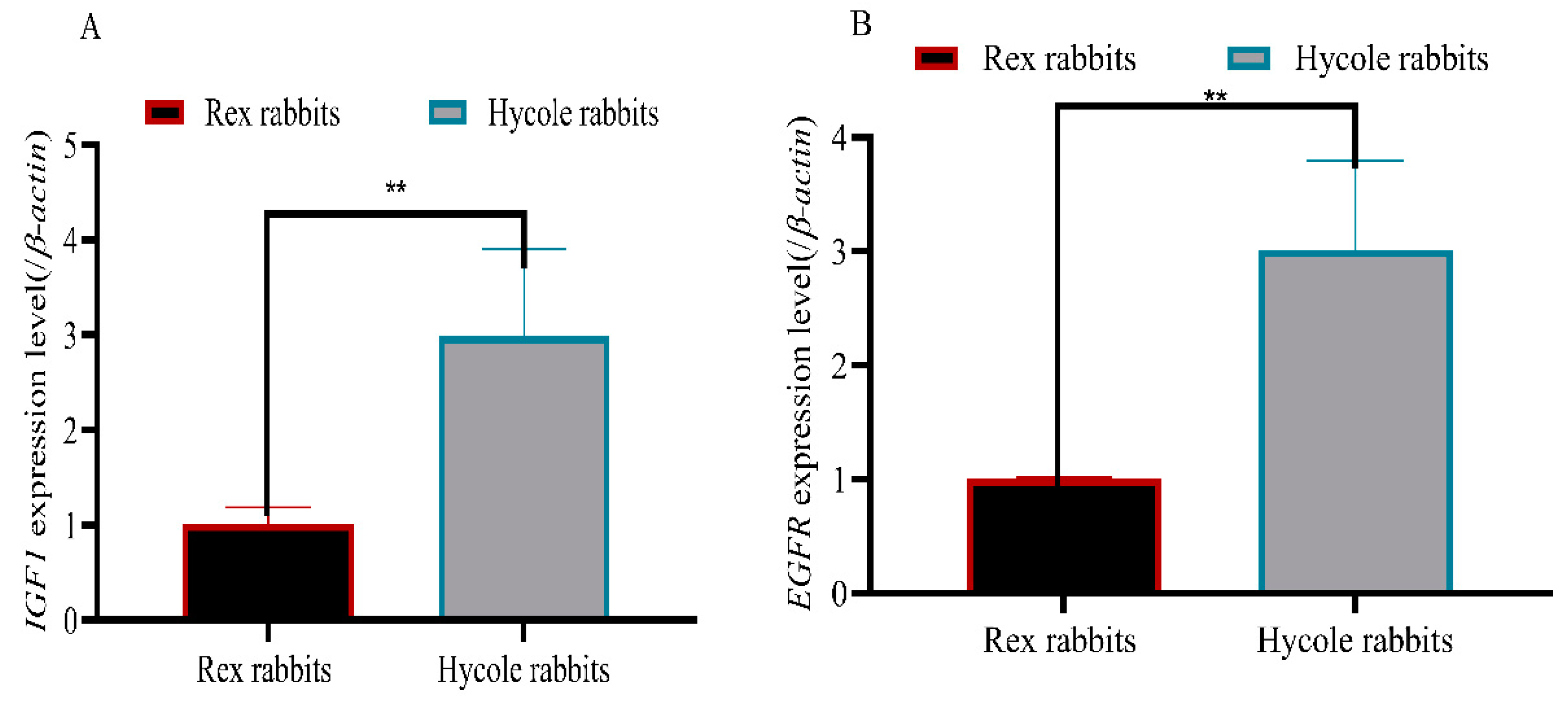
The fragmented mRNA were divided into immunoprecipitation (IP) (95%) and IP control (input) groups (5%). We followed the instructions of the m6A RNA methylation library construction kit (A&D technology, Beijing, China) to IP RNA. Briefly, the cleaved RNA fragments were incubated for 2 h at 4 °C with m6A-specific antibody (No. 202003, Synaptic Systems, Göttingen, Germany) in IP buffer (50 mM Tris-HCl, 750 mM NaCl, and 0.5% Igepal CA-630). Then the IP RNA was reverse transcribed by SuperScript™ II Reverse Transcriptase (Invitrogen, cat. 1896649, Carlsbad, CA, USA). After, we performed the heat-labile UDG enzyme (NEB, cat.m0280, Ipswich, MA, USA) treatment of the U-labeled second-stranded DNAs. Then, the mRNA reacted with the antibody (binding to m6A modification site) with magnetic beads, and sequencing with high-throughput (Illumina Novaseq™6000) (LC-Bio Technology Co., Ltd., Hangzhou, China, 2020) was performed.

2.6. RT-qPCR

The Prime Script RT Reagent Kit (Takara Bio, Saint-Germain-en Laye, France) was used to reverse transcribe the total RNA. RT-qPCR experiments were carried out with a 10 µL system by SYBR Green. β-Actin was used as an internal control. All the primers used for qPCR are listed in Table 1.

2.7. Data Analysis

After sequencing, we used fastp (https://github.com/OpenGene/fastp, accessed on 29 May 2022) [24], the comparison tool bowtie2 [25], and HISAT2 (2.2.1.0) (http://daehwankimlab.github.io/hisat2, accessed on 24 July 2020) [26] software to filter, remove, and compare raw reads separately. The R-Pack exomepeak2 (https://bioconductor.org/packages/edgeR, accessed on 19 May 2022) [27] and DiffBind (3.5) [28] software were used to merge peaks between groups and calculate the abundance of peaks in each sample. StringTie (2.2.1) (https://ccb.jhu.edu/software/stringtie) was used to perform the expression level on 20 May 2022 for all mRNAs from Input libraries by calculating the FPKM (total exon fragments /mapped reads (millions) × exon length (kB)). The differentially expressed mRNAs were selected with log2 (fold change) > 1 or log2 (fold change) < −1 and p value < 0.05 by R package edgeR (4.0.1) (https://bioconductor.org/packages/edgeR on 20 May 2022). Genomes (KEGG) pathway analysis was performed using the database for annotation, visualization, and integrated discovery [29]. Hair and follicle data results were presented as the mean ± standard deviation (SD). GraphPad Prism7 (GraphPad Software, La Jolla, CA, USA) was used to assess the difference. The Student’s t-test was used to analyze the significance of the different levels.

3. Results

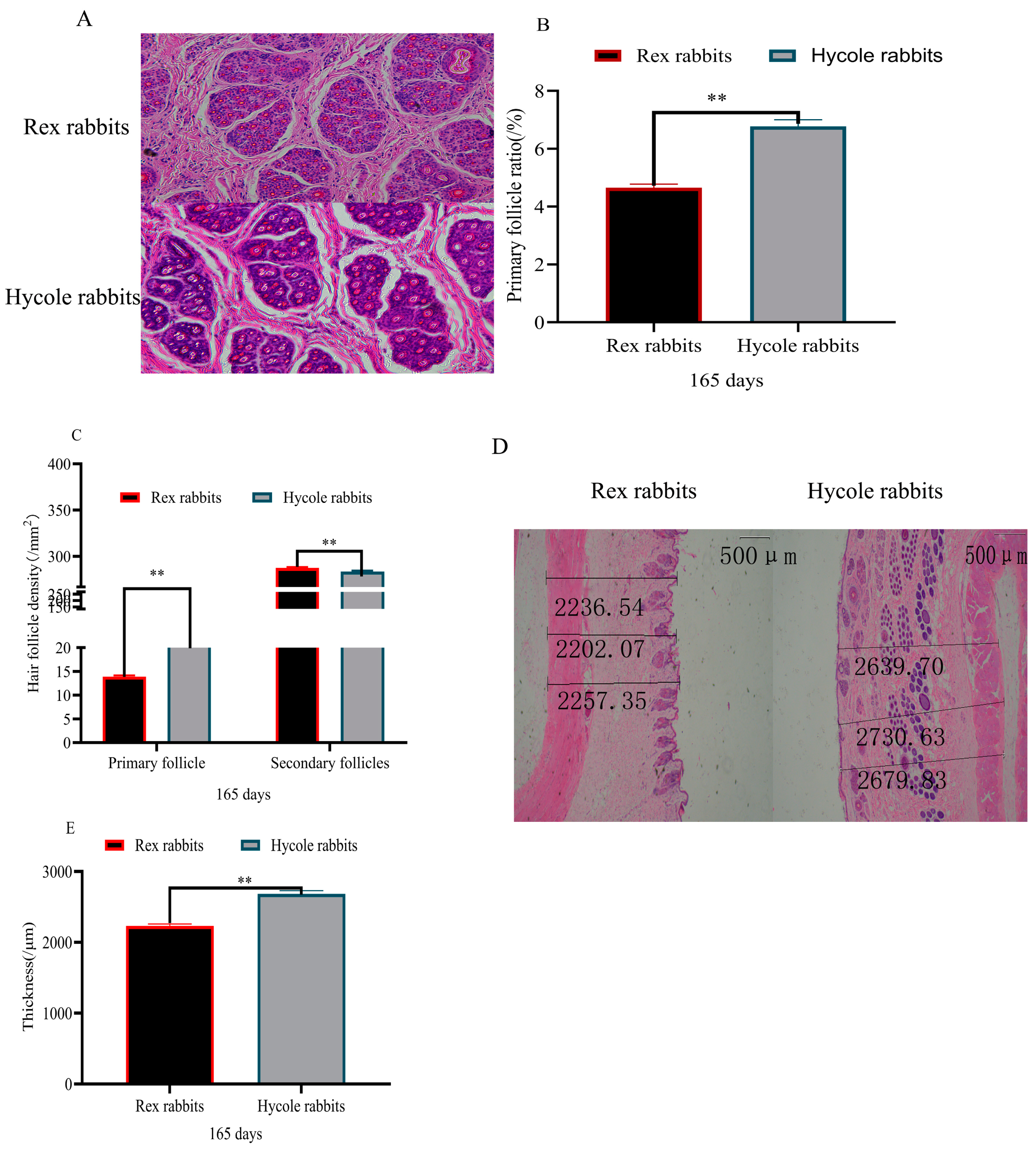
3.1. Difference in Hair Follicles between Rex and Hycole Rabbits

Based on the pictures of the Rex rabbits and Hycole rabbits, we found that the fur of the Rex rabbits was different from that of Hycole rabbits (Figure 1A). The results showed that the hair of the Hycole rabbits was significantly longer than that of Rex rabbits (p < 0.01) (Figure 1B). In addition. the coarse hair rate of the Hycole rabbits was also significantly higher than those of the Rex rabbits (p < 0.01) (Figure 1C). The proportion of primary hair follicles of the Rex rabbits was significantly lower than that of the Hycole rabbits (p < 0.01) (Figure 2A,B). At the same time, the primary hair follicle ratio of Hycole rabbits was significantly higher than that of the Rex rabbits, whereas the ratio of the secondary hair follicles of the Rex rabbits was significantly higher than that of the Hycole rabbits (p < 0.01) (Figure 2C). The results showed that the skin of the Rex rabbit was significantly thinner than that of the Hycole rabbit (p < 0.01) (Figure 2D,E).

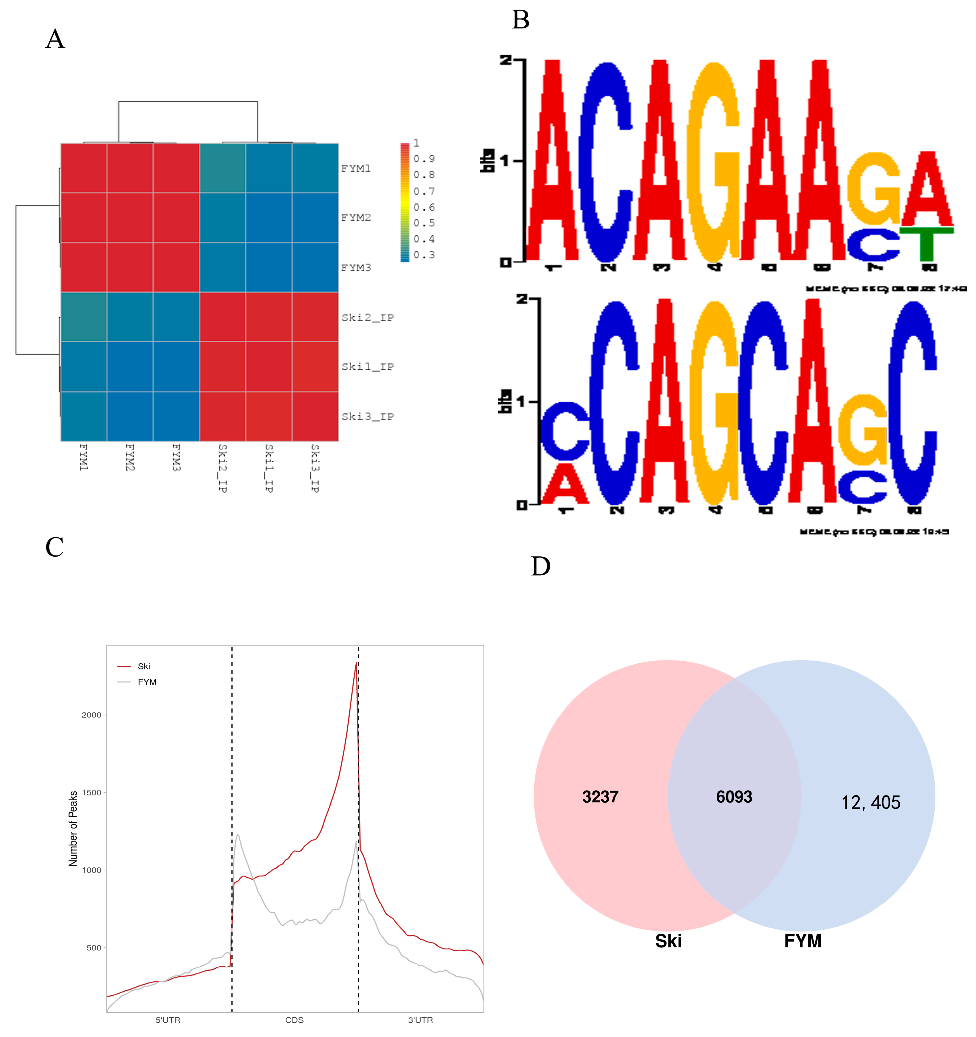
3.2. Summary and Quality Control of Rabbit m6A Sequencing Data

As shown in Supplementary Table S1, MeRIP-seq produced 65,627,376–97,648,830 raw reads from input or IP skin tissues from Rex rabbit (ski) and Hycole rabbit (FYM). We found the GC content in the Rex rabbits’ IP and input was lower than that in the Hycole rabbits. In addition, the proportion of unique mapped reads was higher than 62.30%, and the proportion of multi-mapped reads varied from 3.27% to 25.64% (Supplementary Table S2).

3.3. General Features of Rabbit m6A Methylation

The samples were clustered by calculating the correlation coefficient between the Rex rabbit skin samples and the Hycole rabbit skin samples, which indicated good uniformity within the group (Figure 3A). As shown in Figure 3B, m6A-modified classical sequences RRACH appeared in the sequencing results of the Rex rabbits and Hycole rabbits. According to the statistics, 6093 peaks were methylated both in the Rex rabbit skin and the Hycole rabbit skin. Further, 3237 and 12,405 peaks were specifically methylated in the Rex rabbits’ skin and the Hycole rabbits’ skin, respectively (Figure 3C). To explore the preferential localization of m6A, we counted the distribution of peaks and found that in the CDS, the start and stop codons were the main areas of m6A (Figure 3D). In addition, the peaks enriched in the Rex rabbits were higher than those in the Hycole rabbits in the start codons (Figure 3D). However, the peaks enriched in the Rex rabbits were lower than those in Hycole rabbits in the stop codons (Figure 3D).

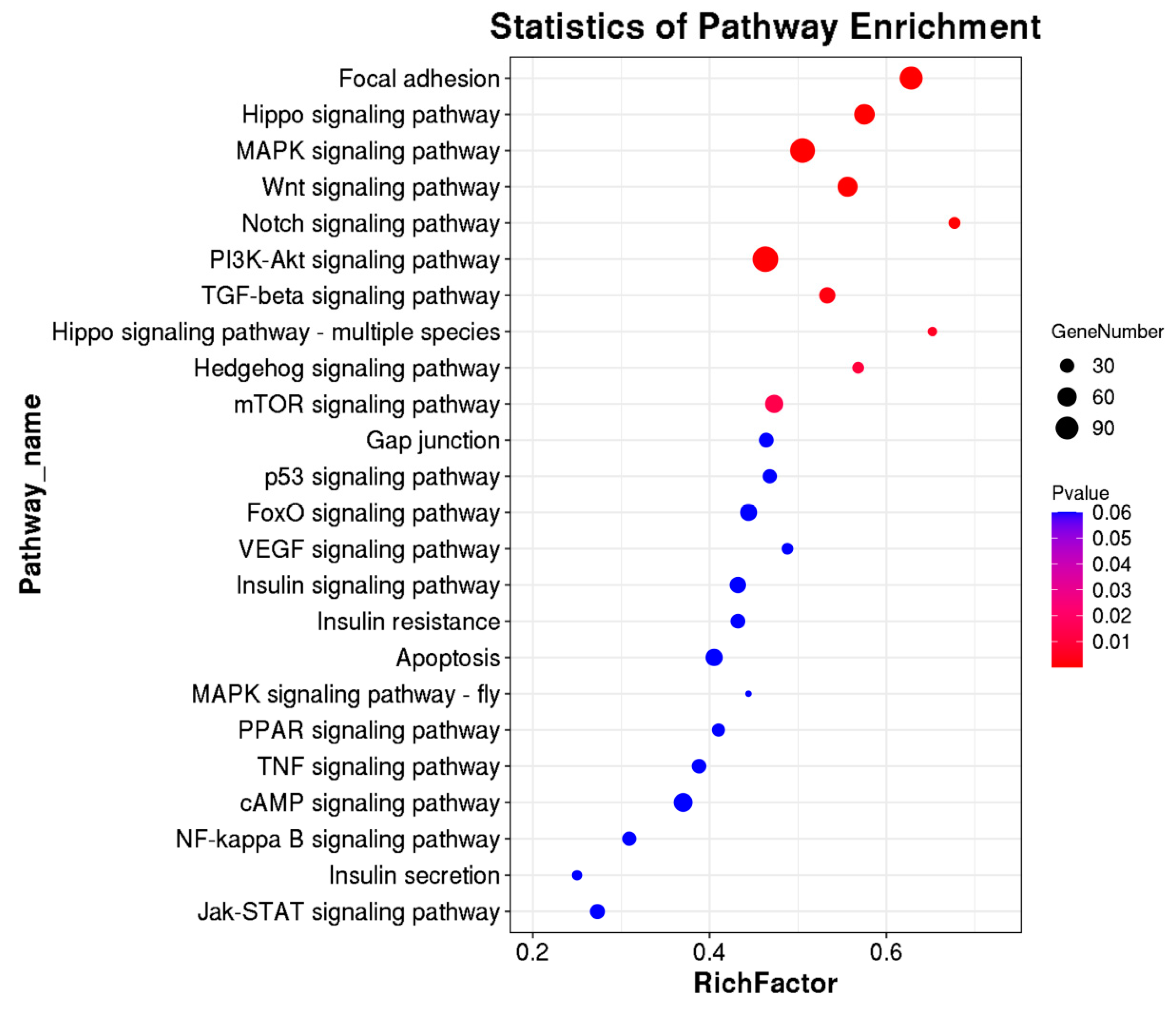
3.4. KEGG Pathway, Methylases, and Methylation Modifying Genes in Rex Rabbit Skin and Hycole Rabbit Skin

To predict the m6A-modified functions associated with hair follicle development, we analyzed the KEGG pathway and found there were 24 differential pathways involved in hair follicle development including Focal adhesion, Hippo, MAPK, WNT, cAMP, and other signaling pathways containing multiple genes (Figure 4). In addition, we found five differential methylation enzymes and 20 differential methylation modification genes were involved in hair follicle development based on the existing study results (Figure 5). To verify the results, we randomly selected two genes (IGF1 and EGFR) for RT-qPCR validation. The expression levels of the IGF1 gene and EGFR gene in the Rex rabbit skin were significantly higher than those in the Hycole rabbit skin (p < 0.01) (Figure 6). In addition, we found the distribution of the m6A modification of 20 genes on chromosomes was significantly different (Figure 7).

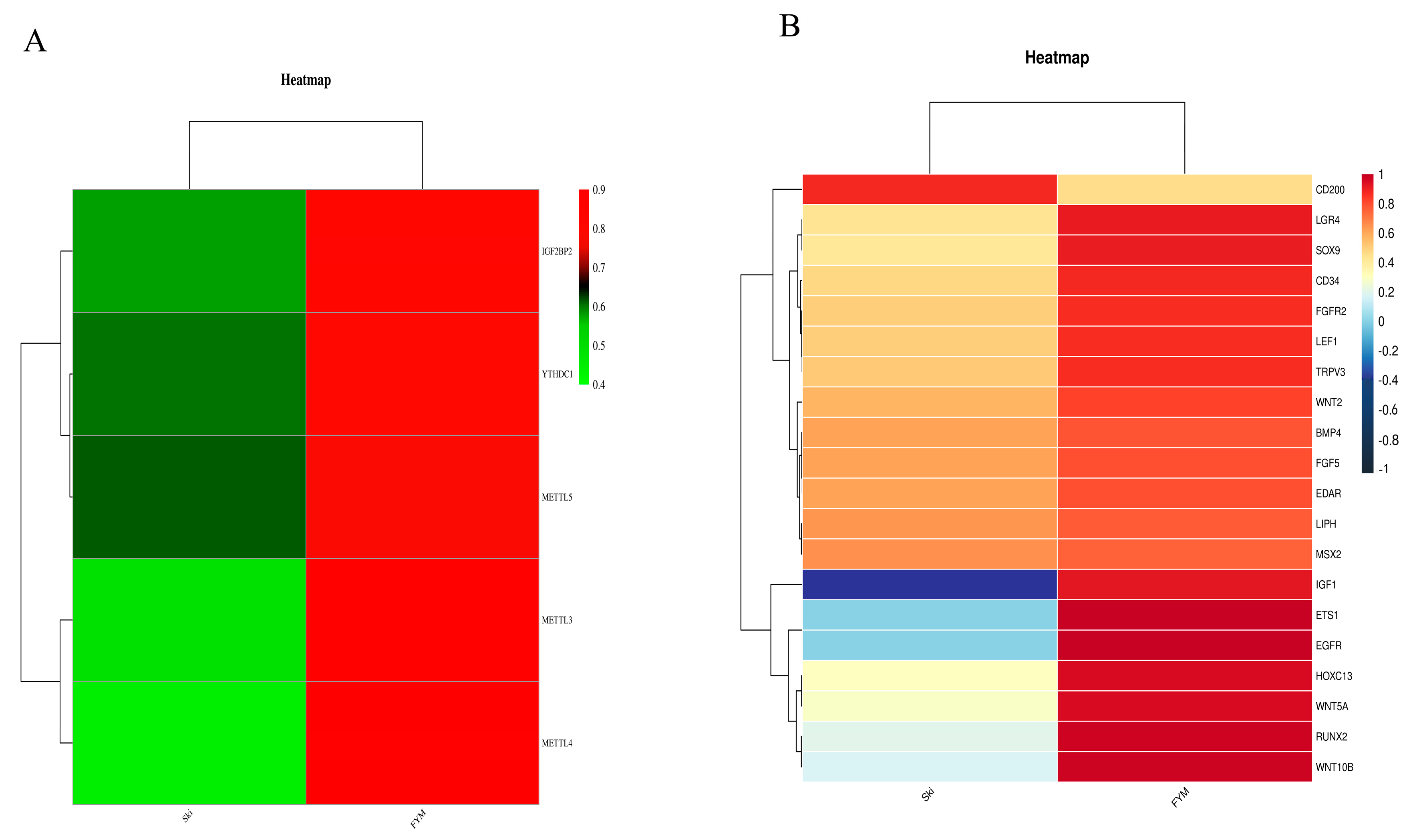
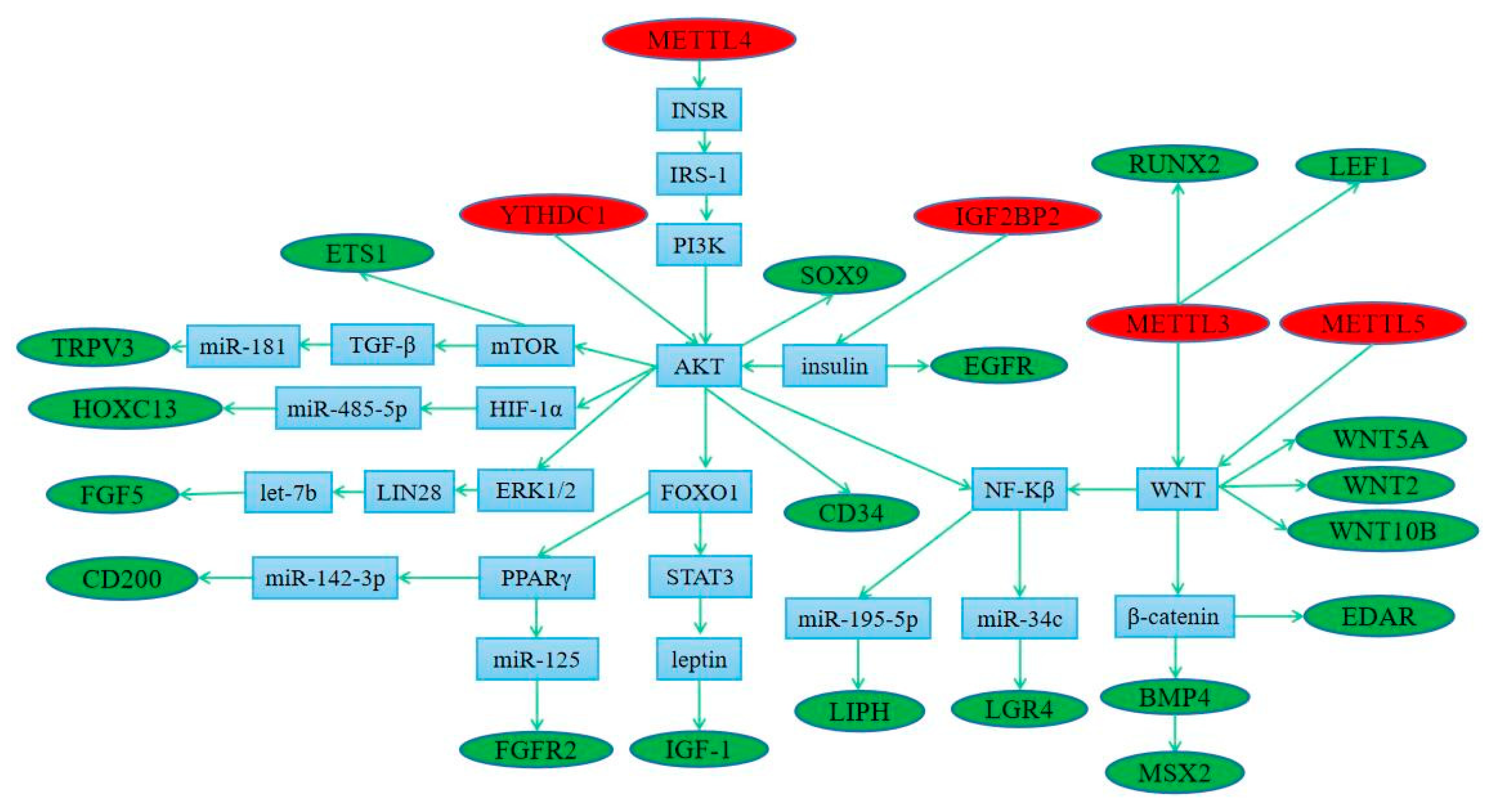
3.5. Regulation of Methylation Modified Genes by Methylase

As shown in Supplementary Figure S1, we found there were many m6A modification sites in 20 different genes. According to the existing research, YTHDC1 can directly regulate the expression of AKT (Figure 8). At the same time, METTL4 and IGF2BP2 can regulate AKT expression through PI3K and insulin, respectively. AKT can directly regulate the expression of CD34 and SOX9 and indirectly regulate the expression of the ETS1, TRPV3, HOXC13, FGF3, CD200, FGFR2, IGF1, LIPH, and LGR4 genes (Figure 8). Another regulatory pathway shows that IGF2BP2 can regulate the expression of the EGFR gene through insulin (Figure 8). As shown in Figure 8, METTL3 can indirectly regulate the expression of the RUNX2 and LEF1 genes. At the same time, METTL3 and METTL5 can regulate the expression of the WNT2, WNT10B, WNT5A, EDAR, BMP4, MSX2, LIPH, and LGR4 genes (Figure 8).

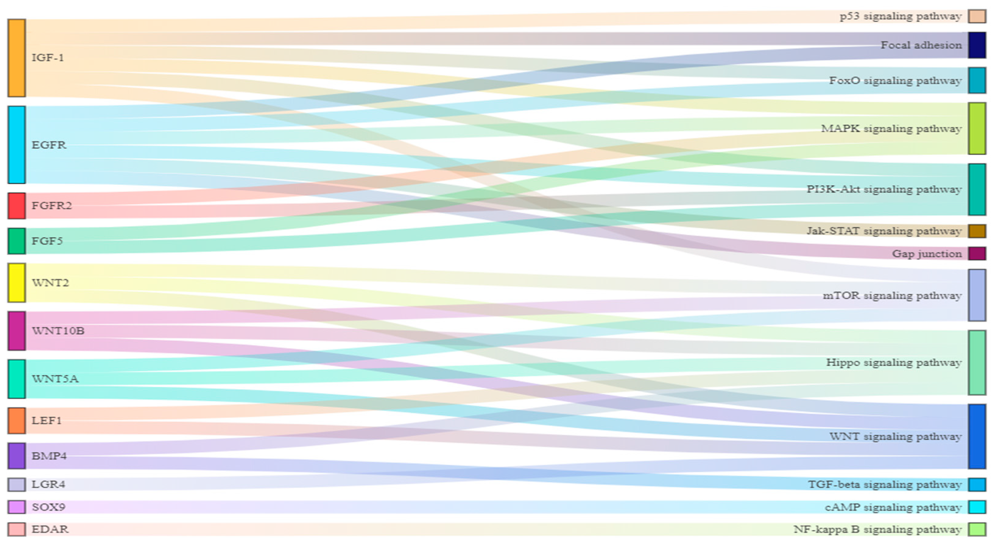
3.6. The Connection between Differential Pathways and Differential Genes

By drawing a Sankey diagram, we found SOX9, LGR4, and EDAR were important regulators in the cAMP, WNT, and NF-Kβ signaling pathways, respectively. BMP4, LEF1, FGF2, and FGFR2 were involved in the regulation of the two signaling pathways. In addition, WNT2, WNT10B, and WNT5A regulated three signal pathways. IGF1 and EGFR regulated six signal pathways (Figure 9). So, We selected IGF1 and EGFR, which had the most regulated signaling pathways, for PCR validation.

4. Discussion

A hair follicle is an important skin derivate with a unique structure and periodic regeneration ability, which plays an important role in hair growth [30]. A hair follicle mainly regulates the growth, color, and fixation of hair [2]. The development of hair follicles plays an important role in the fur rabbit industry [31]. We found that the fur of Rex rabbits was shorter and denser than that of Hycole rabbits. Studies have shown that single nucleotide deletion in exon 9 (1362delA) of LIPH is the reason for the hair phenotype of Rex rabbits [32]. The average hair length, the proportion of primary hair follicles, and the thickness of skin in Hycole rabbits were significantly higher than those in Rex rabbits. The diameter of the primary hair follicles and the volume of their papillae were significantly larger than those of he secondary hair follicles. However, the differentiation and proliferation of secondary hair follicles were faster, which is beneficial for temperature regulation. Therefore, Rex rabbits and Hycole rabbits are two ideal experimental animals to study the differences in hair volume, hair length, and hair follicle growth.
In order to study the mechanism of m6A modification on hair growth and development, we selected the skin tissues of Rex rabbits and Hycole rabbits for MeRIP-seq and found many differences in gene modification. Firstly, we found the typical m6A motif RRACH of animals and plants in two kinds of rabbits [33,34], and the motifs of the two kinds of rabbits are different at many gene loci in Figure 3B. We also found that the location and distribution of the peaks were significantly different in the two breeds of rabbits. Previous studies have shown that m6A methylation regulated the expression of genes related to cashmere growth [35]. These results suggested that m6A regulates hair development.
Based on the existing study results, we found that five methylases can regulate different genes through multiple pathways. METTL3 can directly regulate the expression of RUNX2 and LEF1 [36,37]. In addition, METTL3 and METTL5 both regulated the WNT signaling pathway [38,39]. WNT2, WNT5A, and WNT10B are key factors in the WNT signal pathway. Wnt/β-catenin acts on the upstream of BMP4 [40] and regulates EDAR and MSX2 through β-catenin and BMP4, respectively [41,42]. At the same time, MAPK regulates chondrocytes apoptosis through WNT/NF-κB pathways [43]. NF-κB plays a role in the expression of LGR4 and LIPH through microR-34c and miR-195-5p, respectively [44,45,46,47]. Knockdown of METTL4 led to downregulation and inactivation of the INSR pathway [48], thereby regulating the IRS-1/PI3K/AKT pathway to improve insulin resistance [49]. In addition, AKT regulates the expression of the ETSI [50], TRPV3 [51,52], HOXC13 [53,54,55], and FGF5 [56,57,58] genes through the mTOR, mTOR/TGF-β/miR-181, HIF-1a/miR-485-5p, and ERK1/2/LIN28/let-7b signaling pathways, respectively.
The functions of IGF2BP2 are associated with insulin resistance [59,60], and insulin regulates the EGFR gene to promote the migration of human corneal epithelial cells [61]. In addition, insulin can inhibit hepatic gluconeogenesis by activating the AKT/FOXO1 signaling pathway [62], and FOXO1 inhibits leptin regulation by blocking STAT3 interaction [63]. Leptin and estradiol interact to regulate the expression of IGF-1 [64]. In addition, FOXO1 transrepresses PPARγ transactivation to regulate miR-142-3p, and CD200 is a target gene of miR-142-3p [65,66,67]. At the same time, PPARγ regulates keratinocyte proliferation by targeting FGFR2 with miR-125b [68,69]. MiR-451a represses the AKT/mTOR signaling pathway [70], and AKT regulates SOX9 expression [71] to maintain the imatinib-resistant phenotype of CML CD34+ cells [72]. These existing studies have shown that five methylases can regulate the expression of 20 methylation-modified genes. However, the specific regulation mode is still unclear. This study provides a new possibility for further study on how the five methylases regulate 20 methylated-modified genes.
To further explore the regulation of methylation-modified genes on hair follicle growth and development, we associated methylation-modified genes with differential signaling pathways through a Sankey diagram. As shown in Figure 8, there were 12 genes that regulated 13 differential signaling pathways through different pathways. At the same time, we found that 13 signaling pathways played an important role in the growth and development of hair follicles and hairs. NF-kappaβ promotes hair follicle growth [73]. The FOXO signaling pathway mediates the changes in epidermal morphology, which is closely related to the development of hair follicles [74]. Focal Adhesion Signaling plays an important role in Cochlear Hair Cell Morphology [75]. Some canonical pathways such as WNT, MAPK, TGF-β, and Hippo signaling pathways were detected as promoting the hair follicle growth [76,77,78]. The PI3K/AKT [79], mTOR [79], JAK-STAT [80], Gap junction [81], and cAMP [82] signaling pathways are involved in the development of hair follicles and hairs. In addition, p53 regulated hair follicle regression [83]. So, 12 methylation modification genes regulate the development of hair follicles and hairs through 13 differential signaling pathways.

5. Conclusions

In summary, this study analyzed the modification of m6A methylation in Rex and Hycole rabbit skin tissue. We found five differential methylases, 20 differential genes, and 24 differential signaling pathways related to hair development in Rex and Hycole rabbits. Five methylases regulated the expression of 20 genes related to hair follicle development, of which 12 genes were found to regulate 13 important hair follicle signaling pathways. The development of hair follicles directly affected the growth and density of hair. Studying the effect of m6A on the development of hair follicles lays a theoretical foundation for m6A modification to regulate the development of rabbit hair follicles.

Supplementary Materials

The following supporting information can be downloaded at: https://www.mdpi.com/article/10.3390/biology12111448/s1, Figure S1: The abundance of m6A in the Rex rabbit and Hycole rabbit skin; Table S1: Summary of reads quality control; Table S2: Summary of reads mapping to the rabbit reference genome.

Author Contributions

Conceptualization: Z.R. Formal analysis: G.L., T.Z. and Y.A. Resources: Z.R., Y.A. and R.G. Writing: G.L. All authors have read and agreed to the published version of the manuscript.

Funding

This work was supported by the Innovation integration promotion of technological innovation for improving quality and efficiency in rabbit industry (NYKJ-2021-YL (XN)28v), the Research, development, demonstration and promotion of key technologies for large-scale and standardized healthy rabbit breeding (NYKJ-2020-YL-16v), the and Research and popularization of integrated nutrition technology for meat rabbit mother and calf (NYKJ-2021-YL (XN)26J).

Institutional Review Board Statement

This study was conducted in strict accordance with the ethical standards and approved by the Institutional Animal Care and Use Committee of the College of Animal Science and Technology, Northwest A&F University, Yangling on 15 October 2021, (Permit Number: No. DK-2019008).

Informed Consent Statement

Not applicable.

Data Availability Statement

Raw data have been uploaded to NCBI, which are deposited under SRA BioProject accession PRJNA863730.

Acknowledgments

We thank the staff at our laboratory for their ongoing assistance.

Conflicts of Interest

The authors declare no conflict of interest.

References

  1. Epstein, F.H.; Paus, R.; Cotsarelis, G. The biology of hair follicles. N. Engl. J. Med. 1999, 341, 491–497. [Google Scholar]
  2. Stenn, K.S.; Paus, R. Controls of Hair Follicle Cycling. Physiol. Rev. 2001, 81, 449–494. [Google Scholar] [CrossRef] [PubMed]
  3. Paus, R.; Müller-Rver, S.; Veen, C.V.D.; Maurer, M.; Handjiski, B. A Comprehensive Guide for the Recognition and Classification of Distinct Stages of Hair Follicle Morphogenesis. J. Investig. Dermatol. 1999, 113, 523–532. [Google Scholar] [PubMed]
  4. Ding, H.; Zhao, H.; Cheng, G.; Yang, Y.; Wang, X.; Zhao, X.; Qi, Y.; Huang, D. Analyses of histological and transcriptome differences in the skin of short-hair and long-hair rabbits. BMC Genom. 2019, 20, 140. [Google Scholar] [CrossRef] [PubMed]
  5. Zhao, B.; Chen, Y.; Yan, X.; Hao, Y.; Zhu, J.; Weng, Q.; Wu, X. Gene Expression Profiling Analysis Reveals Fur Development in Rex Rabbits (Oryctolagus cuniculus). Genome 2017, 60, 1060–1067. [Google Scholar] [CrossRef] [PubMed]
  6. Ding, H.; Cheng, G.; Leng, J.; Yang, Y.; Zhao, H. Analysis of histological and microRNA profiles changes in rabbit skin development. Sci. Rep. 2020, 10, 454. [Google Scholar] [CrossRef]
  7. Rishikaysh, P.; Dev, K.; Diaz, D.; Qureshi, W.; Filip, S.; Mokry, J. Signaling Involved in Hair Follicle Morphogenesis and Development. Int. J. Mol. Sci. 2014, 15, 1647–1670. [Google Scholar] [CrossRef] [PubMed]
  8. Roundtree, I.A.; Evans, M.E.; Pan, T.; He, C. Dynamic RNA Modifications in Gene Expression Regulation. Cell 2017, 169, 1187–1200. [Google Scholar] [CrossRef]
  9. Nhan, V.T.; Ernst, F.; Hawley, B.R.; Christiane, Z.; Nathalie, U.; Philipp, H.; Bohnsack, K.E.; Bohnsack, M.T.; Jaffrey, S.R.; Marc, G. The human 18S rRNA m6A methyltransferase METTL5 is stabilized by TRMT112. Nucleic Acids Res. 2019, 49, 7719–7733. [Google Scholar]
  10. Yang, Y.; Hsu, P.J.; Chen, Y.S.; Yang, Y.G. Dynamic transcriptomic m6A decoration: Writers, erasers, readers and functions in RNA metabolism. Cell Res. 2018, 28, 616–624. [Google Scholar] [CrossRef]
  11. Goh, Y.T.; Koh, C.; Sim, D.Y.; Roca, X.; Goh, W. METTL4 catalyzes m6Am methylation in U2 snRNA to regulate pre-mRNA splicing. Nucleic Acids Res. 2020, 48, 9250–9261. [Google Scholar] [CrossRef]
  12. Li, F.; Kennedy, S.; Hajian, T.; Gibson, E.; Seitova, A.; Xu, C.; Arrowsmith, C.H.; Vedadi, M. A Radioactivity-Based Assay for Screening Human m6A-RNA Methyltransferase, METTL3-METTL14 Complex, and Demethylase ALKBH5. J. Biomol. Screen. Off. J. Soc. Biomol. Screen. 2016, 21, 290–297. [Google Scholar] [CrossRef]
  13. Meyer, K.D.; Jaffrey, S.R. Rethinking m6A Readers, Writers, and Erasers. Annu. Rev. Cell Dev. Biol. 2017, 33, 319–342. [Google Scholar] [CrossRef] [PubMed]
  14. Sheng, L.; Mason, C.E. The Pivotal Regulatory Landscape of RNA Modifications. Annu. Rev. Genom. Hum. Genet. 2013, 15, 127–150. [Google Scholar]
  15. Meyer, K.D.; Jaffrey, S.R. The dynamic epitranscriptome: N6-methyladenosine and gene expression control. Nat. Rev. Mol. Cell. Biol. 2014, 15, 313–326. [Google Scholar] [CrossRef] [PubMed]
  16. Ke, S.; Pandya-Jones, A.; Saito, Y.; Fak, J.J.; Vågbø, C.B.; Geula, S.; Hanna, J.H.; Black, D.L.; Darnell, J.E.; Darnell, R.B. m6A mRNA modifications are deposited in nascent pre-mRNA and are not required for splicing but do specify cytoplasmic turnover. Genes Dev. 2017, 31, 990–1006. [Google Scholar] [CrossRef]
  17. Slobodin, B.; Han, R.; Calderone, V.; Vrielink, J.; Loayza-Puch, F.; Elkon, R.; Agami, R. Transcription Impacts the Efficiency of mRNA Translation via Co-transcriptional N6-adenosine Methylation. Cell 2017, 169, 326–337. [Google Scholar] [CrossRef]
  18. Zhang, Z.; Xing, Y.; Gao, W.; Yang, L.; Shi, J.; Song, W.; Li, T. N-6-methyladenosine (m6A) reader IGF2BP2 promotes gastric cancer progression via targeting SIRT1. Bioengineered 2022, 13, 11541–11550. [Google Scholar] [CrossRef] [PubMed]
  19. Sánchez-Vásquez, E.; Jimenez, N.A.; Vázquez, N.; Strobl-Mazzulla, P.H. Emerging role of dynamic RNA modifications during animal development. Mech. Dev. 2018, 154, 24–32. [Google Scholar] [CrossRef]
  20. Wang, X.; Sun, B.; Jiang, Q.; Wu, R.; Cai, M.; Yao, Y.; Liu, Q.; Shi, H.; Feng, J.; Wang, Y. mRNA m6A plays opposite role in regulating UCP2 and PNPLA2 protein expression in adipocytes. Int. J. Obes. 2018, 42, 1912–1924. [Google Scholar] [CrossRef]
  21. Lin, Z.; Hsu, P.J.; Xing, X.; Fang, J.; Lu, Z.; Zou, Q.; Zhang, K.J.; Zhang, X.; Zhou, Y.; Zhang, T. Mettl3-/Mettl14-mediated mRNA N6-methyladenosine modulates murine spermatogenesis. Cell Res. 2017, 27, 1216–1230. [Google Scholar] [CrossRef]
  22. Weber, M.; Davies, J.J.; Wittig, D.; Oakeley, E.J.; Haase, M.; Lam, W.L.; Schübeler, D. Chromosome-wide and promoter-specific analyses identify sites of differential DNA methylation in normal and transformed human cells. Nat. Genet. 2005, 38, 853–862. [Google Scholar] [CrossRef] [PubMed]
  23. Gbolagunte, G. Fibrocartilage embedding hair follicles on the skin of the knee of the camel (Camelus dromedarius): An ideal structure for its comfort. J. Sci. Res. Stud. 2016, 3, 30–33. [Google Scholar]
  24. Chen, S.; Zhou, Y.; Chen, Y.; Gu, J. fastp: An ultra-fast all-in-one FASTQ preprocessor. Bioinformatics 2018, 34, 274100. [Google Scholar] [CrossRef] [PubMed]
  25. Langmead, B.; Salzberg, S.L. Fast gapped-read alignment with Bowtie. Nat. Methods 2012, 9, 357–359. [Google Scholar] [CrossRef]
  26. Siren, J.; Valimaki, N.; Makinen, V. Indexing Graphs for Path Queries with Applications in Genome Research. IEEE/ACM Trans. Comput. Biol. Bioinform. 2014, 11, 375–388. [Google Scholar] [CrossRef]
  27. Meng, J.; Lu, Z.; Liu, H.; Zhang, L.; Zhang, S.; Chen, Y.; Rao, M.K.; Huang, Y. A protocol for RNA methylation differential analysis with MeRIP-Seq data and exomePeak R/Bioconductor package. Methods 2014, 69, 274–281. [Google Scholar] [CrossRef] [PubMed]
  28. Stark, R.; Brown, G. DiffBind differential binding analysis of ChIP-Seq peak data. R Package Version 2011, 100, 3. [Google Scholar]
  29. Kanehisa, M.; Goto, S. KEGG: Kyoto Encyclopedia of Genes and Genomes. Nucleic Acids Res. 2000, 28, 27–30. [Google Scholar] [CrossRef] [PubMed]
  30. Millar, S.E. Molecular mechanisms regulating hair follicle development. J. Investig. Dermatol. 2002, 118, 216–225. [Google Scholar] [CrossRef] [PubMed]
  31. Taha, E.; Hekal, S.; Nasr, A.I. Evaluating skin quality of some rabbit breeds under Egyptian conditions. World Rabbit Sci. 2017, 25, 193–200. [Google Scholar] [CrossRef]
  32. Mathieu, D.; Xavier, M.; Céline, C.-D.; Anne, V.; Gérard, A.; Stéphan, B.; Séverine, D.; Edmond-Paul, C.; Hubert, D.R.; Daniel, A. A deletion in exon 9 of the LIPH gene is responsible for the Rex hair coat phenotype in rabbits (Oryctolagus cuniculus). PLoS ONE 2011, 6, e19281. [Google Scholar]
  33. Duan, H.C.; Wei, L.H.; Zhang, C.; Wang, Y.; Chen, L.; Lu, Z.; Chen, P.R.; He, C.; Jia, G. ALKBH10B is An RNA N6-Methyladenosine Demethylase Affecting Arabidopsis Floral Transition. Plant Cell 2017, 29, 2995–3011. [Google Scholar] [CrossRef] [PubMed]
  34. Dominissini, D.; Moshitch-Moshkovitz, S.; Schwartz, S.; Salmon-Divon, M.; Ungar, L.; Osenberg, S.; Cesarkas, K.; Jacob-Hirsch, J.; Amariglio, N.; Kupiec, M.; et al. Topology of the human and mouse m6A RNA methylomes revealed by m6A-seq. Nature 2012, 485, 201–206. [Google Scholar] [CrossRef] [PubMed]
  35. Wang, Y.; Zheng, Y.; Guo, D.; Zhang, X.; Bai, W. m6A Methylation Analysis of Differentially Expressed Genes in Skin Tissues of Coarse and Fine Type Liaoning Cashmere Goats. Front. Genet. 2020, 10, 1318. [Google Scholar] [CrossRef] [PubMed]
  36. Yan, G.; Yuan, Y.; He, M.; Gong, R.; Yang, L. m6A Methylation of Precursor-miR-320/RUNX2 Controls Osteogenic Potential of Bone Marrow-Derived Mesenchymal Stem Cells. Mol. Ther. Nucleic Acids 2019, 19, 421–436. [Google Scholar] [CrossRef]
  37. Miao, W.; Chen, J.; Jia, L.; Ma, J.; Song, D. The m6A methyltransferase METTL3 promotes osteosarcoma progression by regulating the m6A level of LEF1. Biochem. Biophys. Res. Commun. 2019, 516, 719–725. [Google Scholar] [CrossRef]
  38. Wu, T.; Tang, H.; Yang, J.; Yao, Z.; Bai, L.; Xie, Y.; Li, Q.; Xiao, J. METTL3-m6A methylase regulates the osteogenic potential of bone marrow mesenchymal stem cells in osteoporotic rats via the Wnt signalling pathway. Cell Prolif. 2022, 55, e13234. [Google Scholar] [CrossRef]
  39. Lei, K.; Xu, R.; Wang, Q.; Xiong, Q.; Zhou, X.; Li, Q.; Seriwatanachai, D.; Lin, S.; Zhou, C.; Yuan, Q. METTL5 regulates cranial suture fusion via Wnt signaling. Fundam. Res. 2022, 5, 369–376. [Google Scholar] [CrossRef]
  40. Shu, W.; Guttentag, S.; Wang, Z.; Andl, T.; Ballard, P.; Min, M.L.; Piccolo, S.; Birchmeier, W.; Whitsett, J.A.; Millar, S.E. Wnt/beta-catenin signaling acts upstream of N-myc, BMP4, and FGF signaling to regulate proximal-distal patterning in the lung. Dev. Biol. 2005, 283, 226–239. [Google Scholar] [CrossRef]
  41. Zhang, Y.; Tomann, P.; Andl, T.; Gallant, N.M.; Schmidt-Ullrich, R. Reciprocal Requirements for EDA/EDAR/NF-κB and Wnt/β-Catenin Signaling Pathways in Hair Follicle Induction. Dev. Cell 2009, 17, 49–61. [Google Scholar] [CrossRef]
  42. Hamada, S.; Satoh, K.; Hirota, M.; Kimura, K.; Kanno, A.; Masamune, A.; Shimosegawa, T. Bone morphogenetic protein 4 induces epithelial-mesenchymal transition through MSX2 induction on pancreatic cancer cell line. J. Cell. Physiol. 2010, 213, 768–774. [Google Scholar] [CrossRef] [PubMed]
  43. Zhang, L.B.; Man, Z.T.; Li, W.; Zhang, W.; Wang, X.Q.; Sun, S. Calcitonin protects chondrocytes from lipopolysaccharide-induced apoptosis and inflammatory response through MAPK/Wnt/NF-κB pathways. Mol. Immunol. 2017, 87, 249–257. [Google Scholar] [CrossRef] [PubMed]
  44. Yuan, X.U.; Zhang, A.L.; Xiao, G.; Zhang, Z.; Chen, Z.M.; Zhang, H.; Li, J.-Q. p53 and NFκB regulate microRNA-34c expression in porcine ovarian granulosa cells. J. Integr. Agric. 2016, 15, 1816–1824. [Google Scholar]
  45. Cong, F.; Wu, N.; Tian, X.; Fan, J.; Liu, J.; Song, T.; Fu, H. MicroRNA-34c promotes osteoclast differentiation through targeting LGR4. Gene 2017, 610, 1–8. [Google Scholar] [CrossRef]
  46. Wang, L.; Guo, J.; Zhou, J.; Wang, D.; Zhou, L. NF-κB maintains the stemness of colon cancer cells by downregulating miR-195-5p/497-5p and upregulating MCM2. J. Exp. Clin. Cancer Res. CR 2020, 39, 225. [Google Scholar] [CrossRef]
  47. Gui, X.; Li, Y.; Zhang, X.; Su, K.; Cao, W. Circ_LDLR promoted the development of papillary thyroid carcinoma via regulating miR-195-5p/LIPH axis. Cancer Cell Int. 2020, 20, 241. [Google Scholar] [CrossRef]
  48. Zhang, Z.; Hou, Y.; Wang, Y.; Gao, T.; Du, Q. Regulation of Adipocyte Differentiation by METTL4, a 6 mA Methylase. Sci. Rep. 2020, 10, 8285. [Google Scholar] [CrossRef]
  49. Chen, Y.; Yang, F.; Wang, S.; Wang, D.B.; Xu, J.; Yang, G.Z. Triterpenoid Saponins from Stauntonia chinensis Ameliorate Insulin Resistance via the AMP-Activated Protein Kinase and IR/IRS-1/PI3K/Akt Pathways in Insulin-Resistant HepG2 Cells. Int. J. Mol. Sci. 2014, 15, 10446–10458. [Google Scholar]
  50. Xu, S.; Ge, J.; Zhang, Z.; Zhou, W. MiR-129 inhibits cell proliferation and metastasis by targeting ETS1 via PI3K/AKT/mTOR pathway in prostate cancer. Biomed. Pharmacother. 2017, 96, 634–641. [Google Scholar] [CrossRef]
  51. Wang, Y.; Yu, Y.; Tsuyada, A.; Ren, X.; Wu, X.; Stubblefield, K.; Rankin-Gee, E.K.; Wang, S.E. Transforming growth factor-beta regulates the sphereinitiating stem cell like feature in breast cancer through miRNA-181 and ATM. Oncogene 2011, 30, 1470–1480. [Google Scholar] [CrossRef] [PubMed]
  52. Moroney, J.B.; Vasudev, A.; Pertsemlidis, A.; Zan, H.; Casali, P. Integrative transcriptome and chromatin landscape analysis reveals distinct epigenetic regulations in human memory B cells. Nat. Commun. 2020, 11, 5435. [Google Scholar] [CrossRef] [PubMed]
  53. Wei, Y.; Hu, Y.; Qi, K.; Li, Y.; Chen, J.; Wang, R. Dihydromyricetin improves LPS-induced sickness and depressive-like behaviors in mice by inhibiting the TLR4/Akt/HIF1a/NLRP3 pathway. Behav. Brain Res. 2022, 423, 113775. [Google Scholar] [CrossRef]
  54. Cheng, L.; Peng, R.; Guo, P.; Zhang, H.; Liao, Y. A HIF1A/miR-485-5p/SRPK1 axis modulates the aggressiveness of glioma cells upon hypoxia. Exp. Cell Res. 2021, 402, 112547. [Google Scholar] [CrossRef] [PubMed]
  55. Yang, H.; Fu, J.; Jiao, G.; Zhu, Y.; Wang, Z. LINC00659 Regulates Colorectal Cancer Migration and Invasion by Targeting miR-485-5p/HOXC13 Axis. Res. Sq. 2021, 1–21. [Google Scholar] [CrossRef]
  56. Yu, J.; Wang, L.; Akinyi, M.; Li, Y.; Fan, G. Danshensu protects isolated heart against ischemia reperfusion injury through activation of Akt/ERK1/2/Nrf2 signaling. Int. J. Clin. Exp. Med. 2015, 8, 14793–14804. [Google Scholar] [PubMed]
  57. Xu, X.; Bao, Z.; Liu, Y.; Jiang, K.; Zhi, T.; Dong, W.; Fan, L.; Ning, L.; Jing, J. PBX3/MEK/ERK1/2/LIN28/let-7b positive feedback loop enhances mesenchymal phenotype to promote glioblastoma migration and invasion. J. Exp. Clin. Cancer Res. 2018, 37, 158. [Google Scholar] [CrossRef]
  58. Wang, T.; Zhang, Y.; Wang, H.D.; Shen, Y.; Liu, N.; Cao, J.; Yu, X.J.; Dong, C.S.; He, X.Y. Alpaca fiber growth is mediated by microRNA let-7b via down-regulation of target gene FGF5. Genet. Mol. Res. 2015, 14, 13754–13763. [Google Scholar] [CrossRef]
  59. Cao, J.; Mu, Q.; Huang, H. The Roles of Insulin-Like Growth Factor 2 mRNA-Binding Protein 2 in Cancer and Cancer Stem Cells. Stem Cells Int. 2018, 2018, 4217259. [Google Scholar] [CrossRef]
  60. Ruchat, S.M.; Elks, C.E.; Loos, R.J.F.; Vohl, M.C.; Weisnagel, S.J.; Rankinen, T.; Bouchard, C.; Pérusse, L. Association between insulin secretion, insulin sensitivity and type 2 diabetes susceptibility variants identified in genome-wide association studies. Acta Diabetol. 2009, 46, 217–226. [Google Scholar] [CrossRef]
  61. Joo, C.K.; Lee, K.S.; Lyu, J. Insulin Enhances Cell Migration Through EGFR in Human Corneal Epithelial Cells. Investig. Ophthalmol. Vis. Sci. 2006, 47, 5031. [Google Scholar]
  62. Gomez-Valades, A.G.; Mendez-Lucas, A.; Vidal-Alabro, A.; Blasco, F.X.; Chillon, M.; Bartrons, R.; Bermudez, J.; Perales, J.C. Pck1 Gene Silencing in the Liver Improves Glycemia Control, Insulin Sensitivity, and Dyslipidemia in db/db Mice. Diabetes 2008, 57, 2199–2210. [Google Scholar] [CrossRef]
  63. Yang, G.; Lim, C.Y.; Li, C.; Xiao, X.; Radda, G.K.; Li, C.; Cao, X.; Han, W. FoxO1 Inhibits Leptin Regulation of Pro-opiomelanocortin Promoter Activity by Blocking STAT3 Interaction with Specificity Protein 1. J. Biol. Chem. 2009, 284, 3719–3721. [Google Scholar] [CrossRef]
  64. Shetty, A.; Padmanaban, S. A synergy of estradiol with leptin modulates the long non-coding RNA NEAT1/ mmu-miR-204-5p/IGF1 axis in the uterus of high-fat-diet-induced obese ovariectomized mice. J. Steroid Biochem. Mol. Biol. 2021, 209, 105843. [Google Scholar] [CrossRef] [PubMed]
  65. Fan, W.; Imamura, T.; Sonoda, N.; Sears, D.D.; Patsouris, D.; Kim, J.J.; Olefsky, J.M. FOXO1 transrepresses PPARγ transactivation, coordinating an insulin-induced feed-forward response in adipocytes. J. Biol. Chem. 2009, 284, 12188–12197. [Google Scholar] [CrossRef]
  66. Yuan, Z.; Luo, G.; Li, X.; Chen, J.; Wu, J.; Peng, Y. PPAR gamma inhibits HMGB1 expression through upregulation of miR-142-3p in vitro and in vivo. Cell. Signal. 2016, 28, 158–164. [Google Scholar] [CrossRef] [PubMed]
  67. Kumar, P.M.; Tembhurne, P.A.; Raja, P.; Ingle, V.C. Deciphering the role of gga-miR-142-3p on target gene CD200 and its contribution towards signal transduction and immune response. Appl. Biol. Res. 2017, 19, 132–137. [Google Scholar] [CrossRef]
  68. Pei, G.; Yan, L.; Chen, D.; Ji, L.; Hua, Z.C. FAK regulates E-cadherin expression via p-SrcY416/p-ERK1/2/p-Stat3Y705 and PPARγ/miR-125b/Stat3 signaling pathway in B16F10 melanoma cells. Oncotarget 2017, 8, 13898–13908. [Google Scholar] [CrossRef] [PubMed]
  69. Xu, N.; Brodin, P.; Wei, T.; Meisgen, F.; Eidsmo, L.; Nagy, N.; Kemeny, L.; Ståhle, M.; Sonkoly, E.; Pivarcsi, A. MiR-125b, a microRNA downregulated in psoriasis, modulates keratinocyte proliferation by targeting FGFR2. J. Investig. Dermatol. 2011, 131, 1521–1529. [Google Scholar] [CrossRef]
  70. Cao, D.; Ge, S.; Li, M. MiR-451a promotes cell growth, migration and EMT in osteosarcoma by regulating YTHDC1-mediated m6A methylation to activate the AKT/mTOR signaling pathway. J. Bone Oncol. 2022, 33, 100412. [Google Scholar] [CrossRef]
  71. Cheng, C.; Uchiyama, Y.; Hiyama, A.; Gajghate, S.; Shapiro, I.M.; Risbud, M.V. PI3K/AKT regulates aggrecan gene expression by modulating Sox9 expression and activity in nucleus pulposus cells of the intervertebral disc. J. Cell. Physiol. 2009, 221, 668–676. [Google Scholar] [CrossRef]
  72. Wang, W.Z.; Pu, Q.H.; Lin, X.H.; Liu, M.Y.; Wu, L.R.; Wu, Q.Q.; Chen, Y.H.; Liao, F.F.; Zhu, J.Y.; Jin, X.B. Silencing of miR-21 sensitizes CML CD34+ stem/progenitor cells to imatinib-induced apoptosis by blocking PI3K/AKT pathway. Leuk. Res. 2015, 39, 1117–1124. [Google Scholar] [CrossRef] [PubMed]
  73. Tomann, P.; Paus, R.; Millar, S.E.; Scheidereit, C.; Schmidt-Ullrich, R. Lhx2 is a direct NF-kappa B target gene that promotes primary hair follicle placode down-growth. Development 2016, 143, 1512–1522. [Google Scholar]
  74. Günschmann, C.; Stachelscheid, H.; Akyüz, M.; Schmitz, A.; Missero, C.; Brüning, J.; Niessen, C. Insulin/IGF-1 Controls Epidermal Morphogenesis via Regulation of FoxO-Mediated p63 Inhibition. Dev. Cell 2013, 26, 176–187. [Google Scholar] [CrossRef] [PubMed]
  75. Yao, Q.; Wang, H.; Chen, H.; Li, Z.; Jiang, Y.; Li, Z.; Wang, J.; Xing, Y.; Liu, F.; Yu, D.; et al. Essential Role of Sptan1 in Cochlear Hair Cell Morphology and Function Via Focal Adhesion Signaling. Mol. Neurobiol. 2022, 59, 386–404. [Google Scholar] [CrossRef]
  76. Li, C.; Feng, C.; Ma, G.; Fu, S.; Chen, M.; Zhang, W.; Li, J. Time-course RNA-seq analysis reveals stage-specific and melatonin-triggered gene expression patterns during the hair follicle growth cycle in Capra hircus. BMC Genom. 2022, 23, 140. [Google Scholar] [CrossRef] [PubMed]
  77. Kim, J.; Kim, S.R.; Choi, Y.H.; Shin, J.Y.; Lee, S. Quercitrin Stimulates Hair Growth with Enhanced Expression of Growth Factors via Activation of MAPK/CREB Signaling Pathway. Molecules 2020, 25, 4004. [Google Scholar] [CrossRef]
  78. Jin, J.H.; Ho, J.K.; Eun, K.J.; Kang, H. Various Wavelengths of Light-Emitting Diode Light Regulate the Proliferation of Human Dermal Papilla Cells and Hair Follicles via Wnt/β-Catenin and the Extracellular Signal-Regulated Kinase Pathways. Ann. Dermatol. 2017, 29, 747–754. [Google Scholar]
  79. Zhang, Y.; Yu, J.; Shi, C.; Wang, Y.; Yang, J.; Yang, T. Regulatory effect of β-catenin on proliferation of hair follicle stem cells involves PI3K/Akt pathway. J. Appl. Biomed. 2013, 11, 131–141. [Google Scholar] [CrossRef]
  80. Harel, S.; Higgins, C.A.; Cerise, J.E.; Dai, Z.; Chen, J.C.; Clynes, R.; Christiano, A.M. Pharmacologic inhibition of JAK-STAT signaling promotes hair growth. Sci. Adv. 2015, 1, e1500973. [Google Scholar] [CrossRef]
  81. Risek, B.; Klier, F.G.; Gilula, N.B. Multiple gap junction genes are utilized during rat skin and hair development. Development 1992, 116, 639. [Google Scholar] [CrossRef] [PubMed]
  82. Hoffmann, R.; Eicheler, W.; Wenzel, E.; Happle, R. Interleukin-1beta-Induced Inhibition of Hair Growth In Vitro Is Mediated by Cyclic AMP. J. Investig. Dermatol. 1997, 108, 40–42. [Google Scholar] [CrossRef] [PubMed]
  83. Botchkarev, V.A.; Komarova, E.A.; Siebenhaar, F.; Botchkareva, N.V.; Sharov, A.A.; Komarov, P.G.; Maurer, M.; Gudkov, A.V.; Gilchrest, B.A. p53 Involvement in the Control of Murine Hair Follicle Regression. Am. J. Pathol. 2001, 158, 1913–1919. [Google Scholar] [CrossRef] [PubMed]
Figure 1. Hair difference between Rex Rabbit and Hycole rabbit. (A) Pictures of Rex rabbits and Hycole rabbits; (B) hair length of Rex rabbits and Hycole rabbits; (C) coarse wool ratio of Rex rabbits and Hycole rabbits (“**”, p ≤ 0.01).
Biology 12 01448 g001
Figure 2. Hair follicle distribution and skin thickness. (A) HE staining cross section of skin; (B) proportion of primary hair follicles; (C) primary and secondary follicle density; (D) HE staining longitudinal section of skin; (E) skin thickness (longitudinal section of skin in Rex rabbits and Hycole rabbits) (“**”, p ≤ 0.01).
Biology 12 01448 g002
Figure 3. (A) Cluster analysis heat map; (B) sequence logo identified from Rex rabbit and Hycole rabbit skin; (C) overlap of m6A peaks from Rex rabbit and Hycole rabbit skin; (D) distribution of m6A peaks across the length of mRNA.
Biology 12 01448 g003
Figure 4. Enrichment pathway of the m6A peak related to hair growth and development.
Biology 12 01448 g004
Figure 5. (A) Cluster heat map of differential methylases from Rex rabbit and Hycole rabbit skin; (B) cluster heat map of differential genes related to hair development from Rex rabbit and Hycole rabbit skin.
Biology 12 01448 g005
Figure 6. (A) IGF1 expression level in Rex rabbit and Hycole rabbit skin; (B) EGFR expression level in Rex rabbit and Hycole rabbit skin; (“**”, p ≤ 0.01).
Biology 12 01448 g006
Figure 7. Distribution of differential genes on the chromosomes of 20 genes’ m6A modification.
Biology 12 01448 g007
Figure 8. Pathway map of key genes regulated by five methylases.
Biology 12 01448 g008
Figure 9. Sankey diagram of genes and signal pathways.
Biology 12 01448 g009
Table 1. Primers used in this study.
Gene NamePrimer Sequence (5′-3′)Tm (°C)
β-actinGGAGATCGTGCGGGACAT60
GTTGAAGGTGGTCTCGTGGAT
IGF1ACCCACCCTAACCTGCTGTA60
TCCTGTGGGCTTGTTGAAAT
EGFRACCTTGTCATTCAGGGGGATG60
ACACAAGCCATGGTGGAACT
Disclaimer/Publisher’s Note: The statements, opinions and data contained in all publications are solely those of the individual author(s) and contributor(s) and not of MDPI and/or the editor(s). MDPI and/or the editor(s) disclaim responsibility for any injury to people or property resulting from any ideas, methods, instructions or products referred to in the content.

