Chitosan Coating Applications in Probiotic Microencapsulation
by
 ,
,
Lavinia-Florina Călinoiu
1 ,
,
Bianca Eugenia Ştefănescu
1,2,
Ioana Delia Pop
3,*,
Leon Muntean
4,* and
Dan Cristian Vodnar
1,*
1
Department of Food Science and Technology, Institute of Life Sciences, University of Agricultural Sciences and Veterinary Medicine, Cluj-Napoca, Calea Mănăştur 3-5, 400372 Cluj-Napoca, Romania
2
Department of Pharmaceutical Botany, Iuliu Hațieganu University of Medicine and Pharmacy, 12 I. Creangă Street, 400010 Cluj-Napoca, Romania
3
Department of Exact Sciences, Horticulture Faculty, University of Agricultural Sciences and Veterinary Medicine, Cluj-Napoca, Calea Mănăştur 3-5, 400372 Cluj-Napoca, Romania
4
Department of Plant Culture, University of Agricultural Sciences and Veterinary Medicine, Cluj-Napoca, Calea Mănăştur 3-5, 400372 Cluj-Napoca, Romania
*
Authors to whom correspondence should be addressed.
Coatings 2019, 9(3), 194; https://doi.org/10.3390/coatings9030194
Submission received: 8 February 2019
/
Revised: 11 March 2019
/
Accepted: 13 March 2019
/
Published: 16 March 2019
Abstract
:Nowadays, probiotic bacteria are extensively used as health-related components in novel foods with the aim of added-value for the food industry. Ingested probiotic bacteria must resist gastrointestinal exposure, the food matrix, and storage conditions. The recommended methodology for bacteria protection is microencapsulation technology. A key aspect in the advancement of this technology is the encapsulation system. Chitosan compliments the real potential of coating microencapsulation for applications in the food industry due to its physicochemical properties: positive charges via its amino groups (which makes it the only commercially available water-soluble cationic polymer), short-term biodegradability, non-toxicity and biocompatibility with the human body, and antimicrobial and antifungal actions. Chitosan-coated microcapsules have been reported to have a major positive influence on the survival rates of different probiotic bacteria under in vitro gastrointestinal conditions and in the storage stability of different types of food products; therefore, its utilization opens promising routes in the food industry.
1. Introduction
Nowadays, there is an increase in functional probiotic food demand, as well as waste-derived bioactive compounds re-utilization [1,2,3,4,5,6,7,8], based on the consciousness of consumers regarding their health potential [9,10]. Considering the IndustryARC report [11] from 2018, the global probiotic market is estimated to experience a compound annual growth rate of 5.6% through 2020.
According to the FAO/WHO, probiotics are characterized as living microorganisms which, when ingested in certain amounts, provide health benefits to the host [12]. Some of these health benefits include antagonistic effects against harmful bacteria in humans and immune effects [13]. Their usage positively influences the growth of targeted microorganisms, eliminates harmful bacteria, and boosts the host’s naturally occurring defense actions [14]. In 1993, Ziemer and Gibson [15] were amongst the first researchers to sustain the presence of health-related bacteria in soured milk with an impact on intestinal health. For the past two decades, these bioactive ingredients have been at the forefront of many studies [16,17,18]. Two of the most common types of microbes extensively used as probiotics are the bacteria belonging to the genera Bifidobacterium and Lactobacillus [13,19,20,21,22]. Considering the above, probiotic-enriched food products should reach the recommended level at the time of consumption, which was agreed upon as being 106–107 CFU (colony forming units) of viable probiotic bacteria per gram of food [23].
Administered probiotics must resist the harsh gastric conditions [24] and reach the colon in sufficient amounts to be able to sustain colonization, and hence to bring positive benefits to the human body [19,25]. Unfortunately, the free bacteria’s inability to survive in high numbers during exposure to the host’s gastrointestinal (GI) tract’s conditions [26] and/or during exposure to oxygen while a functional food product on a shelf represents the main issues with probiotics [27]. Therefore, their efficiency is highly correlated with their quantity and their viability during storage and product shelf-life [28,29].
Microencapsulation represents the main modern solution for preserving probiotic viability. By definition, microencapsulation represents an incorporation process of probiotic bacteria into a specific material or membrane that has the ability to reduce cell injury or cell loss, derived from environmental factors, with a controlled-release rate under specific conditions [30,31]. Therefore, this technique has been extensively studied during the last decade, since it can maintain the beneficial properties even for sensitive bacteria during storage and absorption [25]. Many studies and reviews have been conducted to investigate and summarize the protective role of this technique [32,33,34,35].
Based on the literature available so far, chitosan appears to be one of the most promising coating materials among the most common polymers used for microencapsulation to improve the stability of probiotics [31,36,37,38,39]. Moreover, chitosan has a significant protective role against external damages in food products. The antimicrobial ability of chitosan has been observed in numerous studies, of which some resulted in the creation of biodegradable labels, such as the one obtained with chitosan and green tea extract which presents a decontamination effect on the surface of studied fruits and vegetables [40]. Another study even showed its ability to extend the validity of fruit products [41]. Since chitosan is a biopolymer with no or very little sensory influence on food, and considering all the above-mentioned findings, it presents applicability in the food industry [42,43].
The existing literature highlights the high interest in probiotic bacteria microencapsulation, and the importance of coatings for efficient protection and an increased number of probiotics in the GI tract. For the current review, we extracted, evaluated, interpreted, and summarized data related to future trends and implications for applications of chitosan as coating material in probiotic microencapsulation, its performance efficiency on maintaining probiotic viability, protection, and intestinal delivery, as well as its food incorporation aspects.
2. Coatings for Probiotic Microencapsulation
The encapsulation matrix must be food grade and possess suitable physical and chemical properties to deliver protection for the incorporated bacteria [44]. Selection of capsule materials and suitable techniques for tailoring probiotic microcapsules is crucial because it confers the final morphological and functional characteristics of the probiotics [39]. According to Krasaekoopt et al. [45], polymer coatings can significantly increase the chemical and mechanical stability, therefore improving the performance of the microencapsulation materials. Regarding the technologies applied for microencapsulation, emulsion, spray-drying, layer-by-layer (LbL), and extrusion are extensively used and applied at both the laboratory and industrial scales [46,47,48,49]. In coating-based encapsulation technology, a major importance is to control the permeability of the coating. Therefore, the LbL approach is a recommended technique since it sustains the permeation of small molecules, while it traps larger molecules. Moreover, the semi-permeable nature of LbL-based coatings can be regulated by the experimental parameters upon assembly [50]. It is important to keep in mind that a combination of these technologies is applied frequently for a higher rate of success.
According to the literature, food-grade coatings like bio-polymers (i.e., alginate, chitosan, pectin, starch, carrageenan, and milk proteins) are the most suitable materials for bacteria microencapsulation due to their high protective rate under certain stress conditions (e.g., gastric pH, bile salts, enzymes) by creating effective physical barriers. Their availability, low-cost, and biocompatibility are major advantages [51,52,53]. Other compounds, such as proteins and lipids with or without addition of plasticizers and/or surfactants have been proposed and tested as coating materials [33]. The polysaccharide coatings have the ability to prevent oxygen, odor, and oil from entering the capsule (possessing important mechanical characteristics); but, due to their hydrophilic properties, polysaccharides have a big disadvantage, namely moisture permeability [54]. The abovementioned coating materials have been used in combination with alginate-based encapsulation matrices to improve the viability of Lactobacillus and Bifidobacterium spp. during exposure to acidic conditions. In particular, the alginate–chitosan combination provided efficient protection due to chitosan’s strong cationic nature in relation to the anionic alginate [36]. In Table 1 below, a comparison is provided between several coatings for microencapsulation of probiotic bacteria reporting the pros and the cons of each matrix.
The type of coating material has a very significant role in microencapsulation, for example, glucomannan was by far the less effective of all, whereas chitosan coating was reported to provide better protection in simulated gastric conditions than poly-l-lysine (PLL) or alginate coating [73]. The double-layer coating was shown to be significantly better than the single-layer coating. Since each individual coating material possesses some unique, but limited functions, a combination of different encapsulation materials can be more effective.
3. Chitosan-Based Coating Microencapsulation
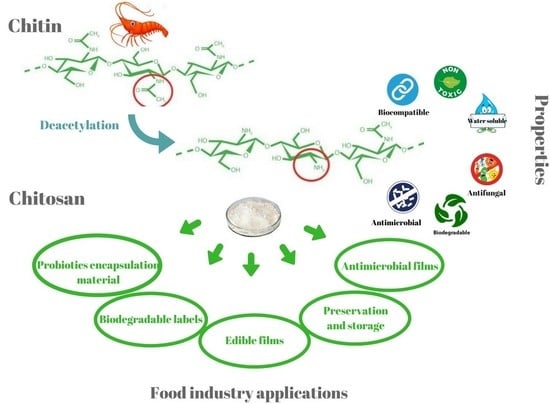
The microcapsule should be stable and retain its integrity throughout the digestive tract passage until it arrives at its target destination, where the capsule should disintegrate and release its contents [74]. Coating adds an extra protective layer on the microcapsule surface, therefore resulting in improved mechanical strength and a strong barrier function. This process involves the immersion of the hydrogel particles into a solution of coating polymer [35]. The main advantages of chitosan coating are unique cationic character, high biocompatibility, non-toxicity, and biodegradability; therefore, it is quite suitable for use in the food and pharmaceutical industries. Its origin lies in the shell waste of crab, shrimp, and crawfish [25]. The origin source influences the molecular weight of chitosan, which is responsible for its crystallinity, degradation, tensile strength, and moisture content, but can be decreased with processing for increasing the deacetylation [75]. This type of coating is of a major interest in the field of targeted release of probiotics due to its high compatibility with living cells [76]. Chemically speaking, chitosan (Figure 1) is a polysaccharide composed of (1, 4)-linked 2-amino-deoxy-b-d-glucan, a deacetylated derivative of chitin. Chitosan ranges second after cellulose in terms of its availability in nature [77]. The degree of deacetylation of chitin represents the major aspect in chitosan’s characterization [78]. For example, when the degree of deacetylation of chitin overcomes 50%, chitosan becomes soluble in aqueous acidic conditions [79]. Moreover, the homogeneous or heterogeneous deacetylation conditions have an important impact on chitosan’s microstructure [80], which mainly determines its solubility and applications (i.e., drug or food carriers) [78]. Figure 1 below illustrates the differences between chitin and chitosan, as chemical structures.
It has been reported [73] that coating alginate beads with chitosan develops a complexation of chitosan with alginate resulting in several important properties, such as alginate beads with reduced porosity, reduced leakage of the encapsulated bacteria, and stability at various pH ranges. The negatively charge property of alginate in contact with the positive charge of chitosan develops a semi-permeable membrane, therefore the resulting capsules possess a smoother surface with a reduced permeability to water soluble molecules [73]. However, since the survival of probiotic cells was shown to not be satisfactory and it was reported to have an inhibitory effect against some bacteria (L. lactis) [55], chitosan is mostly used as a coating/shell, and not as the capsule itself [81]. In fact, encapsulation of probiotic bacteria with chitosan and alginate coating provides protection in simulated GI conditions, and it is a good way of delivering viable bacterial cells to the colon [37]. Considering the above, chitosan-coated alginate microspheres represents a good alternative for probiotic microorganism oral delivery [82]. The chitosan-specific chemical structure allows important changes at the C-2 position with no difficulties [79]. Based on its aqueous acidic solubility, it allows for many applications in the solution and hydrogel fields, due to its gel-forming abilities. The electrical properties such as the surface potential (ζ-potential) of chitosan-coated alginate microgels or other types of chitosan-based microgels can be evaluated by different methods, e.g., electrophoretic light scattering. For instance, a study [83] published in 2016 evaluated the alginate and chitosan microgels for B. longum encapsulation. The particle size of the microbeads was also evaluated using static light scattering, resulting in a higher particle size of the chitosan-coated alginate beads due to the additional coating of alginate or because of some aggregation of the microgels [83].
3.1. Effectiveness of Improving Cell Survival
In order to improve the effectiveness of bacteria survival, researchers have focused on several microencapsulation technologies considering novel combinations of supporting matrices. Several studies conducted on different bacterial strains [84,85,86] have shown that the use of chitosan-coated microcapsules significantly contributes to the survival of probiotic bacteria during simulated GI conditions. Therefore, experimental studies reported the chitosan-coated alginate microcapsules as the best technology for probiotic bacteria protection (such as Lactobacillus and Bifidobacterium spp.) against all conditions tested [73,84]. Another study demonstrated that L. bulgaricus immobilized by chitosan-coated alginate microencapsulation proved increased storage stability in comparison to free cells [85]. A similar effect was observed in a study conducted by Vodnar and Socaciu [86] on L. casei and L. plantarum. Moreover, another study highlighted that chitosan coating provided the best protection of probiotic bacteria under simulated GI conditions and their survival increased (p < 0.05). A recent study [87] from 2017 showed that pectin–chitosan capsules can protect L. casei from the acidic conditions of the stomach and resulted in higher number of viable cells in the intestine [87]. These results are in line with other studies that reported that there was a correlation between the increased concentration of microencapsulating material and the increase in the survival rate of probiotic bacteria under simulated GI conditions [88].
Additionally, it is considered that the probiotic’s efficiency and efficacy can be improved by a combination between probiotics and their growth substrate–prebiotics, by a significant colonization of cells in the human gut, since these non-absorbable carbohydrates are a selective energy source for probiotics [86]. This combination was termed as “synbiotics” [89]. Addition of a prebiotic matrix is a promising approach for effective probiotic protection. Therefore, several studies proposing the probiotic–prebiotic chitosan-coated encapsulation system are described below. A simple representation of the concept is illustrated in Figure 2.
In the study by Varankovich et al. [90], the novel pea protein–alginate microcapsules with a chitosan coating were produced by extrusion. These microcapsules were tested for immobilization and survivability of L. rhamnosus R0011 and L. helveticus R0052 during storage and exposure to in vitro GI conditions. The results indicated the chitosan coating was responsible for an increased cell viability during nine weeks of storage at room temperature, significantly improving the microcapsule performance when compared to non-chitosan coated microcapsules. Under GI conditions, the microcapsule formulation provided high protection for cells, while refrigerated storage had no negative effects on the microcapsule protection performance. In addition, the chitosan coating did not increase the microcapsule size.
Different investigations have demonstrated that selenium-enriched green tea co-encapsulated with probiotic bacteria in chitosan-coated alginate beads offer a compelling approach to expanding the lifespan and viability of probiotic cells in simulated GI juices and refrigerated storage [84,86]. The study by Vodnar and Socaciu [86] on the survival of probiotic bacteria belonging to L. casei and L. plantarum strains tested during storage at 4 °C demonstrated significantly higher numbers (p < 0.05) of survival bacteria encapsulated in chitosan-coated microspheres with selenium-enriched green tea (2 g/100 mL). These results, together with previous findings [84], suggest that immobilization of bacterial strains in chitosan coating improve their viability during refrigeration storage. The chitosan exerts a protective effect on these living microorganisms and the microencapsulation with selenium-enriched green tea was complementary in maintaining the bacteria stability and increased their viability by storage at refrigeration temperature for 30 days. The protective effect of green tea was further demonstrated by sustaining the growth of Lactobacillus ssp. and Bifidobacterium ssp. during simulated conditions [91].
Chavarri et al. [82] microencapsulated L. gasseri and B. bifidum using quercetin as prebiotics and chitosan as coating material in alginate microparticles and reported improved survival during in vitro gastrointestinal conditions. Other studies reported resistant starch as prebiotics and chitosan as coating material for encapsulation of different probiotic bacteria and found increased viability up to 6 months at room temperature [65].
According to de Araújo Etchepare et al. [58], the use of the prebiotic Hi–maize (1%) and chitosan (0.4%) in alginate beads by extrusion technique significantly improved the viability of the microencapsulated bacteria L. acidophilus in both the GI and storage conditions of moist and freeze-dried microcapsules.
In a study by Jantarathin et al. [92], L. acidophilus TISTR 1338 was separately co-encapsulated with two types of prebiotics, inulin and Jerusalem artichoke, within a chitosan-coated sodium–alginate matrix. After testing the capsules’ performance in freeze-drying and high-temperature conditions, the results showed an increase in cells’ viability in chitosan double-coated microcapsules, and this increase was maintained after the freeze-dry process. The high-temperature conditions involved the capsules’ exposure to 70 °C for 60 min and 90 °C for 5 min, and the findings indicated a 3% prebiotic with 3% alginate and 0.8% chitosan as the most efficient combination for increased viability of microcapsules during heat processing, whereas free cells were destroyed. This novel combination could represent an efficient approach for probiotic bacteria protection during functional food processing that involves heating and freeze-dry processes.
As inulin is one of the most used prebiotics, another study tested the influence of different chain lengths, in co-encapsulation with L. casei in chitosan-coated alginate beads. The combination of inulin and chitosan-coating proved to enhance cell viability against gastric and bile salt exposure with 2.7–2.9 log reduction for L. casei, where long-chain inulin showed the highest survival rate (2.7 log reduction) [93].
Another efficient approach to improving the viability of probiotic bacteria under GI conditions for targeted release proposes the use of chitosan and enteric polymers in the formulation of microencapsulated beads [94]. For instance, B. animalis subsp. lactis was incorporated in alginate, alginate–chitosan, alginate–chitosan–sreteric, and alginate–chitosan–aryl–eze. The results indicated that the use of chitosan and enteric polymers in the formulation of the beads, especially aryl–eze, improved the survival rate of B. animalis, while promoting the controlled intestinal delivery of bifidobacteria.
3.2. Microcapsules Size and Protection Performance
The size of microcapsules is of major importance in probiotic protection. Many recent studies on probiotic encapsulation dealth with particle size reduction due to the negative impact of large particle size on the sensorial and textural characteristics of the product [37]. Heidebach et al. [35] showed that a range of size particles between 0.2 mm and 3 mm can provide strong protection for probiotics during GI exposure. A particle size smaller than 100 µm provided the best sensorial properties. Therefore, considering all these, several solutions have been proposed to eliminate these limitations. For example, a spray-drying technique, very accessible in the food industry, can provide small capsules with average diameters below 100 µm at comparably low costs. Besides, there is a direct relationship between adding a chitosan coating and the microcapsules’ diameter [95].
Application of a coating material on the microcapsules’ surface is among the proposed solutions for increasing their probiotic performance. The coating materials belong to different type of class compound, and, in some cases, can coincide with the capsules’ matrix [56]. By interacting with the capsule surface, the coating will create an extra layer on the microcapsule [35], which can be translated into increased probiotic protection. The coating has the ability to reduce the permeability of the capsule, and implicitly the oxygen exposure of the probiotics, therefore increasing their stability under harsh conditions, such as high temperatures and low pH [35,96]. Other authors have used the coatings for establishing new adhesion properties for the microparticles or to optimize the targeted delivery of the cells [97].
For an increased protection performance under different harsh conditions, multiple combinations of different coating materials and techniques have been applied. For instance, the LbL assembly involves the immersion of microcapsules in polymer solution resulting in the coating, while coacervation implies the formation of a coacervate between the microcapsules’ surface and a coating. Regarding the coating development, a major aspect for consideration is the control of the layer’s thickness, which, according to previous studies, have no influence on the increase of the capsules’ size. Cook et al. [98] demonstrated that the thickness of a chitosan-coated alginate microcapsule is directly correlated with the immersion time, with a minimal value of 8 µm after 1 min, and a value of 24 µm after 2400 min, on microcapsules with a diameter of 1 mm.
In a recent review article by Ramos et al. [54], an in-depth comparative analysis of protection performance for different coatings was investigated. The conclusion suggested that the coatings with better protection performance considering hard digestion conditions were chitosan, alginate, poly-L-lysine (PLL), and whey protein; however, among all, chitosan showed the best efficiency due to its ability to resist and protect the probiotic viable cell during in vitro digestion. In addition, the authors’ conclusions underlined the idea that more coatings do not always imply better protection when compared to mono-coated microcapsules.
Chitosan demonstrated to be the most satisfying material to protect microencapsulated probiotics, having efficient results in a variety of alginate microcapsules (performed by different techniques and with different types of alginate), probiotics strains, and exposure conditions. The improved capsule stability and efficient protection was due to the strong ionic interactions between alginate (anionic group) and chitosan (cationic group). Figure 3a illustrates more details regarding this process, where initial microcapsules produced by an anionic encapsulation material (e.g., alginate) was consecutively coated by a cationic material (e.g., chitosan) and after that by another anionic material. The electrostatic forces involved, due to the polyelectrolyte properties of the biopolymers, will contribute to the layer formation that will coat the probiotic-loaded microcapsule [97]. Their ionic interaction representation is illustrated in Figure 3b.
3.3. Chitosan Application According to the Technology and Bio-Based Matrices Used for the Microencapsulation
Different technologies and core materials can be used to develop probiotic encapsulation with chitosan coatings resulting in microcapsules with different characteristics in terms of range in sizes of particles and of types of capsule, as well as protection efficiency. Novel chitosan bio-based matrices were developed with the aim of an increased bacteria protection making use of the most efficient microencapsulation techniques. In most of the studies, chitosan coating proved to have a high efficiency in different probiotic strain protection against harsh conditions, maintaining a proper concentration of viable cells for intestinal delivery. Regarding technologies, spray-dried particles coated with chitosan are recommended as significantly effective capsules in delivering viable bacterial cells to the colon and stable particles during refrigerated storage [99]. Although, one of the most studied encapsulation technologies regarding chitosan coating is extrusion LbL.
In a study by Singht et al. [100], novel bio-based matrices of carboxymethyl cellulose–chitosan (CMC-Cht) were used for the encapsulation of the probiotic bacteria L. rhamnosus GG via a nozzle-spray method. The hybrid micro- and macroparticles results confirmed their potential for encapsulation and delivery, being the first successful encapsulation of L. rhamnosus GG in CMC-Cht particles with an acceptable survival rate. Li et al. [101] encapsulated L. casei ATCC 393 with alginate, chitosan, and carboxymethyl chitosan matrices by an extrusion method, and the system increased the cells’ viability up to 108 cfu/g in a dry state after 4 weeks of storage at 4 °C. After exposure to GI conditions, the encapsulated bacteria maintained its probiotic effect, indicating that alginate–chitosan–carboxymethyl chitosan microcapsules could efficiently protect L. casei against harsh conditions and may represent a novel route for delivery of probiotic cultures as a functional food.
In a study by Zou et al. [66], the encapsulation of B. bifidum F-35 in alginate microspheres developed by emulsification/internal gelation technique was reinforced by addition of pectin/starch or coating with chitosan/PLL to enhance protection for probiotic bacteria. By comparison, the chitosan-coated alginate microspheres showed the highest protection for microencapsulated bacteria under in vitro GI conditions and during 1 month of storage at 4 °C, being an efficient approach for bifidobacteria intestinal colonization.
Cook et al. [102] investigated the LbL coating of alginate matrices with chitosan–alginate for encapsulation of B. breve with the aim of improving bacteria survival under low-pH conditions, and implicitly, intestinal delivery. The experimental study proved that multilayer-coated alginate matrices increased cells’ viability during exposure to in vitro gastric conditions, precisely from <3 log(CFU)/mL, reported in free cells, up to a maximum of 8.84 ± 0.17 log(CFU)/mL in the 3-layer coated matrix, while also providing a targeted gradual intestinal release over 240 min. There are also other studies reporting that chitosan-coated alginate microparticles for probiotic encapsulations allowed better viability [65,103]. Chitosan and alginate have been tested many times for coating abilities in microencapsulation and protection of different probiotics (such as B. bifidum, B. breve, and L. gasseri) [82,98,102,104]. Chitosan and alginate possess high-charge densities, being able to increase the capsule’s residence in targeted areas of release. Therefore, they provide probiotic intestinal delivery [98].
Fareez et al. [105] successfully implemented the microencapsulation of L. plantarum LAB12 in chitosan–alginate–xanthan gum–β–cyclodextrin (Alg–XG–β–CD–Ch) beads considering a survival rate of 95% at pH 1.8 with facilitated release at pH 6.8. Moreover, the microcapsules maintained the cells’ viability >7 log CFU/g during 4-week storage at 4 °C and had reduced viable cell loss at 75 °C and 90 °C. Considering this, the Alg–XG–β–CD–Ch approach may be suitable for application as heat- and pH-stable polymeric beads that incorporate lactobacilli species as efficient transport vehicles crossing gastric conditions for final intestinal colonization, as heat resistant coating up to 90 °C is a significant property in product manufacturing. Therefore, the Alg–XG–β–CD–Ch applications for probiotics are wide and target the health, food, and agro-industries. In 2015, the same author, Fareez et al. [106], demonstrated that incorporation of the same probiotic bacteria into chitosan-coated alginate–xanthan gum (Alg–XG) beads was a feasible physicochemical driven approach for delivering new functional food ingredients [106].
Falco et al. [107], using the LbL technique, developed a chitosan and sulfated β-glucan encapsulation matrix for L. acidophilus considering their prebiotic property for further novel applications, such as carriers for probiotics and sensitive nutraceuticals. Compared to uncoated cells, the viability of cells with four layers of chitosan and sulfated β-glucan decreased only by 2 log CFU/mL. Under in vitro GI exposure, the protection of the coatings was partially degraded, but resisted under acidic gastric conditions. The Hi–maize (1.0% w/v) prebiotic addition to microcapsules containing Lactobacillus spp. coated with chitosan considerably improved (p < 0.05) the viability of cells after GI exposure, and in stored yogurt, in comparison with alginate-based microcapsules [65].
According to Bepeyeva et al. [87], encapsulation of L. casei into calcium–pectinate–chitosan beads provided protection of cells under GI exposure. The beads were prepared by extrusion of amidated pectin into calcium chloride with additional chitosan coating, resulting in high levels of viable bacteria with intestinal delivery application. According to Kanmani et al. [108], the encapsulation of LAB Enterococcus faecium MC13 into chitosan-coated alginate microcapsules demonstrated an improved delivery of viable cells and good resistance to harsh gastro-intestinal conditions. Trabelsi et al. [109] reported that encapsulated L. plantarum TN8 on alginate coated with chitosan during 8 weeks of storage at 4 °C was effective in maintaining the stability of the probiotic bacteria.
The experimental results of Zaeim et al. [110] proposed wet-electrospraying as a successful and novel technique for encapsulation of probiotic bacteria (L. plantarum) inside Ca–alginate/chitosan hydrogel microcapsules by single- and double-stage procedure with an encapsulation yield of almost 98%. The cells’ viability increased with 1 log cycle compared to the free cells under simulated GI conditions, while the outer layer of chitosan, which was deposited on Ca–alginate microcapsules by double-stage procedure, more efficiently protected bacteria at low pH environments.
Table 2 below illustrates the survival rate of different probiotic bacteria in chitosan-coated microcapsules prepared by extrusion-LbL technology.
3.4. Food Applications of Probiotic Microencapsulated in Chitosan-Based Coatings
The most important food applications of chitosan include the encapsulating material for probiotic stability in the production of functional food products [44], formation of biodegradable films, enzymes binding, conservation of foods from microbial deterioration, nutritional supplements, and other applications (additives, emulsifier agents, etc.) [113]. Belonging mainly to lactic acid bacteria (LAB), probiotics are widely used in the production of fermented dairy foods such as yoghurt, cheese, korut, and kefir, being the richest sources of probiotic foods available on the market [39], but in recent years, the focus of using probiotic microencapsulation techniques have moved to fruit juices [114], cereal-based products, chocolate products [115], and cookies—this being a real challenge considering the product matrix [19]. Furthermore, a screening of dairy products, beverages, and other products developed with incorporation of probiotics microencapsulated in chitosan-based coating is presented.
3.4.1. Dairy Products
The microcapsules developed by distinctive technologies with an extra coating represent a technological step recommended to increase protection of the bioactive compounds from external damage factors such as acidity, oxygen, and gastric conditions [25] while incorporated in dairy products. Since the incorporation of microcapsules in yogurt products do not alter the sensory quality [116], chitosan is the perfect candidate for the role of coating material, due to its non-impairing adverse sensory properties to food [117]. In the beginning of the 20th century, the challenge of using chitosan to incorporate LAB was addressed [118], and since then, different bacterial strains were taken under investigation.
One report concluded that L. delbrueckii subsp. bulgaricus immobilized by chitosan-coated alginate maintained cell stability for 4 weeks of storage at 4 °C and 22 °C in skim milk [119]. Studies performed on strains belonging to L. bulgaricus, L. gasseri, and B. bifidum [82,85] loaded in chitosan-coated alginate microspheres showed higher storage stability than free cell cultures. Moreover, in a previous study [120], a comparison was made between the survival rate of bifidobacteria encapsulated in alginate beads containing chitosan and that of the bacteria immobilized only in alginate beads. The results obtained showed that chitosan-based capsules provided higher protection for probiotic cells than alginate matrix in yogurt products and under simulated GI exposure [120]. The study by Urbanska et al. [121] demonstrated the effectiveness of chitosan-coated alginate microcapsules for delivery of probiotic L. acidophilus live cells in yogurt. Moreover, the results reported a structural integrity of microcapsules after 76 h of mechanical agitation in culture broth media and after 24 h in in vitro GI conditions. Krasaekoopt et al. [45] microencapsulated L. acidophilus 547, B. bifidum ATCC 1994, and L. casei 01 in chitosan-coated alginate beads with incorporation in yoghurt from UHT and conventionally treated milk for investigating their survival during storage at 4 °C for 4 weeks. The survival of encapsulated probiotic bacteria was higher than free cells, while the probiotic effect was maintained, the viable cells’ number being above the recommended therapeutic level during storage, except for B. bifidum.
In formulated yoghurt products, the viability of probiotics was improved by applying sodium alginate beads, which were processed with chitosan as an effective microencapsulation to maintain stability under storage at refrigeration temperature. A four times higher viability in yoghurt-applied capsules compared to cells in a saline suspension was observed [122]. This reinforces the fact that microencapsulation with chitosan coating represents an important alternative. Moreover, it is very effective in providing the colon with higher numbers of viable bacterial cells and keeping their survival in dairy products under refrigeration conditions.
Obradović et al. [123] investigated the protection of chitosan coating on cell viability of microencapsulated probiotic starter culture (containing S. salivarius ssp. thermophilus, L. delbrueckii ssp. bulgaricus, L. acidophilus, and B. bifidum) in fermented whey beverages against fermentation process conditions and product storage. The chitosan coating’s influence on the mechanical stability of core encapsulation material was also assessed. Sodium–alginate beads were made using the extrusion technique. The results revealed an increased cell viability with chitosan coatings, as well as improved elastic and strength properties of beads during food storage.
The combination of two lactobacilli (L. acidophilus and/or L. reuteri) were successfully microencapsulated in alginate and alginate–chitosan beads for addition to milk and blackberry jam set-style yogurt [124]. After storage at 5 °C for 30 days, followed by simulated GI conditions exposure, the results indicated that alginate–chitosan encapsulation provided better protection than alginate alone, and increased bacteria survival during storage, with cell counts higher than ≥107 CFU/g, while after GI simulation, the alginate–chitosan system prevented lactobacilli loss and had favorable intestinal releases. The presence of capsules in blackberry jam set-style yogurt had no sensorial influence, while it did in milk. These two types of dairy products can promote microcapsule stability and lactobacilli viability.
The new encapsulation system xanthan–chitosan and xanthan–chitosan–xanthan, where chitosan was applied as coating, improved storage stability of B. bifidum BB01 in yogurt during 21 days at both 4 and 25 °C, providing high probiotic survival during GI tract conditions [125].
3.4.2. Beverages
One study [126] looked at the effect of multi-layer coating of alginate beads on the viability of immobilized L. plantarum under in vitro gastric conditions and during storage in pomegranate juice (a highly acidic juice) at 4 °C. The examined beads were either uncoated, single, or double coated in chitosan. The results obtained showed an improvement in the cells’ survival rate in the case of chitosan-coated beads, under simulated gastric solution (pH 1.5) by 0.5–2 logs compared to the control (uncoated beads). The strong protection of chitosan may be the result of electrostatic interactions between chitosan and alginate beads. This was the first study that researched this double-coated process for immobilization of probiotics with the aim of increasing their survival and resistance, proving to be better than the single-coated process [126]. Moreover, in a later study, the same authors [68] confirmed that the use of double-chitosan-coated alginate beads yielded a cell concentration of 107 CFU/mL and 105 CFU/mL for L. plantarum and B. longum, respectively, after 6 weeks of storage in pomegranate juice and cranberry juice. Therefore, the chitosan coating offered a significant additional protection to that of the encapsulation matrix on the bacteria during storage of microcapsules inside the juice products. This supports the statement that more than one chitosan-layer coating is a promising approach to be used for improving the survival of probiotic cells in strong acidic food matrices [68].
García-Ceja et al. [124] developed a probiotic peach nectar by addition of microencapsulated L. acidophilus and L. reuteri in an alginate–chitosan system for efficient protection. The results revealed that alginate–chitosan beads protected lactobacilli viability in acidic peach nectar, thus, representing a strong alternative for functional beverage products considering the combination of two lactobacilli, therefore providing more health benefits to consumers.
As described in all the abovementioned publications, the survival of probiotic bacteria in alginate beads containing chitosan was better than in alginate beads alone; therefore, this indicates that this may be used for enhancing the survival of strains. Moreover, consumer health issues and environmental consciousness play important roles in the design of next generation encapsulation matrices and technology, and since chitosan is biocompatible, non-toxic, and biodegradable, further research on the usage of chitosan as a coating material for probiotics will benefit the development of novel functional food products.
3.4.3. Other Food Products
Malmo et al. [116] developed a probiotic chocolate soufflé with L. reuteri DSM 17938 microencapsulation via a chitosan-coated alginate system, incorporating it into the dough matrix prior to baking at 180 °C for 10 min (80 °C in the core of product). The authors reported a survival percentage of 10% of the probiotic population after baking and only 1% for free cells. Moreover, the study showed a significant resistance of microencapsulated bacteria when exposed to high temperatures in real food testing compared to the in vitro conditions, indicating a possible extra-protective layer of the food matrices on probiotic cells.
Microencapsulated L. acidophilus LA-5 was successfully incorporated in probiotic jelly dessert by Talebzadeh and Sharifan [127]. When compared to free bacteria and alginate beads, the chitosan-coated alginate beads showed increased physical stability, spherical shape, and metabolic activity in GI testing. Moreover, the number of viable coated bacteria maintained above 6 log (10) CFU/g after 42 days of storage and the probiotic jelly provided high-sensory attributes.
The combination of chitosan coating with calcium–alginate and Hi–maize resistant starch microcapsules via emulsion techniques delivered increased viable probiotics: L. acidophilus LA-5 and L. casei 431 in baked breads [128]. The authors developed synbiotic bread, namely, hamburger buns and white pan breads by inulin addition. Results showed that this microencapsulation system can be used to develop probiotic bakery products with enhanced cell viability against high-thermal conditions with no negative impact on texture or taste, considering that hamburger buns had a higher probiotic survival rate and L. casei 431 was more resistant to high temperature than L. acidophilus LA-5.
The most recent study byde Farias et al. [129] used a calcium alginate–chitosan microencapsulation system via extrusion method to incorporate L. rhamnosus ASCC 290 and L. casei ATCC in yellow mombin ice cream. The authors compared the behavior and viability of free and encapsulated cells inside the food matrix against storage at low-temperature condition (−18 °C for 150 days) and GI exposure. Results revealed that free L. casei (−1.64 log) had a higher resistance to freezing than free L. rhamnosus (−1.92 log), while encapsulated L. rhamnosus and L. casei presented protection efficiencies of 73.8% and 79.5%, respectively. In the GI simulation, 86.2% L. rhamnosus (−0.83 log) and 84% L. casei (−1.3 log) were protected by the alginate–chitosan capsules. Therefore, for preparing probiotic yellow mombin ice cream, the encapsulation process is not advantageous for all probiotic bacteria, namely, L. rhamnosus, whose survival rate was higher in free form than in microencapsulation, but advantageous for L. casei.
The hydrocolloids used in probiotic microencapsulation is a widely-used method for enhancing survival in ice cream during frozen storage. The study by Zanjani et al. [130] indicates that the microencapsulation of probiotics via calcium alginate, wheat, rice, and high-amylose corn (hylon VII) starches coated by chitosan and PLL enhanced probiotic bacteria survival, namely, L. casei ATCC 39392 and B. adolescentis ATCC 15703, in ice cream after storage at −30 °C for 100 days. Chitosan and PLL coatings significantly increased cell viability during the storage of ice cream, as well as the size of microcapsules. This is due to the integrated microcapsule structure provided by hylon starch. Moreover, sensory evaluation of probiotic ice cream indicated no significant effect on organoleptic properties during the storage period at −30°.
4. Conclusions and Future Perspectives
There is a constant concern that free bacteria might not survive in sufficient numbers during their passage through the GI tract in order to exert its probiotic effect. The physical protection of probiotics by microencapsulation with chitosan-coated alginate beads is an efficient approach to improve the probiotics’ survival during GI passage and to achieve a controlled delivery in the intestine. Moreover, multi-stage coating was shown to further increase bacterial survival in acidic food products.
Since the incorporation of probiotics into food matrices is among the challenging areas of research in food technology, and probiotics are quite sensitive to environmental conditions, such as oxygen, light or temperature, and food matrix interactions, the protection of cells is of major importance for the next generation of probiotic foods. Another major challenge is to improve the viability of probiotics during the manufacturing processes, particularly heat processing while considering the perspective of producing thermoresistant probiotic microorganisms as new solutions needed in future research. Therefore, discovering new strains of probiotic bacteria that are heat resistant, either naturally or which have been genetically modified, and creating a microencapsulation system that acts just as “insulation material” are among the most feasible routes. For developing novel encapsulation systems, there is a need for understanding the thermal conductivity properties of most efficient food-grade biopolymers and lipids that are used as encapsulating core materials and coatings, individually and in combination.
Nevertheless, microencapsulation represents the best alternative since it offers a wide range of food application. In a wider sense, encapsulation may be used for plenty of applications within the food industry, such as: production of novel food products, extending the shelf life of functional products, protecting compounds against nutritional loss, controlling the oxidative reaction during storage, providing sustained or controlled release in the gastro-intestinal environment, maintaining the sensory attributes of probiotic-based food products, formation of biodegradable films, and edible labels.
Due to the abundant amino groups, chitosan provides many positive charges in acidic medium, and represents an efficient biopolymer for microencapsulation and delivery systems for the food industry. Moreover, considering its specific physicochemical attributes, biodegradability, and biocompatibility with human tissues, chitosan compliments the real potential of this technology for applications in the food industry. This biopolymer has no negative effects in the amounts used in food because it is natural, non-toxic, and non-allergenic. As the probiotic–prebiotic synergy is well perceived among future trends, insoluble fibers, like β-glucan, are another bio-based natural polysaccharide source less exploited until now, which is available in high quantities in cereal wastes, has biological activities in the human body, and can be fermented by human gut microbiota. β-glucan and chitosan can represent a future delivery system for bioactive molecules and probiotics being a responsive material suitable for targeted release in the intestine.
Dairy products are the main carriers of probiotics and have led the market for many years, but the continuous interest towards improving lifestyle through nutrition led towards the expansion of functional foods variety (beverages, chocolate bars, etc.); therefore, the legislation frame regarding probiotic foods should allow and sustain manufacturers a more effective probiotic food production. New studies must be carried out in order to assess the impact of the chitosan-coated microencapsulated bacteria into a vast range of non-dairy food products, for favoring the needs of particular groups of consumers such as vegetarians, vegans, and lactose-intolerants. Moreover, a deep investigation into the existing material properties for coated capsule production is of major importance for an efficient protection of the probiotic bacteria.
Certainly, the need for in vivo studies evaluating the viability of the incorporated probiotics under GI conditions for establishing the real level of delivered probiotics, and implicitly, the health effects, is a future research direction. Nevertheless, another research trend in this area is to find industrial encapsulation technologies that guarantee the survival of probiotics. In order to achieve these research goals, an integrated approach that combines microencapsulation techniques suitable for the selected food carriers is one of the solutions, as well as consumer behavior assessments toward novel foods considering their future increased demand. Therefore, nowadays, many studies are focusing on reducing the particle size for non-influence on sensorial and textural properties of the product.
Author Contributions
All authors contributed to the preparation of the manuscript, as follows: Conceptualization and design of the work: L.F.C., D.C.V., and I.D.P.; Writing—Manuscript preparation: L.F.C. and B.E.S.; Writing—review and editing: D.C.V., I.D.P., and L.M. All authors read and approved the final manuscript.
Funding
This work was supported by two grants from the Ministry of Research and Innovation, as follows: CNCS–UEFISCDI, project number PN-III-P1-1.1-TE-2016-0661, within PNCDI III; and MCI-UEFISCDI, project number 37 PFE-2018-2020 “Cresterea performantei institutionale prin mecanisme de consolidare si dezvoltare a directiilor de cercetare din cadrul USAMVCN”.
Acknowledgments
We kindly thank Vasile Coman for image support and critical peer review; Bernadette E. Teleky, and Martău Adrian for image support.
Conflicts of Interest
The authors declare that they have no competing interests.
References
- Vodnar, D.C.; Călinoiu, L.F.; Dulf, F.V.; Ştefănescu, B.E.; Crişan, G.; Socaciu, C. Identification of the bioactive compounds and antioxidant, antimutagenic and antimicrobial activities of thermally processed agro-industrial waste. Food Chem. 2017, 231, 131–140. [Google Scholar] [CrossRef] [PubMed]
- Calinoiu, L.-F.; Mitrea, L.; Precup, G.; Bindea, M.; Rusu, B.; Dulf, F.-V.; Stefanescu, B.-E.; Vodnar, D.-C. Characterization of Grape and Apple Peel Wastes’ Bioactive Compounds and Their Increased Bioavailability After Exposure to Thermal Process. Bull. Univ. Agric. Sci. Vet. Med. Cluj-Napoca-Food Sci. Technol. 2017, 74, 80–89. [Google Scholar] [CrossRef]
- Szabo, K.; Cătoi, A.-F.; Vodnar, D.C. Bioactive Compounds Extracted from Tomato Processing by-Products as a Source of Valuable Nutrients. Plant Foods Hum. Nutr. 2018. [Google Scholar] [CrossRef] [PubMed]
- Călinoiu, L.F.; Vodnar, D.C. Whole Grains and Phenolic Acids: A Review on Bioactivity, Functionality, Health Benefits and Bioavailability. Nutrients 2018, 10, 1615. [Google Scholar] [CrossRef] [PubMed]
- Călinoiu, L.F.; Mitrea, L.; Precup, G.; Bindea, M.; Rusu, B.; Szabo, K.; Dulf, F.V.; Ştefănescu, B.E.; Vodnar, D.C. Sustainable use of agro-industrial wastes for feeding 10 billion people by 2050. In Professionals in Food Chains; Wageningen Academic Publishers: Wageningen, The Netherlands, 2018; pp. 482–486. ISBN 978-90-8686-321-1. [Google Scholar]
- Mitrea, L.; Trif, M.; Catoi, A.-F.; Vodnar, D.-C. Utilization of biodiesel derived-glycerol for 1,3-PD and citric acid production. Microb. Cell Factories 2017, 16, 190. [Google Scholar] [CrossRef] [PubMed]
- Dulf, F.V.; Unguresan, M.L.; Vodnar, D.C.; Socaciu, C. Free and Esterified Sterol Distribution in Four Romanian Vegetable Oil. Not. Bot. Horti Agrobot. Cluj-Napoca 2010, 38, 91–97. [Google Scholar]
- Coldea, T.E.R.; Socaciu, C.; Parv, M.; Vodnar, D. Gas-Chromatographic Analysis of Major Volatile Compounds Found in Traditional Fruit Brandies from Transylvania, Romania. Not. Bot. Horti Agrobot. Cluj-Napoca 2011, 39, 109–116. [Google Scholar] [CrossRef] [Green Version]
- Andreicut, A.-D.; Parvu, A.E.; Mot, A.C.; Parvu, M.; Fodor, E.F.; Catoi, A.F.; Feldrihan, V.; Cecan, M.; Irimie, A. Phytochemical Analysis of Anti-Inflammatory and Antioxidant Effects of Mahonia aquifolium Flower and Fruit Extracts. Oxid. Med. Cell. Longev. 2018, 2879793. [Google Scholar] [CrossRef]
- Andreicu, A.-D.; Pârvu, A.E.; Pârvu, M.; Fischer-Fodor, E.; Feldrihan, V.; Florinela, A.; Cecan, M.; Irimie, A. Anti-Inflammatory and Antioxidant Effects of Mahonia Aquifolium Leaves and Bark Extracts. Farmacia 2018, 66, 49–58. [Google Scholar]
- Probiotics Market Research Report: Market size, Industry outlook, Market Forecast, Demand Analysis, Market Share, Market Report 2018–2023. Available online: https://industryarc.com/Report/7492/probiotics-market-analysis.html?gclid=EAIaIQobChMIm--8urnb4AIVyOF3Ch1E7AwNEAAYAiAAEgJlh_D_BwE (accessed on 27 February 2019).
- Food and Agriculture Organization of the United Nations; World Health Organization. Probiotics in Food: Health and Nutritional Properties and Guidelines for Evaluation; Food and Agriculture Organization of the United Nations, World Health Organization: Rome, Italy, 2006; ISBN 978-92-5-105513-7. [Google Scholar]
- Solanki, H.K.; Pawar, D.D.; Shah, D.A.; Prajapati, V.D.; Jani, G.K.; Mulla, A.M.; Thakar, P.M. Development of microencapsulation delivery system for long-term preservation of probiotics as biotherapeutics agent. BioMed Res. Int. 2013, 2013. [Google Scholar] [CrossRef] [PubMed]
- Dunne, C. Adaptation of bacteria to the intestinal niche: probiotics and gut disorder. Inflamm. Bowel Dis. 2001, 7, 136–145. [Google Scholar] [CrossRef]
- Ziemer, C.J.; Gibson, G.R. An overview of probiotics, prebiotics and synbiotics in the functional food concept: Perspectives and future strategies. Proc. Int. Dairy J. 1998, 8, 473–479. [Google Scholar] [CrossRef]
- Vodnar, D.C.; Venus, J.; Schneider, R.; Socaciu, C. Lactic Acid Production by Lactobacillus paracasei 168 in Discontinuous Fermentation Using Lucerne Green juice as Nutrient Substitute. Chem. Eng. Technol. 2010, 33, 468–474. [Google Scholar] [CrossRef]
- Pop, O.L.; Brandau, T.; Schwinn, J.; Vodnar, D.C.; Socaciu, C. The influence of different polymers on viability of Bifidobacterium lactis 300b during encapsulation, freeze-drying and storage. J. Food Sci. Technol.-Mysore 2015, 52, 4146–4155. [Google Scholar] [CrossRef]
- Rotar, A.M.; Vodnar, D.C.; Bunghez, F.; Catunescu, G.M.; Pop, C.R.; Jimborean, M.; Semeniuc, C.A. Effect of Goji Berries and Honey on Lactic Acid Bacteria Viability and Shelf Life Stability of Yoghurt. Not. Bot. Horti Agrobot. Cluj-Napoca 2015, 43, 196–203. [Google Scholar] [CrossRef]
- Rokka, S.; Rantamäki, P. Protecting probiotic bacteria by microencapsulation: Challenges for industrial applications. Eur. Food Res. Technol. 2010, 231, 1–12. [Google Scholar] [CrossRef]
- Capozzi, V.; Arena, M.P.; Crisetti, E.; Spano, G.; Fiocco, D. The hsp 16 Gene of the Probiotic Lactobacillus acidophilus Is Differently Regulated by Salt, High Temperature and Acidic Stresses, as Revealed by Reverse Transcription Quantitative PCR (qRT-PCR) Analysis. Int. J. Mol. Sci. 2011, 12, 5390–5405. [Google Scholar] [CrossRef]
- Taranu, I.; Marin, D.E.; Braicu, C.; Pistol, G.C.; Sorescu, I.; Pruteanu, L.L.; Berindan Neagoe, I.; Vodnar, D.C. In Vitro Transcriptome Response to a Mixture of Lactobacilli Strains in Intestinal Porcine Epithelial Cell Line. Int. J. Mol. Sci. 2018, 19, 1923. [Google Scholar] [CrossRef] [PubMed]
- Calinoiu, L.-F.; Vodnar, D.-C.; Precup, G. The Probiotic Bacteria Viability under Different Conditions. Bull. Univ. Agric. Sci. Vet. Med. Cluj-Napoca-Food Sci. Technol. 2016, 73, 55–60. [Google Scholar] [CrossRef]
- Nazzaro, F.; Fratianni, F.; Coppola, R.; Sada, A.; Orlando, P. Fermentative ability of alginate-prebiotic encapsulated Lactobacillus acidophilus and survival under simulated gastrointestinal conditions. J. Funct. Foods 2009, 1, 319–323. [Google Scholar] [CrossRef]
- Pop, O.L.; Dulf, F.V.; Cuibus, L.; Castro-Giráldez, M.; Fito, P.J.; Vodnar, D.C.; Coman, C.; Socaciu, C.; Suharoschi, R. Characterization of a Sea Buckthorn Extract and Its Effect on Free and Encapsulated Lactobacillus casei. Int. J. Mol. Sci. 2017, 18, 2513. [Google Scholar] [CrossRef] [PubMed]
- Anal, A.K.; Singh, H. Recent advances in microencapsulation of probiotics for industrial applications and targeted delivery. Trends Food Sci. Technol. 2007, 18, 240–251. [Google Scholar] [CrossRef]
- Dimitrellou, D.; Kandylis, P.; Petrović, T.; Dimitrijević-Branković, S.; Lević, S.; Nedović, V.; Kourkoutas, Y. Survival of spray dried microencapsulated Lactobacillus casei ATCC 393 in simulated gastrointestinal conditions and fermented milk. LWT Food Sci. Technol. 2016, 71, 169–174. [Google Scholar] [CrossRef]
- Arena, M.P.; Caggianiello, G.; Russo, P.; Albenzio, M.; Massa, S.; Fiocco, D.; Capozzi, V.; Spano, G. Functional Starters for Functional Yogurt. Foods 2015, 4, 15–33. [Google Scholar] [CrossRef] [PubMed] [Green Version]
- Li, X.Y.; Chen, X.G.; Cha, D.S.; Park, H.J.; Liu, C.S. Microencapsulation of a probiotic bacteria with alginategelatin and its properties. J. Microencapsul. 2009, 26, 315–324. [Google Scholar] [CrossRef] [PubMed]
- Kim, S.J.; Cho, S.Y.; Kim, S.H.; Song, O.J.; Shin, I.S.; Cha, D.S.; Park, H.J. Effect of microencapsulation on viability and other characteristics in Lactobacillus acidophilus ATCC 43121. LWT Food Sci. Technol. 2008, 41, 493–500. [Google Scholar] [CrossRef]
- Desai, K.G.H.; Jin Park, H. Recent Developments in Microencapsulation of Food Ingredients. Dry. Technol. 2005, 23, 1361–1394. [Google Scholar] [CrossRef]
- Pavli, F.; Tassou, C.; Nychas, G.-J.E.; Chorianopoulos, N. Probiotic Incorporation in Edible Films and Coatings: Bioactive Solution for Functional Foods. Int. J. Mol. Sci. 2018, 19, 150. [Google Scholar] [CrossRef]
- Shori, A.B. Microencapsulation Improved Probiotics Survival During Gastric Transit. HAYATI J. Biosci. 2017, 24, 1–5. [Google Scholar] [CrossRef]
- Martín, M.J.; Lara-Villoslada, F.; Ruiz, M.A.; Morales, M.E. Microencapsulation of bacteria: A review of different technologies and their impact on the probiotic effects. Innov. Food Sci. Emerg. Technol. 2015, 27, 15–25. [Google Scholar] [CrossRef]
- Riaz, Q.U.A.; Masud, T. Recent Trends and Applications of Encapsulating Materials for Probiotic Stability. Crit. Rev. Food Sci. Nutr. 2013, 53, 231–244. [Google Scholar] [CrossRef] [PubMed]
- Heidebach, T.; Först, P.; Kulozik, U. Microencapsulation of Probiotic Cells for Food Applications. Crit. Rev. Food Sci. Nutr. 2012, 52, 291–311. [Google Scholar] [CrossRef]
- Cook, M.T.; Tzortzis, G.; Charalampopoulos, D.; Khutoryanskiy, V.V. Microencapsulation of probiotics for gastrointestinal delivery. J. Controlled Release 2012, 162, 56–67. [Google Scholar] [CrossRef] [Green Version]
- Morales, M.E.; Ruiz, M.A. 16—Microencapsulation of probiotic cells: applications in nutraceutic and food industry. In Nutraceuticals; Grumezescu, A.M., Ed.; Nanotechnology in the Agri-Food Industry; Academic Press: Cambridge, MA, USA, 2016; pp. 627–668. ISBN 978-0-12-804305-9. [Google Scholar]
- Ravi Kumar, M.N.V. A review of chitin and chitosan applications. React. Funct. Polym. 2000, 46, 1–27. [Google Scholar] [CrossRef]
- De Prisco, A.; Mauriello, G. Probiotication of foods: A focus on microencapsulation tool. Trends Food Sci. Technol. 2016, 48, 27–39. [Google Scholar] [CrossRef]
- Nasui, L.; Vodnar, D.; Socaciu, C. Bioactive Labels for Fresh Fruits and Vegetables. Bull. Univ. Agric. Sci. Vet. Med. Cluj-Napoca Food Sci. Technol. 2013, 70, 75–83. [Google Scholar] [CrossRef]
- Dong, H.; Cheng, L.; Tan, J.; Zheng, K.; Jiang, Y. Effects of chitosan coating on quality and shelf life of peeled litchi fruit. J. Food Eng. 2004, 64, 355–358. [Google Scholar] [CrossRef]
- Ye, M.; Neetoo, H.; Chen, H. Control of Listeria monocytogenes on ham steaks by antimicrobials incorporated into chitosan-coated plastic films. Food Microbiol. 2008, 25, 260–268. [Google Scholar] [CrossRef]
- Vodnar, D.C. Inhibition of Listeria monocytogenes ATCC 19115 on ham steak by tea bioactive compounds incorporated into chitosan-coated plastic films. Chem. Cent. J. 2012, 6, 74. [Google Scholar] [CrossRef] [PubMed]
- Burgain, J.; Gaiani, C.; Linder, M.; Scher, J. Encapsulation of probiotic living cells: From laboratory scale to industrial applications. J. Food Eng. 2011, 104, 467–483. [Google Scholar] [CrossRef]
- Krasaekoopt, W.; Bhandari, B.; Deeth, H.C. Survival of probiotics encapsulated in chitosan-coated alginate beads in yoghurt from UHT- and conventionally treated milk during storage. LWT Food Sci. Technol. 2006, 39, 177–183. [Google Scholar] [CrossRef]
- Anselmo, A.C.; McHugh, K.J.; Webster, J.; Langer, R.; Jaklenec, A. Layer-by-Layer Encapsulation of Probiotics for Delivery to the Microbiome. Adv. Mater. 2016, 28, 9486–9490. [Google Scholar] [CrossRef] [Green Version]
- Arslan-Tontul, S.; Erbas, M. Single and double layered microencapsulation of probiotics by spray drying and spray chilling. LWT Food Sci. Technol. 2017, 81, 160–169. [Google Scholar] [CrossRef]
- Ashwar, B.A.; Gani, A.; Gani, A.; Shah, A.; Masoodi, F.A. Production of RS4 from rice starch and its utilization as an encapsulating agent for targeted delivery of probiotics. Food Chem. 2018, 239, 287–294. [Google Scholar] [CrossRef]
- Huq, T.; Khan, A.; Khan, R.A.; Riedl, B.; Lacroix, M. Encapsulation of Probiotic Bacteria in Biopolymeric System. Crit. Rev. Food Sci. Nutr. 2013, 53, 909–916. [Google Scholar] [CrossRef]
- Schönhoff, M. Layered polyelectrolyte complexes: physics of formation and molecular properties. J. Phys. Condens. Matter 2003, 15, R1781–R1808. [Google Scholar] [CrossRef]
- Valencia-Chamorro, S.A.; Palou, L.; del Río, M.A.; Pérez-Gago, M.B. Antimicrobial Edible Films and Coatings for Fresh and Minimally Processed Fruits and Vegetables: A Review. Crit. Rev. Food Sci. Nutr. 2011, 51, 872–900. [Google Scholar] [CrossRef] [PubMed]
- Cerqueira, M.A.; Bourbon, A.I.; Pinheiro, A.C.; Martins, J.T.; Souza, B.W.S.; Teixeira, J.A.; Vicente, A.A. Galactomannans use in the development of edible films/coatings for food applications. Trends Food Sci. Technol. 2011, 22, 662–671. [Google Scholar] [CrossRef] [Green Version]
- Šuput, D.Z.; Lazić, V.L.; Popović, S.Z.; Hromiš, N.M. Edible films and coatings: Sources, properties and application. Food Feed Res. 2015, 42, 11–22. [Google Scholar] [CrossRef] [Green Version]
- Ramos, P.E.; Cerqueira, M.A.; Teixeira, J.A.; Vicente, A.A. Physiological protection of probiotic microcapsules by coatings. Crit. Rev. Food Sci. Nutr. 2018, 58, 1864–1877. [Google Scholar] [CrossRef]
- Groboillot, A.F.; Champagne, C.P.; Darling, G.D.; Poncelet, D.; Neufeld, R.J. Membrane formation by interfacial cross-linking of chitosan for microencapsulation of Lactococcus lactis. Biotechnol. Bioeng. 1993, 42, 1157–1163. [Google Scholar] [CrossRef]
- Krasaekoopt, W.; Bhandari, B.; Deeth, H. Evaluation of encapsulation techniques of probiotics for yoghurt. Int. Dairy J. 2003, 13, 3–13. [Google Scholar] [CrossRef]
- Raafat, D.; Sahl, H.-G. Chitosan and its antimicrobial potential – a critical literature survey. Microb. Biotechnol. 2009, 2, 186–201. [Google Scholar] [CrossRef] [Green Version]
- de Araújo Etchepare, M.; Raddatz, G.C.; de Moraes Flores, É.M.; Zepka, L.Q.; Jacob-Lopes, E.; Barin, J.S.; Ferreira Grosso, C.R.; de Menezes, C.R. Effect of resistant starch and chitosan on survival of Lactobacillus acidophilus microencapsulated with sodium alginate. LWT Food Sci. Technol. 2016, 65, 511–517. [Google Scholar] [CrossRef] [Green Version]
- Shah, U.; Naqash, F.; Gani, A.; Masoodi, F.A. Art and Science behind Modified Starch Edible Films and Coatings: A Review. Compr. Rev. Food Sci. Food Saf. 2016, 15, 568–580. [Google Scholar] [CrossRef] [Green Version]
- Gharsallaoui, A.; Roudaut, G.; Chambin, O.; Voilley, A.; Saurel, R. Applications of spray-drying in microencapsulation of food ingredients: An overview. Food Res. Int. 2007, 40, 1107–1121. [Google Scholar] [CrossRef]
- Saravanan, M.; Rao, K.P. Pectin–gelatin and alginate–gelatin complex coacervation for controlled drug delivery: Influence of anionic polysaccharides and drugs being encapsulated on physicochemical properties of microcapsules. Carbohydr. Polym. 2010, 80, 808–816. [Google Scholar] [CrossRef]
- Weinbreck, F.; Bodnár, I.; Marco, M.L. Can encapsulation lengthen the shelf-life of probiotic bacteria in dry products? Int. J. Food Microbiol. 2010, 136, 364–367. [Google Scholar] [CrossRef]
- Doherty, S.B.; Gee, V.L.; Ross, R.P.; Stanton, C.; Fitzgerald, G.F.; Brodkorb, A. Development and characterisation of whey protein micro-beads as potential matrices for probiotic protection. Food Hydrocoll. 2011, 25, 1604–1617. [Google Scholar] [CrossRef]
- Gunasekaran, S.; Ko, S.; Xiao, L. Use of whey proteins for encapsulation and controlled delivery applications. J. Food Eng. 2007, 83, 31–40. [Google Scholar] [CrossRef]
- Iyer, C.; Kailasapathy, K. Effect of co-encapsulation of probiotics with prebiotics on increasing the viability of encapsulated bacteria under in vitro acidic and bile salt conditions and in yogurt. J. Food Sci. 2005. [Google Scholar]
- Zou, Q.; Zhao, J.; Liu, X.; Tian, F.; Zhang, H.; Zhang, H.; Chen, W. Microencapsulation of Bifidobacterium bifidum F-35 in reinforced alginate microspheres prepared by emulsification/internal gelation. Int. J. Food Sci. Technol. 2011, 46, 1672–1678. [Google Scholar] [CrossRef]
- Cui, J.-H.; Goh, J.-S.; Kim, P.-H.; Choi, S.-H.; Lee, B.-J. Survival and stability of bifidobacteria loaded in alginate poly-l-lysine microparticles. Int. J. Pharm. 2000, 210, 51–59. [Google Scholar] [CrossRef]
- Nualkaekul, S.; Cook, M.T.; Khutoryanskiy, V.V.; Charalampopoulos, D. Influence of encapsulation and coating materials on the survival of Lactobacillus plantarum and Bifidobacterium longum in fruit juices. Food Res. Int. 2013, 53, 304–311. [Google Scholar] [CrossRef]
- Woo, J.-W.; Roh, H.-J.; Park, H.-D.; Ji, C.-I.; Lee, Y.-B.; Kim, S.-B. Sphericity Optimization of Calcium Alginate Gel Beads and the Effects of Processing Conditions on Their Physical Properties. Food Sci. Biotechnol. 2007, 16, 715–721. [Google Scholar]
- Stummer, S.; Salar-Behzadi, S.; Unger, F.M.; Oelzant, S.; Penning, M.; Viernstein, H. Application of shellac for the development of probiotic formulations. Food Res. Int. 2010, 43, 1312–1320. [Google Scholar] [CrossRef]
- Buch, K.; Penning, M.; Wächtersbach, E.; Maskos, M.; Langguth, P. Investigation of various shellac grades: additional analysis for identity. Drug Dev. Ind. Pharm. 2009, 35, 694–703. [Google Scholar] [CrossRef]
- Shi, L.-E.; Li, Z.-H.; Zhang, Z.-L.; Zhang, T.-T.; Yu, W.-M.; Zhou, M.-L.; Tang, Z.-X. Encapsulation of Lactobacillus bulgaricus in carrageenan-locust bean gum coated milk microspheres with double layer structure. LWT Food Sci. Technol. 2013, 54, 147–151. [Google Scholar] [CrossRef]
- Krasaekoopt, W.; Bhandari, B.; Deeth, H. The influence of coating materials on some properties of alginate beads and survivability of microencapsulated probiotic bacteria. Int. Dairy J. 2004, 14, 737–743. [Google Scholar] [CrossRef]
- Vodnar, D.C.; Socaciu, C.; Rotar, A.M.; Stãnilã, A. Morphology, FTIR fingerprint and survivability of encapsulated lactic bacteria (Streptococcus thermophilus and Lactobacillus delbrueckii subsp. bulgaricus) in simulated gastric juice and intestinal juice. Int. J. Food Sci. Technol. 2010, 45, 2345–2351. [Google Scholar] [CrossRef]
- Yuan, Y.; Chesnutt, B.M.; Haggard, W.O.; Bumgardner, J.D. Deacetylation of Chitosan: Material Characterization and in vitro Evaluation via Albumin Adsorption and Pre-Osteoblastic Cell Cultures. Materials 2011, 4, 1399–1416. [Google Scholar] [CrossRef] [Green Version]
- de Vos, P.; Faas, M.M.; Spasojevic, M.; Sikkema, J. Encapsulation for preservation of functionality and targeted delivery of bioactive food components. Int. Dairy J. 2010, 20, 292–302. [Google Scholar] [CrossRef]
- Aider, M. LWT Food Science and Technology Chitosan application for active bio-based films production and potential in the food industry: Review. LWT Food Sci. Technol. 2010, 43, 837–842. [Google Scholar] [CrossRef]
- Estevinho, B.N.; Rocha, F.; Santos, L.; Alves, A. Microencapsulation with chitosan by spray drying for industry applications—A review. Trends Food Sci. Technol. 2013, 31, 138–155. [Google Scholar] [CrossRef]
- Rinaudo, M. Chitin and chitosan: Properties and applications. Prog. Polym. Sci. Oxf. 2006, 31, 603–632. [Google Scholar] [CrossRef]
- George, A.; Roberts, F. Structure of chitin and chitosan. In Chitin Chemistry; Palgrave: London, UK, 1992; pp. 85–91. [Google Scholar]
- Mortazavian, A.; Razavi, S.H.; Ehsani, M.R.; Sohrabvandi, S. Principles and Methods of Microencapsulation of Probiotic Microorganisms. Iran. J. Biotechnol. 2007, 5, 1–18. [Google Scholar]
- Chávarri, M.; Marañón, I.; Ares, R.; Ibáñez, F.C.; Marzo, F.; del Carmen Villarán, M. Microencapsulation of a probiotic and prebiotic in alginate-chitosan capsules improves survival in simulated gastro-intestinal conditions. Int. J. Food Microbiol. 2010, 142, 185–189. [Google Scholar]
- Yeung, T.W.; Üçok, E.F.; Tiani, K.A.; McClements, D.J.; Sela, D.A. Microencapsulation in alginate and chitosan microgels to enhance viability of Bifidobacterium longum for oral delivery. Front. Microbiol. 2016, 7. [Google Scholar] [CrossRef]
- Vodnar, D.C.; Socaciu, C. Green tea increases the survival yield of Bifidobacteria in simulated gastrointestinal environment and during refrigerated conditions. Chem. Cent. J. 2012, 6, 61. [Google Scholar] [CrossRef]
- Koo, S.M.; Cho, Y.H.; Huh, C.S.; Baek, Y.J.; Park, J. Improvement of the stability of Lactobacillus casei YIT 9018 by microencapsulation using alginate and chitosan. J. Microbiol. Biotechnol. 2001, 11, 376–383. [Google Scholar]
- Vodnar, D.C.; Socaciu, C. Selenium enriched green tea increase stability of Lactobacillus casei and Lactobacillus plantarum in chitosan coated alginate microcapsules during exposure to simulated gastrointestinal and refrigerated conditions. LWT Food Sci. Technol. 2014, 57, 406–411. [Google Scholar] [CrossRef]
- Bepeyeva, A.; de Barros, J.M.S.; Albadran, H.; Kakimov, A.K.; Kakimova, Z.K.; Charalampopoulos, D.; Khutoryanskiy, V.V. Encapsulation of Lactobacillus casei into Calcium Pectinate-Chitosan Beads for Enteric Delivery. J. Food Sci. 2017, 82, 2954–2959. [Google Scholar] [CrossRef]
- Anekella, K.; Orsat, V. Optimization of microencapsulation of probiotics in raspberry juice by spray drying. LWT Food Sci. Technol. 2013, 50, 17–24. [Google Scholar] [CrossRef]
- Cook, M.T.; Tzortzis, G.; Charalampopoulos, D.; Khutoryanskiy, V.V. Microencapsulation of a synbiotic into PLGA/alginate multiparticulate gels. Int. J. Pharm. 2014, 466, 400–408. [Google Scholar] [CrossRef] [Green Version]
- Varankovich, N.; Martinez, M.F.; Nickerson, M.T.; Korber, D.R. Survival of probiotics in pea protein-alginate microcapsules with or without chitosan coating during storage and in a simulated gastrointestinal environment. Food Sci. Biotechnol. 2017, 26, 189–194. [Google Scholar] [CrossRef]
- Molan, A.L.; Flanagan, J.; Wei, W.; Moughan, P.J. Selenium-containing green tea has higher antioxidant and prebiotic activities than regular green tea. Food Chem. 2009, 114, 829–835. [Google Scholar] [CrossRef]
- Jantarathin, S.; Borompichaichartkul, C.; Sanguandeekul, R. Microencapsulation of probiotic and prebiotic in alginate-chitosan capsules and its effect on viability under heat process in shrimp feeding. Mater. Today Proc. 2017, 4, 6166–6172. [Google Scholar] [CrossRef]
- Darjani, P.; Hosseini Nezhad, M.; Kadkhodaee, R.; Milani, E. Influence of prebiotic and coating materials on morphology and survival of a probiotic strain of Lactobacillus casei exposed to simulated gastrointestinal conditions. LWT 2016, 73, 162–167. [Google Scholar] [CrossRef]
- Liserre, A.M.; Ré, M.I.; Franco, B.D.G.M. Microencapsulation of Bifidobacterium animalis subsp. lactis in Modified Alginate-chitosan Beads and Evaluation of Survival in Simulated Gastrointestinal Conditions. Food Biotechnol. 2007, 21, 1–16. [Google Scholar] [CrossRef]
- Khosravi Zanjani, M.A.; Tarzi, B.G.; Sharifan, A.; Mohammadi, N. Microencapsulation of probiotics by calcium alginate-gelatinized starch with chitosan coating and evaluation of survival in simulated human gastro-intestinal condition. Iran. J. Pharm. Res. 2014, 13, 843–852. [Google Scholar]
- Corona-Hernandez, R.I.; Álvarez-Parrilla, E.; Lizardi-Mendoza, J.; Islas-Rubio, A.R.; de la Rosa, L.A.; Wall-Medrano, A. Structural Stability and Viability of Microencapsulated Probiotic Bacteria: A Review. Compr. Rev. Food Sci. Food Saf. 2013, 12, 614–628. [Google Scholar] [CrossRef] [Green Version]
- Borges, J.; Mano, J.F. Molecular Interactions Driving the Layer-by-Layer Assembly of Multilayers. Chem. Rev. 2014, 114, 8883–8942. [Google Scholar] [CrossRef]
- Cook, M.T.; Tzortzis, G.; Charalampopoulos, D.; Khutoryanskiy, V.V. Production and evaluation of dry alginate-chitosan microcapsules as an enteric delivery vehicle for probiotic bacteria. Biomacromolecules 2011, 12, 2834–2840. [Google Scholar] [CrossRef]
- Pinto, G.L.D.; Campana-Filho, S.P.; de Almeida, L.A. Frontiers in Biomaterials: Chitosan Based Materials and its Applications; Bentham Science Publishers: Sharjah, UAE, 2017; ISBN 978-1-68108-485-5. [Google Scholar]
- Singh, P.; Medronho, B.; Alves, L.; da Silva, G.J.; Miguel, M.G.; Lindman, B. Development of carboxymethyl cellulose-chitosan hybrid micro- and macroparticles for encapsulation of probiotic bacteria. Carbohydr. Polym. 2017, 175, 87–95. [Google Scholar] [CrossRef]
- Li, X.Y.; Chen, X.G.; Sun, Z.W.; Park, H.J.; Cha, D.-S. Preparation of alginate/chitosan/carboxymethyl chitosan complex microcapsules and application in Lactobacillus casei ATCC 393. Carbohydr. Polym. 2011, 83, 1479–1485. [Google Scholar] [CrossRef]
- Cook, M.T.; Tzortzis, G.; Khutoryanskiy, V.V.; Charalampopoulos, D. Layer-by-layer coating of alginate matrices with chitosan–alginate for the improved survival and targeted delivery of probiotic bacteria after oral administration. J. Mater. Chem. B 2013, 1, 52–60. [Google Scholar] [CrossRef] [Green Version]
- Kamalian, N.; Mirhosseini, H.; Mustafa, S.; Manap, M.Y.A. Effect of alginate and chitosan on viability and release behavior of Bifidobacterium pseudocatenulatum G4 in simulated gastrointestinal fluid. Carbohydr. Polym. 2014, 111, 700–706. [Google Scholar] [CrossRef]
- Zhang, F.; Li, X.Y.; Park, H.J.; Zhao, M. Effect of microencapsulation methods on the survival of freeze-dried Bifidobacterium bifidum. J. Microencapsul. 2013, 30, 511–518. [Google Scholar] [CrossRef]
- Fareez, I.M.; Lim, S.M.; Lim, F.T.; Mishra, R.K.; Ramasamy, K. Microencapsulation of Lactobacillus SP. Using Chitosan-Alginate-Xanthan Gum-β-Cyclodextrin and Characterization of its Cholesterol Reducing Potential and Resistance Against pH, Temperature and Storage. J. Food Process Eng. 2017, 40, e12458. [Google Scholar] [CrossRef]
- Fareez, I.M.; Lim, S.M.; Mishra, R.K.; Ramasamy, K. Chitosan coated alginate–xanthan gum bead enhanced pH and thermotolerance of Lactobacillus plantarum LAB12. Int. J. Biol. Macromol. 2015, 72, 1419–1428. [Google Scholar] [CrossRef]
- Yucel Falco, C.; Sotres, J.; Rascón, A.; Risbo, J.; Cárdenas, M. Design of a potentially prebiotic and responsive encapsulation material for probiotic bacteria based on chitosan and sulfated β-glucan. J. Colloid Interface Sci. 2017, 487, 97–106. [Google Scholar] [CrossRef]
- Kanmani, P.; Satish Kumar, R.; Yuvaraj, N.; Paari, K.A.; Pattukumar, V.; Arul, V. Effect of cryopreservation and microencapsulation of lactic acid bacterium Enterococcus faecium MC13 for long-term storage. Biochem. Eng. J. 2011, 58–59, 140–147. [Google Scholar] [CrossRef]
- Trabelsi, I.; Bejar, W.; Ayadi, D.; Chouayekh, H.; Kammoun, R.; Bejar, S.; Ben Salah, R. Encapsulation in alginate and alginate coated-chitosan improved the survival of newly probiotic in oxgall and gastric juice. Int. J. Biol. Macromol. 2013, 61, 36–42. [Google Scholar] [CrossRef]
- Zaeim, D.; Sarabi-Jamab, M.; Ghorani, B.; Kadkhodaee, R.; Tromp, R.H. Electrospray assisted fabrication of hydrogel microcapsules by single- and double-stage procedures for encapsulation of probiotics. Food Bioprod. Process. 2017, 102, 250–259. [Google Scholar] [CrossRef]
- Krasaekoopt, W.; Watcharapoka, S. Effect of addition of inulin and galactooligosaccharide on the survival of microencapsulated probiotics in alginate beads coated with chitosan in simulated digestive system, yogurt and fruit juice. LWT Food Sci. Technol. 2014, 57, 761–766. [Google Scholar] [CrossRef]
- Prisco, A.D.; Maresca, D.; Ongeng, D.; Mauriello, G. Microencapsulation by vibrating technology of the probiotic strain Lactobacillus reuteri DSM 17938 to enhance its survival in foods and in gastrointestinal environment. LWT Food Sci. Technol. 2015, 2, 452–462. [Google Scholar] [CrossRef]
- Ramos, V.; Albertengo, L.; Agullo, E. Present and Future Role of Chitin and Chitosan in Food. Macromol. Biosci. 2003, 3, 521–530. [Google Scholar]
- Gandomi, H.; Abbaszadeh, S.; Misaghi, A.; Bokaie, S.; Noori, N. Effect of chitosan-alginate encapsulation with inulin on survival of Lactobacillus rhamnosus GG during apple juice storage and under simulated gastrointestinal conditions. LWT Food Sci. Technol. 2016. [Google Scholar] [CrossRef]
- Malmo, C.; La Storia, A.; Mauriello, G. Microencapsulation of Lactobacillus reuteri DSM 17938 Cells Coated in Alginate Beads with Chitosan by Spray Drying to Use as a Probiotic Cell in a Chocolate Soufflé. Food Bioprocess Technol. 2013, 6, 795–805. [Google Scholar] [CrossRef]
- Kailasapathy, K. Survival of free and encapsulated probiotic bacteria and their effect on the sensory properties of yoghurt. LWT Food Sci. Technol. 2006, 39, 1221–1227. [Google Scholar] [CrossRef]
- Champagne, C.P.; Fustier, P. Microencapsulation for the improved delivery of bioactive compounds into foods. Curr. Opin. Biotechnol. 2007, 18, 184–190. [Google Scholar] [CrossRef]
- Le-Tien, C.; Millette, M.; Mateescu, M.-A.; Lacroix, M. Modified alginate and chitosan for lactic acid bacteria immobilization. Biotechnol. Appl. Biochem. 2004, 39, 347–354. [Google Scholar] [CrossRef]
- Lee, J.S.; Cha, D.S.; Park, H.J. Survival of freeze-dried Lactobacillus bulgaricus KFRI 673 in chitosan-coated calcium alginate microparticles. J. Agric. Food Chem. 2004, 52, 7300–7305. [Google Scholar] [CrossRef]
- Yu, W.-K.; Yim, T.-B.; Lee, K.-Y.; Heo, T.-R. Effect of skim milk-alginate beads on survival rate of bifidobacteria. Biotechnol. Bioprocess Eng. 2001, 6, 133–138. [Google Scholar] [CrossRef]
- Urbanska, A.M.; Bhathena, J.; Prakash, S. Live encapsulated Lactobacillus acidophilus cells in yogurt for therapeutic oral delivery: preparation and in vitro analysis of alginate–chitosan microcapsules. Can. J. Physiol. Pharmacol. 2007, 85, 884–893. [Google Scholar]
- Brinques, G.B.; Ayub, M.A.Z. Effect of microencapsulation on survival of Lactobacillus plantarum in simulated gastrointestinal conditions, refrigeration, and yogurt. J. Food Eng. 2011, 103, 123–128. [Google Scholar] [CrossRef]
- Obradović, N.S.; Krunić, T.Ž.; Trifković, K.T.; Bulatović, M.L.; Rakin, M.P.; Rakin, M.B.; Bugarski, B.M. Influence of Chitosan Coating on Mechanical Stability of Biopolymer Carriers with Probiotic Starter Culture in Fermented Whey Beverages. Available online: https://www.hindawi.com/journals/ijps/2015/732858/abs/ (accessed on 28 February 2019).
- García-Ceja, A.; Mani-López, E.; Palou, E.; López-Malo, A. Viability during refrigerated storage in selected food products and during simulated gastrointestinal conditions of individual and combined lactobacilli encapsulated in alginate or alginate-chitosan. LWT Food Sci. Technol. 2015, 63, 482–489. [Google Scholar] [CrossRef]
- Chen, L.; Yang, T.; Song, Y.; Shu, G.; Chen, H. Effect of xanthan-chitosan-xanthan double layer encapsulation on survival of Bifidobacterium BB01 in simulated gastrointestinal conditions, bile salt solution and yogurt. LWT Food Sci. Technol. 2017, 81, 274–280. [Google Scholar] [CrossRef]
- Nualkaekul, S.; Lenton, D.; Cook, M.T.; Khutoryanskiy, V.V.; Charalampopoulos, D. Chitosan coated alginate beads for the survival of microencapsulated Lactobacillus plantarum in pomegranate juice. Carbohydr. Polym. 2012, 90, 1281–1287. [Google Scholar] [CrossRef]
- Talebzadeh, S.; Sharifan, A. Developing Probiotic Jelly Desserts with Lactobacillus Acidophilus. J. Food Process. Preserv. 2017, 41, e13026. [Google Scholar] [CrossRef]
- Seyedain-Ardabili, M.; Sharifan, A.; Ghiassi Tarzi, B. The Production of Synbiotic Bread by Microencapsulation. Food Technol. Biotechnol. 2016, 54, 52–59. [Google Scholar] [CrossRef]
- de Farias, T.G.S.; Ladislau, H.F.L.; Stamford, T.C.M.; Medeiros, J.A.C.; Soares, B.L.M.; Stamford Arnaud, T.M.; Stamford, T.L.M. Viabilities of Lactobacillus rhamnosus ASCC 290 and Lactobacillus casei ATCC 334 (in free form or encapsulated with calcium alginate-chitosan) in yellow mombin ice cream. LWT 2019, 100, 391–396. [Google Scholar] [CrossRef]
- Zanjani, M.A.K.; Ehsani, M.R.; Tarzi, B.G.; Sharifan, A. Promoting Lactobacillus casei and Bifidobacterium adolescentis survival by microencapsulation with different starches and chitosan and poly L-lysine coatings in ice cream. J. Food Process. Preserv. 2018, 42. [Google Scholar] [CrossRef]
Figure 1.
Comparison between the chemical structures of fully acetylated chitin and fully deacetylated chitosan.
Figure 1.
Comparison between the chemical structures of fully acetylated chitin and fully deacetylated chitosan.

Figure 2.
Schematic representation of synbiotics microencapsulation.

Figure 3.
The layer-by-layer (LbL) technique scheme on probiotic microcapsules via coatings (a); ionic interaction between alginate and chitosan (b).
Figure 3.
The layer-by-layer (LbL) technique scheme on probiotic microcapsules via coatings (a); ionic interaction between alginate and chitosan (b).


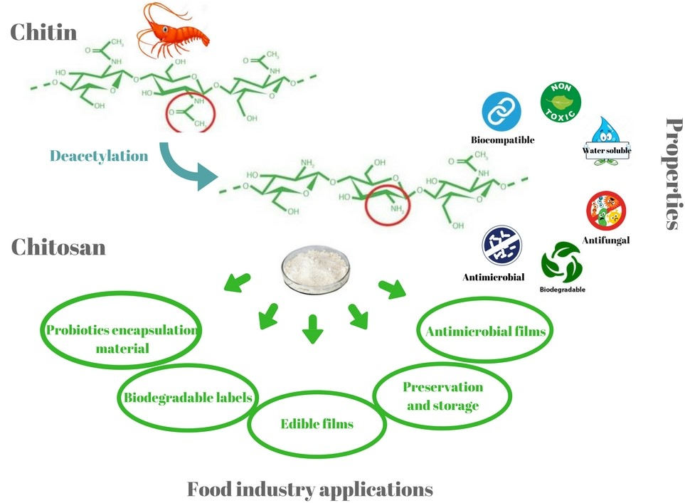
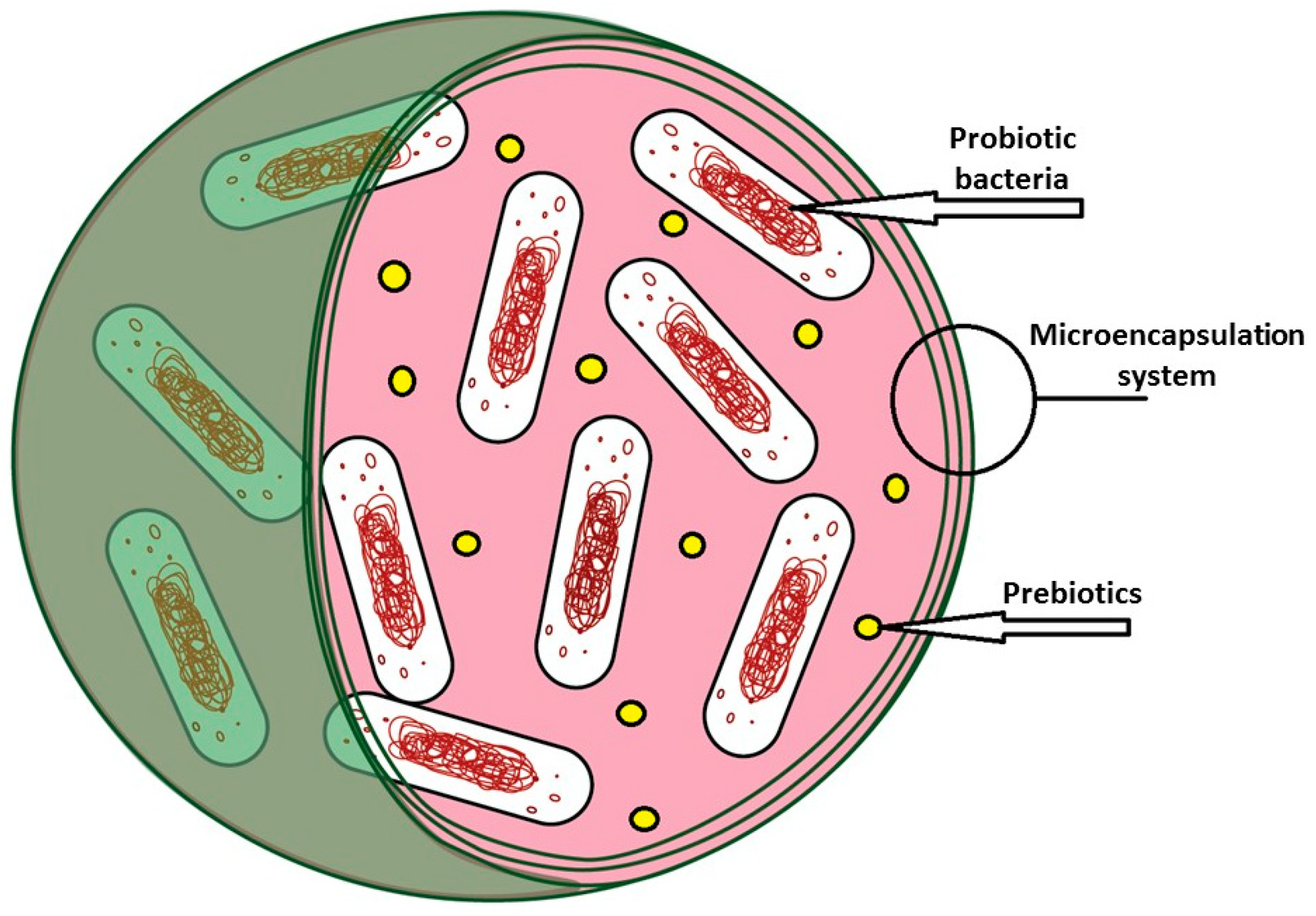
Table 1.
Type of coatings for microencapsulation of probiotic bacteria: pros and cons.
| Coating | Core | Technique | Pros | Cons | Ref. |
|---|---|---|---|---|---|
| Chitosan | Alginate, pectin | Extrusion, layer-by-layer (LbL), Emulsion | unique cationic property and high resistance to acidic environment; excellent film-forming abilities; high biocompatibility with living cells and broad antimicrobial activity; tolerance against the deteriorative effects of calcium chelating and anti-gelling agent; dens and strong beads | increases the excretion of sterols and produces a reduction in the digestibility of ileal fats; reported to have inhibitory effects on lactic acid bacteria (LAB) as core material | [31,37,45,55,56,57] |
| Alginate | Pectin | Extrusion | simplicity, non-toxicity, biocompatibility and low cost | sensitive in acidic environment; low stability in the presence of chelating agents | [37,45,54,56] |
| Resistant starch (corn, potato, cassava etc.) | Alginate | Extrusion, emulsion | inexpensive, abundant, biodegradable and easy to use; transparent, odorless, tasteless and colorless; low permeability to oxygen at low-to-intermediate relative humidity; resistant to pancreatic enzymes (amylases), therefore provides good enteric delivery characteristic; is an ideal surface for the adherence of the probiotic cells to the starch granules and this can enhance probiotic delivery | too high viscosity in solution for most of the encapsulation processes | [58,59,60] |
| Gelatin | Alginate, pectin | Extrusion | able to form complexes with anionic polymers, such as pectin and alginate | very soluble in aqueous systems | [31,37,61] |
| Whey protein | Pectin, alginate | Fluidized bed, extrusion | great gelation properties; biocompatible with probiotics; high nutritional value; improvement in the survival of probiotic after exposure to gastric conditions. | difficult to master, longer duration; do not confer additional protection to probiotics when exposed to simulated intestinal conditions | [62,63,64] |
| Poly-l-lysine (PLL) | Alginate | food-grade status, active properties and charged behavior | high porosity; does not have a strong capacity to be used as a microcapsule coating for probiotics protection against harsh media | [65,66,67] | |
| Glucomannan | Sodium alginate | is abundant in nature; is not hydrolyzed by human digestive enzymes | is a non-ionic polymer, so any coating would have to be the result of a non-ionic interaction, such as hydrogen bonding; there is very little work using glucomannan as a coating material for beads | [68,69] | |
| Shellac | Sodium alginate | Fluid bed | natural origin, therefore acceptable as coating material for food supplement products; good resistance to gastric fluid | the low solubility of shellac in the intestinal fluid, especially in the case of enteric coating of hydrophobic substances | [70,71] |
| Cellulose acetate phthalate (CAP) | Alginate | Emulsion | insoluble in acid media (pH ≤ 5) but it is soluble when the pH is ≥6 as a result of the presence of phthalate groups resulting in an effective way of delivering large numbers of viable bacterial cells to the colon | [44] | |
| k-carrageenan | Milk, alginate | Extrusion, emulsion | low susceptibility to the organic acids, good efficiency in lactic fermented products (such as yogurt); natural products | dissolves only at high temperatures (60–80 °C) for 2%–5% concentration; irregular shapes and poor mechanical characteristics; the produced gels are not able to withstand stresses | [33,37,72] |
Table 2.
Technology-matrix chitosan-coated encapsulation and its applications.
| Microencapsulation Technique | Encapsulation Material | Chitosan-Coating | Probiotic Bacteria | Capsule Size (μm) | Application | Survivability (G: gastric; I: intestinal; colony) | Ref. |
|---|---|---|---|---|---|---|---|
| Extrusion; layer-by-layer (LbL) | Alginate (2%)+ 0.05 M CaCl2 | Chitosan (0.4%) | Bifidobacterium breve NCIMB 8807 | n.a. | In vitro GI exposure | (log colony forming units (CFU)/mL) (G; I) 7.3; 6.8 | [108] |
| Extrusion; LbL | Alginate (2%)+ 0.5 M CaCl2 + galactooligosaccharides and inulin | Chitosan (0.4%) | Lactobacillus acidophilus 5 and Lactobacillus casei 01 | 1830–1850 | In vitro GI exposure | (log CFU/mL) 2.7 and 2.3 > 107 CFU/g−1 | [111] |
| Refrigerated storage for 4 weeks in yogurt and juice | |||||||
| Extrusion; LbL | Alginate (1.8%) + 0.1 M CaCl2 + Hi–maize concentration of up to 1.0% (w/v) | Chitosan Poly-l-lysine (PLL) Alginate | L. acidophilus CSCC 2400 or CSCC 2409 | 500 | In vitro GI exposure | (log CFU, app) Chitosan: 9.1 PLL: 7.3 Alginate: 6 | [65] |
| Extrusion; LbL | Alginate (2%) + 0.5 M CaCl2 | Chitosan (0.7%) | Lactobacillus reuteri DSM 17938 | 110 ± 5 | 8 days storage in different solutions at 4 and 20 °C | log CFU/mL) (G; I) 9.15; 9.3 | [112] |
| In vitro GI exposure | |||||||
| Osmotic stress conditions |
© 2019 by the authors. Licensee MDPI, Basel, Switzerland. This article is an open access article distributed under the terms and conditions of the Creative Commons Attribution (CC BY) license (http://creativecommons.org/licenses/by/4.0/).
Share and Cite
MDPI and ACS Style
Călinoiu, L.-F.; Ştefănescu, B.E.; Pop, I.D.; Muntean, L.; Vodnar, D.C. Chitosan Coating Applications in Probiotic Microencapsulation. Coatings 2019, 9, 194. https://doi.org/10.3390/coatings9030194
AMA Style
Călinoiu L-F, Ştefănescu BE, Pop ID, Muntean L, Vodnar DC. Chitosan Coating Applications in Probiotic Microencapsulation. Coatings. 2019; 9(3):194. https://doi.org/10.3390/coatings9030194
Chicago/Turabian StyleCălinoiu, Lavinia-Florina, Bianca Eugenia Ştefănescu, Ioana Delia Pop, Leon Muntean, and Dan Cristian Vodnar. 2019. "Chitosan Coating Applications in Probiotic Microencapsulation" Coatings 9, no. 3: 194. https://doi.org/10.3390/coatings9030194
Note that from the first issue of 2016, this journal uses article numbers instead of page numbers. See further details here.