Sharply Reduced Biofilm Formation from Cobetia marina and in Black Sea Water on Modified Siloxane Coatings

1
Department of Polymer Engineering, University of Chemical Technology and Metallurgy, 1756 Sofia, Bulgaria
2
Department of Adhesive Bonding Technology and Surfaces, Fraunhofer Institute for Manufacturing Technology and Advanced Materials (IFAM), 28359 Bremen, Germany
3
School of Pharmaceutical Science, University of São Paulo, São Paulo 05508-000, Brazil
*
Author to whom correspondence should be addressed.
Coatings 2018, 8(4), 136; https://doi.org/10.3390/coatings8040136
Submission received: 6 February 2018
/
Revised: 2 April 2018
/
Accepted: 5 April 2018
/
Published: 11 April 2018
(This article belongs to the Special Issue Novel Marine Antifouling Coatings)
Abstract
:Siloxane fouling release coatings are currently the only viable non-toxic commercial alternative to toxic biocide antifouling paints. However, they only partially inhibit biofouling since biofilms remain a major issue. With the aim to improve the bacterial resistance of siloxane coatings modified with non-ionic surfactant (NIS), antioxidant (AO) or both NIS/AO, the ability of PEG-silane co-cross-linker was investigated to reduce Cobetia marina adhesion and multispecies biofilm formation from natural seawater. Surface physical-chemical and physical-mechanical parameters relevant to bio-adhesion were estimated before the testing of the biofilm formation. Slightly reduced biofilm from C. marina and sharply reduced multispecies biofilm, formed in natural sea water, were found on the PEG-silane co-cross-linked coatings without modifying additives. However, both C. marina growth and biofilm formation from natural sea water were sharply reduced on the PEG-silane co-cross-linked coatings containing NIS or AO, even more, no C. marina adhesion was seen on the coating containing NIS and AO simultaneously. Possible explanations of the observed effects are presented in this article. It was concluded that the PEG-silane co-cross-linker, toghether with NIS and AO, can be used as an efficient tool to additionally reduce the bioadhesion of Gram-negative marine bacteria and multispecies biofilm formation on siloxane antifouling coatings.
1. Introduction
Surface modification by deposition of relevant coatings is one the most often studied approaches in the creation of materials that reduce biofilm formation. Siloxane fouling release coatings are currently the only viable commercial non-toxic alternative to the effective yet toxic biocide-containing antifouling paints, which have been banned due to their toxicity since 2008. Milne [1] was among the first researchers who pointed out the antifouling properties of siloxane (silicone) polymers and observed that the low molecular silicone oils greatly enhance their fouling release properties [2]. These early observations constitute the bases of most siloxane fouling release coatings that facilitate only weak adhesion of macro-fouling organisms and ensure the self-cleaning of high speed moving (about 15 knot and higher) ships by easy detachment (release). Siloxane composition coatings that totally prevent the macro-fouling of any submerged surfaces, including those that are statically immersed, were developed by us [3]. However, the current siloxane fouling release coatings only partially inhibit biofouling since biofilms with their own negative impact remain a major issue. Variety approaches to the reduction of marine biofilm formation are currently known, including physical, physical-chemical, and enzymatic ones, mostly biomimetic and/or based on use of natural derivatives, such as natural biocides, surfactants, and quorum sensing inhibitors [4,5]. Unfortunately, no report could be found in the special literature about a surface that is able to completely stop the development of marine biofilms, even if it contains biocide. We proved the anti-biofilm activity of non-toxic antioxidant and first reported it in 2013 [6]. Now, antioxidant coatings are already discussed as a new environmentally friendly alternative to biocide paints [7,8]. Non-ionic surfactants, non-toxic antioxidants, or combinations thereof in modified siloxane composition coatings were developed by us that significantly reduce marine biofilm formation in both laboratory and field conditions [6,9], whereby the effect is most pronounced using a combination of surfactant and antioxidant. The multi-species biofilm developing on such coatings is sparse and easily removed by washing with running water or gently wiping [9]. On the other hand, PEG-silanes were also included in siloxane coatings to create protein-resistant surfaces for medical and microbiological applications [10,11,12,13,14,15]. Incorporation of PEG-silane in the vulcanization network of siloxane elastomers has been used to create a micro-heterogeneous structure with hydrophilic and hydrophobic zones, known to be preferable to achieve decreased bio-fouling [16,17,18,19]. Our idea was to additionally improve the anti-bio-fouling activity formerly developed by us in siloxane composition coatings [3,6,9] by employing a PEG-silane co-cross-linker in an optimal combination with a non-ionic surfactant and non-toxic antioxidant. Hence, the aim of this investigation was to prepare such coatings, analyze their characterization and evaluate the mono-species biofilms’ formation of C. marina, a Gram-negative bacterium extensively used as a model in marine bio-fouling research, as well as multi-species biofilms formation in natural sea water.
2. Materials and Methods
2.1. Coating Compositions
The coating compositions used in this investigation were based on a combination of room temperature vulcanizing (RTV) siloxane elastomers (Gelest, Morrisville, PA, USA), a crosslinking agent (ES40, PSI-021, Gelest, Morrisville, PA, USA), the catalyst dibutylethin dilaurate (SND 3260, Gelest, Morrisville, PA, USA), and the modifying agent non-ionic surfactant (NIS) alkoxylated trisiloxane (Super spreader Y17112, Momentive Comp, Albany, NY, USA) or antioxidant (AO) α-tocopherol (E307, Panteley Toshev Ltd., Sofia, Bulgaria), or their combination, at a weight ratio of 1:3. Chemically bonding PEG-silane (methoxy(polyethyleneoxy)6-9-propyltrimethoxysilane (Gelest Co., Ltd., Morrisville, PA, USA) was used in this investigation as a co-cross-linker to create some micro heterogeneity. All coating compositions were prepared as described in [3].
2.2. Coated Test-Samples
Glass plates (10 × 10 × 2 mm3 or 100 × 150 × 2 mm3) were spin-coated (at 400 min−1) with a primer consisting of ethyltriacetoxysilane (50 wt % toluene solution) and a catalyst (3 wt % dibutyltin dilaurate) to provide good adhesion of the coating to the glass surface. The primed dry glass plates were then covered with a corresponding composition by spin-coating under the same conditions. The prepared test-samples were kept under ambient room conditions for 30 days prior to testing. The thickness of the dry coating was 220–240 µm and measured by a stereomicroscope Leica MZ16 FA (Leica, Wetzlar, Germany).
2.3. Water Contact Angle (WCA) and Surface Energy (γ)
The contact angle measuring instrument Easy Drop (Kruss, Hamburg, Germany) was employed for static contact angle measurements (angle resolution ±0.10) using three liquids with known surface tension: water, ethylene glycol, and n-hexadecane. The surface energy (γc) was calculated according to Fowkes’ method [20].
2.4. Atomic Force Microscopy (AFM)
An Easyscan 2 apparatus equipped with a Pointprobe Contr-10 silicone SPM sensor (Nanosurf, Liestal, Switzerland) (dimensions of 2 × 450 × 50 µm3), was employed to obtain plane and 3D images of the investigated dry surfaces operating in contact mode. A Diamond Viker’s pyramid (Nanosurf, Liestal, Switzerland) with a pike angle of 1360 was used for all measurements at room temperature, with a loading speed of 0.250 mN/s.
2.5. Depth Sensing Indentation (DSI)
A dynamic Ultra Micro-Hardness Meter DUH-211 S (Shimatzu, Kyoto, Japan) was employed for the evaluation of the indentation hardness, HIT, Vicker’s hardness (VIH), and indentation elastic modulus (EIT) under the following conditions: test force 0.30 mN; loading speed 6.0 (0.0250)mN/s; depth of penetration 20 nm.
2.6. Elastic Modulus (E)
The elastic modulus (E) was estimated according to ISO R37 using samples with a thickness of 2 mm, vulcanized under ambient room conditions for at least 30 days.
2.7. Characteristics of Single-Species Biofilms Formed by C. marina
Bacterial Adhesion Test
For this test, 10 × 10 × 2 mm3 coated glass samples were used. C. marina (Gram-negative, aerobic, rod-shaped marine bacterium; size of about ±2 µm; grows in the temperature range of 10 to 42 °C) was the test bacterium for this study. The test procedure included the sterilization of all the samples with isopropanol 70% and 30 min under ultraviolet light, 1 h of incubation with 4.0 × 10⁷ cfu/mL Cobetia marina in 5 a mL suspension on an orbital shaker (50 rpm), then washed in artificial seawater (ASW; Tropic Marine®, pH 7, 33.3 g/L of ultrapure water, Dr. Biener GmbH, Wartenberg, Germany). The culture of Cobetia marina was diluted in a minimal medium (1:100 marine broth to ASW) in order to obtain an optical density (O.D.) of 0.1 at a wavelength of 600 nm, which corresponds to 4.0 × 10⁷ cfu/mL. Bare sterile glass and coated sterile glass samples exposed to a suspension of C. marina were the negative control, while bare sterile glass and the corresponding coated sterile samples exposed to a suspension without C. marina were the positive control. After exposure time, the bacterial suspensions were removed and all the samples were quickly immersed in amorphous salted water (ASW) to remove the excess of non-adhered cells.
2.8. Bacterial Biofilm Visualization (Fluorescence Microscopy–Live/Dead Staining)
The green fluorescent dye SYTO 9 (Molecular Probes, Eugene, OR, USA) was used to determine the viability of the bacteria after 60 min of incubation. The green-fluorescent nucleic acid stain SYTO9 (ex. max.:480 nm; em. max.: 500 nm) stains all bacteria. The test plates were fixed with 5 mL of glutaraldehyde (2.5% in ASW) for 20 min at room temperature; afterwards they were washed once again with ASW for 1 min on the plate shaker. After 24 h drying at room temperature, the plates were stained with 0.6 µL/mL SYTO 9 in NaCl (0.85%) and covered with a glass coverslip. The observation of the adhered bacteria to the test surfaces was made with a fluorescent microscope (Zeiss Axio Imager M.1, Carl Zeiss, Jena, Germany) using an objective with 40× magnification. SYTO 9 bonds to nucleic acids and passes through the membranes of both intact and dead cells, and therefore the fluorescence of all cells, living and dead, is observed in a green color.
2.9. Quantitative Estimation the Bacterial Biofilm
Adenosine triphosphate (ATP) is a universal energy carrier in biological systems, so the measurement of cellular ATP levels is invaluable for assessing metabolic state in living cells. An ATP bioluminescence test system was used to measure the concentration of ATP as relative light units (RLU). In an enzyme based reaction with luciferase, the ATP reacts with oxygen and the light-emitting pigment luciferin resulting in the emission of green-yellow light. The amount of ATP is directly proportional to the amount of light emitted and can be determined. To measure the amount of ATP, which is correlated with the number of bacteria, the Clean-Trace™ NGi Luminometer was used together with Clean-Trace™ ATP Surface Test UXC sticks (3M Deutschland GmbH, Neuss, Germany). The experiments have been performed according to the user manual of the supplier. The measured readout is given in RLU-relative light units. All experiments have been carried out in triplicates.
2.10. Multi-Species Biofilms
Multi-Species Biofilm Formation in a Black Sea Aquarium (BSA)
A BSA aquarium was used to preliminarily observe multi-species biofilm formation on a control coating cross-linked with a conventional cross-linker ES 40 as well as on coatings containing PEG-silane. Two vulcanized samples of each coating composition with a thickness of 3 mm were used for the evaluation of micro-fouling in natural Black Sea water (salinity of 1.8%, and pH of 8.0), collected from Yacht harbor, Nessebar, Bulgaria, at a temperature of 24–25 °C, and a luminance of 1.5 × 10³ lx (cool white LED panel). The cycle of illumination was 12 h light and 12 h dark. The biofilm formation was observed by eye.
2.11. Biofilm Formation in a Field Experiment and Its Characterization
One year of exposure was carried out in the Black sea, Yacht harbor, Nessebar, Bulgaria for the period January 2017–December 2017. Five samples of each coating (100 × 150 × 2 mm3) were fixed on a common plane plate and vertically immersed at a depth of 2 m. The biofilm of all five samples was carefully removed by means of a plastic spatula, followed by rinsing the samples in distilled water as long as no biofilm was seen an under magnifying glass (10×) and placed in 15 mL Falkon type test-tube. 5 mL of distilled water was added to each test-tube. Homogenization of the biofilm mass was performed for 5 min, directly in the test-tube by dipping an ultrasound rood (Bandelin electronic UW 3100) and cooling in ice bath.
2.12. Total Chlorophyll (A and B) and Carotenoids (Carotenes and Xanthophylls)
The total chlorophyll and carotenoids were spectrometrically estimated from ethanol extract, employing the Pharmacia Biotech Ultraspec 3000, (Pharmacia Biotech, Jena, Germany) apparatus. The pigments’ extraction from the mass of the biofilm was performed as follows: 1 mL from the homogenized biofilm suspension was carefully, drop-way placed onto glass filter (pore size of 1.6 µm). The reach of the pigments cell elements were held on the filter and each filter was placed in a 2 mL Eppendorf type centrifuge test-tube. 1.5 mL 95% ethanol was added and the containers were dark-kept for 4 h under mixing on IKA KS250 shaker (IKA-Werke GmbH, Staufen im Br., Germany). The complete extraction was followed by 5 min centrifuging at 28,000× g for clearing of the extract. 1 mL of each clear extract was used to calculate the content of carotenoids-from spectrometering at 470 nm; chlorophyll B at 649 nm; chlorophyll A at 664 nm; and for the turbidity estimation and the other absorbance’s correction at 730 nm with an UV-Vis spectrophotometer (Specord 200, Analytik Jena, Jena, Germany).
2.13. Biofilm Dry Mass
The biofilm dry mass was estimated gravimetrically as follows: 3 mL of each suspension was placed in a preliminary weight (±0.0001 g) Falkon test-tube (dried for 24 h at 1050 °C and conditioned for 4 h at normal conditions) and centrifuged at 5200× g for 5 min. The liquid phase was separated and 5 mL isopropanol (99.7%) was added to remove the residual siloxane oil from the sediment. After centrifuging and the separation of the isopropanol, the test-tube with the sediment was dried for 5 h at 90 °C and conditioned for 4 h at normal conditions and weight (±0.0001 g). The biofilm dry mass was estimated as the difference between this weight and the initial weight of the empty test-tube.
3. Results
3.1. Characteristics of the Test Surfaces
It is known that microbial colonization on solid surfaces can be affected by diverse surface parameters, such as hydrophilic/hydrophobic balance, surface tension, topography, and roughness [18,19] as well as surface mechanical factors [20]. Expecting that they can be influenced by the used modifying agents (PEG-silane, NIS and AO) and surface characterization of each test surface was carried out before testing the bacterial adhesion and biofilm formation. The results are presented in Table 1 and in Figure 1. A comparison of coatings 2 and 3, containing 2 wt % PEG-silane co-cross-linker to the conventionally cross-linked, control coating 1 (Table 1) demonstrates that the PEG-silane slightly alters the hydrophobicity. Insignificant differences are observed only between the WCA and surface tension, γc of samples 1, 2, and 3. This is probably a result of a too short time of measurement, which does not allow an orientation of the PEG-chains to the surface. Thus, they stay hidden in the polysiloxane matrix [17,18,19].
A comparison of the surface roughness average arithmetical deviation from the base line, Ra; average quadratic deviation from the base line, Rq; the highest point, Sp; the dippiest point, Sv and the distance between the highest and the dippiest point, Sy (Table 1, compare coatings 2 and 3 to coating 1) as well as the pictures in Figure 1 demonstrate that the presence of the PEG-silane causes some inhomogeneity, as it was expected to do. The surface physical-mechanical parameters, HMV, HIT, and EIT show a 2.5 to 4.5 fold increase for the coatings containing PEG-silane (Table 1, compare coating 2 and 3 to coating 1). The relationship is the opposite for the bulk elastic modulus E (Table 1), which decreases slightly for the samples containing PEG-silane. The last one indicates the formation of a different vulcanization network on the surface and in the bulk of the coatings.
A comparison of the surface characteristics of coatings 4, 5, and 6 to those of coating 2 containing the same amount PEG-silane (Table 1, compare coatings 4, 5, and 6 to coating 2) demonstrates the following:
As was expected, the presence of NIS, NO, or NIS/NO sharply decreases the WCA, respectively the hydrophobicity. This is most strongly expressed at sample 4, containing NIS, followed by sample 6, containing NIS/AO. Coatings 4 and 6 are already moderately hydrophilic, whereas sample 5 stays on the hydrophilic/hydrophobic border. The moderate hydrophylicity of coatings 4 and 6 can be due to an easier PEG-chains orientation in the presence of NIS, as indicated by the wetting kinetic curves (not presented here), which demonstrate a significant drop during the contact angle measurement for 300 s.
Deviations are observed in the surface nano-roughness, Ra, Rq, Sp, Sv, and Sy if one compares the data for coatings 4, 5, and 6 to those for coating 2 (Table 1 and Figure 1), indicating an influence of the modifying agents NIS, NO, or NIS/NO on these parameters. All of the modified coatings (Table 1, coatings 2–6) demonstrate higher nano-roughness compared to that of control coating 1. However, it is far below the C. marina size of 2 µm and the ‘valleys’ present do not allow the microbe species to reside in a protected area [21,22].
The surface physical-mechanical parameters HMV, HIT, and EIT of coatings 4, 5, and 6 (Table 1) are slightly lower compared to those of the sample 2 that contain the same amount of PEG-silane without NIS and NO (Table 1) and varies slightly according to the type of the modifying agent. This indicates that the formed vulcanization network is affected by the corresponding modifying agent (NIS, NO, or the NIS/NO combination). The observed differences in the surface characteristics of the studied coatings could be a reason for a different resistance to bacterial growth. Decreased EIT could be a reason for the decreased bioadhesion [23].
3.2. Bacterial Biofilms
Two types of bacterial biofilms were studied: (i) single-species biofilms formed from the culture of the Gram-negative marine bacterium C. marina, and multi-species biofilms formed by a mixed bacterial assemblage from the natural Black sea water.
3.2.1. Bacterial Adhesion of C. marina
Fluorescent microscopy with SYTO 9 staining was used to visualize the bacterial biofilms formed after 1 h incubation of C. marina. Representative views of these biofilms showing a monolayer of cells with some three dimensional aggregates are shown in Figure 2 (on glass; 1 on control coating without PEG-silane; 2 and 3 on coatings, containing 2 wt % PEG-silane) and Figure 3 (coatings containing 2 wt % PEG-silane and 4–NIS; 5–NAO and 6–NIS/NAO).
Oil-like blemishes are observed on the pictures of coatings 5 and 6 (Figure 3), both containing NAO in the corresponding siloxane composition. Most probably, they are formed by NAO that has migrated to the surface and does not succeed in completely wetting the surface because of the relatively low surface energy of the cross-linked siloxane coating. Fine fibril elements (the longest of about 2–3 µm) are observed in all PEG-silane containing coatings (Figure 2, samples 2, and 3; Figure 3, samples 4, 5, and 6). Due to the lack of green fluorescence they cannot be bacteria and it is most likely that they represent PEG-silane, as a separated phase in the siloxane matrix. As it is evident from Figure 2 and Figure 3, only the PEG-silane co-cross-linked coating 6, containing simultaneously NIS and NAO, demonstrates any visible growth of C. marina.
3.2.2. Adenosine Triphosphate Bioluminescence
The bacterial biofilm on the tested coatings, formed after 1 h incubation in 4 × 10⁷ cfu/mL C. marina strain, was quantitatively estimated by ATP bioluminescence , since the intensity of the emitted light is directly proportional in the enzyme linked assay to the ATP quantity and, respectively, to the amount of bacterial cells. In Figure 4, the relative amounts of fluorescent light are presented, which are produced by the bacterial films for 5 s, 30 s, and 60 s after the addition of the reagents. It is observed that the glass surface (Figure 4, glass with bacteria) is in the middle regarding C. marina growth, whereas on the control siloxane coating, conventionally cross-linked with ES40 (Figure 4, coating 1), the bacterial growth is at the maximum and about twice as high. Coatings 2 and 3 are also hydrophobic, with WCA similar to that of sample 1 (Table 1). Generally, C. marina growth is relatively high on these three hydrophobic surfaces, which is in agreement with some reports on favorable bacterial adhesion on hydrophobic surfaces [24,25].
Coatings 2 and 3, both containing 2 wt % PEG-silane but with different amounts of the conventional cross-linker ES40, have similar amounts of bacterial biofilm, albeit slightly decreased in comparison to that on the control without the PEG-silane coating 1 (Figure 4, compare coatings 2 and 3 with coating 1). This indicates that some of the PEG chains succeed in orienting in the direction of the water face. Hence they influence bioadhesion during the long period of incubation (1 h). Whereas such orientation of the PEG chains is missing for the short time of the WCA measurement and WCA was almost the same for coatings 1, 2, and 3, despite the presence of PEG in coatings 2 and 3 (Table 1, WCA of coatings 1, 2 and 3). The addition of 0.5 wt % NIS turns the PEG-silane co-cross-linked with strong hydrophobic coating 2 (Table 1, WCA = 103.0°; γc =22.8 mN/m; EIT = 8.13 N/mm2) into a moderate hydrophilic one with a decreased EIT (coating 4 in Table 1, WCA = 51.5°; γc = 30.5 mN/m; EIT = 4.99 N/mm2). This leads to some increase in the C. marina biofilm growth on coating 4 as compared to coating 2, which has the same composition but without NIS (Figure 4, coatings 4 and 2). The increased biofilm growth is in compliance with the fact that the surface tension of 30.5 mN/m is outside of the Bayer window and the fact that an area of moderate hydrophilicity is also favorable for cell attachments, although the reason is still not fully understood [26]. It has long been theoretically predicted and experimentally proven that the occurrence of adhesion in water media approaches zero when WCA or surface tension approaches zero, i.e. when the surface is strong hydrophilic, water-like, or strong-/super hydrophobic [27].
The presence of the antioxidant in PEG-silane co-cross-linked coating 5 significantly reduces the amount of the adhered bacteria compared to that on coating 2 with the same composition but without AO (Figure 4, coatings 5 and 2). The AO decreases the WCA down to the border of hydrophobicity/hydrophilicity (WCA = 90.6°, Table 1) and increases the surface tension up to the border of the Bayer window (γc = 24.4 mN/m, Table 1). These alterations are negative for low biofouling and indicate that maybe the used AO intervenes somehow in the cross-linking of secreted C. marina EPSs. There are few investigations on the effects of α-tocopherol on bacterial adhesion. However, this result correlates with some observations of orthopedic implants made by ultra high molecular weight polyethylene (UHMPE) containing tocopherol as an antioxidant preserving the polymer from destruction during processing and exploitation. Less post-operative infections and reduced biofilm formation on their surface were observed [28,29,30,31]. It was demonstrated that the inclusion of tocopherol in PE significantly decreased the adhesion ability of St. epidermidis, St. aureus, and E. coli. A similar anti-biofilm action of tocopherol against St. epidermidis and St. aureus has also been observed when it is included in polylactic acid [32]. Although there is a generally accepted lack of antibacterial activity of tocopherol, some studies have demonstrated such activity against St. epidermidis, St. aureus, E. coli, and Ps. aeroginosa [33]. So far, there has been no convincing explanation for the anti-bioadhesion properties of tocopherol.
The used NIS/AO combination as a modifying agent in a PEG-silane co-cross-linked siloxane coating 6 demonstrates a synergetic effect. Many folds reduced C. marina growth, as is evident from Figure 5 (compare coating 6 to coating 2). Coating 6 creates a moderate hydrophilic surface (WCA = 60°) with a surface tension (γc = 32.1 mN/m) outside of the Bayer window (Table 1) that is contra visional of the biofouling resistant surfaces. The explanation of this result is difficult and only some suppositions can be made.
3.2.3. Sea Biofouling and Multi-Species Biofilms Formation
Prior the start of a field experiment, biofilm formation was observed on PEG-silane co-cross-linked samples in a Black sea aquarium. Since the used PEG-silane co-cross-linker was three-functional, a trial was conducted to partially (2 wt %) or totally (6 wt %) replace the conventionally used four-functional ethyl-silicate ES40 using an equal amount of 0.5 wt % of the catalyst dibutylthin dilaurate. The three types of vulcanized samples prepared in this way were exposed to biofouling in a Black Sea aquarium for up to 6 months, which aimed to understand if the biofilm formation would be different in the presence of PEG-silane. Figure 5 presents the pictures of the biofouling. Important differences are observed in the micro-fouler growth on the tested samples. The single sample (2), almost without growth is cross-linked with 4 wt % ES40 and 2 wt % PEG-silane, whereas the two others (1 and 1’) are almost totally covered by algae.
It is well known that PEG-chains in water media are strongly solvated, which makes non-specific protein adsorption difficult [5]. Probably for this reason, the presence of PEG-chains covalently bonded to the poly(siloxane) matrix creates the strong antigrowth effect of coating 2. The samples without a conventional cross-linker, coating 1’, which contains three times more PEG-silane (6 wt %), do not demonstrate anti-growth properties as good as those of sample 2. The low degree of cross-linking of coating 1’ is the probable reason. At a low degree of cross-linking, (i) a large amount of free silanol groups (–Si–OH) exist, which are a prerequisite for strong molecular interactions with extracellular polymers of the microorganisms, and (ii) a glue-like surface creation to which biofouling organisms attach easily. Weakly cross-linked siloxanes are even used in the preparation of contact medical glues [34]. The results of the preliminary testing in the Black Sea aquarium give reason to continue the following experiments with composition coatings containing the conventional cross-linker ES40, albeit in reduced amounts.
We aim to compare the results of the laboratory experiments, which solely investigated a single species (C. marina) under controlled conditions, with field experiments that employed a mixed species marine environment under natural conditions. The field experiments were carried out in costal Black sea water, Yacht harbor, Nessebar. To evaluate the net effect of the PEG-silane co-cross-linker, conventionally cross-linked and PEG-silane co-cross-linked coatings with one the same composition were simultaneously exposed. No mactrofoulers, only biofilm formation was observed on all studied coatings after one year of exposure. Several parameters, such as total chlorophyll, carotenoids content, and biofilm dry mass were used to evaluate the formed multi-species biofilms. The results are presented in Figure 6.
Figure 6 demonstrates reduced multispecies biofilms formation on conventionally cross-linked and PEG-silane co-cross-linked siloxane coatings, the effect depending on the type of the modifying agent (NIS, AO or NIS/AO) and always sharply expressed at the PEG-silane co-cross-linked coating (compare dark blue to corresponding light blue columns). These results are in agreement with those found earlier by us about a multispecies biofilm formation on conventionally cross-linked transparent siloxane coatings containing the same modifying agents [6]. It is interesting to note that the tree parameters, total chlorophyll, carotenoids content, and biofilm dry mass, were several folds lower for all PEG-silane co-cross-linked coatings compared to the conventionally cross-linked ones, demonstrating in this way the strong reducing effects of PEG-silane onto the multispecies biofilm formation in natural sea water. PEG-silane co-cross-linked coating, containing NIS/AO, sharply reduces most marine biofilm formation, as it was in the case of the ES40 cross-linked one, which contained the same modifying agent. On the contrary to the relative good mono-culture bacterial adhesion on PEG-silane co-cross-linked coatings, excluding coating 6 containing NIS/AO, a scarpe multi-species biofilm only was observed on all studied coatings after one year of immersion in seawater. This could be due to the different mechanisms of mono- and multi-species biofilm formation. In the second case, it is most probably due to a concurrent microbial attachment as a result of a concurrent microbial EPS adsorption, a phenomenon similar to the concurrent blood plasma proteins adsorption, well-known as the Vroman effect [35].
4. Discussion
In this work, surface physical-chemical and physical-mechanical properties relevant for bio-adhesion were investigated of PEG-silane co-cross-linked modified siloxane composition coatings containing NIS, AO of both NIS and AO as modifying agents (Table 1, Figure 1). Improved anti-biofilm properties of all PEG-silane co-cross-linked coatings as compared to the corresponding ones, cross-linked conventionally with ES40 (modified with NIS, AO or NIS/AO as well as without modifying agent), were observed for multispecies biofilm formation from natural sea water (Figure 6). The mono-species biofilms formed from bacteria C. marina were insignificantly reduced whereas (Figure 4) the multi-species biofilms formed from natural Black sea water (Figure 6) were significantly reduced on the PEG-silane co-cross-linked coatings in the absence of modifying agents. The best reduction ability for both C. marina and multi-species biofilms from natural sea water demonstrated PEG-silane co-cross-linked coating, modified with NIS/AO (Figure 4 and Figure 6). Our results show in general that not always does a correlation exist between bioadhesion and the surface characteristics, including the knowledge that the adhesion will correlate better with (γc.E)1/2 than with either surface tension, γc or elastic modulus, E on their own [23] because of altered surface chemistry in the presence of a co-cross-linker (PEG-silane) and modifying agents (NIS, AO and NIS/AO). Discussing the interaction of solid surface and bacteria, as well as bacterial biofilm formation, one should have for, in addition to the surface characteristics, the complex mode of biofilm development, which involves a transport of organic and inorganic molecules and microbial cells to the surface, a subsequent adsorption to the surface, and finally, irreversible attachment aided by the production of EPSs [36]. The adhesion of microbial cells is mediated by EPSs, and the composition and quantity varies depending on the type of microorganisms, the age of the biofilm, and the environmental conditions [37]. The chemical constituents of the EPS, as well as the mode of attachment, vary depending also on the type of substrate and its surface characteristics [38,39,40,41].
4.1. Single Species Biofilms of C. marina
C. marina that was used as a model bacterium in this investigation is a hydrophilic species Gram-negative bacterium that produces large quantities of EPSs, composed mainly of uronic acids. Some polysaccharides of such bacteria are neutral or polyanionic [18,19,42]. As for other bacterial cells, the adhesion to hydrophilic and hydrophobic surfaces will be affected by the different composition of the EPSs produced by different C. marina strains on substrates with different characteristics [43]. It is known, for example, that C. marina strain KMM MC-296 produces an alkaline phosphatase with a very high specific activity (15,000 DE U/1 mg of protein) [43], the Cobetia sp. strain MM1IDA2H-1 produces a bio-surfactant that interferes with the quorum sensing of fish pathogens by signal hijacking [44], and the C. marina DSMZ 4741 strain synthesizes an unexpected K-antigen-like exopolysaccharide [45]. Therefore, the explanation of the mechanism of C. marina attachment and biofilm formation on modified siloxane coatings with different composition and surface characteristics is difficult. It seems that the presence of PEG chains from the PEG-silane co-cross-linker, which create some inhomogeneity and slight nano-roughness (Table 1, coatings 2 and 3), slightly influence the C. marina EPS secretion and biofilm formation, respectively (Figure 5, coatings 2 and 3). The presence of NIS eases the orientation of the PEG chains to the surface and thus turns the surface moderately hydrophilic (WCA = 51.5°, Table 1), slightly increasing biofilm formation (Figure 5, coating 4). It is known that C. marina is able to secret different EPSs that increase the adherence to hydrophilic or to hydrophobic surfaces [42]. In this case perhaps the secreted EPSs are from the first type. The presence of AO in the PEG-silane co-cross-linked coating 5 (Figure 5) reduces significantly the C. marina biofilm. It can be accepted as an indication for secretion by C. marina of EPS (with unknown exactly chemistry) that is able to oxidative cross-linking in which AO intervenes somehow. There could be some oxidative cross-linking undergoing adhesive protein, presenting a very small amount to be detected (so far there are no reports about such proteins in EPSs secreted by C. marina) in large volume exopolysaccarides, but in a very active conformation [46]. Reduced biofilm formation on siloxane coatings containing the same AO was earlier observed and firstly reported by us in 2013. Then, this effect was ascribed to some restoration (by the same antioxidant) of oxidative cross-linking of the secreted EPS [6]. The inhibition action of antioxidants is outside of doubt now and antioxidant coatings are already under intense study as a future non-toxic alternative to biocide paints [7]. The observed, sharply reduced single-species biofilm on coating 6 (Figure 5) due to a synergetic effect of NIS/AO is very interesting. Similar effects on multi-species biofilm formations were already observed at NIS/AO modified siloxane coatings, which were conventionally cross-linked with ES40 [9]. NIS usually makes difficult the initial microbial attachment, whereas AO intervenes in oxidative cross-linking processes. Both together contribute to scare biofilm formation on the NIS/AO modified coatings. In the case of C. marina, the action of NIS was “suppressed” by the PEG-chains in the absence of AO (coating 4). In addition, the effect of AO was less pronounced in the absence of NIS (coating 5). This gives reason to accept that the NIS assists somehow the actions of the used antioxidant, most likely supporting its migration to the surface and better spreading on the surface, where contact with the C. marina EPSs happens.
4.2. Multi-Species Biofilms
Significantly reduced multi-species biofilms were observed on all the coatings co-cross-linked with PEG-silane as compared to those conventionally cross-linked with ES40 (Figure 6, the light and dark blue columns). This result demonstrates the high efficiency of PEG-chains, included in the vulcanization network of modified siloxane coatings against multi-species biofilms formation in natural sea water. The multi-species biofilm on the modified coating containing NIS/AO, both conventionally cross-linked and PEG-silane co-cross-linked (Figure 6, coatings 7 and 8, respectively) is scarce (insignificant on the second one) and easily cleaned by running water or gently wiping. Multi-species biofilm formation in natural sea water is mediated by EPSs secreted form a mix of microbial cells. The composition of such EPSs is very complicated and varies depending on environment conditions and the substrate characteristics. Little data could be found about EPSs secreted by mixed microbe strains. The mechanisms of multi-species biofilms formation are more complicated and difficult for studying. We can only make some suggestions and identify some surface characteristics of impact. It seems that the observed alterations in the surface characteristics (Figure 1) created by the PEG-silane co-cross-linker favors the reduced multi-species biofilm formation on the studied siloxane coatings. The modifying agents, NIS, AO, and NIS/AO also contribute in this direction, an effect most strongly expressed at NIS/AO (Table 1, Figure 1). It is likely that the NIS makes the initial bacterial attachment difficult and enables an easy dispersion of the biofilm, whereas AO intervenes with some oxidative cross-linking processes of the very complex EPSs secreted by the mixed microbe cells in the sea water. The discovery of the true reason requires very complicated and profound microbiological and biochemical investigations. All of the experimental data obtained so far demonstrate that, as a co-cross-linker in siloxane antifouling coatings, PEG-silane slightly reduces single species C. marina biofilm formation (Figure 4, compare coatings 2 and 3 to coating 1). In opposite, the effect is well expressed at multi-species biofilms from natural sea water (Figure 6). PEG-silane co-cross-linker affect very significantly both single-species and multi-species biofilms formation on modified siloxane coatings containing NIS, AO, or NI/AO. The different effects of the PEG-silane co-cross-linker on single-species and multispecies biofilm formation are attributed to the different composition of the mediating bioadhesion EPSs secreted by C. marina and a mix of microbial species in the Black sea water.
5. Conclusions
PEG-silane co-cross-linker and modifying agents, non-ionic surfactant (NIS), antioxidant (AO), or a combination of both (NIS/AO), affect the vulcanization network of the corresponding coatings and hence surface characteristics influencing bioadhesion.
PEG-silane co-cross-linker reduces sharply both single-species (C. marian) and multi-species biofilms formation on modified siloxane coatings. No bacterial adhesion of C. marina was seen on the coating containing NIS/AO and multi-species biofilm formation in natural sea water was reduced 15–16-fold down.
The effects of PEG-silane co-cross-linker in the modified siloxane coating are explained in light of the altered surface characteristic and composition of EPSs secreted on their surfaces. Intervention in oxidative cross-linking is supposed for the antioxidant.
Using whiteout to obtain profound knowledge about the mechanism of this action, this study has proved that PEG-silane co-cross-linker in combination with relevantly selected surfactant and antioxidant can be used for an additional sharp reduction of mono-species Gram-negative bacterial and multi-species marine biofilms formation on modified siloxane composition coatings.
Acknowledgments
This work was performed in the frame of COST Action CA1526 ENBA. The authors gratefully acknowledged the National Scientific Fund, Bulgaria for its financial support (Grant No. DCOST 01/21/2017) and thank Laura Davis for critically reading the manuscript.
Author Contributions
Danail Akuzov and Todorka Vladkova conceived, designed the experiments and performed the experiments on preparation and surface characterization of the studied coatings as well the field experiment; Lucia Franka and Ingo Grunwald performed C. marina bioadhesion testing; all co-authors analyzed the data; Todorka Vladkova wrote the paper.
Conflicts of Interest
The authors declare no conflict of interest. The founding sponsor NSF of Bulgaria had no role in the design of the study; in the collection, analyses, or interpretation of data; in the writing of the manuscript, and in the decision to publish the results.
References
- Milne, A. Coated Marine Surfaces. UK Patent 1470465 A, 24 April 1977. [Google Scholar]
- Milne, A. Antifouling Marine Compositions. U.S. Patent 4025693, 24 May 1978. [Google Scholar]
- Vladkova, T.G.; Dineff, P.D.; Zlatanov, I.; Katiroly, V.; Venkatezan, R.; Murthy, S. Composition Coating for Biofouling Protection. WO Patent 074102 A1, 26 June 2008. [Google Scholar]
- Vladkova, T.G.; Akuzov, D.T.; Klöppel, A.; Brümmer, F. Current approaches to reduction of marine biofilm formation. J. Chem. Technol. Metall. 2014, 49, 345–355. [Google Scholar]
- Becherer, T.; Vieira Nascimento, M.; Sindram, J.; Noeske, P.L.M.; Wei, Q.; Haag, R.; Grunwald, I. Fast and easily applicable glycerol-based spray coating. Prog. Org. Coat. 2015, 87, 146–154. [Google Scholar] [CrossRef]
- Akuzov, D.T.; Brümmer, F.; Vladkova, T.G. Some possibilities to reduce the biofilm formation on transparent siloxane coatings. Colloids Surf. B Biointerfaces 2013, 104, 303–310. [Google Scholar] [CrossRef] [PubMed]
- Jansen, K. Antioxidant coatings could help reduce marine biofouling. Chemical & Engineering News, 29 September 2016. [Google Scholar]
- Del Grosso, C.A.; McCarthy, T.M.; Clark, C.L.; Cloud, J.L.; Wilker, J.J. Managing the redox chemistry to deter marine biological adhesion. Chem. Mater. 2016, 28, 6791–6796. [Google Scholar] [CrossRef]
- Akuzov, D.T.; Vladkova, T.G.; Zamfirova, G.; Gaydarov, V.; Naszimento, M.; Szeglat, N.; Grunwald, I. Polydimetthyl siloxane coatings with superior antifouling efficiency in laboratory and marine conditions. Prog. Org. Coat. 2017, 103, 126–134. [Google Scholar] [CrossRef]
- Chen, H.; Brook, M.; Chen, Y.; Sheardown, H. Surface properties of PEO-silicone composites: Reducing protein adsorption. J. Biomater. Sci. Polym. Ed. 2005, 16, 531–548. [Google Scholar] [CrossRef] [PubMed]
- Murthy, R.; Cox, C.; Hahn, M.; Grunlan, M. Protein-resistant silicones: Incorporation of poly(ethylene oxide) via siloxane tethers. Biomacromolecules 2007, 8, 3244–3255. [Google Scholar] [CrossRef] [PubMed]
- Murthy, R.; Shell, C.; Grunlan, M. The influence of poly(ethylene oxide) grafting via siloxane tethers on protein adsorption. Biomaterials 2009, 30, 2433–2439. [Google Scholar] [CrossRef] [PubMed]
- Hawkins, M.; Grunlan, M. The protein resistance of silicones prepared with a PEO-silane amphiphile. J. Mater. Chem. 2012, 22, 19540–19546. [Google Scholar] [CrossRef]
- Rufin, M.; Barry, M.; Adair, P.; Hawkins, M.; Raymond, J.; Grunlan, M. Protein resistance efficacy of PEO-silane amphiphiles: Dependence on PEO-segment length and concentration in silicone. Acta Biomater. 2016, 41, 247–252. [Google Scholar] [CrossRef] [PubMed]
- Hawkins, M.L.; Faÿ, F.; Réhel, K.; Linossier, I.; Grunlan, M.A. Bacteria and diatom resistance of silicones modified with PEO-silane amphiphiles. Biofouling 2014, 30, 247–258. [Google Scholar] [CrossRef] [PubMed]
- Bioclean ECO. Environmentally-Friendly Biocide-Free Silicone Foul Release Coating System; Chugoku Marine Paints Ltd.: Tokyo, Japan, 2017. [Google Scholar]
- Murthy, R.; Bailey, B.; Valentin-Rodriguez, C.; Ivanisevic, A.; Grunlan, M. Amphiphilic silicones prepared with branched PEO-silanes with siloxane tethers. Polym. Sci. Part A Polym. Chem. 2010, 48, 4108–4119. [Google Scholar] [CrossRef]
- Hawkins, M.; Rufin, M.; Raymond, J.; Grunlan, M. Direct observation of the nanocomplex surface reorganization of antifouling silicones containing a highly mobile PEO-silane amphiphile. J. Mater. Chem. Part B 2014, 2, 5689–5697. [Google Scholar] [CrossRef]
- Rufin, M.; Gruetzner, J.; Hurley, M.; Hawkins, M.L.; Raymond, E.S.; Raymond, J.E.; Grunlan, M.A. Enhancing the protein resistance of silicone via surface-restructuring PEO-silane amphiphiles with variable PEO length. J. Mater. Chem. B 2015, 3, 2816–2825. [Google Scholar] [CrossRef] [PubMed]
- Chan, C.-M. Polymer Surface Modification and Characterisation; Carl Hanser: Munich, Germany; Vienna, Austria; New York, NY, USA, 1993. [Google Scholar]
- Donlan, R.M. Biofilms: Microbial life on surfaces. Emerg. Infect. Dis. 2002, 8, 881–890. [Google Scholar] [CrossRef] [PubMed]
- Flemming, H.C.; Wingender, J. Relevance of microbial extracellular polymeric substances (EPSs)—Part I: Structural and ecological aspects. Water Sci. Technol. 2001, 43, 1–8. [Google Scholar] [PubMed]
- Brady, R.F.; Singer, I.L. Mechanical factors favouring release from fouling release coatings. Biofouling 2000, 15, 73–81. [Google Scholar] [CrossRef] [PubMed]
- Boks, N.; Norde, W.; van der Mei, H.; Busscher, H. Forces involved in bacterial adhesion to hydrophilic and hydrophobic surfaces. Microbiology 2008, 154, 3122–3133. [Google Scholar] [CrossRef] [PubMed]
- Cunliffe, D.; Smart, C.; Alexander, C.; Vulfson, E.N. Bacterial adhesion at synthetic surfaces. Appl. Environ. Microbiol. 1999, 65, 4995–5002. [Google Scholar] [PubMed]
- Altankov, G.P. Interaction of Cells with Biomaterial Surfaces. Ph.D. Thesis, Institute of Biophysics, Bulgarian Academy of Sciences, Sofia, Bulgaria, 2003. [Google Scholar]
- Ikada, Y.; Suzuki, M.; Tamada, Y. Polymer surfaces possessing minimal interaction with blood components. In Polymers as Biomaterials; Shalaby, S.W., Ed.; Plenum Press: New York, NY, USA, 1984. [Google Scholar]
- DL-Alpha Tocopherol, Product Details. Hangzhou Toyond Biotech Co. Ltd. Available online: http://product.lookchem.com/item/315/dl-alpha-tocopherol.html (accessed on 9 April 2018).
- Molina-Manso, D.; Gómez-Barrena, E.; Esteban, J.; Adames, H.; Martínez, M.; Cordero, J.; Fernández-Roblas, R.; Puértolas, J.A. Bacterial adherence on UHMWPE doped with vitamin E: An in vitro study. J. Phys. Conf. Ser. 2010, 252, 012014. [Google Scholar] [CrossRef]
- Banche, G.; Allizond, V.; Bracco, P.; Bistolfi, A.; Boffano, M.; Cimino, A.; Brach del Prever, E.M.; Cuffini, A.M. Interplay between surface properties of standard, vitamin E blended and oxidised ultra high molecular weight polyethylene used in total joint replacement and adhesion of Staphylococcus aureus and Escherichia coli. Bone Joint J. 2014, 96B, 497–501. [Google Scholar] [CrossRef]
- Banche, J.; Bracco, P.; Bistolfi, A.; Allizond, V.; Boffano, M.; Costa, L.; Cimino, A.; Cuffini, A.M.; Brach del Prever, E.M. Vitamin E blended UHMWPE may have the potential to reduce bacterial adhesive ability entered the right name of the paper. Orthop. Res. 2011, 29, 1662–1669. [Google Scholar] [CrossRef] [PubMed]
- Campoccia, D.; Visai, L.; Renò, F.; Cangini, I.; Rizzi, M.; Poggi, A.; Montanaro, L.; Rimondini, L.; Arciola, C.R. Bacterial adhesion to poly-(d,l)lactic acid blended with vitamin E: Toward gentle anti-infective biomaterials. J. Biomed. Mater. Res A 2015, 103, 1447–1458. [Google Scholar] [CrossRef] [PubMed]
- Al-Salih, D.; Jehad, M.; Aziz, F.; Mshimesh, B. Antibacterial effects of vitamin E: In vitro study. J. Biotechnol. Res. Cent. 2013, 7, 17–23. [Google Scholar]
- Thomas, X. Silicone Adhesives in Healthcare Application; Dow Corning Corporation: Midland, NM, USA, 2003. [Google Scholar]
- Vroman, I. Effect of adsorbed proteins on the wettability of hydrophylic and hydrophobic solids. Nature 1962, 196, 476–477. [Google Scholar] [CrossRef] [PubMed]
- Beech, I.B. Corrosion of technical materials in the presence of biofilms—Current understanding and state-of-the art methods of study. Int. Biodeterior. Biodegrad. 2004, 53, 177–196. [Google Scholar] [CrossRef]
- Mayer, C.; Moritz, R.; Kirschner, C.; Borchard, W.; Maibaum, R.; Wingender, J.; Flemming, H.-C. The role of intermolecular interactions: Studies on model systems for bacterial biofilms. Int. J. Biol. Macromol. 1999, 26, 3–16. [Google Scholar] [CrossRef]
- Ahimou, F.; Semmens, M.J.; Haugstad, G.; Novak, P.J. Effect of protein, polysaccharide, and oxygen concentration profiles on biofilm cohesiveness. Appl. Environ. Microbiol. 2007, 73, 2905–2910. [Google Scholar] [CrossRef] [PubMed]
- Gehrke, T.; Telegdi, J.; Thierry, D.; Sand, W. Importance of extracellular polymeric substances from Thiobacillus ferrooxidans for bioleaching. Appl. Environ. Microbiol. 1998, 64, 2743–2747. [Google Scholar] [PubMed]
- Harneit, K.; Goksel, A.; Kock, D.; Klock, J.H.; Gehrke, T.; Sand, W. Adhesion to metal sulfide surfaces by cells of Acidithiobacillus ferrooxidans, Acidithiobacillus thiooxidans and Leptospirillum ferrooxidans. Hydrometallurgy 2006, 83, 245–254. [Google Scholar] [CrossRef]
- Sutherland, I.W. Biofilm exopolysaccharides: A strong and sticky framework. Microbiology 2001, 147, 3–9. [Google Scholar] [CrossRef] [PubMed]
- Shea, C.; Nunley, J.W.; Williamson, J.C.; Smithsomerville, H.E. Comparison of the adhesion properties of Deleya marina and the exopolysaccharide-defective mutant strain DMRt. Appl. Environ. Microbiol. 1991, 57, 3107–3113. [Google Scholar] [PubMed]
- Plisova, E.Y.; Balabanova, L.A.; Ivanova, E.P.; Kozhemyako, V.B.; Mikhailov, V.V.; Agafonova, E.V.; Rasskazov, V.A. A highly active alkaline phosphatase from the marine bacterium cobetia. Mar. Biotechnol. 2005, 7, 173–178. [Google Scholar] [CrossRef] [PubMed]
- Ibacache-Quiroga, C.; Ojeda, J.; Espinoza-Vergara, G.; Olivero, P.; Cuellar, M.; Dinamarca, M.A. The hydrocarbon-degrading marine bacterium Cobetia sp. strain MM1IDA2H-1 produces a biosurfactant that interferes with quorum sensing of fish pathogens by signal hijacking. Microb. Biotechnol. 2013, 6, 394–405. [Google Scholar] [CrossRef] [PubMed]
- Lelchat, F.; Cérantola, S.; Brandily, C.; Colliec-Jouault, S.; Baudoux, A.C.; Ojima, T.; Boisset, C. The marine bacteria Cobetia marina DSMZ 4741 synthesizes an unexpected K-antigen-like exopolysaccharide. Carbohydr. Polym. 2015, 124, 347–356. [Google Scholar] [CrossRef] [PubMed]
- Vladkova, T.G.; Krasteva, N.; Kostadinova, A.; Altankov, G. Preparation of PEG-coated surfaces and a study for their interaction with living cells. J. Biomater. Sci. Polym. Ed. 1999, 10, 609–615. [Google Scholar] [CrossRef] [PubMed]
Figure 1.
Nano-topography (49µm × 49 µm surface area) of the studied coatings, containing: (1) 6 wt % conventional cross-linker, ES 40 (control); (2) 4 wt % ES40 + 2 wt % PEG-silane; (3) 6 wt % ES40 + 2 wt % PEG-silane; (4) 4 wt % ES40 + 2 wt % PEG-silane + 0.5 wt % NIS; (5) 4 wt % ES40 + 2 wt % PEG-silane + 2.0 wt % AO; (6) 4 wt % ES40 + 2 wt % PEG-silane + 0.5 wt % NIS + 1.5 wt % AO.
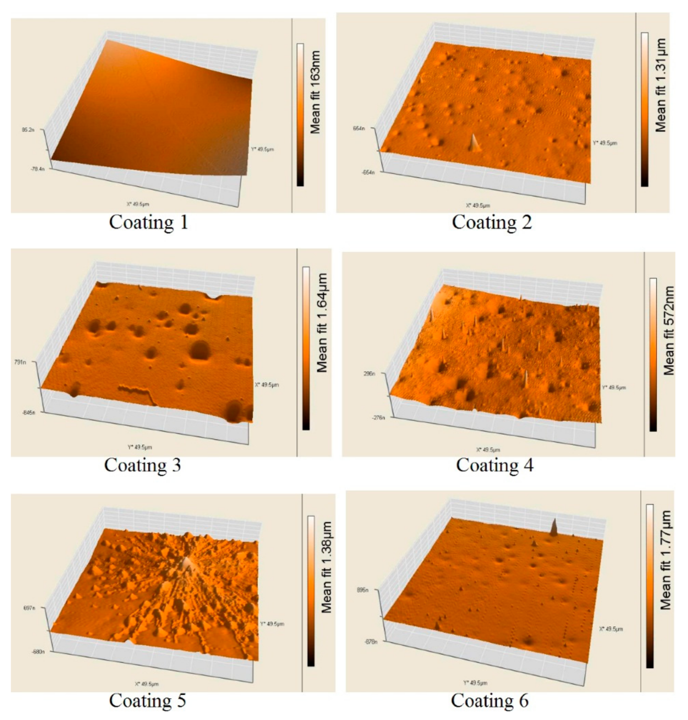
Figure 2.
Fluorescent microscopy pictures of SYTO 9 stained C. marina biofilms formed after 1 h incubation on (NC) negative control, glass slide with staining procedure and without bacteria; and on glass samples with PEG-silane co-cross-linked coatings containing: (1) 6.0 wt % conventional cross-linker, ES 40 (control, bacteria and staining); (2) 4.0 wt % ES40 + 2.0 wt % PEG-silane; (bacteria and staining); (3) 6.0 wt % ES40 + 2.0 wt % PEG-silane, (bacteria and staining). The yellow arrows indicate areas with stained bacteria.
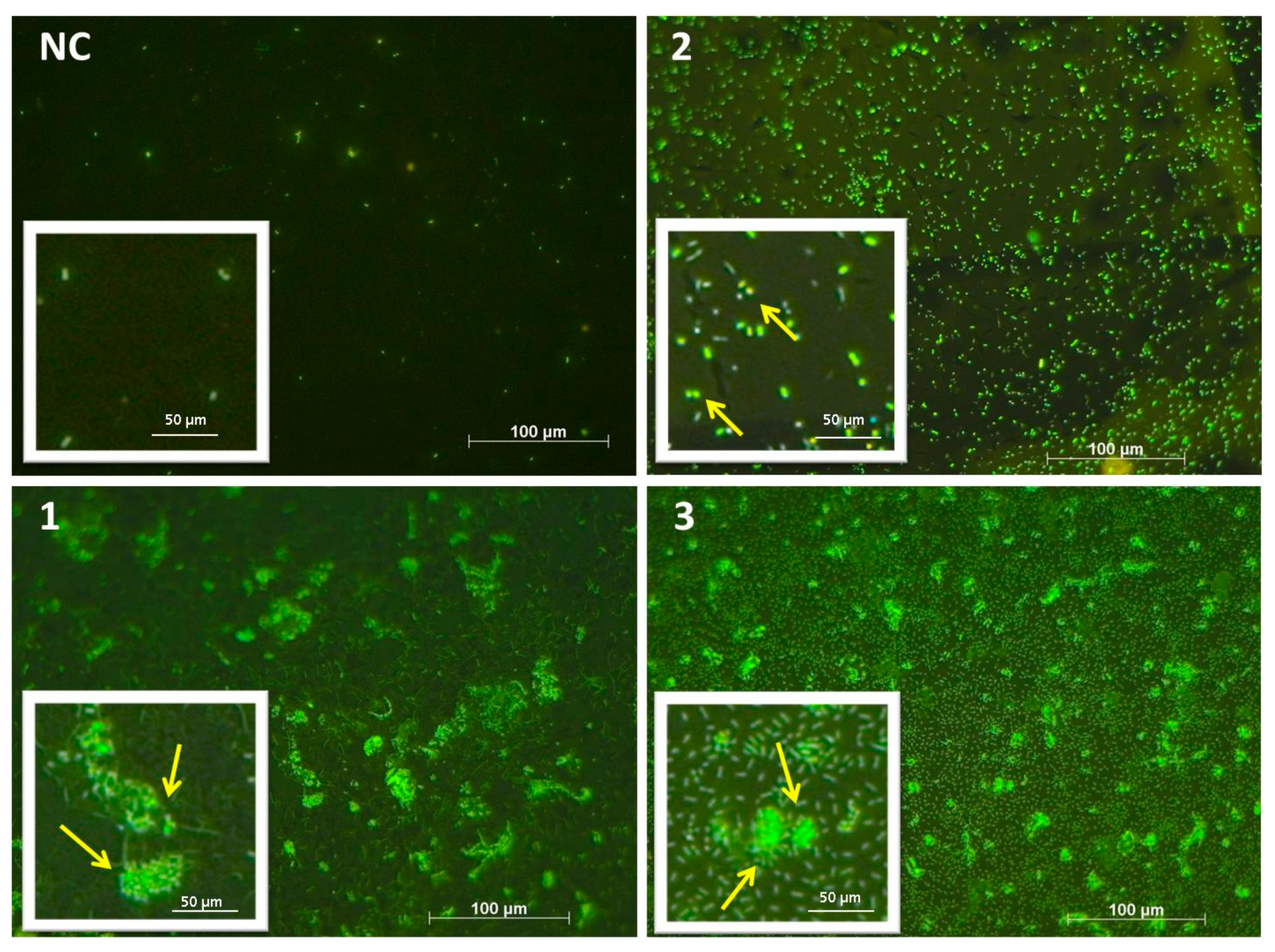
Figure 3.
Fluorescent microscopy pictures of SYTO 9 stained C. marina biofilms formed after a 1 h incubation on (PC) positive control, glass slide with staining procedure and bacteria and on glass samples with PEG-silane co-cross-linked coatings containing modifying agent: (4) 4.0 wt % ES40 + 2.0 wt % PEG-silane + 0.5 wt % NIS; (bacteria and staining); (5) 4.0 wt % ES40 + 2.0 wt % PEG-silane + 2.0 wt % AO; (bacteria and staining); (6) 4.0 wt % ES40 + 2.0 wt % PEG-silane + 0.5 wt % NIS + 1.5 wt % AO, (bacteria and staining). The yellow arrows indicate areas with stained bacteria.

Figure 4.
ATP (Adenosine thriphosphate) luminescence (in absence of biofilm and with biofilm formed after a 1 h incubation of 4.107 cfu/mL C. marina) on non-covered glass and glass covered with a coating containing: (1) 6 wt % conventional cross-linker, ES 40 (control); (2) 4 wt % ES40 + 2 wt % PEG-silane; (3) 6 wt % ES40 + 2 wt % PEG-silane; (4) 4 wt % ES40 + 2 wt % PEG-silane + 0.5 wt % NIS; (5) 4 wt % ES40 + 2 wt % PEG-silane + 2.0 wt % AO; (6) 4 wt % ES40 + 2 wt % PEG-silane + 0.5 wt % NIS + 1.5 wt % AO.

Figure 5.
Microbial growth after four (a) and six (b) months of exposure in a Black Sea aquarium on vulcanized siloxane coatings, cross-linked with: (1) 6 wt % conventional cross-linker, ES40 (control); (2) 6 wt % PEG-silane instead of 6 wt % ES40; (1’) 4 wt % ES40 + 2 wt % PEG-silane.
Figure 5.
Microbial growth after four (a) and six (b) months of exposure in a Black Sea aquarium on vulcanized siloxane coatings, cross-linked with: (1) 6 wt % conventional cross-linker, ES40 (control); (2) 6 wt % PEG-silane instead of 6 wt % ES40; (1’) 4 wt % ES40 + 2 wt % PEG-silane.
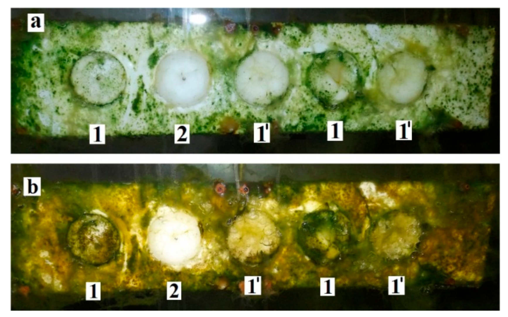
Figure 6.
Total chlorophyll (a), carotenoids content (b) and dry mass (c) of biofilm formed on coatings cross-linked with ES40 (columns 1, 3, 5, 7) or with ES 40 and 2 wt % PEG-silane (columns 2, 4, 6 and 8) without additives: columns 1 and 2, or containing: 0.5 wt % NIS (columns 3 and 4); 2.0 wt % AO (columns 5 and 6); 0.5 wt % NIS and 1.5 wt % AO simultaneously (columns 7 and 8).


Table 1.
Surface physical-chemical characteristics: water contact angle (WCA), surface energy (γc) and its disperse (γd) and polar (γp) components; surface roughness (Ra, Rq, Sp, Sv, Sy) and physical-mechanical parameters: dynamic Vicker’s hardness (HMV), indentation hardness (HIT), indentation elastic modulus (EIT), and elastic modulus (Et) of the studied coatings: (1) control (6 wt % ES40,without PEG-silane); (2) (4 wt % ES40 + 2 wt % PEG-silane); (3) (6 wt % ES40 + 2 wt % PEG silane); (4) (4 wt % ES40 + 2 wt % PEG-silane + 0.5 wt % NIS); (5) (4 wt % ES40 + 2 wt % PEG-silane + 2.0 wt % AO); (6) (4 wt % ES40 + 2 wt % PEG-silane + 0.5 wt % NIS + 1.5 wt % AO).
| Parameter | Sample No. | |||||
|---|---|---|---|---|---|---|
| 1 | 2 | 3 | 4 | 5 | 6 | |
| WCA | 104.1 ± 0.3 | 103.0 ± 0.5 | 104.1 ± 0.4 | 51.5 ± 0.6 | 90.6 ± 0.4 | 60.0 ± 0.6 |
| γc, mN/m | 22.4 | 22.8 | 22.1 | 30.5 | 24.4 | 32.1 |
| γd, mN/m | 22 | 22.6 | 22 | 20.1 | 22 | 21 |
| γp, mN/m | 0.4 | 0.7 | 0.1 | 10.4 | 2.3 | 11.1 |
| Ra, nm | 12 ± 4 | 12 ± 6 | 33 ± 9 | 21 ± 6 | 49 ± 10 | 51 ± 10 |
| Rq, nm | 15 ± 7 | 18 ± 7 | 42 ± 9 | 27 ± 2 | 63 ± 16 | 61 ± 6 |
| Sp, nm | 56 ± 15 | 428 ± 28 | 151 ± 21 | 232 ± 31 | 458 ± 58 | 737 ± 73 |
| Sv, nm | −38 ± 12 | −158 ± 16 | −275 ± 11 | −120 ± 13 | −193 ± 14 | 151 ± 18 |
| Sy, nm | 94 ± 11 | 640 ± 48 | 426 ± 26 | 352 ± 58 | 651 ± 64 | 888 ± 83 |
| HMV (N/mm2) | 0.13 ± 0.02 | 0.46 ± 0.03 | 0.45 ± 0.02 | 0.34 ± 0.01 | 0.34 ± 0.02 | 0.27 ± 0.01 |
| HIT (N/mm2) | 0.34 ± 0.01 | 0.82 ± 0.01 | 0.82 ± 0.07 | 0.77 ± 0.03 | 0.63 ± 0.02 | 0.51 ± 0.03 |
| EIT (N/mm2) | 1.76 ± 0.06 | 8.13 ± 0.10 | 7.88 ± 0.10 | 4.99 ± 0.08 | 5.87 ± 0.06 | 4.41 ± 0.02 |
| E (N/mm2) | 0.48 ± 0.01 | 0.31 ± 0.01 | 0.35 ± 0.03 | – | – | – |
© 2018 by the authors. Licensee MDPI, Basel, Switzerland. This article is an open access article distributed under the terms and conditions of the Creative Commons Attribution (CC BY) license (http://creativecommons.org/licenses/by/4.0/).
Share and Cite
MDPI and ACS Style
Akuzov, D.; Franca, L.; Grunwald, I.; Vladkova, T. Sharply Reduced Biofilm Formation from Cobetia marina and in Black Sea Water on Modified Siloxane Coatings. Coatings 2018, 8, 136. https://doi.org/10.3390/coatings8040136
AMA Style
Akuzov D, Franca L, Grunwald I, Vladkova T. Sharply Reduced Biofilm Formation from Cobetia marina and in Black Sea Water on Modified Siloxane Coatings. Coatings. 2018; 8(4):136. https://doi.org/10.3390/coatings8040136
Chicago/Turabian StyleAkuzov, Danail, Lia Franca, Ingo Grunwald, and Todorka Vladkova. 2018. "Sharply Reduced Biofilm Formation from Cobetia marina and in Black Sea Water on Modified Siloxane Coatings" Coatings 8, no. 4: 136. https://doi.org/10.3390/coatings8040136
Note that from the first issue of 2016, this journal uses article numbers instead of page numbers. See further details here.