Article

Stachys Species: Comparative Evaluation of Phenolic Profile and Antimicrobial and Antioxidant Potential

1
Department of Pharmacognosy, “Iuliu Hațieganu” University of Medicine and Pharmacy, 12 I. Creanga Street, 400010 Cluj-Napoca, Romania
2
Department of Pharmaceutical Chemistry, “Iuliu Hațieganu” University of Medicine and Pharmacy, 41 V. Babeş Street, 400012 Cluj-Napoca, Romania
3
Department of Pharmaceutical Botany, “Iuliu Hațieganu” University of Medicine and Pharmacy, 12 I. Creanga Street, 400010 Cluj-Napoca, Romania
4
Department of Pharmaceutical Technology and Biopharmacy, “Iuliu Hațieganu” University of Medicine and Pharmacy, 41 V. Babeş Street, 400012 Cluj-Napoca, Romania
5
Department of Chemistry and Chemical Engineering, Babes-Bolyai University, 11 A. Janos Street, 400028 Cluj-Napoca, Romania
6
State Animal Health and Safety Veterinary Laboratory, 1 Piata Marasti Street, 400609 Cluj-Napoca, Romania
7
Department of Physiology, “Iuliu Hațieganu” University of Medicine and Pharmacy, 1 Clinicilor Street, 400006 Cluj-Napoca, Romania
8
Department of Clinical Sciences, University of Agricultural Sciences and Veterinary Medicine Cluj-Napoca, 400372 Cluj-Napoca, Romania
*
Author to whom correspondence should be addressed.
These authors contributed equally to this work.
Antibiotics 2023, 12(11), 1644; https://doi.org/10.3390/antibiotics12111644
Submission received: 24 October 2023 / Revised: 14 November 2023 / Accepted: 18 November 2023 / Published: 20 November 2023
(This article belongs to the Special Issue Antimicrobial Activity of Natural Products and Plants Extracts)

Abstract

:
This study aimed to investigate the polyphenolic composition and antioxidant and antimicrobial potential of six Romanian Stachys species: S. officinalis, S. germanica, S. byzantina, S. sylvatica, S. palustris, and S. recta. The LC-MS/MS method was used to analyze the polyphenolic profile, while the phenolic contents were spectrophotometrically determined. The antioxidant activity was evaluated using the following methods: DPPH, FRAP, nitrite-induced autooxidation of hemoglobin, inhibition of cytochrome c-catalyzed lipid peroxidation, and electron paramagnetic resonance spectroscopy. The in vitro antimicrobial properties were assessed using agar-well diffusion, broth microdilution, and antibiofilm assays. Fifteen polyphenols were identified using LC-MS and chlorogenic acid was the major component in all the samples (1131.8–6761.4 μg/g). S. germanica, S. palustris, and S. byzantina extracts each displayed an intense antiradical action in relation to high contents of TPC (6.40 mg GAE/mL), flavonoids (3.90 mg RE/mL), and caffeic acid derivatives (0.89 mg CAE/mL). In vitro antimicrobial and antibiofilm properties were exhibited towards Candida albicans, Gram-positive and Gram-negative strains, with the most intense efficacy recorded for S. germanica and S. byzantina when tested against S. aureus. These results highlighted Stachys extracts as rich sources of bioactive compounds with promising antioxidant and antimicrobial efficacies and important perspectives for developing phytopharmaceuticals.

1. Introduction

As a member of the Lamiaceae family, the Stachys L. genus (wound plants) has garnered scientific interest regarding its chemical composition and therapeutic properties. In fact, numerous species of this genus have been used empirically in different regions of the world for their medicinal and economic potential since ancient times [1,2].
A recently published paper reviewed the existing information on various traditional uses of 25 species and 6 subspecies of Stachys genus, conditioned mostly as herbal tea, infusion, and decoction, but also as extracts (oily and alcoholic) and edible food (tubers) [2]. This study also described the phytochemistry and pharmacological profile of distinct Stachys sp. originating from the Mediterranean region, Asia, America, and southern Africa.
Overall, a complex chemical composition including secondary metabolites such as flavonoids (baicalin, apigenin, isoscutellarein 7-O-β-D-glucopyranoside), phenolcarboxylic acids (caffeic, rosmarinic, chlorogenic acids), tannins, phenylethanoid glucosides (acteoside, martinosid), lignans, iridoids, diterpenes (kauran, clerodane), and triterpenes; essential oils characterize Stachys genus [1,2].
Moreover, several Stachys species have been studied in vitro and in vivo to scientifically justify their traditional use. The pharmacological assessment demonstrated important therapeutic properties, namely the following: antioxidant (S. recta subsp. recta, S. palustris, S. byzantina, S. sylvatica), anti-inflammatory (S. recta, S. germanica, S. officinalis, S. alpina), analgesic, anxiolytic, antidepressant, antiproliferative (S. lavandulifolia), antibacterial (S. byzantina, S. officinalis, S. sylvatica), renoprotective, and hepatoprotective (S. pilifera) [1,2].
Although Stachys sp. are spreading in the Romanian flora [3,4], only a few references are available [5,6,7,8]. In this regard, common and Latin names, botanical descriptions, and geographical distribution are presented for 14 species of Stachys, all perennial herbaceous plants, in the Flora of Romania [3,4]. Among these, Stachys officinalis (L.) Trevis. (sin. Betonica officinalis L.) is the only Stachys species documented by Romanian traditional medicine. Phytopreparations obtained from its leaves and aerial parts are mentioned for tonic, antipyretic, analgesic, and cicatrizing efficacy, and have been traditionally recommended for various disorders such as rhinitis, headache, hemoptysis, chest pain, wounds, and varicose ulcers [5,6]. Nowadays, most of the empirical therapeutic applications involve S. officinalis tea in atonic dyspepsia and liver disorders based on antispastic, astringent, carminative, cholagogue, digestive stimulant, emmenagogue, anthelmintic, and diuretic properties. The tea is also listed for anxiolytic and sedative effects, being able to reduce anxiety and irritability [6]. Regarding other Stachys species, S. byzantine is a very adaptable and decorative plant with brightly colored flowers and is commonly cultivated for ornamental purposes [3].
Previous studies on Romanian Stachys extracts addressed the following species: S. officinalis [8], S. byzantina, and S. sylvatica [7,8].
The current study objective was to evaluate the polyphenolic composition, antioxidant, and in vitro antimicrobial potential of Stachys species frequently found in the Romania flora: S. officinalis, S. germanica, S. byzantina, S. sylvatica, S. palustris, and S. recta. These data are required to complete these species’ pharmacognostic profiles aiming to ensure a well-deserved place in phytotherapy. Thus, additional chemical and pharmacotherapeutic data are needed to reveal the most promising Stachys species for medicinal use.

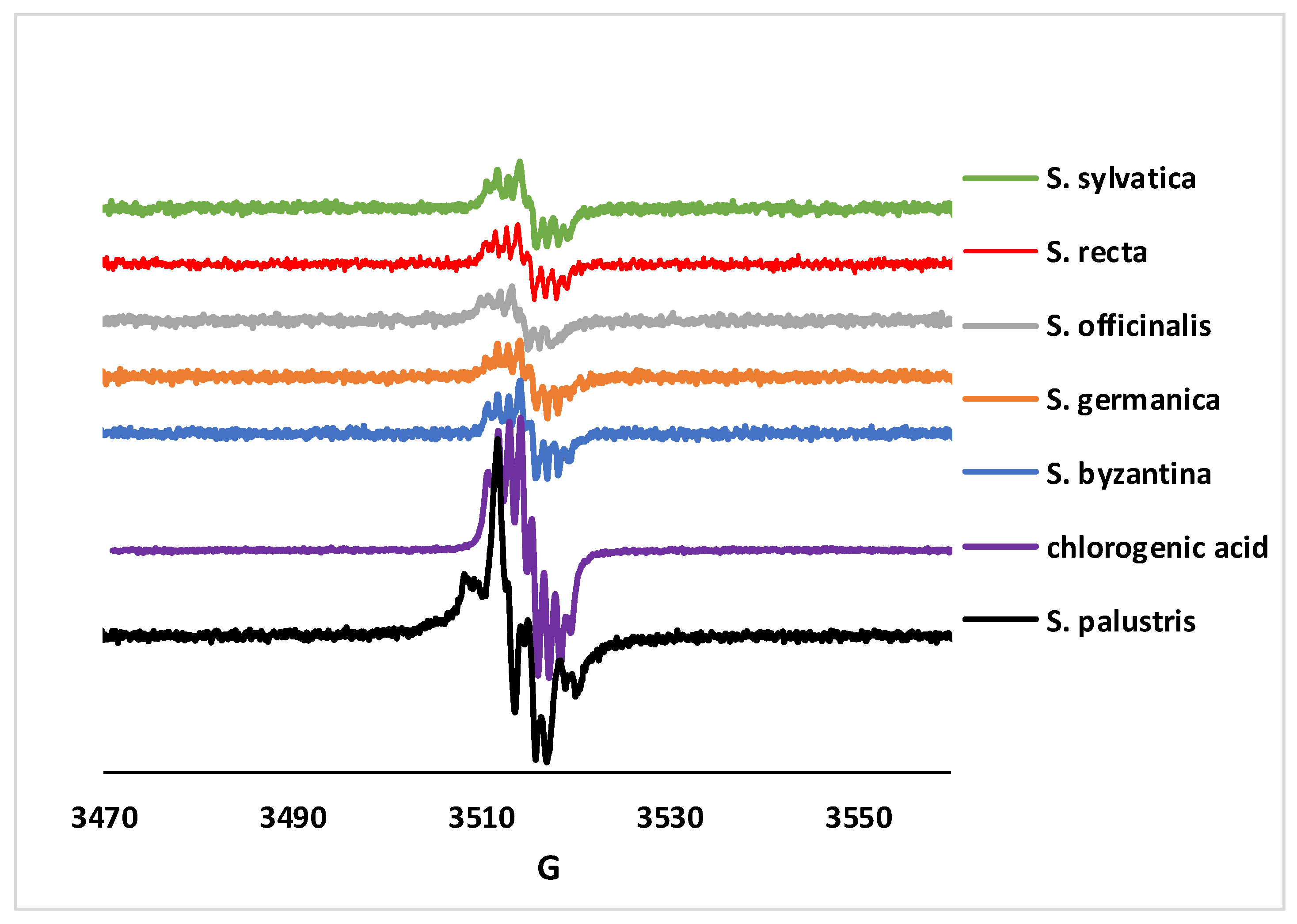
2. Results and Discussions

2.1. Chromatographic Analysis

To characterize plant extracts rich in polyphenols, the most widely used technique for the separation and individual quantification of phenolic compounds is liquid chromatography coupled with tandem mass spectrometry (LC-MS/MS) [9,10]. The amounts of compounds detected in the six Stachys sp.-derived extracts were expressed in μg/g dry weight (dw) of vegetal product (Table 1).
Fifteen polyphenolic compounds, including ten phenolic acids (gallic, rosmarinic, protocatehuic, gentisic, caftaric, chlorogenic, vanillic, syringic, p-coumaric, ferulic acids) and five flavonoids (isoquercitrin, rutin, quercitrin, luteolin, apigenin), were detected by LC-MS in all Stachys extracts (Table 1).
Furthermore, all extracts presented high amounts of chlorogenic acid (1131.8–6761.4 μg/g dw). Chlorogenic acid with multiple therapeutic potentials (neuroprotective; cardio-, hepato-, gastro-, renoprotective; anticancer) is a phenolic acid widely distributed in many species of the Stachys genus [2,11]. The extracts richest in chlorogenic acid were from S. germanica (4205.2 μg/g dw), S. sylvatica (5552.7 μg/g dw), and S. recta (6761.4 μg/g dw). High concentrations of chlorogenic acid were measured in Turkish S. germanica [12], but this active principle was not found in S. recta [13]. The other two indigenous Stachys: S. officinalis (1131.8 μg/g dw) and S. byzantina (2012.1 μg/g dw) showed lower concentrations of chlorogenic acid compared to the other species, but higher than some published values [14]. The results obtained for the S. officinalis extract are similar to those previously reported by other authors [15].
S. officinalis presented higher amounts of vanilic acid (83.5 μg/g dw), syringic acid (82.7 μg/g dw), ferulic acid (11.1 μg/g dw), and apigenin (84.2 μg/g dw) than the other Stachys species. Bączek et al. determined ferulic acid and apigenin in Polish S. officinalis in higher concentrations [16] and Paun et al. have published lower values of apigenin than ours [17]. Regarding protocatechuic and p-coumaric acids, the S. recta extract was found to be the richest in these compounds, at 157.8 μg/g dw and 32.0 μg/g dw, respectively.
Caftaric acid was determined only in S. officinalis (4.7 μg/g dw) and gallic acid only in S. palustris (141.5 μg/g dw).
The S. officinalis extract (82.7 μg/g dw) was the richest in syringic acid, compared to the S. sylvatica (8.6 μg/g dw), S. byzantina (7.0 μg/g dw), and S. palustris (4.4 μg/g dw) extracts. Four species of Stachys (S. officinalis, S. recta, S. sylvatica, S. germanica) contained isoquercitrin; the richest species was S. germanica (233.1 μg/g dw). Isoquercitrin, also identified by other authors in S. recta [13], develops numerous chemoprotective effects in vitro and in vivo against oxidative stress, diabetes, allergies, cancer, etc. [17].
Rutin and quercitrin were found in very small quantities, below the limit of detection in the extracts of S. officinalis (rutin,) and S. palustris (quercitrin).
The characterization of polyphenolic profiles of the six Stachys species showed differences in the presence of key compounds, but also in what concerns the concentrations of individual phenols.
Numerous medicinal species in the Lamiaceae family contain rosmarinic acid (RA), a natural polyphenolic molecule with antioxidant, antidiabetic, antimicrobial, hepatoprotective, cardioprotective, nephroprotective, anti-aging, anti-inflammatory, antiviral, and anticancer therapeutic properties [18]. Data from the literature confirm the presence of RA in other species of Stachys: S. thirkei [19], S. tmolea [20], S. cretica [21], S. lavandulifolia, S. byzantina [22], and S. officinalis [15,23].
In this case, we chose to use the LC-MS/MS method, published by us, to determine the RA content of the six Stachys species [24].
The ESI source mass spectrometer, in the negative mode, was set to isolate and fragment the deprotonated RA molecule with m/z = 359. The quantification of RA was performed with a tR of 2.2 min. In the range of 40–640 ng/mL, a linear calibration curve was performed (R2 = 0.999). The RA content values (μg/g dw) are presented in Table 1; this amount varied between 1.9–43.4 μg/g dw of vegetal product.
The S. officinalis extract showed a higher RA content (43.4 μg/g dw) compared to the other Stachys species extracts studied by us, but lower than the results published by other authors [15,16]. From the data in the literature accessed by us until now, we can say that RA was found for the first time in the extracts of S. germanica (7.8 μg/g dw), S. recta (2.6 μg/g dw), S. palustris (2.4 μg/g dw), and S. sylvatica (1.9 μg/g dw). Of the six spontaneous native species of Stachys, S. officinalis can be considered as an important source of rosmarinic acid.
The ethanolic extracts of Stachys sp. were examined to determine the polyphenolic TPC), flavonoid, and caffeic acid derivatives contents. The results obtained in this study were expressed as gallic acid (GA), rutin (R), and caffeic acid (CA) equivalents, respectively (Table 2).
Medicinal plants produce polyphenols with various chemical structures, such as flavonoids, phenolic acids, with important antioxidant, cytotoxic, anti-inflammatory, antihypertensive, and antidiabetic properties [25]. The TPC contents of these six Stachys sp. extracts, determined using the Folin−Ciocâlteu reagent [26], are shown in Table 2. According to this analysis, S. byzantina extract had the highest concentration of TPC (6.40 mg GAE/mL) and the S. palustris and S. germanica extracts had similar values (5.35 mg GAE/mL and 5.98 mg GAE/mL, respectively). The other three species showed lower values, between 2.51 mg/mL and 4.00 mg GAE/mL, the S. officinalis extract being the lowest in TPC (2.51 mg GAE/mL). In terms of TPC, the extract of S. byzantina was the richest in these active compounds. The statistical analysis of these data (Table 2) showed highly significant differences between S. byzantina (SB) and other Stachys sp.: SO, SG, SS, and SR (p < 0.001) and significant differences between S. byzantina (SB) and S. palustris (p = 0.011). Our results were similar to those published by other authors for S. byzantina and S. germanica species, [27,28] while other authors described lower values than ours for S. officinalis [15].
The total flavonoid contents were determined using the AlCl3 method [29]. The highest content of flavonoids, with values between 3.14 and 3.90 mg RE/mL (p > 0.05), was found in ethanolic extracts of S. palustris, S. germanica, and S. byzantina. The other extracts of S. officinalis, S. sylvatica, and S. recta extracts had lower amounts of flavonoids, around 1 mg/mL (1.13, 1.18, and 1.22 mg RE/mL, respectively, in relation with S. byzantina (0.001 < p < 0.05). Comparing different studies from the literature, some authors have reported lower levels of flavonoids in S. officinalis [15], S. germanica, S. sylvatica, and S. recta [30]. Sytar et al. found higher concentrations of flavonoids in S. byzantina [14] compared to our results, and Nikolova recorded similar values for S. officinalis [30].
Caffeic acid derivatives, which are widespread in many plant species, have been extensively studied; this has revealed a wide range of potential therapeutic applications. Regarding the contents of caffeic acid derivatives for the Stachys sp. extracts, values ranged between 0.20–0.89 mg CAE/mL (Table 2). The highest values were obtained for the ethanolic extracts of S. palustris (0.72 mg CAE/mL), S. germanica (0.88 CAE mg/mL), and S. byzantina (0.89 CAE mg/mL), while the lowest were measured in the extracts of S. officinalis (0.20 mg CAE/mL), S. sylvatica (0.20 mg CAE/mL), and S. recta (0.23 mg CAE/mL), compared to S. byzantina (0.001 < p < 0.05). The literature provides data only for S. germanica caffeic acid derivatives’ content; our results varied slightly compared to those published by Mitic et al. [28].
Qualitative and quantitative evaluation of the phenolic profile indicated S. germanica, S. byzantina, and S. palustris as the species with the richest content in these active compounds including flavonoids and caffeic acid derivatives.

2.2. Antioxidant Activity of Stachys sp. Extracts

Bioactive compounds of the polyphenol type are important natural antioxidants due to their redox characteristics, as a result of hydroxyl groups that facilitate the elimination of free radicals and thus offer many benefits to human health [25]. In this regard, extracts of Stachys sp. with polyphenols, have been evaluated by several antioxidant methods: the DPPH radical scavenging method, the FRAP test (the ferric-reducing ability of plasma), nitrite-induced autooxidation of hemoglobin (NHA), inhibition of cytochrome c-catalyzed lipid peroxidation, and EPR spectroscopy assays (Table 3).
The results of DPPH scavenging activity revealed that five Stachys extracts presented high antioxidant activity: S. byzantina (IC50 = 53.61 µg/mL), S. germanica (IC50 = 54.18 µg/mL) and S. palustris (IC50 = 54.82 µg/mL) extracts, followed by extracts of S. sylvatica (IC50 = 76.04 µg/mL) and S. recta (IC50 = 84.94 µg/mL), but lower compared to Trolox (a p < 0.001). In contrast, the S. officinalis extract showed a higher value of IC50 (139.16 µg/mL), which indicates a lower activity. Other authors have obtained IC50 values higher than ours (S. officinalis, S. germanica, S. sylvatica, S. byzantina) [13,30]. Similar results were obtained for S. officinalis from Romania [15], as well as for the Bulgarian S. recta species [30].
The FRAP values for the investigated extracts were reported in Table 3, so that S. germanica and S. palustris extracts showed antioxidant activity (688.21 and 502.43 μM TE/mL), in relation to high polyphenol contents (5.77 and 5.56 mg GAE/mL), but also with the values obtained by the DPPH method (53.83 and 55.66 µg/mL). For the other four Stachys sp. extracts, lower values were obtained (between 218.17and 354.88 μM TE/mL) than S. germanica (b p < 0.001). The NHA results on all Stachys extracts offer values in the same order and proportion as the FRAP results (e.g., highest results for S. germanica and lowest results— for S. sylvatica, by about three times). This excellent agreement between FRAP and NHA was not mirrored by the DPPH results. Thus, in the DPPH measurements S. sylvatica was far from being the weakest antioxidant, while S. germanica was narrowly exceeded as the strongest oxidant by S. byzantina—even though the two had widely different activities in the NHA assay. Interestingly, the NHA data does not correlate with either the total phenolic of flavonoid contents or with the content of any particular component (including the major ones—rosmarinic acid, chlorogenic acid, and caffeic derivatives). The NHA assay tests a nitrosative stress process, while FRAP was based on oxidative stress agents. The excellent agreement between NHA and FRAP, together with the fact that NHA is based on a physiological reaction employing a protein from blood, may be taken as argument that these two assays have a particularly important biomedical/physiological relevance. Regarding the FRAP values published by other authors, important results were obtained for S. germanica subsp. heldreichii [12]; the species S. byzantina and S. sylvatica showed a lower antioxidant activity [27,31,32,33].
An antioxidant assay based on the inhibition of cytochrome-catalyzed lipid autooxidation was also attempted. However, in this case the Stachys extracts exhibited effects that were too narrowly similar to each other—although S. germanica was still found at the most-active end of the range, while S. sylvatica was at the least-active end.
Exposure of phenolic compounds to basic pH under aerobic conditions allows for the generation of phenol-based free radicals via autooxidation processes. Such radicals can be detected by EPR spectroscopy and can be used as fingerprints for assessing the dominant components in polyphenolic-containing natural extracts. Figure 1 shows that a clear complex signal was detected for all the Stachys extracts, which is very similar to that of pure chlorogenic acid samples. This observation was in line with the fact chlorogenic acid was by far the most abundant polyphenolic compound in these samples, that according to the chromatographic data in Table 1; while up to a sixfold difference in chlorogenic acid concentrations is seen in Table 1, Figure 1 offers a spectra of intensities that were essentially equal among all samples. A single exception exists—namely, the S. palustris sample, for which the EPR spectrum was no longer identical to that of chlorogenic acid. This implies that S. palustris contains a unique antioxidant species, not shared with the other Stachys extracts. Table 1 and Table 2 reveal only one such antioxidant—namely, gallic acid, which is present only in S. palustris. Indeed, the shape of the S. palustris EPR spectrum was consistent with that of a gallate radical imposed over chlorogenic acid [34].
In the present study, it was observed that the extracts of S. germanica, S. palustris, and S. byzantina had the highest antiradical action, in a general relation to a high content of TPC, flavonoids, and caffeic acid derivatives. The S. recta and S. sylvatica extracts with a medium content of polyphenols also presented a high antioxidant capacity. Thus, it can be concluded from the evaluation of the phenolic content and the antioxidant activity that these species of Stachys can be recommended as valuable natural antioxidants for various nutritional functions and to preserve health.

2.3. Antimicrobial Activity of Stachys sp. Extracts

The use of natural herbal preparations as antimicrobial agents is a therapeutic alternative, especially in the context of the increasing bacterial resistance to antibiotics. In addition, it is known from the literature that Stachys species have many pharmacological activities (anti-inflammatory, antianxiety, antinephritic, antioxidant, cytotoxic) [12,27,35,36] and antimicrobial properties have been demonstrated for some species, such as S. byzantina, S. guyoniana, S. officinalis, S. sylvatica, S. cretica, S. scardica, S. recta, and S. germanica. [3,23,37,38,39,40,41].

2.3.1. Antibacterial Activity Using the Agar-Well Diffusion Method

Using the agar-well diffusion method, we evaluated the antimicrobial susceptibility of significant bacterial isolates of the six indigenous Stachys species (Table 4) [42]. Our extracts showed a strong antibacterial activity, especially against S. aureus and L. monocytogenes, with areas of inhibition ranging from 14 and 20 mm, compared to gentamicin (IZD = 18 mm), used as a reference antibiotic (0.001 < p < 0.05). The activity on S. aureus was almost the same as gentamicin for S. officinalis, S. sylvatica, and S. recta (p < 0.05) and was superior for S. germanica and S. byzantina with IZD = 20 mm (p < 0.001). The same IZD (18 mm) was measured for the S. palustris extract as for the antibiotic standard (p = 1). All extracts showed lower activity on L. monocytogenes (p < 0.001) compared to gentamicin used as reference antibiotic.
Regarding the antibacterial activity against Gram-negative strains, Stachys extracts showed limited activity against E. coli and S. enteritidis, statistically lower compared to gentamicin (p < 0.001). These results were consistent with those published by other authors, according to which Gram-negative bacteria display less sensitivity to the effect of Stachys extracts compared to Gram-positive ones [3,32,38,43]. Also, similarly to previous reports, our Stachys extracts showed a relatively low anti-C. albicans activity compared to Fluconazole (p < 0.001), which was used as the reference antifungal [38,44].

2.3.2. Antibacterial Activity Using the Broth Microdilution Method

Moreover, the results obtained using the broth microdilution method (Table 5) showed Stachys extracts higher inhibitory efficacy towards Gram-positive bacteria, S. aureus, and L. monocytogenes. The lowest values of MIC were recorded for S. germanica and S. byzantina. These two extracts were found also with the lowest MBC values established against S. aureus, suggesting their superior bactericidal potential towards this bacterial species. All Stachys extracts presented in vitro inhibitory and bactericidal potential against E. coli and S. enteritidis, but only at the highest tested concentrations (Table 5). C. albicans growth was inhibited by all tested extracts and a two- and threefold higher fungicidal effect was noticed for MBC values than for MIC values, respectively.

2.3.3. Antibiofilm Activity

The inhibitory activity of Stachys sp. extracts against biofilm formation is presented in Table 6.
Both the biofilm attachment (T0) and destruction of 24 h pre-formed biofilm (T24) were inhibited by certain extracts, the most intense effect (++) was still noticed against the planktonic cells. The highest efficacy was recorded for S. germanica, S. byzantina, S. sylvatica, and S. palustris-derived extracts.
According to the literature, S. byzantina, S. officinale, and S. sylvatica showed strong antibacterial activity, S. aureus being very susceptible to these extracts [3,32,38]. The antibacterial activity of the Stachys sp. extracts could be due to the presence of chlorogenic acid, which is found in large quantities in our extracts and according to studies can increase the permeability of the outer membrane and plasma and thus lead to the loss of the barrier function. It also depletes intracellular potential and releases cytoplasmic macromolecules, which can lead to bacterial cell death [46,47].
The results of this study pointed out that Stachys extracts were able to display an important in vitro antimicrobial potential against Gram-positive strains, with the extracts of S. germanica, S. byzantina, and S. palustris achieving very good antistaphylococcal activity. This aspect is relevant as S. aureus is regarded worldwide as one of the most important human bacteria, with both colonizing and opportunistic pathogen valences and elevated levels of multidrug resistance [48]. In this regard, the results indicated S. germanica and S. byzantina extracts for their superior antibiofilm properties against S. aureus.

3. Materials and Methods

3.1. Plant Material

The aerial parts from six Stachys species: S. officinalis—SO, S. germanica—SG, S. byzantina—SB, S. sylvatica—SS, S. palustris—SP, and S. recta—SR (Voucher numbers: 106-111) were collected from the spontaneous flora of Cluj County, Romania (Podeni village: 46°41′4″ N 23°1′45″ E), during the flowering period. The plant material was identified by Ph.D. A. Mocanu (“Iuliu Haţieganu” University of Medicine and Pharmacy Cluj-Napoca).

3.2. Chemicals

All high-quality purity chemicals (substances and reagents) used in this study were acquired from the following companies: Sigma, St. Louis, MO, USA (chlorogenic, cafeic, p-coumaric acids, isoquercitrin, quercitrin, hyperoside, rutin, apigenin, myricetin, quercetin, kaempferol, fisetin); Roth, Karlsruhe, Germany (sinapic, ferulic, gallic, gentisic, rosmarinic acids, patuletin, luteolin); Dalton, Toronto, ON, Canada (caftaric, cichoric acids); Alfa-Aesar, Karlsruhe, Germany (sodium hydroxide, sodium carbonate, and iron (III) chloride); Sigma-Aldrich, Steinheim, Germany (soy lecithin, bovine hemoglobin, protocatechuic, vanillic and syringic acids, (−)-epicatechin, (+)-catechin), 6-hydroxy-2,5,7,8-tetramethylchroman-2-carboxylic acid (Trolox), 2,4,6-Tris (2-pyridyl)-s-triazine (TPTZ), sodium acetate buffer solution); Merck, Darmstadt, Germany (Arnow’s reagent, Folin−Ciocâlteu reagent, methanol, ethanol, aluminum chloride, sodium acetate, hydrochloric acid, 2,2-diphenyl-1-picrylhydrazyl; the methanol, acetonitrile, ammonium acetate, and acetic acid were all of the HPLC grade).
The Gram-positive bacteria: Staphylococcus aureus (ATCC 6538P) and Listeria monocytogenes (ATCC 13932); Gram-negative bacteria: Escherichia coli (ATCC 25922) and Salmonella typhimurium (ATCC14028); and one fungal strain, Candida albicans (ATCC 10231), were distributed by MicroBioLogics®, St. Cloud, MN, USA.
The spectrophotometric data were acquired using a Jasco V-530 UV–Vis spectrophotometer (Jasco International Co., Ltd., Tokyo, Japan).

3.3. Preparation of Stachys sp. Extracts

Ten grams of each powdered plant material was extracted with 100 mL of 70% ethanol using ultrasound (Polsonic Palczyski Sp. J., Sonic 3, Warsaw, Poland) at 60 °C, for 30 min. The ethanolic extracts were filtered through paper filters into 100 mL flasks and then centrifuged (at 4500 rpm, 10 min) and the supernatant solutions were collected [29,49].

3.4. LC-MS/MS Analysis of Polyphenols

The study of flavonoids, as well as of the phenolic acids present in the six extracts of Stachys species was performed using the LC-MS/MS analysis on an Agilent 1100 HPLC Series system (Agilent, Santa Clara, CA, USA) equipped with: degasser (G1322A), binary gradient pump (G13311A), autosampler (G1313A), column thermostat, and UV detector (G1316A) that was coupled with an Agilent Ion Trap 1100 SL mass spectrometer (LC/MSD Ion Trap VL), according to the method previously published [24,50]. The standards used were from the class of phenolic acids (caftaric, gentisic, caffeic, chlorogenic, p-coumaric, ferulic, synapic acids), but also from the class of flavonoids (rutin, hyperoside, isoquercitrin, quercitrin, quercetin, myricetol, fisetin, patuletin, luteolin, kaempferol, apigen). The following parameters were used to separate the polyphenolic principles from plant extracts: a reversed-phase HPLC columns (Zorbax Agilent, Santa Clara, CA, USA, SB-C18, 100 mm × 3.0 mm id, 3.5 µm), mobile phase with methanol: 0.1% acetic acid (v/v), and a binary solvent gradient; the detection was performed using UV and MS with an electrospray ion source in negative mode. Other HPLC parameters: flow rate = 1 mL/min, injection volume = 5 µL, and column temperature = 48 °C with combined detection: UV (330 nm, 370 nm) and MS mode. The quantitative results were processed using the Agilent ChemStation software B01.03 and DataAnalysis version 5.3.
Next, another previously described LC-MS method was used to determine 6 other polyphenols: epicatechin, catechin, syringic, gallic, protocatechuic, and vanillic acids [51]. The separation of the phenolic components was performed under the same chromatographic conditions mentioned above but with a different binary gradient, starting with 3% methanol and increasing to 8% in 3 min, 20% methanol for 8.5 min to 10 min, and finally, 3% methanol to re-equilibrate the column. An electrospray ion source operating in negative mode was used by the MS system in both analyses.
The polyphenolic substances in the Stachys extracts were identified by comparing their retention times and recorded electrospray mass spectra with those of the standards under the same conditions. Subsequently, the phenolic compounds were measured using their peak area and the matching standard’s calibration curve. The results were calculated in µg polyphenol/g dry weight (dw) of vegetal product.

3.5. LC-MS/MS Analysis of Rosmarinis Acid

Rosmarinic acid (RA) was quantified by the LC-MS/MS method described by Prof. L. Vlase [24]. An Agilent 1100 HPLC Series system (Agilent, Santa Clara, CA, USA) equipped as in Section 3.4 was used. The mobile phase used was different and prepared with acetonitrile and ammonium acetate in water (1 mM), the gradient elution: start with 5% acetonitrile and 3.3 min 25% acetonitrile. The flow rate was 1 mL/min. The autosampler injection volume was set at 25 μL. The results obtained were expressed as µg RA/g dry weight (dw) of vegetal product.

3.6. Total Polyphenols Content

Spectrophotometry is one of the most used methods for quantifying phenolic compounds in medicinal plants. The total polyphenol (TPC) contents of the seven Stachys extracts were measured by the Folin−Ciocâlteu method, according to the European Pharmacopoeia [52]. A similar method has been described previously [53]. Brief method: the extracts were treated with the Folin−Ciocâlteu reagent, and after dilution with distilled water, with a solution of sodium carbonate (290 g/L). After 30 min, the absorbance was measured at 760 nm. After performing the gallic acid calibration curve (R2 = 0.999), the results were calculated and expressed as mg of gallic acid equivalent (GAE)/mL plant extract [26,52]. Absorbance readings were taken in triplicates.

3.7. Flavonoid Content

The total flavonoid contents were spectrophotometrically determined, using a method previously described by us and based on the color reaction of the flavonoids with the aluminum chloride reagent (25 g/L). The absorbance of the yellow solution was measured at 430 nm [29]. Using a calibration curve (R2 = 0.999), the results were presented as mg of rutin equivalents (RE)/mL plant extract. Absorbance readings were taken in triplicates.

3.8. Caffeic Acid Derivative Content

The quantification of caffeic acid derivatives can be evaluated by colorimetric method with Arnow’s reagent (10.0 g sodium nitrite and 10.0 g sodium molybdate), according to the rules of the Romanian Pharmacopoeia X edition [31]. Absorbance was measured at 500 nm with the values of phenolic acids content of the Stachys extracts, expressed as caffeic acid equivalent (mg CAE/mL plant extract), using an equation derived from the calibration curve of caffeic acid (R2 = 0.994). Absorbance readings were taken in triplicates [29,53].

3.9. Antioxidant Activity

The antioxidant activity of the six Stachys extracts was examined by several methods: the DPPH radical scavenging method, the FRAP test (the ferric-reducing ability of plasma), nitrite-induced autooxidation of hemoglobin (NHA), inhibition of cytochrome c-catalyzed lipid peroxidation, and an electron paramagnetic resonance (EPR) spectroscopy assay [9,54,55].

3.9.1. DPPH Radical Scavenging Assay

The antioxidant potentials of the Stachys sp. extracts were evaluated according to the DPPH method previously described by the authors [56,57]. This method measures the ability to remove stable free radicals DPPH by the natural antioxidant compounds (e.g., polyphenols) from plant extracts. A stock solution of 0.10 g/L DPPH in methanol (purple color) was prepared and 2.0 mL of this solution was added to 2.0 mL of extract solution (or standard) in methanol at different concentrations (12.50–250 μg/mL; yellow color). The absorbance of the Stachys extracts (As) and the control solutions (Ac = absorbance of DPPH with methanol, containing all reagents except the extracts) were spectrophotometrically measured at 517 nm, after 30 min (three replicates per treatment). Trolox was used as an antioxidant reference. The total antioxidant activity of the six extracts was expressed as IC50 (μg/mL): the concentration of plant material required to cause a 50% DPPH inhibition. The % of inhibition was plotted against concentration, and from the graphs, IC50 (R2: 0.999, 0.997, 0.996, 0.998, 0.999, 0.997) was calculated.

3.9.2. FRAP Assay

The antioxidant capacity of the Stachys extracts was measured using the FRAP method [54,56,58]. After the preparation and treatment of the FRAP reagent with the samples, the absorbance of the obtained solutions was measured at 450 nm. The results were calculated in μmol Trolox Equivalent/mL Stachys sample.

3.9.3. Nitrite-Induced Autooxidation of Hemoglobin

Hemoglobin and nitrite’s reaction was measured at 540 nm in a 50 mM pH 7 phosphate buffer. A total of 166 µM nitrite and 40 µM oxyhemoglobin were combined together with the samples. Origin 8 was utilized to calculate the inflection time (ti). The results are given in mg CAE/g (chlorogenic acid) plant for Statcys extracts. The linearity of the calibration curve is very good for chlorogenic acid (R2 = 0.99) as well as for catechin (R2 = 0.98) [59].

3.9.4. Inhibition of Lipid Peroxidation Catalyzed by Cytochrome c

By sonication of lecithin (5.0 mg/mL) solubilized in 10 mM phosphate and at neutral pH, liposomes were produced. They were mixed with cytochrome c (2.0 µM) and Stachys extract (8.3 µg/mL). The reaction was monitored at 235 nm [60].

3.9.5. Free Radical Generation Experiment

For the EPR experiment, the extracts were diluted at 0.05% and then mixed with 5 mM NaOH, in EtOH—90%. With a Bruker ELEXSYS E580 spectrometer (Bruker BioSpin GmbH, Rheinstetten, Germany) operating at room temperature and recording continuous waves at the X band, the EPR spectra were obtained [60].

3.10. Determination of Antimicrobial Activity

3.10.1. Agar-Well Diffusion Method

The Stachys ethanolic extracts were evaluated for in vitro antibacterial and antifungal activities using the agar-well diffusion method [42]. These extracts were tested against five reference strains of microorganisms including two Gram-positive (S. aureus, L. monocytogenes), two Gram-negative (S. enteritidis, E. coli) bacteria and one yeast (C. albicans). Mueller–Hinton (MH) and Sabouraud dextrose (SD) broth and agar were used for bacteria and C. albicans, respectively. The plates containing specific agar were inoculated with a standardized inoculum prepared from pure colonies that were suspended in sterile broth to obtain a turbidity equivalent to 0.5 McFarland (1.0 × 10⁶ CFU/mL) standard (bio-Meriuex, Marcy l’Etoile, France). Six-millimeter diameter wells were cut from the inoculated agar plates using a sterile cork borer to allow the addition of tested extracts (50 μL) (three wells for each extract). After incubation at 37 °C for 24 h for bacteria and 48 h for C. albicans, the growth inhibition zones diameters were measured. Tests were conducted in triplicate, and clear halos greater than 10 mm were considered positive results. Gentamicin (10 µg) and fluconazole (25 µg) commercial disks were included as positive controls (standard antibiotics and antifungals agents). The negative control was 70% methanol with a diameter = 6 mm.

3.10.2. Broth Microdilution Method

The in vitro antimicrobial efficacy was further investigated in terms of minimum inhibitory (MIC), bactericidal (MBC), and fungicidal (MFC) concentrations performing the broth microdilution method [45,58,61]. To determine these concentrations, each Stachys sp. extract was subjected to twofold serial dilutions using 100 µL of specific broth (MH and SD for bacteria and C. albicans, respectively). Each dilution was placed in two wells of sterile flat-bottomed 96-well microtiter plates (Deltalab, Barcelona, Spain) and incubated with a volume of 5.0 µL microorganism inoculum for 24 h at 37 °C. At the end of the incubation period, the wells were visually examined compared to the controls (the two types of broths used to culture bacterial and fungal species (MH and SD, respectively). No turbidity indicated an inhibitory effect, with the highest dilution associated with it determined as the MIC value. Next, a volume of 10.0 µL from each well was cultured on the corresponding agar plates. No visible growth on these plates after 24 h and 48 h for bacteria and C. albicans, respectively, pointed out the MBC and MFC values. This assay included controls such as gentamicin 50 mg/mL (Sigma-Aldrich, St. Louis, MO, USA), and fluconazole (10–1000 μM) (Sigma-Aldrich, St. Louis, MO, USA) and was performed in duplicates for each tested product.

3.10.3. Anti-Biofilm Assay

The inhibitory activity of Stachys sp. extracts against two stages of biofilm formation, biofilm attachment (T0) and destruction of 24 h pre-formed biofilm (T24), respectively, was investigated using previously reported protocols [45,61,62]. The bacterial and fungal inoculums were prepared as presented for the disk diffusion; for T0 the inoculums were placed in sterile flat-bottomed 96-well microtiter plates to be incubated with equal volumes (100 μL) of each Stachys extract for 24 h at 37 °C without shaking. In the case of T24, each inoculum was initially cultured for 24 h to obtain the biofilm and further treated with equal volumes of each Stachys extract. Controls such as organisms + specific broth, bacteria + MH broth + gentamicin, C. albicans + SD broth + fluconazole were included. Following 24 h incubation, the biofilm biomass was quantified using the crystal violet staining (CVS) assay. The content of each well was removed and the 96-well microtiter plates were washed three times using sterile distilled water, a step required to remove the unattached microorganism cells. The plates were dried and added with 96% methanol (150 μL) for 20 min and the fixed adhered cells were stained with 100 μL of 0.1% crystal violet solution (Sigma-Aldrich, St. Louis, MO, USA) for 20 min at room temperature. The plates were washed five times with sterile distillated water. The crystal violet stain bound to the adherent cells of each well was re-solubilized with 150 μL of 100% ethanol. After gentle shaking, the plates’ absorbance (optic density, OD) was read at 490 nm using a microplate reader Sunrise™ (Tecan, Männedorf, Switzerland). and the results were expressed as percentage inhibition using the equation below [45].
Inhibition (%) = (ODcontrol − ODExtract)/ODcontrol × 100. Based on the inhibition (%) calculated values, the tested extract activity was described as good (above 50%, ++), poor (0–50%, +), and no inhibition, or enhancement of biofilm development and growth (<0, -) [45].

3.11. Statistical Data Analysis

All experiments were performed in triplicate or more and all data were expressed as mean ± standard deviation using the Excel software package 2016. The statistical analysis of differences was performed using one-way analysis of variance (ANOVA), with p < 0.05 as the threshold value for statistical significance: very significant (p < 0.001), significant (0.001 < p < 0.05), and insignificant (p > 0.05).

4. Conclusions

This is a detailed report on the phytochemical composition and antioxidant and antimicrobial properties of extracts obtained from six medicinal species of Stachys from Romania: S. officinalis, S. germanica, S. byzantina, S. sylvatica, S. palustris, and S. recta. Fifteen phenolic compounds were found in the six species using LC-MS, of which seven substances were common: protocatechuic acid, rosmarinic acid, gentisic acid, vanillic acid, p-coumaric acid, ferulic acid, luteolin, and apigenin. Chlorogenic acid was found in large amounts; the most abundant species were S. germanica (4205.2 μg/g) S. sylvatica (5552.7 μg/g), and S. recta (6761.4 μg/g). Furthermore, significant antioxidant properties were demonstrated using conventional and innovative methods for S. byzantina, S. germanica and S. palustris-derived extracts. These extracts also displayed the most intense in vitro antimicrobial potential. These results provide a scientific foundation and open important perspectives for developing phytopharmaceuticals indicated in various pathologies that involve antioxidant and antimicrobial mechanisms.

Author Contributions

Conceptualization, D.B.; methodology, D.B., C.P., R.S.-D., L.V., A.-M.V., D.H., B.T., I.O., M.N. and M.D.; experimental work, D.B., C.P., R.S.-D., L.V., A.-M.V., D.H., B.T., A.N., M.N., I.O. and M.D.; software, D.B., C.P., R.S.-D., C.C.L. and L.V.; validation, D.B.; formal analysis, D.B., C.C.L. and B.T.; resources, D.B., R.S.-D., L.V., D.H. and M.D.; writing—original draft preparation, D.B., C.P., R.S.-D. and L.V.; writing—review and editing, D.B., C.P., R.S.-D., L.V., M.N., D.H. and I.O.; supervision D.B.; project administration, D.B., L.V., D.H., R.S.-D. and M.D.; funding acquisition, D.B. All authors have read and agreed to the published version of the manuscript.

Funding

This research received no external funding.

Institutional Review Board Statement

Not applicable.

Informed Consent Statement

Not applicable.

Data Availability Statement

Data are contained within the article.

Acknowledgments

We would like to thank Cezera Zagrean-Tuza (UBB Cluj) for the useful discussions, Andrei Mocan (UMF Cluj), as well as Larisa Silvia Ionaș.

Conflicts of Interest

The authors declare no conflict of interest. The funders had no role in the design of the study; in the collection, analyses, or interpretation of data; in the writing of the manuscript; or in the decision to publish the results.

References

  1. Kanjevac, M.; Zlatić, N.; Bojović, B.; Stanković, M. Pharmaceutical and biological properties of Stachys species: A review. Braz. J. Pharm. Sci. 2022, 58, e20211. [Google Scholar] [CrossRef]
  2. Tomou, E.M.; Barda, C.; Skaltsa, H. Genus Stachys: A review of traditional uses, phytochemistry and bioactivity. Medicines 2020, 7, 63. [Google Scholar] [CrossRef]
  3. Sârbu, I.; Stefan, N.; Oprea, A. Plante Vasculare din România. Determinator Ilustrat de Teren (Vascular Plants of Romania); Publishing House: Bucharest, Romania, 2013; pp. 656–660. [Google Scholar]
  4. Gille, E.; Cretu, R.M.; Stefanache, C.P.; Gavril, G.L.; Sidoroff, M.E. Medicinal and Aromatic Plants from the Wild Flora of Dobrogea (Romania); Piatra Neamt, Romania, Cuza University: Iassy, Romania, 2020. [Google Scholar]
  5. Ardelean, A.; Mohan, G. Flora Medicinala a Romaniei; Editura ALL: Bucuresti, Romania, 2008; p. 20. [Google Scholar]
  6. Dragulescu, C.; Marculescu, A. Plantele in Medicina Populara Romaneasca; Editura Universitatii Transilvania: Brasov, Romania, 2020; pp. 89–90. [Google Scholar]
  7. Stegarus, D.I.; Lengyel, E.; Apostolescu, G.F.; Botoran, O.R.; Tanase, C. Phytochemical analysis and biological activity of three Stachys species (Lamiaceae) from Romania. Plants 2021, 10, 2710. [Google Scholar] [CrossRef]
  8. Imbrea, I.; Butnariu, M.; Nicolin, A.; Imbrea, F.; Prodan, M. Valorising the species Stachys officinalis (L.) Trevis. from south-western Romania. Res. J. Agric. Sci. 2011, 43, 198. [Google Scholar]
  9. López-Fernández, O.; Domínguez, R.; Pateiro, M.; Munekata, P.; Rocchetti, G.; Lorenzo, J. Determination of polyphenols using Liquid Chromatography-Tandem Mass Spectrometry Technique (LC–MS/MS): A Review. Antioxidants 2020, 9, 479. [Google Scholar] [CrossRef] [PubMed]
  10. Muresan, M.; Benedec, D.; Vlase, L.; Oprean, R.; Toiu, A.; Oniga, I. Screening of polyphenolic compounds, antioxidant and antimicrobial properties of Tanacetum vulgare from Transylvania. Stud. Univ. Babes-Bolyai Chem. 2015, 60, 127–138. [Google Scholar]
  11. Lu, H.; Tian, Z.; Cui, Y.; Liu, Z.; Ma, X. Chlorogenic acid: A comprehensive review of the dietary sources, processing effects, bioavailability, beneficial properties, mechanisms of action, and future directions. Compr. Rev. Food Sci. Food Saf. 2020, 19, 3130–3158. [Google Scholar] [CrossRef] [PubMed]
  12. Sarikurkcu, C.; Ceylan, O.; Benabdallah, A.; Tepe, B. Stachys germanica subsp. heldreichii (Boiss.) Hayek: Phytochemical analysis, antioxidant and enzyme inhibitory activities. S. Afr. J. Bot. 2020, 143, 291–300. [Google Scholar] [CrossRef]
  13. Vundac, V.B. Taxonomical and phytochemical characterisation of 10 Stachys taxa recorded in the Balkan Peninsula flora: A review. Plants 2019, 8, 32. [Google Scholar] [CrossRef]
  14. Sytar, O.; Hemmerich, I.; Zivcak, M.; Rauh, C.; Brestic, M. Comparative analysis of bioactive phenolic compounds composition from 26 medicinal plants. Saudi J. Biol. Sci. 2018, 25, 631–641. [Google Scholar] [CrossRef]
  15. Paun, G.; Neagu, E.; Moroeanu, V.; Ungureanu, O.; Cretu, R.; Ionescu, E.; Tebrencu, C.E.; Ionescu, R.; Stoica, I.; Radu, G.L. Phytochemical analysis and in vitro biological activity of Betonica officinalis and Salvia officinalis extracts. Rom. Biotechnol. Lett. 2017, 22, 12751–12761. [Google Scholar]
  16. Bączek, K.; Kosakowska, O.; Przybył, J.L.; Węglarz, Z. Accumulation of phenolic compounds in the purple betony herb (Stachys officinalis L.) originated from cultivation. Herba Pol. 2016, 62, 7–16. [Google Scholar] [CrossRef]
  17. Valentova, K.; Vrba, J.; Bancirova, M.; Ulrichova, J.; Kren, V. Isoquercitrin: Pharmacology, toxicology, and metabolism. Food Chem. Toxicol. 2014, 68, 267–282. [Google Scholar] [CrossRef]
  18. Nadeem, M.; Imran, M.; Aslam Gondal, T.; Imran, A.; Shahbaz, M.; Muhammad Amir, R.; Wasim Sajid, M.; Batool Qaisrani, T.; Atif, M.; Hussain, G. Therapeutic potential of rosmarinic acid: A comprehensive review. Appl. Sci. 2019, 9, 3139. [Google Scholar] [CrossRef]
  19. Ertas, A.; Yener, I. A comprehensive study on chemical and biological profiles of three herbal teas in Anatolia; rosmarinic and chlorogenic acids. S. Afr. J. Bot. 2020, 130, 274–281. [Google Scholar] [CrossRef]
  20. Elfalleh, W.; Kirkan, B.; Sarikurkcu, C. Antioxidant potential and phenolic composition of extracts from Stachys tmolea: An endemic plant from Turkey. Ind. Crops Prod. 2019, 127, 212–216. [Google Scholar] [CrossRef]
  21. Carev, I.; Sarikurkcu, C. LC-MS/MS profiles and in vitro biological activities of extracts of an endemic species from Turkey: Stachys cretica ssp. anatolica. Plants 2021, 10, 1054. [Google Scholar] [CrossRef]
  22. Bahadori, M.B.; Zengin, G.; Dinparast, L.; Eskandani, M. The health benefits of three Hedgenettle herbal teas (Stachys byzantina, Stachys inflata, and Stachys lavandulifolia)-profiling phenolic and antioxidant activities. Eur. J. Integr. Med. 2020, 36, 101134. [Google Scholar] [CrossRef]
  23. Paun, G.; Neagu, E.; Moroeanu, V.; Albu, C.; Ursu, T.M.; Zanfirescu, A.; Radu, G.L. Anti-inflammatory and antioxidant activities of the Impatiens noli-tangere and Stachys officinalis polyphenolic-rich extracts. Rev. Bras. Farmacog. 2018, 28, 57–64. [Google Scholar] [CrossRef]
  24. Vlase, L.; Benedec, D.; Hanganu, D.; Damian, G.; Csillag, I.; Sevastre, B.; Mot, A.C.; Silaghi-Dumitre scu, R.; Tilea, I. Evaluation of antioxidant and antimicrobial activities and phenolic profile for Hyssopus officinalis, Ocimum basilicum and Teucrium chamaedrys. Molecules 2014, 19, 5490–5507. [Google Scholar] [CrossRef]
  25. Rana, R.; Samtiya, M.; Dhewa, T.; Mishra, V.; Aluko, R.E. Health benefits of polyphenols: A concise review. Food Biochem. 2022, 46, e14264. [Google Scholar] [CrossRef]
  26. Singleton, V.L.; Orthofer, R.; Lamuela-Raventos, R.M. Analysis of total phenols and other oxidation substrates and antioxidants by means of Folin-Ciocalteu reagent. Methods Enzymol. 1999, 299, 152–178. [Google Scholar] [CrossRef]
  27. Khanavi, M.; Hajimahmoodi, M.; Cheraghi-Niroomand, M.; Kargar, Z.; Ajani, Y.; Hadjiakhoondi, A.; Oveisi, M.R. Comparison of the antioxidant activity and total phenolic contents in some Stachys species. Afr. J. Biotechol. 2009, 8, 1143–1147. [Google Scholar]
  28. Mitic, S.S.; Stojkovic, M.B.; Pavlovic, J.L.; Mitic, M.N.; Stojanovic, B.T. Antioxidant activity, phenolic and mineral content of Stachys germanica L. (Lamiaceae). Oxid. Commun. 2012, 35, 1011–1020. [Google Scholar]
  29. Romanian Pharmacopoeia Commission National Medicines Agency. Romanian Pharmacopoeia, 10th ed.; Medical Publishing House: Bucharest, Romania, 1993; p. 335. [Google Scholar]
  30. Nikolova, M. Screening of radical scavenging activity and polyphenol content of Bulgarian plant species. Pharmacogn. Res. 2011, 3, 256–259. [Google Scholar] [CrossRef] [PubMed]
  31. Hajimehdipoor, H.; Gohari, A.R.; Ajani, Y.; Saeidnia, S. Comparative study of the total phenol content and antioxidant activity of some medicinal herbal extracts. Res. J. Pharmacog. (RJP) 2014, 1, 21–25. [Google Scholar]
  32. Toplan, G.G.; Taşkin, T.; Mataraci, K.E.; Genc, G.E. Antioxidant, and antimicrobial activities of various extracts from Stachys cretica subsp. bulgarica Rech.f., Stachys byzantina K. Koch and Stachys thirkei K. Koch. Istanb. J. Pharm. 2021, 51, 341–347. [Google Scholar] [CrossRef]
  33. Hajdari, A.; Novak, J.; Mustafa, B.; Franz, C. Essential oil composition and antioxidant activity of Stachys sylvatica L. (Lamiaceae) from different wild populations in Kosovo. Nat. Prod. Res. 2012, 26, 1676–1681. [Google Scholar] [CrossRef] [PubMed]
  34. Zagrean-Tuza, C.; Dorneanu, S.; Mot, A.C. The strange case of polyphenols inhibiting the Briggs-Rauscher reaction: PHmodulated reactivity of the superoxide radical. Free Radic. Biol. Med. 2020, 46, 189–197. [Google Scholar] [CrossRef] [PubMed]
  35. Rabbani, M.; Sajjadi, S.E.; Zarei, H.R. Anxiolytic effects of Stachys lavandulifolia Vahl on the elevated plus-maze model of anxiety in mice. J. Ethnopharmacol. 2003, 89, 271–276. [Google Scholar] [CrossRef] [PubMed]
  36. Hayashi, K.; Nagamatsu, T.; Ito, M.; Hattori, T.; Suzuki, Y. Acotoside, a component of Stachys sieboldii MIQ, may be a promising antinephritic agent. 1) Effects of acetoside on crescentic-type anti-GBM nephritis in rats. Jpn. J. Pharmacol. 1994, 65, 143–151. [Google Scholar] [CrossRef]
  37. Lazarevic, J.S.; Ðordevic, A.S.; Kitic, D.V.; Zlatkovic, B.K.; Stojanovic, G.S. Chemical composition and antimicrobial activity of the essential oil of Stachys officinalis (L.) Trevis. (Lamiaceae). Chem. Biodivers. 2013, 10, 1335–1349. [Google Scholar] [CrossRef] [PubMed]
  38. Saeedi, M.; Morteza-semnani, K.; Mahdavi, M.R.; Rahimi, F. Antimicrobial studies on extracts of four species of Stachys. Indian J. Pharm. Sci. 2008, 70, 403–406. [Google Scholar] [CrossRef]
  39. Serbetci, T.; Demirci, B.; Güzel, C.B.; Kültür, S.; Ergüven, M.; Baser, K.H. Essential oil composition, antimicrobial and cytotoxic activities of two endemic Stachys cretica subspecies (Lamiaceae) from Turkey. Nat. Prod. Commun. 2010, 5, 1369–1374. [Google Scholar] [CrossRef] [PubMed]
  40. Skaltsa, H.D.; Demetzos, C.; Lazari, D.; Sokovic, M. Essential oil analysis and antimicrobial activity of eight Stachys species from Greece. Phytochemistry 2003, 64, 743–752. [Google Scholar] [CrossRef]
  41. Ferhat, M.; Erol, E.; Beladjila, K.A.; Çetintaş, Y.; Duru, M.E.; Öztürk, M.; Kabouche, A.; Kabouche, Z. Antioxidant, anticholinesterase, and antibacterial activities of Stachys guyoniana and Mentha aquatica. Pharm. Biol. 2017, 55, 324–329. [Google Scholar] [CrossRef] [PubMed]
  42. Reeves, D.S.; White, L.O. Principles of methods of assaying antibiotics. In Pharmaceutical Microbiology, 3rd ed.; Hugo, W.B., Russel, A.D., Eds.; Blackwell Scientific Publication: Oxford, UK, 1983; pp. 140–162. [Google Scholar]
  43. Napolitano, A.; Di Napoli, M.; Castagliuolo, G.; Badalamenti, N.; Cicio, A.; Bruno, M.; Piacente, S.; Maresca, V.; Cianciullo, P.; Capasso, L.; et al. The chemical composition of the aerial parts of Stachys spreitzenhoferi (Lamiaceae) extract growing in Kythira Island (Greece) and its antioxidant, antimicrobial, anti-inflammatory and antiproliferative properties. Phytochemistry 2022, 203, 113373. [Google Scholar] [CrossRef]
  44. Dulger, G.; Aki, C. Antimicrobial activity of the leaves of endemic Stachys pseudopinardii in Turkey. Trop. J. Pharm. Res. 2009, 8, 371–375. [Google Scholar] [CrossRef]
  45. Olawuwo, O.S.; Famuyide, I.M.; McGaw, L.J. Antibacterial and antibiofilm activity of selected medicinal plant leaf extracts against pathogens implicated in poultry diseases. Front. Vet. Sci. 2022, 9, 820304. [Google Scholar] [CrossRef]
  46. Lou, Z.; Wang, H.; Zhu, S.; Ma, C.; Wang, Z. Antibacterial activity and mechanism of action of chlorogenic acid. J. Food Sci. 2011, 76, 98–403. [Google Scholar] [CrossRef]
  47. Kabir, F.; Katayama, S.; Tanji, N.; Nakamura, S. Antimicrobial effects of chlorogenic acid and related compounds. J. Korean Soc. Appl. Biol. Chem. 2014, 57, M359–M365. [Google Scholar] [CrossRef]
  48. Cheung, G.Y.C.; Bae, J.S.; Otto, M. Pathogenicity and virulence of Staphylococcus aureus. Virulence 2021, 12, 547–569. [Google Scholar] [CrossRef] [PubMed]
  49. Hanganu, D.; Olah, N.K.; Mocan, A.; Vlase, L.; Benedec, D.; Raita, O.; Toma, C. Comparative polyphenolic content and antioxidant activities of two Romanian Lysimachia species. Rev. Chim. 2016, 67, 227–231. [Google Scholar]
  50. Gligor, O.; Clichici, S.; Moldovan, R.; Muntean, D.; Vlase, A.M.; Nadăș, G.C.; Matei, I.A.; Filip, G.A.; Crișan, G. The effect of extraction methods on phytochemicals and biological activities of green coffee beans extracts. Plants 2023, 12, 712. [Google Scholar] [CrossRef]
  51. Rusu, M.; Gheldiu, A.-M.; Mocan, A.; Moldovan, C.; Popa, D.-S.; Tomuta, I.; Vlase, L. Process optimization for improved phenolic compounds recovery from walnut (Juglans regia L.) septum: Phytochemical profile and biological activities. Molecules 2018, 23, 2814. [Google Scholar] [CrossRef]
  52. Council of Europe. European Pharmacopoeia, 5th ed.; Council of Europe: Strasbourg, France, 2005; p. 221. [Google Scholar]
  53. Oniga, I.; Vlase, L.; Toiu, A.; Benedec, D.; Duda, M. Evaluation of phenolic acid derivatives and essential oil content in some Melissa officinalis L. varieties. Farmacia 2010, 58, 764–769. [Google Scholar]
  54. Benzie, I.F.F.; Strain, J.J. The ferric reducing ability of plasma (FRAP) as a measure of “Antioxidant Power”: The FRAP Assay. Anal. Biochem. 1996, 239, 70–76. [Google Scholar] [CrossRef]
  55. Slinkard, K.; Singleton, V.L. Total phenol analyses: Automation and comparison with manual methods. Am. J. Enol. Vitic. 1977, 28, 49–55. [Google Scholar] [CrossRef]
  56. Thaipong, K.; Boonprakob, U.; Crosby, K.; Cisneros-Zevallos, L.; Hawkins Byrne, D. Comparison of ABTS, DPPH, FRAP, and ORAC assays for estimating antioxidant activity from guava fruit extracts. J. Food Compos. Anal. 2006, 19, 669–675. [Google Scholar] [CrossRef]
  57. Vundac, V.B.; Brantner, A.H.; Miško, P. Content of polyphenolic constituents and antioxidant activity of some Stachys taxa. Food Chem. 2007, 104, 1277–1281. [Google Scholar] [CrossRef]
  58. Niculae, M.; Hanganu, D.; Oniga, I.; Benedec, D.; Ielciu, I.; Giupana, R.; Sandru, C.D.; Ciocârlan, N.; Spinu, M. phytochemical profile and antimicrobial potential of extracts obtained from Thymus marschallianus Willd. Molecules 2019, 24, 3101. [Google Scholar] [CrossRef] [PubMed]
  59. Hathazi, D.; Scurtu, F.; Bischin, C.; Mot, A.; Attia, A.; Kongsted, J.; Silaghi-Dumitrescu, R. The Reaction of oxy hemoglobin with nitrite: Mechanism, antioxidant-modulated effect, and implications for blood substitute evaluation. Molecules 2018, 23, 350. [Google Scholar] [CrossRef]
  60. Obasi, T.C.; Benedec, D.; Hanganu, D.; Gheldiu, A.M.; Vlase, L.; Oniga, I.; Pușcaș, C.; Silaghi-Dumitrescu, R.; Oprean, R. Free radical scavenging activity and total polyphenol content of Securidaca Longipedunculata roots and leaves extracts. Farmacia 2020, 68, 116–120. [Google Scholar] [CrossRef]
  61. Soliman, S.S.M.; Semreen, M.H.; El-Keblawy, A.A.; Abdullah, A.; Uppuluri, P.; Ibrahim, A.S. Assessment of herbal drugs for promising anti-Candida activity. BMC Complement. Altern. Med. 2017, 17, 257. [Google Scholar] [CrossRef]
  62. Meccatti, V.M.; Santos, L.F.; de Carvalho, L.S.; Souza, C.B.; Carvalho, C.A.T.; Marcucci, M.C.; Abu Hasna, A.; de Oliveira, L.D. Antifungal action of herbal plants’ glycolic extracts against Candida species. Molecules 2023, 28, 2857. [Google Scholar] [CrossRef] [PubMed]
Figure 1. EPR spectra of the extracts treated with NaOH. Conditions: extracts—0.5%, NaOH—5 mM, and Et-OH—90%; G—magnetic field strength.
Antibiotics 12 01644 g001
Table 1. Phenolic compounds identified by LC-MS/MS in Stachys sp. extracts (μg/g dw).
Phenolic
Compounds
m/z
Value
(Precursor)
m/z
Value
(Daughter Ions; Collision
Energy)
tR ± SD
(min)
S.
officinalis
S.
germanica
S.
byzantina
S.
sylvatica
S.
palustris
S.
recta
Gallic acid169169 (0.5 V)1.50 ± 0.01----141.5 ± 8.4-
Rosmarinic acid359160.7; 178.6; 196.7 (0.9 V)2.20 ± 0.0143.4 ± 0.57.8 ± 0.013.0 ± 0.021.9 ± 0.02 2.4 ± 0.01 2.6 ± 0.05
Protocatechuic
acid
153153 (0.5 V)2.80 ± 0.0194.6 ± 0.583.5 ± 0.428.6 ± 0.3100.7 ± 9.294.2 ± 1.7157.8 ± 2.1
Gentisic acid153108.7 (0.9 V)3.52 ± 0.04<0.2<0.2<0.2<0.2<0.2<0.2
Caftaric acid311148.6; 178.6
(0.9 V)
3.54 ± 0.054.7 ± 0.2-----
Chlorogenic acid353178.7; 190.7
(0.9 V)
5.62 ± 0.051131.8 ± 15.84205.2 ± 44.72012.1 ± 37.85552.7 ± 47.23595.2 ± 24.76761.4 ± 58.5
Vanillic acid167167 (0.5 V)6.70 ± 0.0183.5 ± 0.721.1 ± 0.116.4 ± 0.316.6 ± 0.26.2 ± 0.0820.3 ± 0.5
Syringic acid197197 (0.5 V)8.40 ± 0.0182.7 ± 0.2-7.0 ± 0.068.6 ± 0.14.4 ± 0.3-
p-Coumaric acid163118.7 (0.9 V)9.48 ± 0.0827.8 ± 0.16.2 ± 0.084.4 ± 0.086.2 ± 0.314.0 ± 0.432.0 ± 0.9
Ferulic acid193133.7; 148.7; 177.6 (0.9 V)12.80 ± 0.1011.1 ± 0.45.0 ± 0.023.5 ± 0.043.0 ± 0.066.0 ± 0.2<0.2
Isoquercitrin463254.9; 270.9; 300.7; 342.8
(1.2 V)
19.60 ± 0.118.9 ± 0.1233.1 ± 6.8-86.2 ± 3.7-72.8 ± 2.1
Rutin609254.9; 270.9; 300.7; 342.8
(1.2 V)
20.20 ± 0.15<0.2-----
Quercitrin447178.8; 300.7
(1.2 V)
23.64 ± 0.13----<0.2-
Luteolin 285150.6; 174.6; 198.6; 240.7
(1.5 V)
29.10 ± 0.1915.7 ± 0.226.8 ± 0.112.2 ± 0.1<0.23.9 ± 0.2<0.2
Apigenin269148.6; 150.6; 224.7; 226.7
(1.5 V)
33.10 ± 0.1584.2 ± 1.721.4 ± 0.227.3 ± 0.613.6 ± 0.2<0.2<0.2
“-” not found, below limit of detection. Values are the mean ± SD (n = 3).
Table 2. Polyphenolic contents of Stachys sp. extracts.
Stachys sp.
Extracts
TPC
(mg GAE/mL)
Flavonoids
(mg RE/mL)
Caffeic Acid Derivatives (mg CAE/mL)
S. officinalis (SO)2.51 ± 0.09 a1.18 ± 0.01 d0.20 ± 0.01 g
S. germanica (SG)5.98 ± 0.21 a3.30 ± 0.11 f0.88 ± 0.01 i
S. byzantina (SB)6.40 ± 0.113.90 ± 0.210.89 ± 0.05
S. sylvatica (SS)4.00 ± 0.13 a1.22 ± 0.06 d,f0.20 ± 0.03 h
S. palustris (SP)5.35 ± 0.29 b,c3.14 ± 0.17 f0.72 ± 0.06 g,i
S. recta (SR)3.78 ± 0.17 a,c1.13 ± 0.09 e0.23 ± 0.08 h,i
Each value is the mean ± SD of three independent measurements; GAE, RE, CAE: gallic acid, rutin, caffeic acid equivalents. Lowercase letters in the same row indicate significant differences (p < 0.05). a p < 0.001 (SB vs. SO, SG, SS, SR); b p < 0.05 (SB vs. SP); c p > 0.05 (SG vs. SP, SS vs. SR); d p < 0.001 (SB vs. SO, SS); e p < 0.05 (SB vs. SR; SG vs. SR); f p > 0.05 (SO vs. SS, SO vs. SP, SB vs. SP, SG); g p < 0.001 (SB vs. SO, SP); h p < 0.05 (SG vs. SS, SR; SB vs. SS, SR; SS vs. SR); i p > 0.05 (SO vs. SP, SR, SB vs. SG, SP vs. SR).
Table 3. Antioxidant activity of Stachys sp. extracts.
Stachys sp.
Extracts
DPPH
(IC50 µg/mL)
FRAP
(μM TE/mL)
NHA
mg CATE/g
S. officinalis139.16 ± 3.80 a280.17 ± 1.83 b23.44 ± 3.24
S. germanica54.18 ± 1.16 a688.21 ± 5.91 66.59 ± 5.37
S. byzantina53.61 ± 1.19 a354.88 ± 3.11 b27.06 ± 3.00
S. sylvatica76.04 ± 0.35 a218.01 ± 0.98 b20.01 ± 6.78
S. palustris54.82 ± 1.33 a502.43 ± 5.56 b59.38 ± 2.54
S. recta84.94 ± 1.45 a305.15 ± 2.84 b37.49 ± 1.96
Trolox11.19 ± 0.09--
Each value is the mean ± SD of three independent measurements; IC50: half-maximal inhibitory concentration; TE: Trolox equivalents; NHA: nitrite-induced hemoglobin autooxidation; CATE: Catechin equivalents. Lowercase letters in the same row indicate statistical differences: a p < 0.001 (Trolox vs. SO, SG, SB, SS, SP, SR); b p < 0.001 (SG vs. SO, SB, SS, SP, SR).
Table 4. In vitro antibacterial activity of Stachys sp. ethanolic extracts by agar-well diffusion method.
Inhibition Zone Diameter (IZD, mm)
Stachys sp. ExtractsStaphylococcus aureusListeria
monocytogenes
Escherichia coliSalmonella enteritidisCandida albicans
S. officinalis16 ± 0.50 b14 ± 1.00 a10 ± 1.00 a10 ± 0.25 a12 ± 0.50 d
S. germanica20 ± 0.25 a18 ± 0.50 a8 ± 0.25 a10 ± 0.50 a10 ± 0.50 d
S. byzantina20 ± 0.25 a18 ± 0.00 a8 ± 0.50 a8 ± 0.50 a12 ± 1.00 d
S. sylvatica16 ± 0.75 b16 ± 0.25 a8 ± 1.00 a8 ± 1.00 a10 ± 0.50 d
S. palustris18 ± 0.50 c16 ± 1.00 a8 ± 0.00 a8 ± 0.25 a12 ± 0.50 d
S. recta16 ± 0.00 b16 ± 1.00 a10 ± 0.25 a12 ± 0.50 b12 ± 0.00 d
Gentamicin18 ± 0.2522 ± 0.5018 ± 0.25 19 ± 1.00-
Fluconazole----21 ± 0.00
Values are means of triplicate determination (n = 3) ± standard deviations. Lowercase letters in the same row indicate significant differences (p < 0.05). a p < 0.001 (extracts vs. gentamicin); b p < 0.05 (S. sylvatica, S. recta vs. gentamicin); c p > 0.05 (S. palustris vs. gentamicin); d p < 0.001 (extracts vs. fluconazole); gentamicin (10 μg/disk) and fluconazole (25 μg/disk) were used as positive controls.
Table 5. In vitro antibacterial activity of Stachys sp. ethanolic extracts using the broth microdilution method.
Stachys sp.
Extracts
Staphylococcus aureusListeria monocytogenesEscherichia coliSalmonella
enteriditis
Candida albicans
MICMBCMICMBCMICMBCMICMBCMICMFC
S. officinalis0.7051.4111.4111.4111.4111.4111.4111.4110.3521.411
S. germanica0.4230.4230.8471.6953.393.393.393.391.6953.39
S. byzantina0.4680.4681.8753.753.753.753.753.750.9373.75
S. sylvatica1.202.401.202.402.402.402.402.401.202.40
S. palustris0.8171.6351.6353.273.273.273.273.270.8173.27
S. recta1.122.241.122.242.242.242.242.240.562.24
Gentamicin
MIC (mg/L)
3433-
Fluconazole
MIC (mg/L)
----8
“-” = not active; MIC: minimum inhibitory concentration (μmol GAE/mL); MBC: minimum bactericidal concentration (μmol GAE/mL); MFC: minimum fungicidal concentration (μmol GAE/mL).
Table 6. Antibiofilm activity of Stachys sp. ethanolic extracts.
% Inhibition
Stachys sp. ExtractsStaphylococcus aureusListeria monocytogenesEscherichia
coli
Salmonella
enteriditis
Candida albicans
T0T24T0T24T0T24T0T24T0T24
S. officinalis+-+-+--++++
S. germanica++++++++--+++
S. byzantina+++++++--+++++
S. sylvatica++++++++--++-
S. palustris+++++++--+++-
S. recta+++-+---++-
Gentamicin++++++-++-++--
Fluconazole--------++++
The extracts antibiofilm activity was described based on the inhibition (%) calculated values, as good (above 50%, ++), poor (0–50%, +), and no inhibition, or enhancement of biofilm development and growth (<0, -) [45].
Disclaimer/Publisher’s Note: The statements, opinions and data contained in all publications are solely those of the individual author(s) and contributor(s) and not of MDPI and/or the editor(s). MDPI and/or the editor(s) disclaim responsibility for any injury to people or property resulting from any ideas, methods, instructions or products referred to in the content.

