Carbamazepine Degradation Mediated by Light in the Presence of Humic Substances-Coated Magnetite Nanoparticles

 , and
, and
Abstract
:
1. Introduction
2. Materials and Methods
2.1. Reagents
2.2. Synthesis and Characterization of Magnetic Nanoparticles
2.3. Kinetics of Carbamazepine (CBZ) Photodegradation
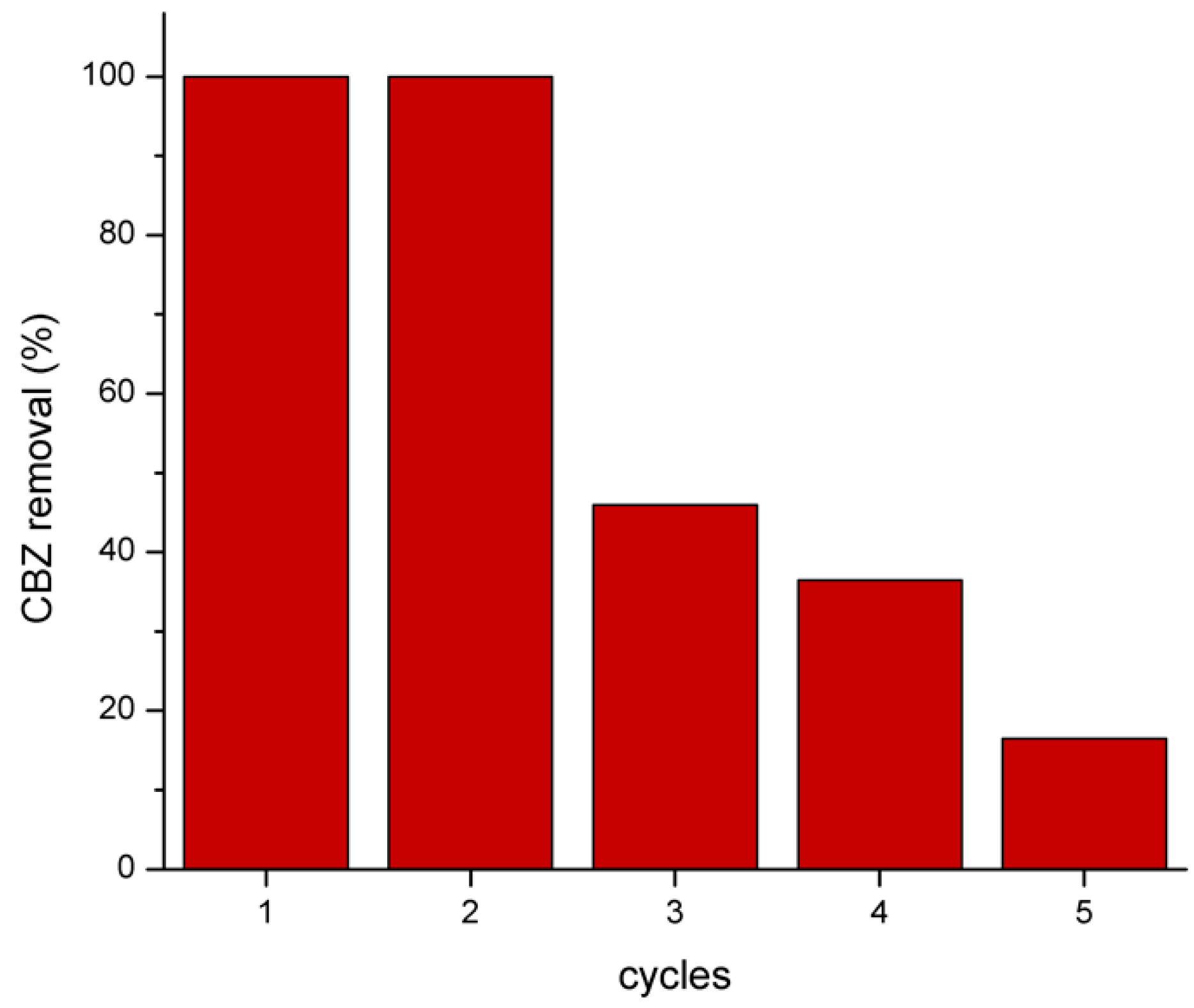
2.4. Reuse Test
2.5. Product Analysis
3. Results
3.1. Characterization of Magnetic Nanoparticles
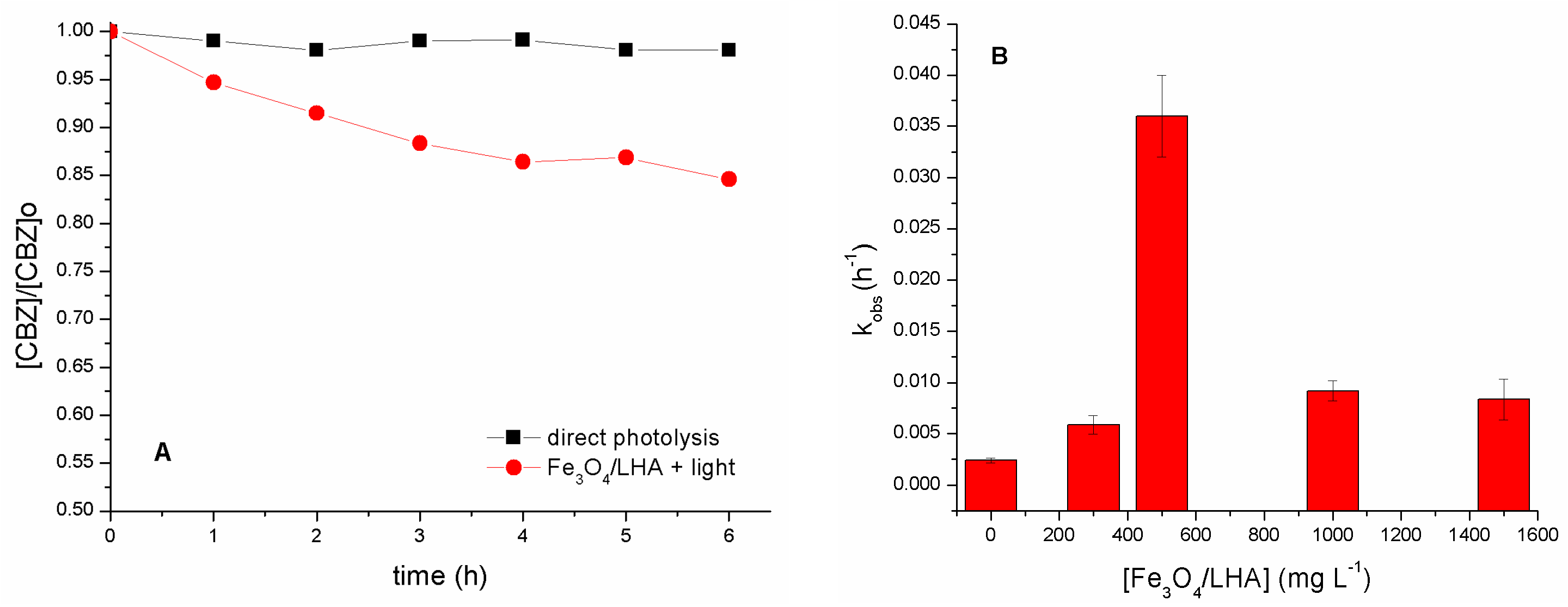
3.2. Fe3O4/Leonardite Humic Acid (LHA) as Photosensitizers
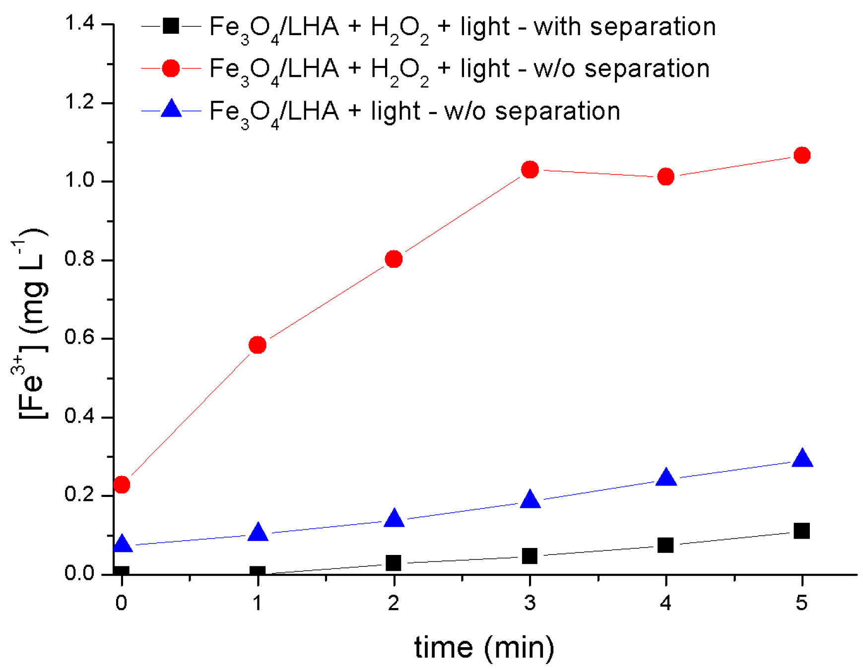
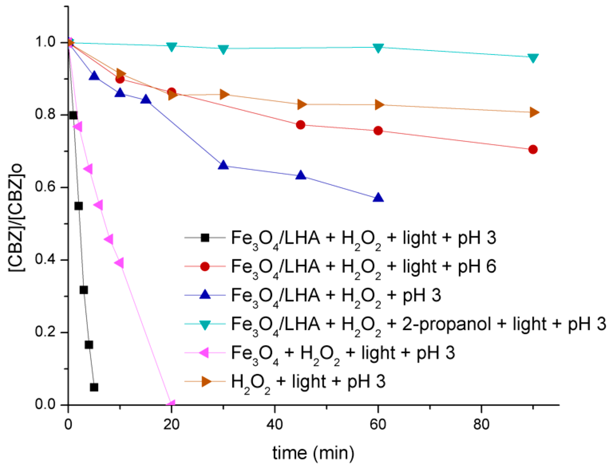
3.3. Fe3O4/LHA as Iron Sources in Photon-Fenton Treatment
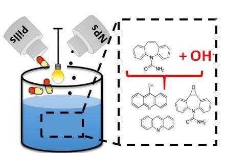
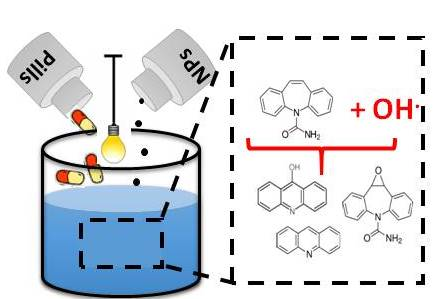
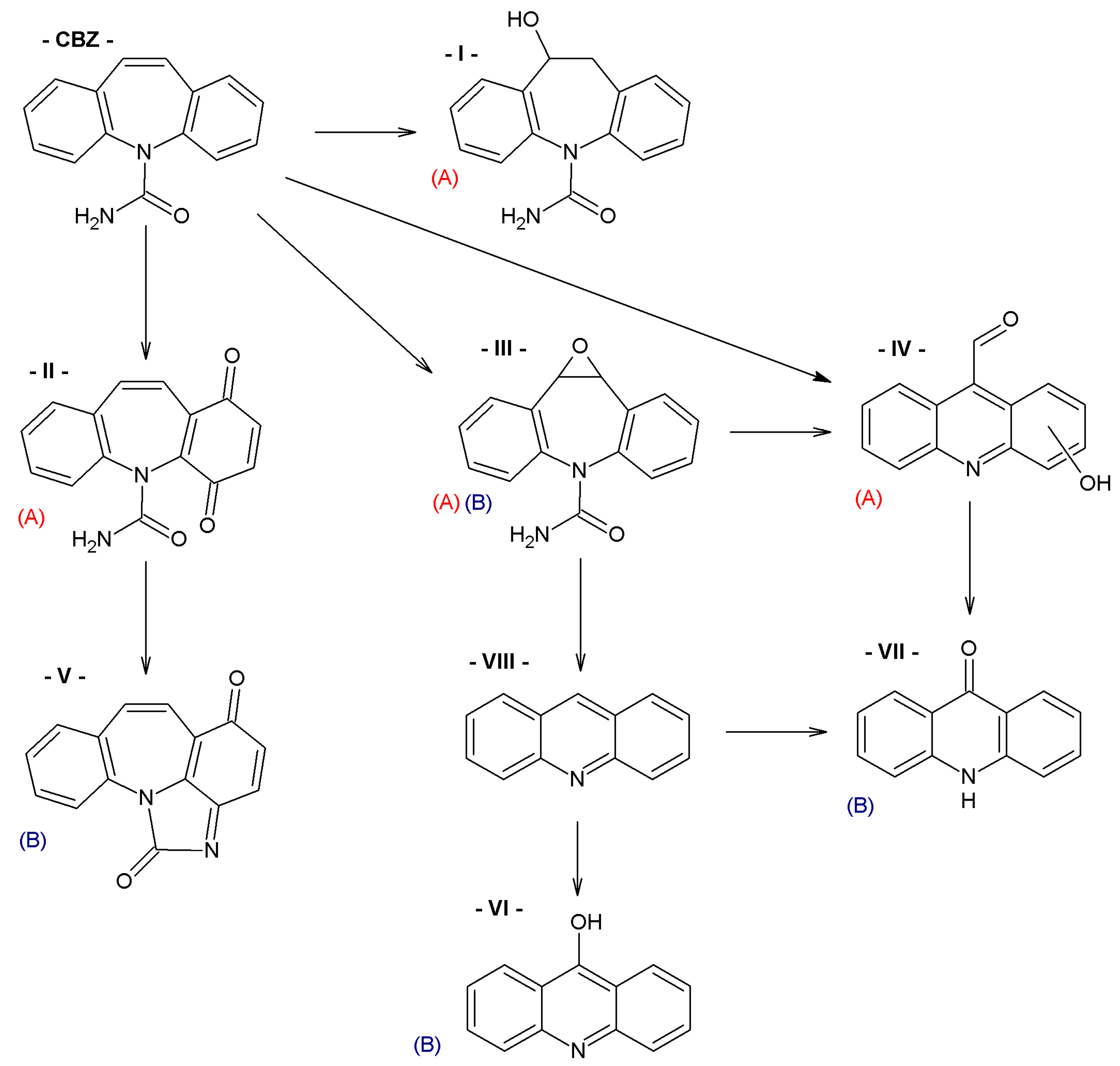
3.4. Product Analysis
4. Conclusions
Supplementary Materials
Author Contributions
Funding
Conflicts of Interest
References
- Petrovic, M.; Barceló, D. Liquid chromatography–mass spectrometry in the analysis of emerging environmental contaminants. Anal. Bioanal. Chem. 2006, 385, 422–424. [Google Scholar] [CrossRef] [PubMed]
- Richardson, S.D. Environmental mass spectrometry: Emerging contaminants and current issues. Anal. Chem. 2012, 84, 747–778. [Google Scholar] [CrossRef] [PubMed]
- Petrie, B.; Barden, R.; Kasprzyk-Hordern, B. A review on emerging contaminants in wastewaters and the environment: Current knowledge, understudied areas and recommendations for future monitoring. Water Res. 2015, 72, 3–27. [Google Scholar] [CrossRef] [PubMed]
- Huerta, B.; Rodriguez-Mozaz, S.; Lazorchak, J.; Barcelo, D.; Batt, A.; Wathen, J.; Stahl, L. Presence of pharmaceuticals in fish collected from urban rivers in the U.S. EPA 2008–2009 national rivers and streams assessment. Sci. Total Environ. 2018, 634, 542–549. [Google Scholar] [CrossRef] [PubMed]
- Martínez-Alcalá, I.; Guillén-Navarro, J.M.; Fernández-López, C. Pharmaceutical biological degradation, sorption and mass balance determination in a conventional activated-sludge wastewater treatment plant from Murcia, Spain. Chem. Eng. J. 2017, 316, 332–340. [Google Scholar] [CrossRef]
- Van De Steene, J.C.; Stove, C.P.; Lambert, W.E. A field study on 8 pharmaceuticals and 1 pesticide in Belgium: Removal rates in waste water treatment plants and occurrence in surface water. Sci. Total Environ. 2010, 408, 3448–3453. [Google Scholar] [CrossRef] [PubMed]
- Ebele, A.J.; Abou-Elwafa Abdallah, M.; Harrad, S. Pharmaceuticals and personal care products (PPCPs) in the freshwater aquatic environment. Emerg. Contam. 2017, 3, 1–16. [Google Scholar] [CrossRef]
- Elorriaga, Y.; Marino, D.J.; Carriquiriborde, P.; Ronco, A.E. Human pharmaceuticals in wastewaters from urbanized areas of Argentina. Bull. Environ. Contam. Toxicol. 2013, 90, 397–400. [Google Scholar] [CrossRef]
- Ribeiro, A.R.; Nunes, O.C.; Pereira, M.F.R.; Silva, A.M.T. An overview on the advanced oxidation processes applied for the treatment of water pollutants defined in the recently launched Directive 2013/39/EU. Environ. Int. 2015, 75, 33–51. [Google Scholar] [CrossRef] [PubMed]
- Gligorovski, S.; Strekowski, R.; Barbati, S.; Vione, D. Environmental implications of hydroxyl radicals (OH). Chem. Rev. 2015, 115, 13051–13092. [Google Scholar] [CrossRef]
- Alharbi, S.K.; Price, W.E. Degradation and fate of pharmaceutically active contaminants by advanced oxidation processes. Curr. Pollut. Rep. 2017, 3, 268–280. [Google Scholar] [CrossRef]
- Patel, M.; Kumar, R.; Kishor, K.; Mlsna, T.; Pittman, C.U.; Mohan, D. Pharmaceuticals of emerging concern in aquatic systems: Chemistry, occurrence, effects, and removal methods. Chem. Rev. 2019, 119, 3510–3673. [Google Scholar] [CrossRef] [PubMed]
- Tang, S.C.N.; Lo, I.M.C. Magnetic nanoparticles: Essential factors for sustainable environmental applications. Water Res. 2013, 47, 2613–2632. [Google Scholar] [CrossRef] [PubMed]
- Carlos, L.; García Einschlag, F.S.; González, M.C.; Mártire, D.O. Applications of magnetite nanoparticles for heavy metal removal from wastewater. In Waste Water-Treatment Technologies and Recent Analytical Developments; Carlos, L., García Einschlag, F.S., Eds.; Intech: London, UK, 2013; pp. 63–77. [Google Scholar]
- Zhang, Y.; Wu, B.; Xu, H.; Liu, H.; Wang, M.; He, Y.; Pan, B. Nanomaterials-enabled water and wastewater treatment. NanoImpact 2016, 3, 22–39. [Google Scholar] [CrossRef]
- Iqbal, A.; Iqbal, K.; Li, B.; Gong, D.; Qin, W. Recent advances in iron nanoparticles: Preparation, properties, biological and environmental application. J. Nanosci. Nanotechnol. 2017, 17, 4386–4409. [Google Scholar] [CrossRef]
- Mohammed, L.; Gomaa, H.G.; Ragab, D.; Zhu, J. Magnetic nanoparticles for environmental and biomedical applications: A review. Particuology 2017, 30, 1–14. [Google Scholar] [CrossRef]
- Mamba, G.; Mishra, A. Advances in magnetically separable photocatalysts: Smart, recyclable materials for water pollution mitigation. Catalysts 2016, 6, 79. [Google Scholar] [CrossRef]
- Gómez-Pastora, J.; Dominguez, S.; Bringas, E.; Rivero, M.J.; Ortiz, I.; Dionysiou, D.D. Review and perspectives on the use of magnetic nanophotocatalysts (MNPCs) in water treatment. Chem. Eng. J. 2017, 310, 407–427. [Google Scholar] [CrossRef]
- Carlos, L.; Cipollone, M.; Soria, D.B.; Moreno, M.S.; Ogilby, P.R.; García Einschlag, F.S.; Mártire, D.O. The effect of humic acid binding to magnetite nanoparticles on the photogeneration of reactive oxygen species. Sep. Purif. Technol. 2012, 91, 23–29. [Google Scholar] [CrossRef]
- Polliotto, V.; Pomilla, F.R.; Maurino, V.; Marcì, G.; Bianco Prevot, A.; Nisticò, R.; Magnacca, G.; Paganini, M.C.; Ponce Robles, L.; Perez, L.; et al. Different approaches for the solar photocatalytic removal of micro-contaminants from aqueous environment: Titania vs. hybrid magnetic iron oxides. Catal. Today 2019, 328, 164–171. [Google Scholar] [CrossRef]
- Franzoso, F.; Nisticò, R.; Cesano, F.; Corazzari, I.; Turci, F.; Scarano, D.; Bianco Prevot, A.; Magnacca, G.; Carlos, L.; Mártire, D.O. Biowaste-derived substances as a tool for obtaining magnet-sensitive materials for environmental applications in wastewater treatments. Chem. Eng. J. 2017, 310, 307–316. [Google Scholar] [CrossRef]
- García Einschlag, F.S.; Carlos, L.; Capparelli, A.L.; Braun, A.M.; Oliveros, E. Degradation of nitroaromatic compounds by the UV-H2O2 process using polychromatic radiation sources. Photochem. Photobiol. Sci. 2002, 1, 520–525. [Google Scholar] [CrossRef] [PubMed]
- Tarafder, P.K.; Thakur, A.R. An optimised 1,10-phenanthroline method for the determination of ferrous and ferric oxides in silicate rocks, soils and minerals. Geostand. Geoanal. Res. 2013, 37, 155–168. [Google Scholar] [CrossRef]
- Woods, J.; Mellon, M. Thiocyanate method for iron: A spectrophotometric study. Ind. Eng. Chem. Anal. Ed. 1941, 13, 551–554. [Google Scholar] [CrossRef]
- Maity, D.; Agrawal, D.C. Synthesis of iron oxide nanoparticles under oxidizing environment and their stabilization in aqueous and non-aqueous media. J. Magn. Magn. Mater. 2007, 308, 46–55. [Google Scholar] [CrossRef]
- Wang, X.; Chen, C.; Liu, H.; Ma, J. Preparation and characterization of PAA/PVDF membrane-immobilized Pd/Fe nanoparticles for dechlorination of trichloroacetic acid. Water Res. 2008, 42, 4656–4664. [Google Scholar] [CrossRef]
- Magnacca, G.; Allera, A.; Montoneri, E.; Celi, L.; Benito, D.E.; Gagliardi, L.G.; Gonzalez, M.C.; Mártire, D.O.; Carlos, L. Novel magnetite nanoparticles coated with waste sourced bio-based substances as sustainable and renewable adsorbing materials. ACS Sustain. Chem. Eng. 2014, 2, 1518–1524. [Google Scholar] [CrossRef]
- Mignone, R.A.; Martin, M.V.; Morán Vieyra, F.E.; Palazzi, V.I.; Lopez de Mishima, B.; Martire, D.O.; Borsarelli, C.D. Modulation of optical properties of dissolved humic substances by their molecular complexity. Photochem. Photobiol. 2012, 88, 792–800. [Google Scholar] [CrossRef]
- Carlos, L.; Mártire, D.O.; Gonzalez, M.C.; Gomis, J.; Bernabeu, A.; Amat, A.M.; Arques, A. Photochemical fate of a mixture of emerging pollutants in the presence of humic substances. Water Res. 2012, 46, 4732–4740. [Google Scholar] [CrossRef]
- Bosio, G.N.; David Gara, P.M.; García Einschlag, F.S.; Gonzalez, M.C.; Del Panno, M.T.; Martire, D.O. Photodegradation of soil organic matter and its effect on gram-negative bacterial growth. Photochem Photobiol. 2008, 84, 1126–1132. [Google Scholar] [CrossRef]
- Martin, M.V.; Ruiz, G.T.; Gonzalez, M.C.; Borsarelli, C.D.; Mártire, D.O. Photolytic and radiolytic oxidation of humic acid. Photochem. Photobiol. 2012, 88, 810–815. [Google Scholar] [CrossRef] [PubMed]
- Chiron, S.; Minero, C.; Vione, D. Photodegradation processes of the antiepileptic drug carbamazepine, relevant to estuarine waters. Environ. Sci. Technol. 2006, 40, 5977–5983. [Google Scholar] [CrossRef] [PubMed]
- Vogna, D.; Marotta, R.; Andreozzi, R.; Napolitano, A.; D’Ischia, M. Kinetic and chemical assessment of the UV/H2O2 treatment of antiepileptic drug carbamazepine. Chemosphere 2004, 54, 497–505. [Google Scholar] [CrossRef]
- De Laurentiis, E.; Chiron, S.; Kouras-Hadef, S.; Richard, C.; Minella, M.; Maurino, V.; Minero, C.; Vione, D. Photochemical fate of carbamazepine in surface freshwaters: Laboratory measures and modeling. Environ. Sci. Technol. 2012, 46, 8164–8173. [Google Scholar] [CrossRef] [PubMed]
- Kosjek, T.; Andersen, H.R.; Kompare, B.; Ledin, A.; Heath, E. Fate of carbamazepine during water treatment. Environ. Sci. Technol. 2009, 43, 6256–6261. [Google Scholar] [CrossRef] [PubMed]
- Zhang, Q.; Chen, J.; Dai, C.; Zhang, Y.; Zhou, X. Degradation of carbamazepine and toxicity evaluation using the UV/persulfate process in aqueous solution. J. Chem. Technol. Biotechnol. 2015, 90, 701–708. [Google Scholar] [CrossRef]
- Zhu, Z.; Chen, Y.; Gu, Y.; Wu, F.; Lu, W.; Xu, T.; Chen, W. Catalytic degradation of recalcitrant pollutants by Fenton-like process using polyacrylonitrile-supported iron (II) phthalocyanine nanofibers: Intermediates and pathway. Water Res. 2016, 93, 296–305. [Google Scholar] [CrossRef] [PubMed]
- Liu, N.; Zheng, M.; Sijak, S.; Tang, L.; Xu, G.; Wu, M. Aquatic photolysis of carbamazepine by UV/H2O2 and UV/Fe (II) processes. Res. Chem. Intermed. 2015, 41, 7015–7028. [Google Scholar] [CrossRef]


| Product Number | Formula | Molecular Ions (M + H)+, (m/z) | Specific Fragments (m/z) | Type of Treatment | |
|---|---|---|---|---|---|
| A | B | ||||
| I |  | 255 | 237 | √ | |
| II | 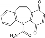 | 267 | 249 | √ | |
| III |  | 253 | 210, 180 | √ | √ |
| IV | 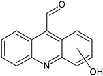 | 224 | 196 | √ | |
| V |  | 251 | 223, 208, 180 | √ | |
| VI | 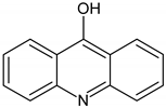 | 196 | 167, 168 | √ | |
| VII |  | 196 | 167, 168 | √ | |
© 2019 by the authors. Licensee MDPI, Basel, Switzerland. This article is an open access article distributed under the terms and conditions of the Creative Commons Attribution (CC BY) license (http://creativecommons.org/licenses/by/4.0/).
Share and Cite
Aparicio, F.; Escalada, J.P.; De Gerónimo, E.; Aparicio, V.C.; García Einschlag, F.S.; Magnacca, G.; Carlos, L.; Mártire, D.O. Carbamazepine Degradation Mediated by Light in the Presence of Humic Substances-Coated Magnetite Nanoparticles. Nanomaterials 2019, 9, 1379. https://doi.org/10.3390/nano9101379
Aparicio F, Escalada JP, De Gerónimo E, Aparicio VC, García Einschlag FS, Magnacca G, Carlos L, Mártire DO. Carbamazepine Degradation Mediated by Light in the Presence of Humic Substances-Coated Magnetite Nanoparticles. Nanomaterials. 2019; 9(10):1379. https://doi.org/10.3390/nano9101379
Chicago/Turabian StyleAparicio, Francisca, Juan Pablo Escalada, Eduardo De Gerónimo, Virginia C. Aparicio, Fernando S. García Einschlag, Giuliana Magnacca, Luciano Carlos, and Daniel O. Mártire. 2019. "Carbamazepine Degradation Mediated by Light in the Presence of Humic Substances-Coated Magnetite Nanoparticles" Nanomaterials 9, no. 10: 1379. https://doi.org/10.3390/nano9101379