Article

Antifungal Activity of Biosynthesized Silver Nanoparticles (AgNPs) against Aspergilli Causing Aspergillosis: Ultrastructure Study

1
Botany and Microbiology Department, Faculty of Science, Al-Azhar University, Cairo 11884, Egypt
2
Regional Center for Mycology and Biotechnology, Al-Azhar University, Cairo 11884, Egypt
3
Department of Botany and Microbiology, Faculty of Science, King Saud University, Riyadh 11451, Saudi Arabia
4
School of Molecular Sciences, The University of Western Australia, Perth, WA 6009, Australia
5
Microbiology and Immunology Department, Faculty of Pharmacy (Boys), Al-Azhar University, Cairo 11884, Egypt
*
Authors to whom correspondence should be addressed.
J. Funct. Biomater. 2022, 13(4), 242; https://doi.org/10.3390/jfb13040242
Submission received: 14 October 2022 / Revised: 3 November 2022 / Accepted: 11 November 2022 / Published: 15 November 2022

Abstract

:
Currently, nanoparticles and nanomaterials are widely used for biomedical applications. In the present study, silver nanoparticles (AgNPs) were successfully biosynthesized using a cell-free extract (CFE) of Bacillus thuringiensis MAE 6 through a green and ecofriendly method. The size of the biosynthesized AgNPs was 32.7 nm, and their crystalline nature was confirmed by XRD, according to characterization results. A surface plasmon resonance spectrum of AgNPs was obtained at 420 nm. Nanoparticles were further characterized using DLS and FTIR analyses, which provided information on their size, stability, and functional groups. AgNPs revealed less cytotoxicity against normal Vero cell line [IC50 = 155 μg/mL]. Moreover, the biosynthesized AgNPs exhibited promising antifungal activity against four most common Aspergillus, including Aspergillus niger, A. terreus, A. flavus, and A. fumigatus at concentrations of 500 μg/mL where inhibition zones were 16, 20, 26, and 19 mm, respectively. In addition, MICs of AgNPs against A. niger, A. terreus, A. flavus, and A. fumigatus were 125, 62.5, 15.62, and 62.5 μg/mL, respectively. Furthermore, the ultrastructural study confirmed the antifungal effect of AgNPs, where the cell wall’s integrity and homogeneity were lost; the cell membrane had separated from the cell wall and had intruded into the cytoplasm. In conclusion, the biosynthesized AgNPs using a CFE of B. thuringiensis can be used as a promising antifungal agent against Aspergillus species causing Aspergillosis.

1. Introduction

The most common spoilage fungi are Aspergillus sp., Fusarium sp., Penicillium sp., and Candida sp.; they can produce mycotoxins, which are extremely harmful to both humans and animals. Symptoms of spoilage fungi can also cause grain discoloration, chemical and nutritional changes, and reduced germination [1,2]. Aspergillus species are widespread and their spores contribute up to 22% of the air spore samples [3]. Although there are more than 250 Aspergillus species, only a small number of them cause problems in humans [4]. Aspergillus infections mostly affect the respiratory system. Inhaled Aspergillus spores can germinate in the presence of ideal lung conditions such as high humidity, oxygen, and carbon dioxide [5,6,7].
The most prevalent aspergillosis-causing species is Aspergillus fumigatus, followed by A. flavus and A. niger, while A. niger is among the most prevalent infections that cause mycosis in humans. These molds can induce systemic invasive infections and kill at least 50% of infected people [8,9]. In addition to contaminating a variety of nuts, fruits, vegetables, and grains, A. flavus also produces aflatoxins, which are mutagenic and carcinogenic [10]. Recently, the emergence of drug-resistant Aspergillus species isolates has been reported as a result of prolonged exposure to antifungals [11]. To avoid the generation of more drug-resistant Aspergillus sps., novel antifungal medications are urgently needed for the treatment of aspergillosis. New antifungal agents can be created using nanotechnology to reduce the effects of newly developed resistant fungi [12].
For successful control and treatment of fungal diseases, recent strategies including the production of nanoparticle-based medicines have been addressed [13]. Nanoparticles, such as silver nanoparticles (AgNPs), have been used for a long time as antimicrobial agents and are highly effective against fungal pathogens [14,15,16,17,18]. AgNPs work through a variety of mechanisms, including binding to phosphate groups in DNA [19] and plasma membrane interactions that cause protons to diffuse and cause cell death [20]. They can also interact with sulfhydryl groups in proteins and enzymes, interfere with the electron transport chain, and disrupt membrane permeability to phosphate groups and protons [21]. The production of AgNPs, which makes up almost half of all manufactured nanomaterials globally, is over 450 tons [22]. The commercialization of AgNPs occurs most frequently in the fields of microelectronics, textiles, food and beverage packaging, and healthcare [23]. The traditional methods of chemical synthesis are expensive and result in environmentally hazardous and poisonous chemicals. Therefore, scientists have investigated the ambient biological processes that often take place in the natural systems. The study of natural systems has led to the development of microbial and plant-extract-based methods that have replaced nanosynthesis. Microorganisms have drawn a lot of interest in the green production of nanomaterials [24,25,26,27]. The biological and physicochemical properties of these nanoparticles are valuable in various applications in many fields, including biomedical and agricultural production [13,28,29,30,31,32]. Several microorganisms, including different bacterial species, have successfully been employed for the bio-fabrication of AgNPs [33]. Herein, this study aimed to biosynthesize AgNPs using bacteria and fully characterize them using UV–vis, TEM, FTIR, XRD, and DLS, as well as to study the antifungal activity of AgNPs against aspergilli causing aspergillosis. In addition, ultrastructure changes of highly sensitive Aspergillus species were investigated.

2. Material and Methods

2.1. Isolation of Bacterial Isolates

Twenty bacterial isolates were isolated from different areas. All isolates were screened and tested for the production of AgNPs. The bacterial isolate coded MAE 6 was the only isolate that has a high-yielding capacity for AgNP biosynthesis, which was determined by UV–Vis spectroscopy. This strain was isolated from a soil sample obtained from Egypt’s eastern desert (N: 29°58′49.06″, E: 32°7′3.39″), in a sterilized 250 mL screw-capped bottle. The bacterial strain was isolated using the serial dilution plate method on Luria–Bertani (LB) agar plates. Briefly, 10 mL of sterile water and 1 g of soil sample were combined, well mixed, and serially diluted. On the plates, streaks of the diluted samples were applied. The plates were then incubated at 37 °C for 24 h. A single discrete colony was picked out and re-cultured into fresh plates to check the purity [34].

2.2. Identification of Bacterial Strain

2.2.1. Morphological and Biochemical Identification

The isolated pure culture was identified by performing morphological and biochemical tests such as Gram staining, motility, endospore staining, oxidase, catalase, indole, and starch hydrolysis, and glucose fermentation tests [35].

2.2.2. Molecular Identification

The isolated strain was categorized at the species level by 16S rRNA gene sequencing. PCR was employed to amplify the 16S rRNA after the bacterial DNA was separated. This was performed using the 27F and 1492R primers (5’-AGAGTTTGATCCTGGCTCAG-3’ and 5’ GGTTACCTTGTTACGACTT-3’, respectively). A PCR purification kit was used to purify the PCR results, and an ABI 3730xl DNA sequencer (GATC Biotech, Germany) was used to sequence them. In order to compare the amplified product sequences, the BLAST (NCBI) search similarity analysis was used. The strain phylogenetic region was verified by creating a phylogenetic tree using the Neighbor-joining approach using MEGA 7.0 software and 1000 bootstrap repetitions.

2.3. Biosynthesis of AgNPs

AgNPs were generated according to a previous protocol [34] by inoculating 200 mL of nutrient broth with the B. thuringiensis MAE 6 culture, followed by 24 h of shaking incubation. After incubation, the culture was centrifuged for 20 min at 3075 g to separate the pellet and supernatant. In a clean 250 mL conical flask, supernatant (100 mL) was added, and 1 mL of silver nitrate solution (1 mM) was added. The second reaction served as a control test without AgNO3. The mixture was then incubated in a shaking incubator at 200 rpm, 37 °C for 24 h in the dark to prevent oxidation of AgNO3. Visual confirmation of AgNP synthesis was investigated by changing the yellow color of the cell-free extract to a brownish color when the precursor was added to it. To create a fine AgNP powder, the supernatant was dried at 60 °C followed by crushing. To completely remove any contaminants from the control flask, AgNPs were thoroughly washed with distilled water and stored for characterization.

2.4. AgNP Characterization

AgNPs were characterized according to methods used by Saied et al. [34]. A reaction mix ranging from light yellow to brownish was formed after 24 h. The formation of AgNPs was thought to be indicated by this color change, in which the color shift of control was not seen as compared to the sample. The results were confirmed using conventional characterization methods, such as a UV–visible spectrophotometer (scanning spectra 109 range from 300 to 700 nm, Shimadzu UV-1700, Kyoto, Japan). The morphology of AgNPs was examined using transmission electron microscopy (JEOL 1010 TEM, Japan). The functional groups involved in reducing, capping, and stabilization of AgNPs were studied using Fourier transform infrared spectroscopy (FTIR) (Perkin-Elmer FTIR-1600, USA) by following the standard operating protocol, and the spectral bands of prepared (crystallized form) AgNPs were determined with 400–4000 cm−1. X-ray diffraction (XRD) (Shimadzu LabX XRD-6000) with a Cu-Kα X-ray source (λ = 1.5418 Å) analysis was used to evaluate the crystallite nature of AgNPs. The average particle size and size distribution of AgNPs in colloidal solutions were determined by dynamic light scattering (DLS) (The Nicomp ZLS Z 3000, USA).

2.5. In Vitro Cytotoxicity of AgNPs against Normal Cell Line

The cytotoxicity of AgNPs and was determined using the MTT protocol [36] with minor modification. An American-type culture collection provided the typical Vero cell line (ATCC). Using the following formula, the total number of cells and the percentage of viable cells were calculated:
Viability   % = Test   OD Control   OD × 100

2.6. In Vitro Antifungal Activity of AgNPs

The antifungal activity of AgNPs was evaluated using agar well diffusion against Aspergillus niger RCMB 02724, A. terreus RCMB 02574, A. flavus RCMB 02782, and A. fumigatus RCMB 02568. All tested fungal strains were grown on PDA plates and incubated for 3–5 days at 30 °C [37,38]. The fungal suspension was prepared in sterilized phosphate buffer solution (PBS) pH 7.0, and then the inoculum was adjusted to 107 spores/mL after counting in a cell counter chamber. One milliliter was uniformly distributed on agar MEA plates. Using a sterile cork-borer, wells (8 mm) were cut; 100 µL of AgNPs and AgNO3 were transferred to each well individually and left for 2 h at 4 °C. Nystatin was used as a standard antifungal, and then the plates were incubated for 3 days at 30 °C. After incubation, the inhibition zones were determined and recorded. Moreover, different concentrations of AgNPs were evaluated as antifungal to detect the minimum inhibitory concentration (MIC).

2.7. Ultrastructure Study

An ultrastructure study was carried out on highly AgNPs-sensitive Aspergillus sp. to AgNPs. Therefore, this study was performed on A. terreus and A. flavus. TEM samples were prepared as follows: fungal specimens (nearly 1 mm3 each) were removed from agar colonies. The samples were fixed in phosphate buffer, washed in 3% glutaraldehyde, and then post-fixed for five minutes at room temperature in potassium permanganate solution. The samples were dehydrated for 30 min in absolute ethanol after 15 min in each ethanol dilution, which ranged from 10% to 90%. They were subsequently submerged in pure resin after being subjected to a graded series of injections of epoxy resin and acetone. Very thin fragments were collected on copper grids. Sections were then stained three times with uranyl acetate and lead citrate. A JEOL-JEM 1010 transmission electron microscope was employed by the Regional Center for Mycology and Biotechnology (RCMB), Al- Azhar University, to examine stained slices at a voltage of 70 kV [39,40,41].

2.8. Statistical Analysis

As all experiments were carried out in triplicate, and the means and standard errors were calculated using SPSS v18 by t-test. The significance levels were set at p < 0.05.

3. Results and Discussion

3.1. Isolation and Identification of the Bacterial Isolate Bacillus sp. MAE 6

Studying of morphological and biochemical characteristics was carried out to identify the bacterial isolate, and molecular identification (16S rDNA) was performed to confirm the morphological identification. The bacterial isolate MAE 6 was morphologically characterized as Gram-positive endospore formers, and showed negative results for β-xylosidase, d-mannitol, d-mannose, β-glucosidase, and methyl-d-xyloside but positive results for d-glucose, d-trehalose, esculin hydrolysis, phenylalanine arylamidase, and tyrosine arylamidase. The biochemical assays of MAE 6 were carried out by the bioMérieux VITAK2 system and exhibited the identical characteristics of B. thuringiensis as described in Bergey’s Manual of Systematic Bacteriology. Supplementary Table S1 displays all of these data. The 16S rDNA fragment of MAE 6 was sequenced for further confirmation of the molecular identity. As illustrated in Figure 1, the MAE 6 phylogenetic tree that was constructed revealed a clustering link between MAE 6 and the B. thuringiensis strains. As a result, the bacterial isolate MAE 6 was identified to be B. thuringiensis MAE 6, and the sequencing was added to the Gene Bank with the accession number MW547437. The 16S rDNA gene contains highly variable sequences that are a powerful tool in species identification [42]. This technique was used by [42], and they identified bacterial strain KFU36 as Bacillus sp. with an identity of 99%. Similarly, Wilson et al. [43] employed the 16S rDNA technique to identify the bacterial isolate (P3) as B. subtilis with a similarity of 100%. Moreover, the 16S rDNA shows that the maximum sequence identity (99.48%) of strain AW1-2 was reached with B. cereus strain CMS7 (Z2) (KX3443935), B. anthracis strain WFS6 (KU977477), and B. thuringiensis WG1 (KU977479) [44]. This method was successfully used by Saied et al. [45] to identify HIS7 as a Lysinibacillus cresolivuorans strain.

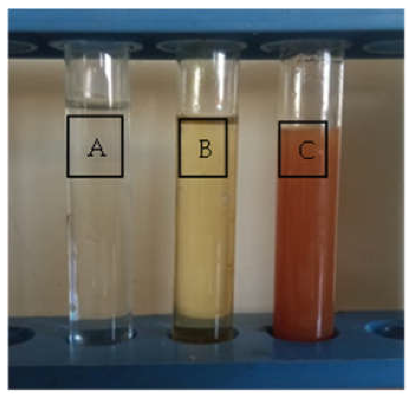
3.2. Biosynthesis of AgNPs Using CFE of B. thuringiensis

The reaction of silver nitrate with the culture supernatant of B. thuringiensis MAE 6 and the gradual color change to brown were visually detected after 24 h of incubation, as shown in Figure 2. As a result, the OH groups of the bioactive metabolites in the cell-free extract (CFE) were electrostatically attracted to the positive-charged Ag ions, which reduced to Ag0 and eventually led to the creation of AgNPs. The control color did not alter. This result was consistent with those reported for a number of Bacillus species, including B. subtilis [46], B. licheniformis [47], B. cereus [48], and B. thuringiensis [49]. Liu et al. [50] showed that they synthesized AgNPs by using Lysinibacillus sphaericus after 24 h. On the other hand, Nallal et al. [51] succeeded in the biosynthesis of AgNPs by using 1 mM of silver nitrate mixed with an A. ampeloprasum bulb extract within 20 min. Additionally, after 24 h of incubation, the green production of AgNPs utilizing A. sydowii was accomplished [52].

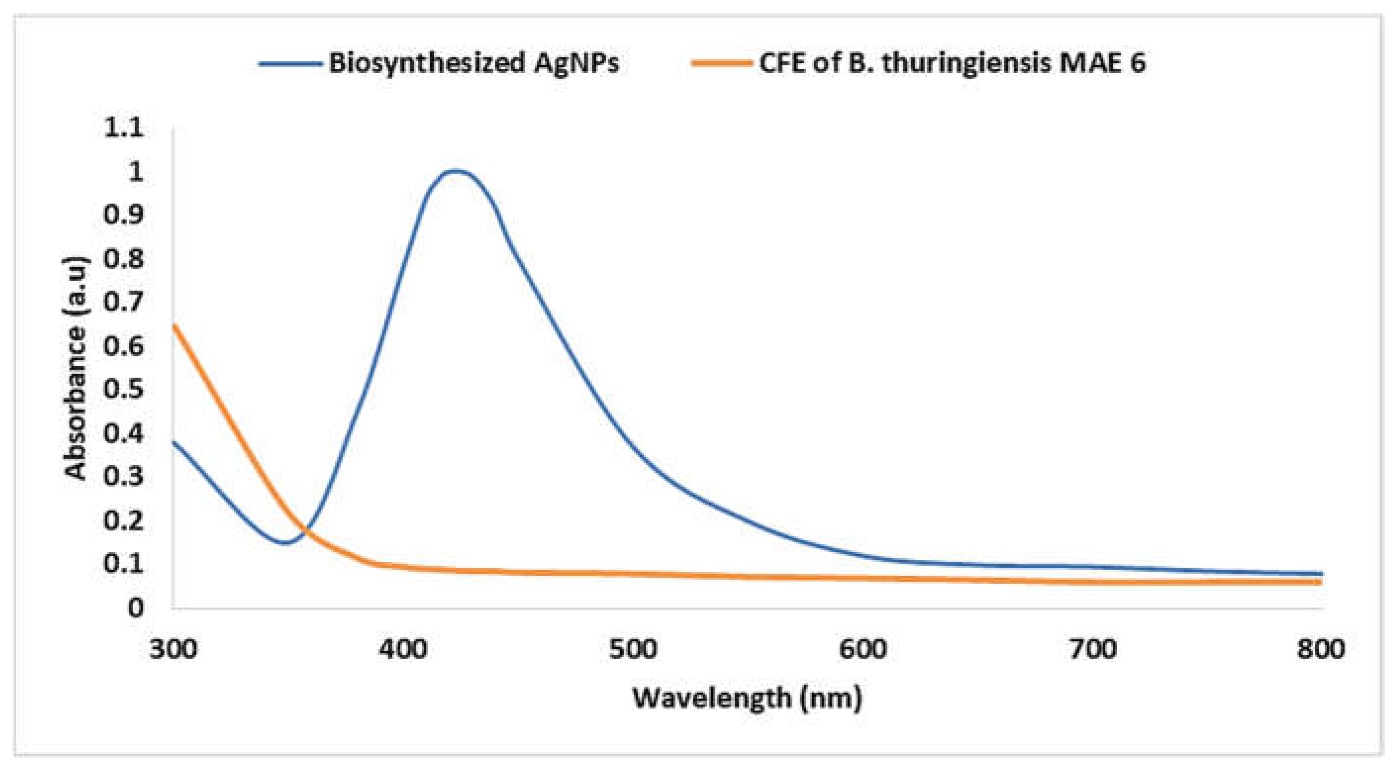
3.3. Characterization of Biosynthesized AgNPs

A UV–Vis spectrophotometer was used to characterize the prepared biosynthesized AgNPs in the preliminary stage, and it produced an absorbance peak at 420 nm (Figure 3). A powerful localized SPR characteristic was the cause of the NP stimulation. According to the previous explanation, the shape and size of AgNPs are often related to the specific SPR state [53]. Here, AgNPs produced by B. thuringiensis MAE 6 had an SPR absorption band at 420 nm. This was accomplished by the free electrons in the AgNPs mutually vibrating with the light wave. SPR values below or higher than 400 nm indicate smaller or bigger nanoparticles, respectively [54]. Similarly, Dawoud et al. [55] reported that the biosynthesis of AgNPs by Nigrospora oryzae occurred at an absorbance peak of 420 nm. Additionally, UV–Vis examination of the produced AgNPs revealed a plasmonic peak at 420 nm [56]. Our results on color change and UV–Vis spectral range resembled the findings of Ahmed et al. [57], who reported the color shift of the reaction mixture from yellow to dense brown and the UV–Vis absorption climax of 418.99 nm in the case of AgNPs produced by using the B. cereus strain. On the other hand, Nallal et al. [51] proposed the effective and quick synthesis of AgNPs by sunlight using an extract of A. ampeloprasum, in which AgNP production was verified by an absorption peak in the UV–Vis spectrum at 446 nm. Using a natural Bacillus sp. strain AW1-2, the absorption peak of UV–Vis spectroscopy was detected at 447 nm for the biogenesis of AgNPs [44]. Moreover, Liu et al. [50] reported that the UV−Vis spectra of the biosynthesized AgNPs by using Lysinibacillus sphaericus showed a prominent absorbance peak at 413 nm.

3.3.1. XRD Analysis

The pure polycrystalline Ag metal was discovered through XRD investigation. AgNPs have been reported to possess a face-centered cubic structure with a comparable crystalline and metallic nature [22,46]. XRD pattern showed the typical Bragg reflections at 2ϴ = 31.5°, 45.2°, 56.3°, and 75.0°, which match the XRD pattern of the indices (111), (200), (220), and (311), as shown in Figure 4. According to the Debye–Scherrer equation, which = 0.9λ/β.cos θ, where 0.1541 nm, the full width at half maximum (FWHM) is the Bragg’s angle in degrees, and D is the average particle size, the average particle size of biogenic AgNPs was determined to be 27.31 nm. The effective production of nanosized particles was proven by the sharp diffraction peak at the lattice plane (111). The XRD measurements obtained agreed with Joint Committee on Powder Diffraction Standards (JCPDS) standard No. 04-0783, confirming that the biosynthesized AgNPs had face-centered cubic (FCC) structures [58,59]. El-Naggar et al. [60] reported the successful fabrication of crystalline AgNPs due to the reduction of Ag ions by phycocyanin, and characterized by face-centered cubic structures. Taran et al. [61] showed that intense peaks corresponding to (111), (200), (220), and (311), which exhibit the formation of AgNPs synthesized by Bacillus sp. HAI4, were crystal and face-centered cubic structure in nature. Furthermore, the capping agents may have stabilized NPs, as seen by the strong diffraction peaks seen in the XRD spectra [62]. Unassigned diffraction peaks in the XRD spectra could be caused by biomolecules covering the surface of the crystallizing NPs [63].

3.3.2. FTIR Analysis

Different biomolecules essential for capping and stabilizing reduced AgNPs as well as reducing silver (Ag+) ions were identified through the use of FTIR analysis. Data represented in (Figure 5) revealed intense absorption peaks at 3430, 2961, 1640, 1405, 1050, 710, 605, and 529 cm−1. OH and the OH stretching group of phenols and alcohol or NH stretching of aliphatic primary amines might be responsible for the large peak shown at 3430 cm−1 [64]. The peak at 2961 cm–1 signified C-H vibrational stretching of aliphatic methyl (CH3). The peak at 1640 cm−1 was associated with I and II amide peptide linkage [65,66]. This amide I band suggests that proteins that may exist in the B. thuringiensis MAE 6 cell-free biomass filtrate have the ability to bind to Ag+ ions through carboxylate ions or free amine groups to assist in the production of AgNPs during a reduction process. The shifting peak at a wavenumber of the peak at 1405 cm–1 can be appointed to C–N stretching vibrations of aliphatic and aromatic amines [67]. The absorption of C-O-C or C-O may be responsible for the peak at 1050 cm−1 that was detected [68]. The stretching peaks which appeared at 710 and 605 cm−1 indicated the amide IV (OCN) stretch bending for protein and alkene (=C-H bending), respectively. In accordance with other evidence, proteins may serve as capping and/or stability agents during the biosynthesis of AgNPs [69,70]. Elbahnasawy et al. [71] discovered that FTIR spectrum analysis suggested that the cell-free extract of Rothia endophytica MAE 11 may function as a reducing and stabilizing agent during the production of AgNPs. Liu et al. [50] showed that FTIR spectra of Lysinibacillus sphaericus mediated the synthesis of AgNPs to identify biomolecules involved in the reduction of Ag+ ions and capping of biosynthesized AgNPs. Almalki and a Khalifa [42] employed FTIR spectra to identify biomolecules found in the CFE of Bacillus sp KFU36, which were used as reducing agents for AgNP synthesis.

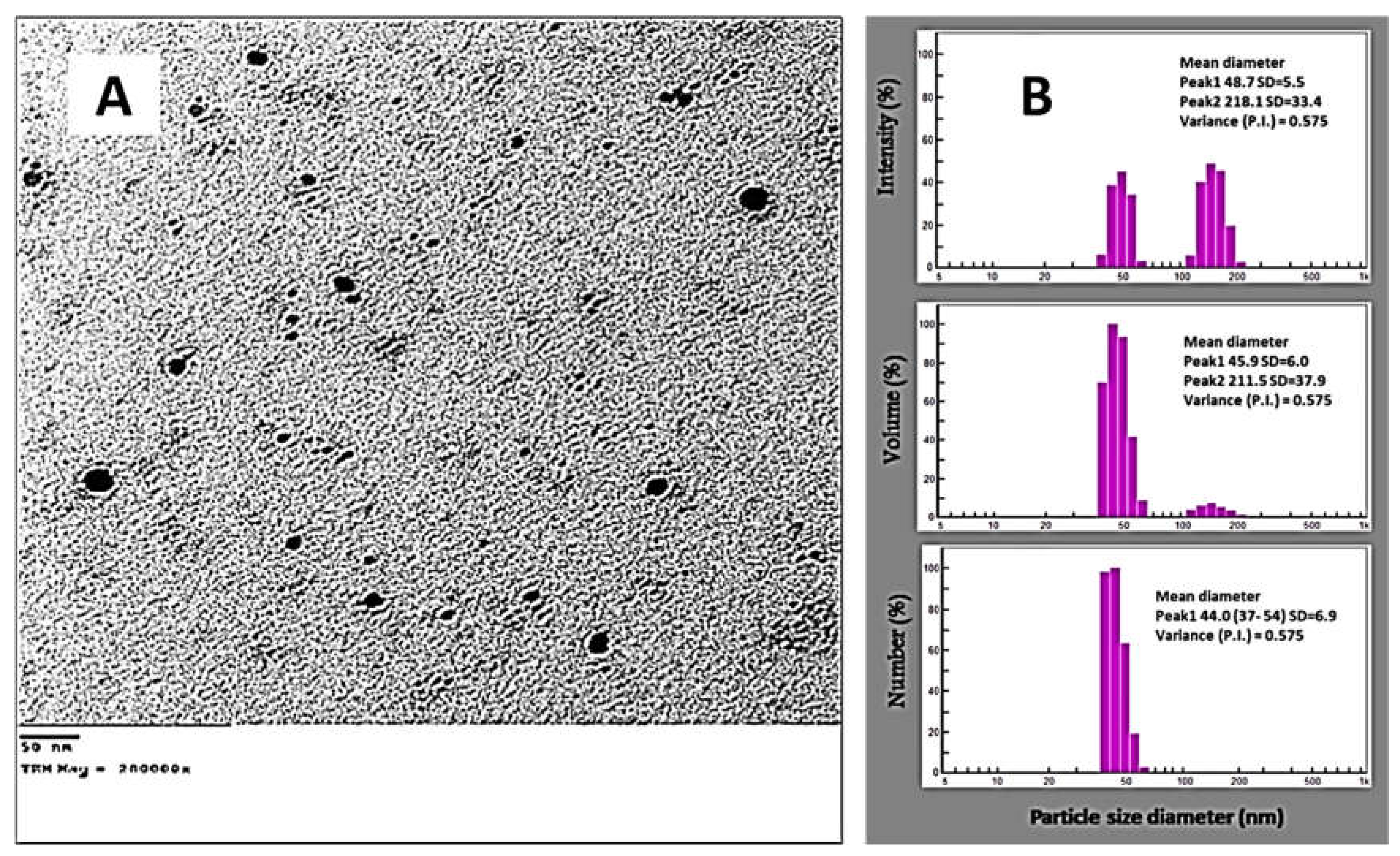
3.3.3. DLS and TEM Analyses

Factors that influence the biotechnological and biological activities of NPs include their size, shape, and distribution [72]. The activity and biocompatibilities of NPs increased as their sizes decreased [63]. Therefore, it is important to examine the NPs’ morphological characteristics. TEM was employed to achieve this objective. Monodispersed spheres were the image of the form of NPs. The size distribution histogram revealed that the average size of the nanoparticles, which ranged in size from 30 to 47 nm, was 32.7 nm (Figure 6A), which was smaller than that formed with B. thuringiensis (43.52–142.97 nm) [73]. The results are consistent with other studies that have been published and show that silver oxide nanoparticles produced using B. linens varied in size from 25 to 70 nm [74]. Additionally, the size of spherical AgNPs synthesized by Bacillus sp KFU36 was extended between 5 and 15 nm [42]. Chi et al. [75] showed that the synthesized BT-AgNPs size ranged from 20.15 to 22.21 nm. Moreover, the AgNPs synthesized by walnut fruit (Juglans regia) extract had an average size of 31.4 nm [76]. The TEM image demonstrates that the metabolites in the culture extract are effective in reducing and capping monodispersed spherical AgNPs without aggregation (Figure 6A). Using the DLS method, AgNP size and particle distribution were assessed. The average particle diameter for AgNPs synthesized by B. thuringiensis MAE 6 was 37.9 nm for a volume, as illustrated in Figure 6B. The electrical double layer that developed on charged AgNPs and/or coating metabolites on the surface of the nanoparticles utilized for capping and stabilizing AgNPs might be responsible for the DLS’s average size being greater than that generated by TEM [77]. AgNPs synthesized by B. subtilis (P3) had an average particle size of 805 nm [43], and this significant disparity could be related to sample variability and synthesis method. A DLS study of silver oxide (Ag2O) nanoparticles produced by B. paramycoides revealed that the majority of the particles are between 50 and 150 nm in size [74]. According to Saied et al. [34], the average diameter of the AgNPs generated by Bacillus sp. was determined to be 55.8 nm for a volume. According to Pavan et al. [78], the DLS examination of the produced AgNPs revealed that the average particle size was 100 nm, and the polydispersity index was 0.224. The average diameter of total particles and stability of green synthesized AgNPs by S. pachycarpa extract were reported as 107–150 nm [79]. Nanoparticles with biomolecules attached to their surfaces offer steric stability and stop the particles from aggregation. The osmotic repulsive force improves when the concentration of adsorbed biomolecules increases, because this enhances the stability of nanostructured particles [80,81].

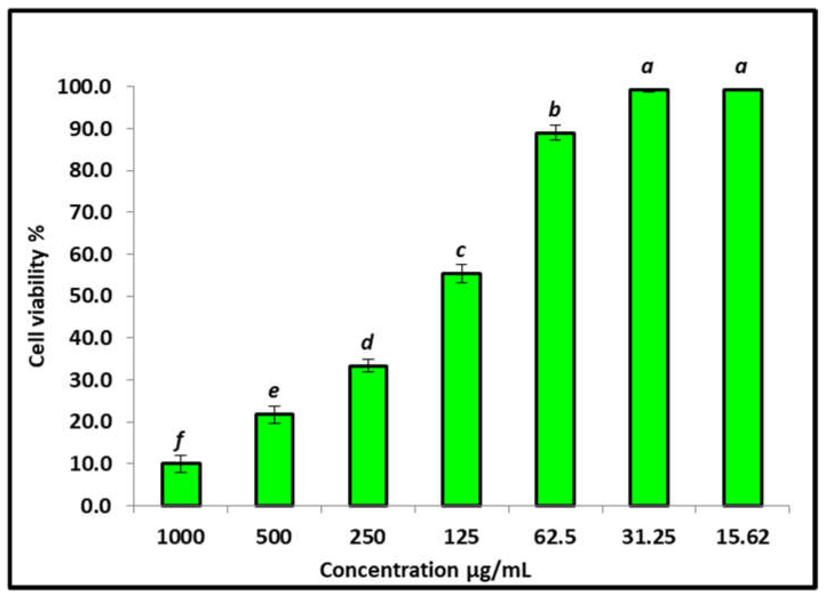
3.4. Effect of the AgNPs on the Vero Cells’ Viability Using MTT Assay

The in vitro cytotoxic effects of biosynthesized AgNPs were evaluated against a Vero normal cell line and the percentage of cell inhibition was confirmed by MTT assay. The MTT in Vero cells treated with AgNPs at different concentrations (15.62–1000 μg/mL) for 24 h is illustrated in Figure 7. The viability of Vero cells decreased when the AgNPs’ concentration increased. The cell viability was recorded as 99.3% at 15.62 μg/mL of AgNPs. However, cell viability was substantially decreased to 10% at 1000 μg/mL of AgNPs, with a toxicity percent of 0.7% and 90% at 15.62 and 1000 μg/mL, respectively, for Vero cells. A substantial inhibition of Vero cell viability by the AgNPs was detected with an IC50 of 155 μg/mL concentrations, indicating cytotoxic effects of these nanoparticles. Similar results agree with data reported by Mohamed et al. [82], who recorded that the normal Vero cell showed 100% viability at 15.62 μg/mL and viability decreased by increasing AgNPs’ concentration: IC50 was recorded at 280 μg/mL. Generally, if the IC50 is ≥ 90 μg/mL, the material is classified as non-cytotoxic [83]. Therefore, the biosynthesized AgNPs in this study are safe for use.

3.5. Antifungal Activity

In this study, the antifungal activity of AgNPs toward Aspergillus species was evaluated using the agar well diffusion method (Figure 8). Results illustrated that biosynthesized AgNPs exhibited promising antifungal activity against all tested fungal strains. AgNPs had the ability to inhibit fungal growth of A. niger, A. terreus, A. flavus, and A. fumigatus at concentrations of 500 μg/mL where inhibition zones were 16, 20, 26, and 19 mm, respectively. Moreover, MICs of AgNPs against A. niger, A. terreus, A. flavus, and A. fumigatus were determined as illustrated in Table 1 MIC results showed that A. flavus was the most sensitive fungus to AgNPs, which inhibited growth at concentrations of 15.6 μg/mL, while for the other fungal species, inhibition of growth was observed at 125 μg/ mL for A. niger and 62.5 μg/mL for A. terreus and A. fumigatus. AgNPs in solution at higher concentrations may be able to adhere to and saturate fungus hyphae, destroying the fungus cells. Such an inhibitory effect can be attributed to Ag+ that primarily affects the function of membrane-associated enzymes such as those found in the respiratory chain. Ag+ may also affect the expression of some microbial proteins and enzymes. Disruption of DNA replication has also been documented. AgNPs can also interact with substrates through a process known as competitive inhibition, inactivating the enzymes and preventing the production of products required for cell activity. At 50 μg/mL nanoparticle concentrations, the production of AgNPs by Bacillus MB353 also exhibited excellent antifungal efficacy against Aspergillus niger, A. fumigatus, and F. soleni [84]. Elbahnasawy et al. [71] reported that the biosynthesized AgNPs using endophytic Rothia endophytica have anticandidal activity against C. albicans, where the MIC and MBC were 62.5 and 125 μg/mL. The antifungal effectiveness against a well-known phytopathogen Colletotrichum falcatum was evaluated by Ajaz et al. [44], where, at a concentration of 20 μg/mL, the ZV-AgNPs greatly inhibited the fungal mycelia in vitro as compared to the untreated control. Particularly, AgNPs prepared by a Penicillium chrysogenum and F. chlamydosporum cell-free culture filtrates showed significant antifungal efficacy against the mycotoxigenic fungus A. flavus and A. ochraceus at a concentration of 47–51 μg/mL [85]. Biogenic AgNPs from the extract of Syzygium cumini leaves at 100 μg/mL show antifungal activity in both strains A. flavus and A. parasiticus [86]. At quantities of 50–200 μg/mL, AgNPs produced by Penicillium verrucosum inhibited the radial growth of F. chlamydosporum and A. flavus, reported by [56]. Wang et al. [52] observed AgNPs produced by A. Sydowii that exhibited antifungal activity against A. fumigatus, A. flavus, and A. terreus. Dashora et al. [87] observed significant antifungal activity against A. alternate at 500 μg/mL of PL-AgNPs mediated by Polyalthia longifolia leaf extract.

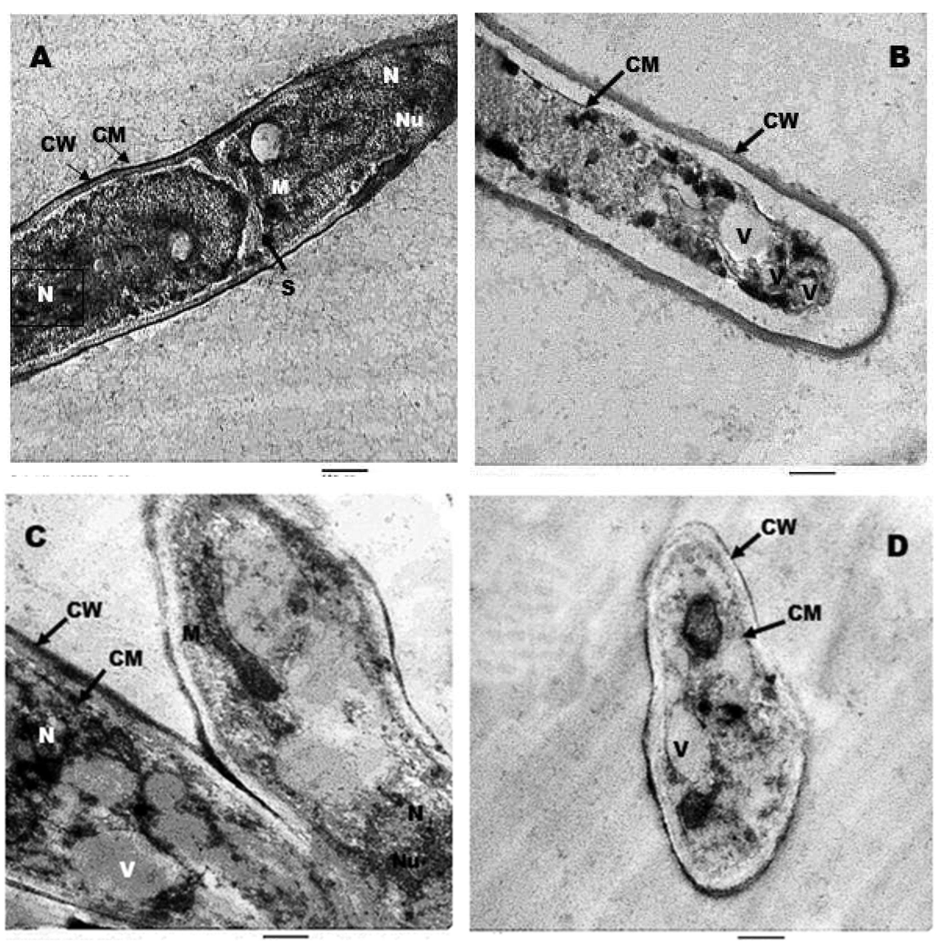
3.6. Ultrastructure Studies by TEM

Ultrathin sections of untreated (control) A. terrus (Figure 9A) and A. flavus (Figure 9C) cells showed normal mycelia with a consistent layout and homogeneous cytoplasm; the cell wall (CW) of the untreated hypha was fully integrated and undamaged. The cell membrane (CM) was entirely connected to the cell wall and was smooth and wrinkle-free. The cytoplasm was dense and uniform, and the intercellular septum (S) was healthy and unbroken, with normal nucleus (N), nucleolus (Nu), and mitochondria (M) identified. Figure 9B,D show the influence of AgNPs on the cell structure of A. terrus and A. flavus, respectively. To some extent, the cell wall’s integrity and homogeneity were lost; the cell membrane had separated from the cell wall and had intruded into the cytoplasm. Other modifications included a cytoplasm density decrease and vacuolization (V).
Hyphal filaments in normal samples of A. terrus have a classical tubular structure where various organelles, including cell wall and cell membrane, appear as well-established and thick structures. In the treated group, using nanoparticles leads to shrinkage in structure, affecting various organelles and causing enlargement of the volume of vacuoles, which agreed with Al-Otibi et al. [88] who studied ultrastructure alteration in Bipolaris sp. upon treatment with AgNPs. Furthermore, photomicrographs of ultrastructure sections of the A. flavus normal group revealed the regular cellular organelles including nuclei, cell wall, and cell membrane, while using AgNPs led to decreased cell size and deformation of various organelles. This is in the same line as [89], who reported the beneficial effect of colloidal AgNPs on pathogenic A. flavus. Our results on ultrastructure studies resembled the findings of previous work [90], which imaged the damage to the integrity of the cell wall and cytoplasmic membrane of Candida albicans through SEM and TEM studies.

4. Conclusions

Green chemistry has been the subject of growing interest due to its being environmentally safe, inexpensive, biocompatible, and that it avoids producing toxic byproducts. In the current study, AgNPs were successfully biosynthesized using the CFE of B. thuringiensis MAE 6. According to FTIR analyses, the generated AgNPs are capped and stabilized with various functional groups. The biosynthesized AgNPs showed low cytotoxic activity against Vero normal cell line, with an IC50 of 155 μg/mL. Moreover, AgNPs exhibited superior performance in antifungal activities against four types of Aspergillus species causing Aspergillosis. Furthermore, an ultrastructure study confirmed the antifungal effect of AgNPs, where the cell wall’s integrity and homogeneity were lost; the cell membrane had separated from the cell wall and had intruded into the cytoplasm. Therefore, the biosynthesized AgNPs using a CFE of B. thuringiensis can be used as a promising antifungal agent against Aspergillus species.

Supplementary Materials

The following supporting information can be downloaded at: https://www.mdpi.com/article/10.3390/jfb13040242/s1, Table S1. Characteristics of B. thuringiensis.

Author Contributions

Conceptualization, A.H.H. and M.A.E.; methodology, A.H.H., M.A.E., B.H.A., E.S. and F.M.E.; data curation, A.H.H. and A.A.A.; writing—original draft preparation, E.S.; writing—review and editing, E.S., A.H.H., M.A.E., F.O.A., A.A.A.-A. and F.M.E.; Funding acquisition, F.O.A. and A.A.A.-A. All authors have read and agreed to the published version of the manuscript.

Funding

This research project was supported by a grant from the Researchers Supporting Project, number (RSP-2021/114), King Saud University, Riyadh, Saudi Arabia.

Institutional Review Board Statement

Not applicable.

Informed Consent Statement

Not applicable.

Data Availability Statement

The data used to support the findings of this study are available from the corresponding author upon request.

Acknowledgments

The authors express their sincere thanks to Faculty of Science, Al-Azhar University, Cairo, Egypt, for providing the necessary research facilities.

Conflicts of Interest

The authors declare no conflict of interest.

References

  1. Habschied, K.; Krstanović, V.; Zdunić, Z.; Babić, J.; Mastanjević, K.; Šarić, G.K. Mycotoxins biocontrol methods for healthier crops and stored products. J. Fungi 2021, 7, 348. [Google Scholar] [CrossRef] [PubMed]
  2. Haque, M.A.; Wang, Y.; Shen, Z.; Li, X.; Saleemi, M.K.; He, C. Mycotoxin contamination and control strategy in human, domestic animal and poultry: A review. Microb. Pathog. 2020, 142, 104095. [Google Scholar] [CrossRef] [PubMed]
  3. Ramírez-Camejo, L.A.; Zuluaga-Montero, A.; Morris, V.; Rodríguez, J.A.; Lázaro-Escudero, M.T.; Bayman, P. Fungal diversity in Sahara dust: Aspergillus sydowii and other opportunistic pathogens. Aerobiologia 2022, 38, 367–378. [Google Scholar] [CrossRef]
  4. Seyedmousavi, S. Aspergillosis in humans and animals. In Recent Trends in Human and Animal Mycology; Springer: Berlin/Heidelberg, Germany, 2019; pp. 81–98. [Google Scholar]
  5. Salazar, F.; Bignell, E.; Brown, G.D.; Cook, P.C.; Warris, A. Pathogenesis of respiratory viral and fungal coinfections. Clin. Microbiol. Rev. 2022, 35, e00094-21. [Google Scholar] [CrossRef] [PubMed]
  6. Kumar, P.; Kausar, M.; Singh, A.; Singh, R. Biological contaminants in the indoor air environment and their impacts on human health. Air Qual. Atmos. Health 2021, 14, 1723–1736. [Google Scholar] [CrossRef] [PubMed]
  7. Dacrory, S.; Hashem, A.H.; Hasanin, M. Synthesis of cellulose based amino acid functionalized nano-biocomplex: Characterization, antifungal activity, molecular docking and hemocompatibility. Environ. Nanotechnol. Monit. Manag. 2021, 15, 100453. [Google Scholar] [CrossRef]
  8. Miller, A.S.; Wilmott, R.W. The Pulmonary Mycoses. In Kendig’s Disorders of the Respiratory Tract in Children; Elsevier: Amsterdam, The Netherlands, 2019; pp. 507–527.e503. [Google Scholar]
  9. Sugui, J.A.; Kwon-Chung, K.J.; Juvvadi, P.R.; Latge, J.-P.; Steinbach, W.J. Aspergillus fumigatus and related species. Cold Spring Harb. Perspect. Med. 2015, 5, a019786. [Google Scholar] [CrossRef] [PubMed] [Green Version]
  10. Tripathi, A.; Alam, A. Mycotoxins, Mycotoxicosis and Managing Mycotoxin Contamination: A Review. Bio-Manag. Postharvest Dis. Mycotoxigenic Fungi 2020, 161–180. [Google Scholar] [CrossRef]
  11. Hendrickson, J.A.; Hu, C.; Aitken, S.L.; Beyda, N. Antifungal resistance: A concerning trend for the present and future. Curr. Infect. Dis. Rep. 2019, 21, 1–8. [Google Scholar] [CrossRef] [PubMed]
  12. Sousa, F.; Ferreira, D.; Reis, S.; Costa, P. Current insights on antifungal therapy: Novel nanotechnology approaches for drug delivery systems and new drugs from natural sources. Pharmaceuticals 2020, 13, 248. [Google Scholar] [CrossRef] [PubMed]
  13. Abdelaziz, A.M.; Dacrory, S.; Hashem, A.H.; Attia, M.S.; Hasanin, M.; Fouda, H.M.; Kamel, S.; ElSaied, H. Protective role of zinc oxide nanoparticles based hydrogel against wilt disease of pepper plant. Biocatal. Agric. Biotechnol. 2021, 35, 102083. [Google Scholar] [CrossRef]
  14. Silva, L.P.; Silveira, A.P.; Bonatto, C.C.; Reis, I.G.; Milreu, P.V. Silver nanoparticles as antimicrobial agents: Past, present, and future. In Nanostructures for Antimicrobial Therapy; Elsevier: Amsterdam, The Netherlands, 2017; pp. 577–596. [Google Scholar]
  15. Salem, S.S.; Ali, O.M.; Reyad, A.M.; Abd-Elsalam, K.A.; Hashem, A.H. Pseudomonas indica-Mediated Silver Nanoparticles: Antifungal and Antioxidant Biogenic Tool for Suppressing Mucormycosis Fungi. J. Fungi 2022, 8, 126. [Google Scholar] [CrossRef]
  16. Hasanin, M.; Elbahnasawy, M.A.; Shehabeldine, A.M.; Hashem, A.H. Ecofriendly preparation of silver nanoparticles-based nanocomposite stabilized by polysaccharides with antibacterial, antifungal and antiviral activities. Biometals Int. J. Role Met. Ions Biol. Biochem. Med. 2021, 34, 1313–1328. [Google Scholar] [CrossRef] [PubMed]
  17. Shehabeldine, A.M.; Salem, S.S.; Ali, O.M.; Abd-Elsalam, K.A.; Elkady, F.M.; Hashem, A.H. Multifunctional Silver Nanoparticles Based on Chitosan: Antibacterial, Antibiofilm, Antifungal, Antioxidant, and Wound-Healing Activities. J. Fungi 2022, 8, 612. [Google Scholar] [CrossRef]
  18. Salem, S.S.; Hashem, A.H.; Sallam, A.-A.M.; Doghish, A.S.; Al-Askar, A.A.; Arishi, A.A.; Shehabeldine, A.M. Synthesis of Silver Nanocomposite Based on Carboxymethyl Cellulose: Antibacterial, Antifungal and Anticancer Activities. Polymers 2022, 14, 3352. [Google Scholar] [CrossRef] [PubMed]
  19. Neves, A.C.; Viana, A.D.; Menezes, F.G.; Neto, A.O.W.; Melo, M.C.N.; Gasparotto, L.H. Biospectroscopy and chemometrics as an analytical tool for comparing the antibacterial mechanism of silver nanoparticles with popular antibiotics against Escherichia coli. Spectrochim. Acta Part A Mol. Biomol. Spectrosc. 2021, 253, 119558. [Google Scholar] [CrossRef]
  20. Salleh, A.; Naomi, R.; Utami, N.D.; Mohammad, A.W.; Mahmoudi, E.; Mustafa, N.; Fauzi, M.B. The potential of silver nanoparticles for antiviral and antibacterial applications: A mechanism of action. Nanomaterials 2020, 10, 1566. [Google Scholar] [CrossRef] [PubMed]
  21. Badmus, J.; Oyemomi, S.; Adedosu, O.; Yekeen, T.; Azeez, M.; Adebayo, E.; Lateef, A.; Badeggi, U.; Botha, S.; Hussein, A. Photo-assisted bio-fabrication of silver nanoparticles using Annona muricata leaf extract: Exploring the antioxidant, anti-diabetic, antimicrobial, and cytotoxic activities. Heliyon 2020, 6, e05413. [Google Scholar] [CrossRef] [PubMed]
  22. Pourzahedi, L.; Vance, M.; Eckelman, M.J. Life cycle assessment and release studies for 15 nanosilver-enabled consumer products: Investigating hotspots and patterns of contribution. Environ. Sci. Technol. 2017, 51, 7148–7158. [Google Scholar] [CrossRef]
  23. Yugay, Y.; Rusapetova, T.; Mashtalyar, D.; Grigorchuk, V.; Vasyutkina, E.; Kudinova, O.; Zenkina, K.; Trifuntova, I.; Karabtsov, A.; Ivanov, V. Biomimetic synthesis of functional silver nanoparticles using hairy roots of Panax ginseng for wheat pathogenic fungi treatment. Colloids Surf. B Biointerfaces 2021, 207, 112031. [Google Scholar] [CrossRef] [PubMed]
  24. Rana, A.; Yadav, K.; Jagadevan, S. A comprehensive review on green synthesis of nature-inspired metal nanoparticles: Mechanism, application and toxicity. J. Clean. Prod. 2020, 272, 122880. [Google Scholar] [CrossRef]
  25. Saratale, R.G.; Karuppusamy, I.; Saratale, G.D.; Pugazhendhi, A.; Kumar, G.; Park, Y.; Ghodake, G.S.; Bharagava, R.N.; Banu, J.R.; Shin, H.S. A comprehensive review on green nanomaterials using biological systems: Recent perception and their future applications. Colloids Surf. B Biointerfaces 2018, 170, 20–35. [Google Scholar] [CrossRef] [PubMed]
  26. Saied, E.; Salem, S.S.; Al-Askar, A.A.; Elkady, F.M.; Arishi, A.A.; Hashem, A.H. Mycosynthesis of Hematite (α-Fe2O3) Nanoparticles Using Aspergillus niger and Their Antimicrobial and Photocatalytic Activities. Bioengineering 2022, 9, 397. [Google Scholar] [CrossRef]
  27. Hasanin, M.; Al Abboud, M.A.; Alawlaqi, M.M.; Abdelghany, T.M.; Hashem, A.H. Ecofriendly Synthesis of Biosynthesized Copper Nanoparticles with Starch-Based Nanocomposite: Antimicrobial, Antioxidant, and Anticancer Activities. Biol. Trace Elem. Res. 2022, 200, 2099–2112. [Google Scholar] [CrossRef]
  28. Salem, S.S.; Fouda, A. Green synthesis of metallic nanoparticles and their prospective biotechnological applications: An overview. Biol. Trace Elem. Res. 2021, 199, 344–370. [Google Scholar] [CrossRef] [PubMed]
  29. Abdelaziz, A.M.; Salem, S.S.; Khalil, A.M.A.; El-Wakil, D.A.; Fouda, H.M.; Hashem, A.H. Potential of biosynthesized zinc oxide nanoparticles to control Fusarium wilt disease in eggplant (Solanum melongena) and promote plant growth. Biometals Int. J. Role Met. Ions Biol. Biochem. Med. 2022, 35, 601–616. [Google Scholar] [CrossRef]
  30. Hashem, A.H.; Al Abboud, M.A.; Alawlaqi, M.M.; Abdelghany, T.M.; Hasanin, M. Synthesis of nanocapsules based on biosynthesized nickel nanoparticles and potato starch: Antimicrobial, antioxidant, and anticancer activity. Starch-Stärke 2022, 74, 2100165. [Google Scholar] [CrossRef]
  31. Abu-Elghait, M.; Hasanin, M.; Hashem, A.H.; Salem, S.S. Ecofriendly novel synthesis of tertiary composite based on cellulose and myco-synthesized selenium nanoparticles: Characterization, antibiofilm and biocompatibility. Int. J. Biol. Macromol. 2021, 175, 294–303. [Google Scholar] [CrossRef]
  32. Hasanin, M.; Hashem, A.H.; Lashin, I.; Hassan, S.A.M. In vitro improvement and rooting of banana plantlets using antifungal nanocomposite based on myco-synthesized copper oxide nanoparticles and starch. Biomass Convers. Biorefinery 2021, 1–11. [Google Scholar] [CrossRef]
  33. Hashem, A.H.; Abdelaziz, A.M.; Askar, A.A.; Fouda, H.M.; Khalil, A.M.A.; Abd-Elsalam, K.A.; Khaleil, M.M. Bacillus megaterium-Mediated Synthesis of Selenium Nanoparticles and Their Antifungal Activity against Rhizoctonia solani in Faba Bean Plants. J. Fungi 2021, 7, 195. [Google Scholar] [CrossRef]
  34. Saied, E.; Hashem, A.H.; Ali, O.M.; Selim, S.; Almuhayawi, M.S.; Elbahnasawy, M.A. Photocatalytic and Antimicrobial Activities of Biosynthesized Silver Nanoparticles Using Cytobacillus firmus. Life 2022, 12, 1331. [Google Scholar] [CrossRef] [PubMed]
  35. Elbahnasawy, M.A.; ElSayed, E.E.; Azzam, M.I. Newly isolated coliphages for bio-controlling multidrug-resistant Escherichia coli strains. Environ. Nanotechnol. Monit. Manag. 2021, 16, 100542. [Google Scholar] [CrossRef]
  36. Van de Loosdrecht, A.; Beelen, R.; Ossenkoppele, G.; Broekhoven, M.; Langenhuijsen, M. A tetrazolium-based colorimetric MTT assay to quantitate human monocyte mediated cytotoxicity against leukemic cells from cell lines and patients with acute myeloid leukemia. J. Immunol. Methods 1994, 174, 311–320. [Google Scholar] [CrossRef]
  37. Hashem, A.H.; Khalil, A.M.A.; Reyad, A.M.; Salem, S.S. Biomedical applications of mycosynthesized selenium nanoparticles using Penicillium expansum ATTC 36200. Biol. Trace Elem. Res. 2021, 199, 3998–4008. [Google Scholar] [CrossRef] [PubMed]
  38. Mohamed Aly Khalil, A.; Hosny Hashem, A. Morphological changes of conidiogenesis in two Aspergillus species. J. Pure Appl. Microbiol. 2018, 12, 2041–2048. [Google Scholar] [CrossRef] [Green Version]
  39. Amin, B.H.; Amer, A.; Azzam, M.; Abd El-Sattar, N.E.; Mahmoud, D.; Al-Ashaal, S.; Al-Khalaf, A.A.; Hozzein, W.N. Antimicrobial and anticancer activities of Periplaneta americana tissue lysate: An in vitro study. J. King Saud Univ. Sci. 2022, 34, 102095. [Google Scholar] [CrossRef]
  40. Amin, B. Isolation and characterization of antiprotozoal and antimicrobial metabolite from Penicillium roqueforti. Afr. J. Mycol. Biotech. 2016, 21, 13–26. [Google Scholar]
  41. Amin, B.H.; Abou-Dobara, M.I.; Diab, M.A.; Gomaa, E.A.; El-Mogazy, M.A.; El-Sonbati, A.Z.; EL-Ghareib, M.S.; Hussien, M.A.; Salama, H.M. Synthesis, characterization, and biological investigation of new mixed-ligand complexes. Appl. Organomet. Chem. 2020, 34, e5689. [Google Scholar] [CrossRef]
  42. Almalki, M.A.; Khalifa, A.Y. Silver nanoparticles synthesis from Bacillus sp KFU36 and its anticancer effect in breast cancer MCF-7 cells via induction of apoptotic mechanism. J. Photochem. Photobiol. B Biol. 2020, 204, 111786. [Google Scholar] [CrossRef]
  43. Wilson, J.J.; Lakshmi, M.P.; Sivakumar, T.; Ponmanickam, P.; Sevarkodiyone, S. Green synthesis of silver nanoparticles using Bacillus subtilis (P3) and its Larvicidal, histopathological and biotoxicity efficacy. S. Afr. J. Bot. 2022. [Google Scholar] [CrossRef]
  44. Ajaz, S.; Ahmed, T.; Shahid, M.; Noman, M.; Shah, A.A.; Mehmood, M.A.; Abbas, A.; Cheema, A.I.; Iqbal, M.Z.; Li, B. Bioinspired green synthesis of silver nanoparticles by using a native Bacillus sp. strain AW1-2: Characterization and antifungal activity against Colletotrichum falcatum Went. Enzym. Microb. Technol. 2021, 144, 109745. [Google Scholar]
  45. Saied, E.; Fouda, A.; Alemam, A.M.; Sultan, M.H.; Barghoth, M.G.; Radwan, A.A.; Desouky, S.G.; Azab, I.H.E.; Nahhas, N.E.; Hassan, S.E.-D. Evaluate the toxicity of pyrethroid insecticide cypermethrin before and after biodegradation by Lysinibacillus cresolivuorans strain HIS7. Plants 2021, 10, 1903. [Google Scholar] [CrossRef]
  46. Alsamhary, K.I. Eco-friendly synthesis of silver nanoparticles by Bacillus subtilis and their antibacterial activity. Saudi J. Biol. Sci. 2020, 27, 2185–2191. [Google Scholar] [CrossRef] [PubMed]
  47. Tufail, S.; Liaqat, I.; Ali, S.; Ulfat, M.; Shafi, A.; Sadiqa, A.; Iqbal, R.; Ahsan, F. Bacillus licheniformis (MN900686) Mediated Synthesis, Characterization and Antimicrobial Potential of Silver Nanoparticles. J. Oleo Sci. 2022, 71, 701–708. [Google Scholar] [CrossRef] [PubMed]
  48. Mujaddidi, N.; Nisa, S.; Al Ayoubi, S.; Bibi, Y.; Khan, S.; Sabir, M.; Zia, M.; Ahmad, S.; Qayyum, A. Pharmacological properties of biogenically synthesized silver nanoparticles using endophyte Bacillus cereus extract of Berberis lyceum against oxidative stress and pathogenic multidrug-resistant bacteria. Saudi J. Biol. Sci. 2021, 28, 6432–6440. [Google Scholar] [CrossRef] [PubMed]
  49. Khaleghi, M.; Khorrami, S.; Ravan, H. Identification of Bacillus thuringiensis bacterial strain isolated from the mine soil as a robust agent in the biosynthesis of silver nanoparticles with strong antibacterial and anti-biofilm activities. Biocatal. Agric. Biotechnol. 2019, 18, 101047. [Google Scholar] [CrossRef]
  50. Liu, P.; Zhao, W.-H.; Song, Y.-P.; Wang, Q.; Kan, Y.-F.; Wang, S.-Y.; Xia, J.-L.; Bilal, M.; Zhu, X.-Y.; Wang, Z.-X. Characterization, antimicrobial, and antioxidant potentialities of first-time isolated silver nanoparticles synthesizing protein secreted by Lysinibacillus sphaericus. Process Biochem. 2022, 122, 230–237. [Google Scholar] [CrossRef]
  51. Nallal, V.U.M.; Prabha, K.; VethaPotheher, I.; Ravindran, B.; Baazeem, A.; Chang, S.W.; Otunola, G.A.; Razia, M. Sunlight-driven rapid and facile synthesis of Silver nanoparticles using Allium ampeloprasum extract with enhanced antioxidant and antifungal activity. Saudi J. Biol. Sci. 2021, 28, 3660–3668. [Google Scholar] [CrossRef]
  52. Wang, D.; Xue, B.; Wang, L.; Zhang, Y.; Liu, L.; Zhou, Y. Fungus-mediated green synthesis of nano-silver using Aspergillus sydowii and its antifungal/antiproliferative activities. Sci. Rep. 2021, 11, 1–9. [Google Scholar] [CrossRef]
  53. Liu, Q.; Kim, Y.J.; Im, G.B.; Zhu, J.; Wu, Y.; Liu, Y.; Bhang, S.H. Inorganic nanoparticles applied as functional therapeutics. Adv. Funct. Mater. 2021, 31, 2008171. [Google Scholar] [CrossRef]
  54. Dube, P.; Meyer, S.; Madiehe, A.; Meyer, M. Antibacterial activity of biogenic silver and gold nanoparticles synthesized from Salvia africana-lutea and Sutherlandia frutescens. Nanotechnology 2020, 31, 505607. [Google Scholar] [CrossRef] [PubMed]
  55. Dawoud, T.M.; Yassin, M.A.; El-Samawaty, A.R.M.; Elgorban, A.M. Silver nanoparticles synthesized by Nigrospora oryzae showed antifungal activity. Saudi J. Biol. Sci. 2021, 28, 1847–1852. [Google Scholar] [CrossRef] [PubMed]
  56. Yassin, M.A.; Elgorban, A.M.; El-Samawaty, A.E.-R.M.; Almunqedhi, B.M. Biosynthesis of silver nanoparticles using Penicillium verrucosum and analysis of their antifungal activity. Saudi J. Biol. Sci. 2021, 28, 2123–2127. [Google Scholar] [CrossRef]
  57. Ahmed, T.; Shahid, M.; Noman, M.; Niazi, M.B.K.; Mahmood, F.; Manzoor, I.; Zhang, Y.; Li, B.; Yang, Y.; Yan, C. Silver nanoparticles synthesized by using Bacillus cereus SZT1 ameliorated the damage of bacterial leaf blight pathogen in rice. Pathogens 2020, 9, 160. [Google Scholar] [CrossRef] [PubMed] [Green Version]
  58. Garibo, D.; Borbón-Nuñez, H.A.; de León, J.N.D.; García Mendoza, E.; Estrada, I.; Toledano-Magaña, Y.; Tiznado, H.; Ovalle-Marroquin, M.; Soto-Ramos, A.G.; Blanco, A. Green synthesis of silver nanoparticles using Lysiloma acapulcensis exhibit high-antimicrobial activity. Sci. Rep. 2020, 10, 12805. [Google Scholar] [CrossRef]
  59. Soliman, A.M.; Abdel-Latif, W.; Shehata, I.H.; Fouda, A.; Abdo, A.M.; Ahmed, Y.M. Green approach to overcome the resistance pattern of Candida spp. using biosynthesized silver nanoparticles fabricated by Penicillium chrysogenum F9. Biol. Trace Elem. Res. 2021, 199, 800–811. [Google Scholar]
  60. El-Naggar, N.E.-A.; Hussein, M.H.; El-Sawah, A.A. Bio-fabrication of silver nanoparticles by phycocyanin, characterization, in vitro anticancer activity against breast cancer cell line and in vivo cytotxicity. Sci. Rep. 2017, 7, 10844. [Google Scholar] [CrossRef] [Green Version]
  61. Taran, M.; Rad, M.; Alavi, M. Characterization of Ag nanoparticles biosynthesized by Bacillus sp. HAI4 in different conditions and their antibacterial effects. J. Appl. Pharm. Sci. 2016, 6, 094–099. [Google Scholar]
  62. Shu, M.; He, F.; Li, Z.; Zhu, X.; Ma, Y.; Zhou, Z.; Yang, Z.; Gao, F.; Zeng, M. Biosynthesis and antibacterial activity of silver nanoparticles using yeast extract as reducing and capping agents. Nanoscale Res. Lett. 2020, 15, 14. [Google Scholar] [CrossRef] [Green Version]
  63. Lashin, I.; Fouda, A.; Gobouri, A.A.; Azab, E.; Mohammedsaleh, Z.M.; Makharita, R.R. Antimicrobial and in vitro cytotoxic efficacy of biogenic silver nanoparticles (Ag-NPs) fabricated by callus extract of Solanum incanum L. Biomolecules 2021, 11, 341. [Google Scholar] [CrossRef]
  64. Jalali, E.; Maghsoudi, S.; Noroozian, E. A novel method for biosynthesis of different polymorphs of TiO2 nanoparticles as a protector for Bacillus thuringiensis from Ultra Violet. Sci. Rep. 2020, 10, 426. [Google Scholar] [CrossRef] [PubMed] [Green Version]
  65. Kavun, V.; van der Veen, M.A.; Repo, E. Selective recovery and separation of rare earth elements by organophosphorus modified MIL-101 (Cr). Microporous Mesoporous Mater. 2021, 312, 110747. [Google Scholar] [CrossRef]
  66. Stani, C.; Vaccari, L.; Mitri, E.; Birarda, G. FTIR investigation of the secondary structure of type I collagen: New insight into the amide III band. Spectrochim. Acta Part A Mol. Biomol. Spectrosc. 2020, 229, 118006. [Google Scholar] [CrossRef] [PubMed]
  67. Csakvari, A.C.; Moisa, C.; Radu, D.G.; Olariu, L.M.; Lupitu, A.I.; Panda, A.O.; Pop, G.; Chambre, D.; Socoliuc, V.; Copolovici, L. Green synthesis, characterization, and antibacterial properties of silver nanoparticles obtained by using diverse varieties of Cannabis sativa leaf extracts. Molecules 2021, 26, 4041. [Google Scholar] [CrossRef]
  68. Fei, P.; Liao, L.; Cheng, B.; Song, J. Quantitative analysis of cellulose acetate with a high degree of substitution by FTIR and its application. Anal. Methods 2017, 9, 6194–6201. [Google Scholar] [CrossRef]
  69. Alshameri, A.W.; Owais, M. Antibacterial and cytotoxic potency of the plant-mediated synthesis of metallic nanoparticles Ag NPs and ZnO NPs: A Review. OpenNano 2022, 8, 100077. [Google Scholar] [CrossRef]
  70. Weng, X.; Yang, K.; Owens, G.; Chen, Z. Biosynthesis of silver nanoparticles using three different fruit extracts: Characterization, formation mechanism and estrogen removal. J. Environ. Manag. 2022, 316, 115224. [Google Scholar] [CrossRef]
  71. Elbahnasawy, M.A.; Shehabeldine, A.M.; Khattab, A.M.; Amin, B.H.; Hashem, A.H. Green biosynthesis of silver nanoparticles using novel endophytic Rothia endophytica: Characterization and anticandidal activity. J. Drug Deliv. Sci. Technol. 2021, 62, 102401. [Google Scholar] [CrossRef]
  72. Saied, E.; Eid, A.M.; Hassan, S.E.-D.; Salem, S.S.; Radwan, A.A.; Halawa, M.; Saleh, F.M.; Saad, H.A.; Saied, E.M.; Fouda, A. The catalytic activity of biosynthesized magnesium oxide nanoparticles (Mgo-nps) for inhibiting the growth of pathogenic microbes, tanning effluent treatment, and chromium ion removal. Catalysts 2021, 11, 821. [Google Scholar] [CrossRef]
  73. Najitha Banu, A.; Balasubramanian, C.; Moorthi, P.V. Biosynthesis of silver nanoparticles using Bacillus thuringiensis against dengue vector, Aedes aegypti (Diptera: Culicidae). Parasitol. Res. 2014, 113, 311–316. [Google Scholar] [CrossRef]
  74. Dharmaraj, D.; Krishnamoorthy, M.; Rajendran, K.; Karuppiah, K.; Annamalai, J.; Durairaj, K.R.; Santhiyagu, P.; Ethiraj, K. Antibacterial and cytotoxicity activities of biosynthesized silver oxide (Ag2O) nanoparticles using Bacillus paramycoides. J. Drug Deliv. Sci. Technol. 2021, 61, 102111. [Google Scholar] [CrossRef]
  75. Chi, N.T.L.; Veeraragavan, G.R.; Brindhadevi, K.; Chinnathambi, A.; Salmen, S.H.; Alharbi, S.A.; Krishnan, R.; Pugazhendhi, A. Fungi fabrication, characterization, and anticancer activity of silver nanoparticles using metals resistant Aspergillus niger. Environ. Res. 2022, 208, 112721. [Google Scholar]
  76. Khorrami, S.; Zarrabi, A.; Khaleghi, M.; Danaei, M.; Mozafari, M. Selective cytotoxicity of green synthesized silver nanoparticles against the MCF-7 tumor cell line and their enhanced antioxidant and antimicrobial properties. Int. J. Nanomed. 2018, 13, 8013. [Google Scholar] [CrossRef] [PubMed] [Green Version]
  77. Rahman, A.; Chowdhury, M.A.; Hossain, N. Green synthesis of hybrid nanoparticles for biomedical applications: A review. Appl. Surf. Sci. Adv. 2022, 11, 100296. [Google Scholar]
  78. Pavan, S.; Venkatesan, J.; Prabhu, A. Anticancer activity of silver nanoparticles from the aqueous extract of Dictyota ciliolata on non-small cell lung cancer cells. J. Drug Deliv. Sci. Technol. 2022, 74, 103525. [Google Scholar]
  79. Kiani, Z.; Aramjoo, H.; Chamani, E.; Siami-Aliabad, M.; Mortazavi-Derazkola, S. In vitro cytotoxicity against K562 tumor cell line, antibacterial, antioxidant, antifungal and catalytic activities of biosynthesized silver nanoparticles using Sophora pachycarpa extract. Arab. J. Chem. 2022, 15, 103677. [Google Scholar] [CrossRef]
  80. Restrepo, C.V.; Villa, C.C. Synthesis of silver nanoparticles, influence of capping agents, and dependence on size and shape: A review. Environ. Nanotechnol. Monit. Manag. 2021, 15, 100428. [Google Scholar] [CrossRef]
  81. Selvamani, V. Stability studies on nanomaterials used in drugs. In Characterization and Biology of Nanomaterials for Drug Delivery; Elsevier: Amsterdam, The Netherlands, 2019; pp. 425–444. [Google Scholar]
  82. Mohmed, A.A.; Saad, E.; Fouda, A.; Elgamal, M.S.; Salem, S.S. Extracellular biosynthesis of silver nanoparticles using Aspergillus sp. and evaluation of their antibacterial and cytotoxicity. J. Appl. Life Sci. Int. 2017, 11, 169–180. [Google Scholar]
  83. Ioset, J.-R.; Brun, R.; Wenzler, T.; Kaiser, M.; Yardley, V. Drug Screening for Kinetoplastids Diseases. A Training Manual for Screening in Neglected Diseases. 2009. Available online: https://dndi.org/wp-content/uploads/2009/04/kinetoplastid_drug_screening_manual_final.pdf (accessed on 11 November 2022).
  84. Khan, T.; Yasmin, A.; Townley, H.E. An evaluation of the activity of biologically synthesized silver nanoparticles against bacteria, fungi and mammalian cell lines. Colloids Surf. B Biointerfaces 2020, 194, 111156. [Google Scholar] [CrossRef]
  85. Khalil, N.M.; Abd El-Ghany, M.N.; Rodríguez-Couto, S. Antifungal and anti-mycotoxin efficacy of biogenic silver nanoparticles produced by Fusarium chlamydosporum and Penicillium chrysogenum at non-cytotoxic doses. Chemosphere 2019, 218, 477–486. [Google Scholar] [CrossRef]
  86. Asghar, M.A.; Zahir, E.; Asghar, M.A.; Iqbal, J.; Rehman, A.A. Facile, one-pot biosynthesis and characterization of iron, copper and silver nanoparticles using Syzygium cumini leaf extract: As an effective antimicrobial and aflatoxin B1 adsorption agents. PLoS ONE 2020, 15, e0234964. [Google Scholar] [CrossRef] [PubMed]
  87. Dashora, A.; Rathore, K.; Raj, S.; Sharma, K. Synthesis of silver nanoparticles employing Polyalthia longifolia leaf extract and their in vitro antifungal activity against phytopathogen. Biochem. Biophys. Rep. 2022, 31, 101320. [Google Scholar] [CrossRef] [PubMed]
  88. Al-Otibi, F.; Alfuzan, S.A.; Alharbi, R.I.; Al-Askar, A.A.; Al-Otaibi, R.M.; Al Subaie, H.F.; Moubayed, N.M. Comparative study of antifungal activity of two preparations of green silver nanoparticles from Portulaca oleracea extract. Saudi J. Biol. Sci. 2022, 29, 2772–2781. [Google Scholar] [CrossRef] [PubMed]
  89. Mustafa, E.A.; Hashem, A.E.G.; Elhifnawi, H.N.; Nada, H.G.; Khattab, R.A. One-pot biosynthesis of silver nanoparticles with potential antimicrobial and antibiofilm efficiency against otitis media–causing pathogens. Eur. J. Clin. Microbiol. Infect. Dis. 2021, 40, 49–58. [Google Scholar] [CrossRef] [PubMed]
  90. Ansari, M.A.; Kalam, A.; Al-Sehemi, A.G.; Alomary, M.N.; AlYahya, S.; Aziz, M.K.; Srivastava, S.; Alghamdi, S.; Akhtar, S.; Almalki, H.D. Counteraction of biofilm formation and antimicrobial potential of Terminalia catappa functionalized silver nanoparticles against Candida albicans and multidrug-resistant Gram-negative and Gram-positive bacteria. Antibiotics 2021, 10, 725. [Google Scholar] [CrossRef] [PubMed]
Figure 1. Neighbor-joining phylogenetic tree for B. thuringiensis MAE 6 using distance matrix analysis of 16S rRNA gene sequences.
Jfb 13 00242 g001
Figure 2. Biosynthesis of AgNPs using B. thuringiensis MAE 6. (A) Silver nitrate (AgNO3) solution only; (B) Cell-free extract (CFE) of B. thuringiensis MAE 6; and (C) AgNO3+ B. thuringiensis MAE 6 CFE showing the brownish color of biosynthesized AgNPs.
Jfb 13 00242 g002
Figure 3. UV–Vis spectra at wavelength 300–800 nm for the biosynthesized AgNPs and CFE of B. thuringiensis MAE 6.
Jfb 13 00242 g003
Figure 4. XRD spectrum of biosynthesized AgNPs.
Jfb 13 00242 g004
Figure 5. FTIR spectra of biosynthesized AgNPs.
Jfb 13 00242 g005
Figure 6. Morphological characterization of biosynthesized AgNPs: (A) TEM; (B) DLS.
Jfb 13 00242 g006
Figure 7. Cytotoxicity of AgNPs against Vero normal cell line (IC50 = 155.2 µg/mL). Letters a>b>c>d<e>f mean the power of siginificance.
Jfb 13 00242 g007
Figure 8. Antifungal activity of AgNPs, AgNO3, and Nystatin (NS) at concentration 500 µg/mL against A. niger, A. terreus, A. flavus, and A. fumigatus using agar well diffusion method.
Jfb 13 00242 g008
Figure 9. TEM micrographs of A. terreus (control (A) and treated (B)) and A. flavus (control (C) and treated (D)). CW, CM, N, and V mean cell wall, cell membrane, nucleus, and vacuole, respectively. Scale bar = 500 nm.
Jfb 13 00242 g009
Table 1. Inhibition zones and MICs of AgNPs against A. niger, A. terreus, A. flavus, and A. fumigatus.
AgNPsAgNO3NS
A. nigerIZ/mm16ND **ND
MIC *125NDND
A. terreusIZ/mm20NDND
MIC62.5NDND
A. flavusIZ/mm26NDND
MIC15.62NDND
A. fumigatusIZ/mm19NDND
MIC62.5NDND
* MIC by µg/mL ** ND means no activity detected.
Publisher’s Note: MDPI stays neutral with regard to jurisdictional claims in published maps and institutional affiliations.

