Article

Viral and Bacterial Epibionts in Thermally-Stressed Corals

1
Institute of Research for Development (IRD), National Center for Scientific Research (CNRS), UMR MARBEC, Montpellier 34095 cedex, France
2
Institute of Oceanography (IO), Vietnam Academy of Science and Technology (VAST), Nha Trang 650000, Vietnam
3
Institute of Biotechnology (IBT), Vietnam Academy of Science and Technology (VAST), Hanoi, 100000, Vietnam
*
Author to whom correspondence should be addressed.
Present address: IRD—Van Phuc Diplomatic Compound, Bldg 2G, Appt 202, 298 Kim Ma, Ba Dinh, Hanoi 100000, Vietnam.
J. Mar. Sci. Eng. 2015, 3(4), 1272-1286; https://doi.org/10.3390/jmse3041272
Submission received: 2 August 2015 / Accepted: 19 October 2015 / Published: 22 October 2015
(This article belongs to the Special Issue Marine Microbial Communities: Biodiversity, Composition and Function)

Abstract

:
The periodic rise in seawater temperature is one of the main environmental determinants of coral bleaching. However, the direct incidence of these episodic thermal anomalies on coral-associated microbiota and their subsequent effects on coral health are still not completely understood. In this study, we investigated the dynamics of three main microbial communities of the coral holobiont (e.g., Symbiodinium, bacteria and viruses), during an experimental thermal stress (+4 °C) conducted on the scleractinian Fungia repanda. The heat-treatment induced coral bleaching after 11 days and resulted in a final elevation of ca. 9, 130 and 250-fold in the abundance of mucosal viruses, bacteria, and Symbiodinium, respectively. On the contrary, the proportion of actively respiring bacterial cells declined by 95% in heat-stressed corals. The community composition of epibiotic bacteria in healthy corals also greatly differed from bleached ones, which also exhibited much higher production rates of viral epibionts. Overall, our results suggest that the shift in temperature induced a series of microbial changes, including the expulsion and transfer of Symbiodinium cells from the coral polyps to the mucus, the collapse of the physiological state of the native bacterial associates, a substantial alteration in their community structure, and accompanied by the development of a cortege of highly active virulent phages. Finally, this study provides new insights into the environmentally-driven microbial and viral processes responsible for the dislocation of the coral holobiont.

Graphical Abstract

1. Introduction

Coral bleaching is a widespread phenomenon in tropical waters that has caused the massive decline of coral cover surface over the last decades [1]. This event typically occurs after the expulsion of the symbiotic dinoflagellates (i.e., Symbiodinium sp.) by the coral animal (the polyp). The disruption of this symbiotic relationship is generally caused by environmental stresses, the most common of which being the elevation of seawater temperature [2,3,4], and, to a lesser extent, changes in solar radiation [5] and in salinity [6], ocean acidification [7], presence of contaminants [8], and eutrophication [9].
Recently, it has been shown that prokaryotes could be also involved in coral bleaching, although the underlying mechanisms still remain unclear and controversial. Coral-associated bacteria through a long history of selection and co-evolution with their host act as a nutrition supplier for corals [10,11]. They also represent a natural barrier against pathogen colonization due to their ability to synthesize antimicrobial compounds, such as peptides and antibiotics [12,13,14,15]. Any alteration in their physiological state, metabolic capacities and/or community structure may then directly affect their ecological functions and in turn impact coral health. For example, the access of the surrounding pathogens to coral surface may be facilitated by a weaker line of bacterial defense. This has been conceptualized in the coral probiotic hypothesis [16,17]. Evidence came from in situ study and laboratory experiments, which revealed the presence of pathogens in bleached corals, namely Vibrio shiloi and Vibrio coralliilyticus in the scleractinian Oculina patagonica and Pocillopora damicornis, respectively [18,19,20,21]. However, these findings faced controversial debates since other studies showed no involvement of pathogens in coral bleaching [22,23]. Nonetheless, several studies have reported remarkable shifts in the entire coral bacteriome during bleaching events in both natural and experimental conditions [24,25,26,27]. However, until now, no studies have clarified whether these shifts are a cause or a consequence of bleaching and whether these changes are also driven by biotic or abiotic factors.
Among the biological sources of bacterial control in marine habitats, viruses are certainly one of the most prominent [28,29]. Recently, viruses have been also recognized for their large abundance in coral mucus [30,31,32,33] and their potential ability to control both bacterial symbionts and pathogens [34,35,36], and also to an unknown extent the zooxanthellae Symbiodinium [26,37,38,39]. By using a complex combination of lytic and lysogenic strategies, viruses have been hypothesized to be capable of either protecting corals from pathogen colonization and viral surinfection, or also conversely hastening their decline, especially during times of adverse conditions (elevated temperature, for instance) [40]. However more data are needed to validate such presumptions. For example, knowledge about viral occurrence in bleached corals still remains limited, and finally little is known about their effective control of epibiotic bacterial communities.
To tentatively clarify the dynamics of mucosal viruses and their bacterial hosts during a bleaching event, we conducted a thermal stress experiment over 11 days using individuals of the free-living coral Fungia repanda, collected in the Nha Trang Bay, Viet Nam. In this study, we targeted the following objectives: (1) to compare the concentrations of the three main communities of the coral holobiont (viruses, bacteria, and Symbodinium) between healthy and bleached corals; (2) to track potential shifts in the physiological activity and community composition of coral-associated bacterial communities during the thermal stress; (3) and to estimate the viral lytic pressure on such associates.

2. Materials and Methods

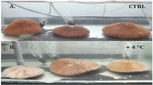
2.1. Experimental Design

The experiment was conducted from the 9th to the 19th of October in 2012 at the Institute of Oceanography of Nha Trang (Nha Trang, Viet Nam). In this study, individuals of the plate coral Fungia repanda were collected in the Nha Trang Bay. This free-living coral is easy to handle and typically produces large quantities of mucus (>10 mL/individual/5 min).
Prior to the experiment, all the corals were kept in tanks filled with sand-filtrated seawater collected at corals’ site of origin for 10 days to allow for acclimatization. The experimental design consisted of two 40-L aquaria with triplicate individuals of F. repanda corals of similar sizes (15–17 cm in diameter). At the end of the acclimatization period, the temperature was gradually increased (over 3 days) to 32 °C in the heat-treatment tank by using an immersion thermostat (LAUDA Ecoline Staredition E200), while the water temperature was kept at ambient temperature (28 °C) in the control aquarium. Water was renewed once a day in both aquaria with seawater previously adjusted to the different tank temperatures. During the experiment, both tanks received aeration to maintain air-saturated conditions. Lighting was provided by a fluorescent lamp (VHO: General Electric, 175 watt), with irradiance of 200 μmoL photons m−2·s−1 (on a 12 h:12 h light:dark cycle). Water temperature, salinity, oxygen concentration, and light intensity were monitored twice a day. HOBO Pendant loggers (Onset, Massachusetts, MA, USA) were used to log light intensity and temperature in the control and heat treatment aquaria. After 11 days under this thermal stress, all the corals were sampled using each set of measurements described below.
Thirty milliliters of water, and 5–7 mL of mucus were collected from each coral at the beginning (T0) and at the end (after 11 days) of incubation (Tend). Mucus was collected by using the desiccation method described in details elsewhere [32,41]. Briefly, the corals were taken out of the water and exposed to air for 1 to 3 min. This stress caused the mucus to be secreted, forming long gel-like threads dripping from the coral surface. The first 30 s of mucus production was discarded to prevent contamination and dilution by seawater. Mucus was then distributed in cryotubes for estimation of (1) viral, bacteria, and Symbiodinium abundance; (2) viral lytic production rate (3) cell physiological activity; and (4) bacterial community composition.

2.2. Enumeration of Symbiodinium, Bacteria, and Viruses

The fixed mucus was processed for viral and bacterial extraction and enumeration by using the potassium citrate method, as recommended by Leruste et al. [31], and adapted from Williamson et al. [42]. Briefly, 100 μL of mucus was eluted into 900 μL of 0.02-μm-pore-size-filtered, pH 7 solution of 1% citrate potassium (10 g potassium citrate, 1.44 g Na2HPO4·7H2O, and 0.24 g KH2PO4 per liter). All tubes were then vortexed at a moderate speed for 5 min before particles were stained and enumerated. The number of viruses and bacteria contained in duplicate subsamples (2 independent counts for each of the six corals) were determined after retention of the particles on 0.02-μm pore-size membranes (Anodisc, GE Healthcare, Little Chalfont, United Kingdom) and staining with the nucleic acid stain SYBR Gold [43]. On each slide, 300–500 bacteria and viruses were counted with an Olympus Provis-AX70 epifluorescence microscope (Olympus SAS, Rungis, France) in 20 fields under blue light excitation (488 nm). Symbiodinium cells, due to their photosynthetic pigments could be also enumerated on the same slides, under the blue light excitation.

2.3. Bacterial Physiological State

The proportion of respiring bacteria that have high rates of metabolism was determined using 5-cyano-2,3-ditolyl tetrazolium chloride (CTC), an indicator of the respiratory electron transport system activity [44]. A stock solution of 50 mmoL l-1 CTC (Tebu-bio SAS, Le Perray-en-Yvelines, France) was prepared at both sampling dates (day = 0 and day = 11), filtered through 0.01 mm filters and kept in the dark at 4 °C until use. CTC stock solution was then added to 0.45 mL of both duplicate fresh mucus and water samples (5 mmoL l-1 final CTC concentration) and incubated for 1.5 h at room temperature in the dark. Formaldehyde (3% final concentration) was used to stop the CTC reaction. Samples were flash frozen in liquid nitrogen and stored at −80 °C freezer until flow cytometer (FCM) analysis (FACSCALIBUR, BD Biosciences, Franklin Lakes, NJ, USA). The red fluorescence of CTC (FL3) and the light scatter SSC were used to discriminate the CTC+ cells from other cells or weak fluorescent particles. The percentage of CTC+ cells, based on triplicate analyses, was calculated relative to the total bacterial counts obtained by epifluorescence microscopy (see 2.2. Enumeration of Symbiodinium, Bacteria and Viruses).

2.4. Viral Lytic Production

The decay, i.e., the decrease in the viral concentration over time, was recorded after inhibition of new viral lytic production (VP) by the addition of potassium cyanide (KCN; final concentration of 2 mM) in both mucus and water samples [45,46]. All incubations for decay experiments were performed in duplicate at in situ temperature, for 12 h. Incubations were stopped after addition of formaldehyde (3% final concentration). Viral abundance was determined in KCN-treated and untreated water and coral mucus, by using SYBR Gold and epifluorescence microscopy. The difference between the abundance of viruses with and without KCN allows the estimation of VP.

2.5. DGGE Analysis of Bacteria Community Structure

The genetic diversity of both Eubacterial communities in water and mucus samples was estimated by using a fingerprinting technique: Denaturing gradient gel electrophoresis (DGGE) [47]. PowerSoil® DNA Isolation Kit (MO BIO, Carlsbad, CA, USA) was used to extract DNA from water and mucus samples. The DNA sequences were then subjected to touchdown PCR using following primers: 341F-GC [48] and 519R [49], which target bacterial 16S gene (178 bp). Thirty-five cycles of amplification were done starting at 93 °C initial denaturation of the dsDNAs followed by a second denaturation phase at 92 °C and the annealing step, which was done at the high temperature of 71 °C minimizing unspecific primer binding. After each cycle, the temperature was lowered by 1/2 of a degree until reaching the touchdown temperature of 61 °C, keeping that temperature for the last 15 cycles. The DGGE was performed with Ingeny U-Phor system (Ingeny, Waltham, MA, USA) in 0.5× TAE buffer (Euromedex, Souffelweyersheim, France) at 60 °C with a constant voltage of 80 V for 18 h. The DNA was then stained with SYBR-Gold. DNA bands were visualized on a UV trans-illumination table with the imaging system GelDoc® XR (Bio-Rad, Hercules, CA, USA) and analyzed using fingerprint and gel analysis Quantity One software (Bio-Rad, Hercules, CA, USA). Band matching was performed with 1.00% position tolerance and 1.00% optimization. After generating a band-matching table, we obtained the binary presence-absence matrix for all the detected bands. The matrix was used to calculate a distance matrix with Sorensen dissimilarity index, which subsequently was used for an ordination analysis—principal coordinate analysis (PCoA) (see data analysis).

2.6. Statistical Analysis

The differences among samples in all variables were tested by one-way ANOVA, followed by a post-hoc analysis Tukey-Kramer for pairwise comparisons of means between samples. All the parameters were normalized prior to test. A level of 0.05 was considered significant. The JMP 9.0 software (SAS institute, Cary, NC, USA) was used for these statistical analyses. In order to evaluate differences and variability in the bacterial community composition, a principal coordinate analysis (PCoA) was applied, using Sorensen dissimilarity matrices as inputs. The analysis and calculation were done using R software version 3.0.2 with the vegan, ade4 and betapart packages.

3. Results

The experiment was stopped after 11 days when clear signs of bleaching were observed in all the three replicates of F. repanda corals in the heat-treated aquarium. At that time, no visible trace of coral bleaching was detected in the control tank.

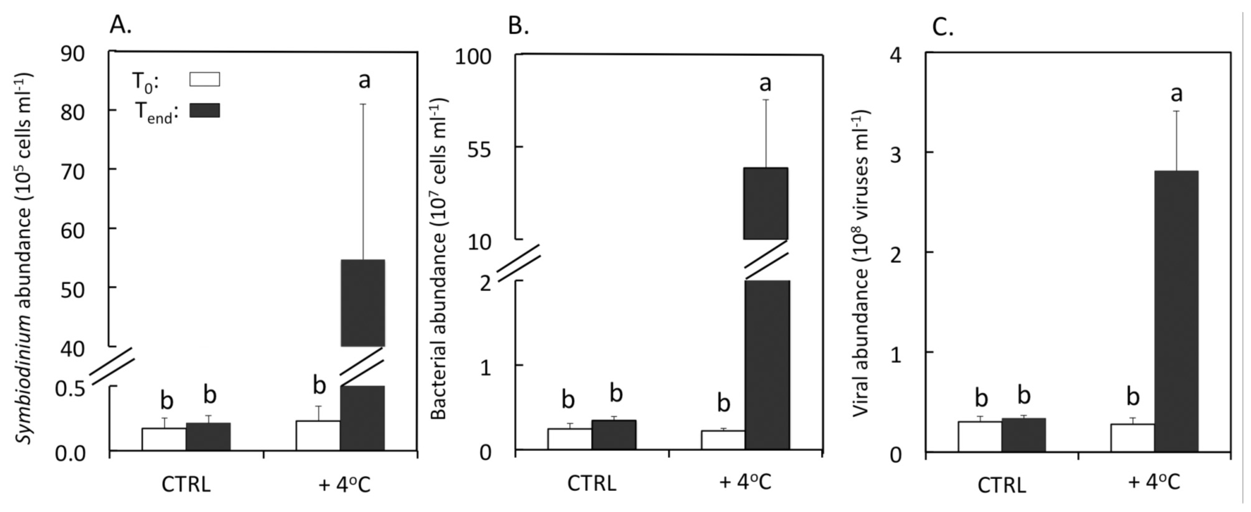
3.1. Abundances of Symbiodinium, Bacteria and Viruses

The heat treatment resulted in a large increase in the abundance of Symbiodinium in the mucus of F. repanda reaching up to 5.5 × 10⁶ cells·mL−1 (one-way ANOVA, F = 12.7, p = 0.002), which was more than 250-fold higher than that observed in the control tank at the end of the experiment (Tukey-Kramer, p < 0.05). On the contrary, the micro-algal concentration remained relatively stable in untreated corals throughout the experiment (Tukey-Kramer, p < 0.05) (Figure 1A). The final abundance of bacteria was also greatly enhanced (by almost 130 times) in heat-stressed corals (Tukey-Kramer, p < 0.05) to reach an average value of 4.5 × 10⁸ cells·mL−1 (Figure 1B). The pattern of viral abundance was highly comparable with that of bacteria, showing a final enhancement factor of almost nine between heat-treated and control tanks, respectively (Tukey-Kramer, p < 0.05) (Figure 1C). As for Symbiodinium cells, bacterial and viral abundance did not vary significantly in the control tank (Figure 1B,C).
Figure 1. Abundances of Symbiodinium (A); bacteria (B), and viruses (C) in mucus samples of F. repanda in both control (CTRL) and heat-treatment (+4 °C) at the beginning (To) and at the end of the experiment (Tend). Error bars represent one standard deviation from the mean (n = 3).Histograms with the same letters are not significantly different at p = 0.05.
Jmse 03 01272 g001
Figure 2. Percentages of CTC + cells in mucus samples of F. repanda in both control (CTRL) and heat-treatment (+4 °C). Error bars represent one standard deviation from the mean (n = 3). Histograms with the same letters are not significantly different at p = 0.05.
Jmse 03 01272 g002

3.2. Physiological State of Bacteria

The percent of metabolically active respiring cells (CTC+) dramatically dropped from 62.4% to 3.1% in the mucus of heat stressed corals (one-way ANOVA, F = 12.2, p = 0.0001) (Figure 2). This value was 17-fold lower than that observed in the control aquarium (mean = 53.0%) at the end of the experiment (Tukey-Kramer, p < 0.05). In control treatment, coral mucus harbored a relatively constant proportion of active cells throughout the experiment (53.0%–59.3%).

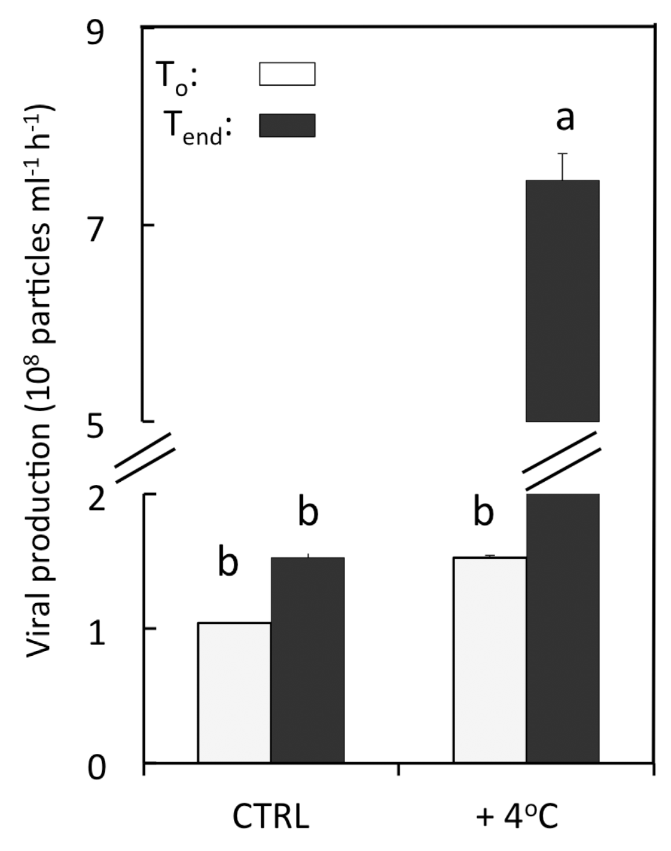
3.3. Viral Lytic Production
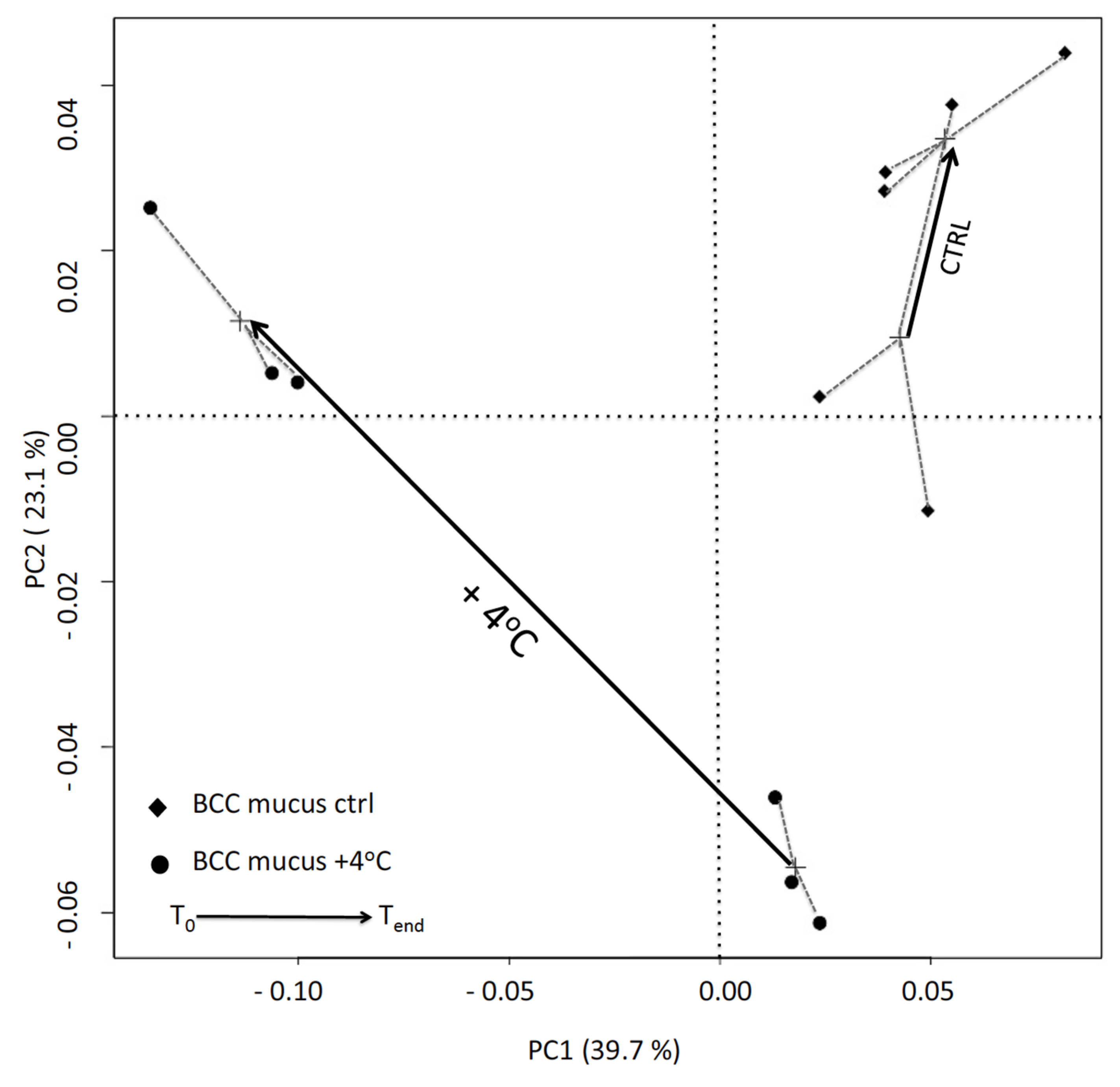
The viral production rate in coral mucus was also strongly stimulated by the elevation of temperature, with values ultimately reaching up to 7.5 × 10⁶ viruses mL−1·h−1 (Figure 3), which was, on average, 4.9 times higher than that observed in the control tank (Tukey-Kramer, p < 0.05). In this aquarium, viral lytic production did not show any significant changes between the beginning and the end of the experiment (Tukey-Kramer, p < 0.05).
Figure 3. Viral lytic production in mucus samples of F. repanda in both control (CTRL) and heat-treatment (+4 °C). Error bars represent one standard deviation from the mean (n = 3). Histograms with the same letters are not significantly different at p = 0.05.
Jmse 03 01272 g003

3.4. DGGE Analysis

The thermal stress also strongly impacted the community structure of epibiotic cells (Figure 4). The first principal coordinate (39.7% of total variations) showed clear discrimination between bleached and healthy corals at T0. In the control tank, the structure of mucosal communities remained relatively stable over the experimental course (Figure 4). Conversely, a significant shift was observed in thermally stressed corals as shown by the length of the specific arrow.
Figure 4. Principal component analysis (PCOA) obtained with the Sorensen dissimilarity index calculated from denaturing gradient gel electrophoresis (DGGE) presence/absence profile. Arrows, drawn from the calculated centroid of all replicates of each sample, show the evolution in the bacterial community structure between the beginning and the end of the experiment, in mucus samples of F. repanda, in both control (CTRL) and heat-treatment (+4 °C).
Jmse 03 01272 g004

4. Discussion

Coral bleaching is typically characterized by the loss of intracellular endosymbionts (Symbodinium) from the coral tissue [2,50]. Here, the bleached Fungia observed at the end of the incubation, exhibited higher concentrations of Symbodinium cells in their mucus than healthy individuals, which clearly confirmed their expulsion from coral tissue to the mucus layer, before their ultimate transfer into the water column.

4.1. High Abundance but Low Metabolic Activity of Bleached-Coral-Associated Bacteria

The mucus of bleached corals contained a much higher abundance of bacteria than healthy ones. At first sight, such observations are not surprising as bacterial growth generally increases with temperature. However, the thermal stress also resulted in a substantial drop of the physiological state of mucus associated bacterial cells, where the proportion of CTC+ respiring bacterial cells decreased from about 60% to 3%. As for all other physiological indicators, respiration is affected by temperature. After the optimum temperature, the Q10-value (index of the temperature sensitivity of bacterial respiration) typically decreases and becomes negative, i.e., respiration decreases with increasing temperature [51]. Our results indicate that the thermal tolerance of mucosal cells was probably low with a disruption point occurring between 28 °C and 32 °C. A significant reduction in the mineralization abilities of Acropora millepora’s associates was also reported after they faced a thermal increase of 1 °C to 3 °C [24,26]. Here, the proliferation of abundant but low active cells could be favored by the degradation of particular key active bacterial associates. For example, recent studies have demonstrated that the antibacterial activity of mucosal bacteria is altered at high temperature [13,15,52]. We then suspect that the decline in cell respiring activity in stressed corals may facilitate opportunist infections. In the absence of a strong line of defense, the alteration of the coral immune systems may then result in a higher susceptibility to colonization by the surrounding planktonic bacteria (pathogenic or not), which are typically less active than their epibiotic counterparts [32]. Alternatively, the drop in cell activity could be also simply explained by the physiological decline of the most thermo-sensitive cells due to their inability to cope with the increase in temperature.
In a changing environment, modifications to the abundance and physiological state of planktonic bacterial communities have been shown to induce dramatic changes in their structure [53]. Likewise, the observed changes in abundance and respiration of coral associated bacteria after they received the heat treatment were coupled with substantial changes in their community composition, as revealed by the principal coordinate analysis (Figure 4). Comparable shifts in bacterial community structure have been observed on several occasions when corals were subjected to elevated temperatures [24,25,26,54]. Vega-Thurber et al. [27], by using a pyrosequencing procedure, demonstrated that a controlled elevation of seawater temperature from a local ambient temperature of 25 °C to 30 °C can impair the microbiome of the coral Porites compressa to more pathogenic taxa. Such findings were corroborated by a recent study where Garren and her colleagues [55] showed that the mucus of heat-stressed corals could produce a high amount of dimethylsulfoniopropionate, which greatly attracts coral pathogens and causes coral bleaching [11]. In this study, the shift in the bacterial community composition could be also caused by changes in physicochemical properties of the mucus layer. In fact, coral mucus composition and production are quantitatively and qualitatively influenced by environmental factors such as temperature and/or irradiance [56,57]. As a result, particular substrates favoring the growth of the resident bacteria may disappear from the mucus and cause their decline or at least a metabolic depression. As suggested above, the surrounding bacteria may then find their chance to invade this new medium and outcompete with the native groups for the new nutrient sources [14].

4.2. High Viral Production and Abundance in the Mucus of Bleached F. repanda

One of the main findings of this research was the almost 5- and 9-fold increase in viral production rates (Figure 3) and abundance (Figure 1A), respectively, between healthy and bleached corals. These findings were expected as bacterial growth typically increases with temperature, which could naturally favor viral proliferation. Indeed, most of the viruses found in coral mucus were actually phages [31,32,58], and therefore their high occurrence in heat-treated corals is probably the result of a lytic activity on the numerous resident bacterial hosts. The high phage production measured in the bleached corals (compared to healthy ones) clearly confirmed this hypothesis. However, other explanations can be provided. For example, the members of the bacterial community found in bleached corals might be more susceptible to viral infections, in comparison with those in healthy ones where virus-bacteria interactions are presumably more stable and where lytic pressure is applied with parsimony to ultimately ensure coral persistence [35,36,40,59]. Also, we know that phage replication is dependent on host metabolism [60,61]. Therefore the high viral production rates measured in bleached corals might be the result of lytic infections of the most active mucosal cells, as demonstrated by Nguyen-Kim et al. [32].
Alternatively, the increase in the abundance and production of mucosal viruses could be due to the thermally-driven induction of the lysogens present in this organic layer. Nguyen-Kim et al. [62], reported that the proportion of lysogenized cells measured in the mucus of two different scleractinian corals (Fungia repanda and Acropora formosa) in a seasonal study was, on average, more than two-fold higher (mean = 8.5% of total bacterial counts) than that measured in the surrounding water (mean = 3.8%). It has been recently hypothesized that lysogeny, by conferring immunity to bacterial symbionts against other lytic surinfection, may therefore represent a vital strategy for corals for their subsistence [40].
The adhesive properties of the coral mucus layer may also represent another explanation for the high occurrence of viruses in bleached corals. Indeed, mucus typically acts as a trap for planktonic particles [63,64], and phages with their specific Ig-proteins located on their capsid are now known for their strong affinity with the mucin-protein of coral mucus [34].
Finally, a fraction of the viral pool might also come from the infected Symbiodinium after their expulsion from the coral tissues. Indeed, these endosymbiotic microalgae have been shown to harbor latent viruses, which can be activated by thermal or UV stresses [39,65] and this could be an additional explanation for the large concentrations of viruses in bleached corals.
Current research priorities include elucidating whether such highly abundant bacteria and viruses in bleached corals are the results of the proliferation of (1) the native mucosal associates after they were stimulated by the elevated temperature; (2) the planktonic communities after their adhesion to the mucus gel and/or (3) the opportunists surrounding cells (pathogens or not) taking advantage of the disruption of the bacterial assemblage.
The questions that also now need to be answered are whether coral bleaching occurs prior or after the reduction of cell activity and the shift in bacterial community structure in coral mucus, and whether the expulsion of Symbiodinium cells is ecologically linked with such mechanisms. The role of viruses in the structuring epibiotic bacteria also need to be clarified. Finally, further experiments should be conducted at a higher temporal resolution to better address these gaps.

5. Conclusions

Overall, our results seem to suggest that the thermal stress of +4 °C could be responsible for substantial changes in bacterial and viral traits, mostly resulting from the alteration of the physiological state of the native cells. The high levels of viral production measured in bleached corals may have strong implications for coral reef ecosystems including, for example, the enrichment of viruses in the water column through the continuous sloughing off of mucus, and which could in turn have a strong local influence on the bacterial stocks, diversity and functions in reef waters, but could also potentially interact with nearby hosts in other corals.

Acknowledgments

We thank the TOTAL foundation and the Hoa Sen Lotus French-Vietnamese Program for their financial support. Thanks to the French Institute of Research for Development (IRD) for PhD grant to HNK. We thank the Ecological Toxicity Laboratory of the Institute of Oceanography (Vietnam) and the MARBEC research unit laboratory (France) for lab space and material support to carry out the experiment. We are grateful to Lam Nguyen-Ngoc, Hai Doan-Nhu for their scientific and logistical support. We also thank Hoang Phan-Kim for diving assistant during the sampling, Callis Amid and Marie Olstedt for helping during the experiment.

Author Contributions

Hanh Nguyen-Kim, Yvan Bettarel, Thierry Bouvier and Huong Le-Lan designed and conducted the experiment with mesocosms; Hanh Nguyen-Kim, and Corinne Bouvier analyzed the samples and realized the statistical tests; Hanh Nguyen-Kim, Yvan Bettarel, Thierry Bouvier, and Ngoc Van-Bui co-wrote the paper.

Conflicts of Interest

The authors declare no conflict of interest.

References

  1. Hoegh-Guldberg, O. Coral reef ecosystems and anthropogenic climate change. Reg. Environ. Chang. 2011, 11, S215–S227. [Google Scholar] [CrossRef]
  2. Brown, B.E. Coral bleaching: Causes and consequences. Coral Reefs 1997, 16, S129–S138. [Google Scholar] [CrossRef]
  3. Glynn, P.W.; Dcroz, L. Experimental evidence for high temperature stress as the cause of El Nino-coincident coral mortality. Coral Reefs 1990, 8, 181–191. [Google Scholar] [CrossRef]
  4. Jokiel, P.L.; Brown, E.K. Global warming, regional trends and inshore environmental conditions influence coral bleaching in hawaii. Glob. Chang. Biol. 2004, 10, 1627–1641. [Google Scholar] [CrossRef]
  5. Lesser, M.P.; Stochaj, W.R.; Tapley, D.W.; Shick, J.M. Bleaching in coral reef anthozoans: Effects of irradiance, ultraviolet radiation and temperature on the activities of protective enzymes against active oxygen. Coral Reefs 1990, 8, 225–232. [Google Scholar] [CrossRef]
  6. Coles, S.L.; Jokiel, P.L. Synergistic effects of temperature, salinity and light on hermatypic coral Montipora verrucosa. Mar. Biol. 1978, 49, 187–195. [Google Scholar] [CrossRef]
  7. Anthony, K.R.N.; Kline, D.I.; Diaz-Pulido, G.; Dove, S.; Hoegh-Guldberg, O. Ocean acidification causes bleaching and productivity loss in coral reef builders. Proc. Natl. Acad. Sci. USA 2008, 105, 17442–17446. [Google Scholar] [CrossRef] [PubMed]
  8. Guzman, H.M.; Jackson, J.B.C.; Weil, E. Short term ecological consequences of a major oil spill on panamanian subtidal reef corals. Coral Reefs 1991, 10, 1–12. [Google Scholar] [CrossRef]
  9. Vega Thurber, R.L.; Burkepile, D.E.; Fuchs, C.; Shantz, A.A.; McMinds, R.; Zaneveld, J.R. Chronic nutrient enrichment increases prevalence and severity of coral disease and bleaching. Glob. Chang. Biol. 2014, 20, 544–554. [Google Scholar] [CrossRef] [PubMed]
  10. Kushmaro, A.; Kramarsky-Winter, E. Bacteria as a Source of Coral Nutrition. In Coral Health and Disease; Rosenberg, E., Loya, Y., Eds.; Springer-Verlag: New York, NY, USA, 2004; pp. 231–241. [Google Scholar]
  11. Rosenberg, E.; Kushmaro, A.; Kramarsky-Winter, E.; Banin, E.; Yossi, L. The role of microorganisms in coral bleaching. ISME J. 2009, 3, 139–146. [Google Scholar] [CrossRef] [PubMed]
  12. de Lima, L.A.; Migliolo, L.; Barreiro e Castro, C.; Pires, D.D.; Lopez-Abarrategui, C.; Goncalves, E.F.; Vasconcelos, I.M.; de Oliveira, J.T.A.; Otero-Gonzalez, A.D.J.; Franco, O.L.; et al. Identification of a novel antimicrobial peptide from brazilian coast coral Phyllogorgia dilatata. Protein Pept. Lett. 2013, 20, 1153–1158. [Google Scholar] [CrossRef] [PubMed]
  13. Kvennefors, E.C.E.; Sampayo, E.; Kerr, C.; Vieira, G.; Roff, G.; Barnes, A.C. Regulation of bacterial communities through antimicrobial activity by the coral holobiont. Microb. Ecol. 2012, 63, 605–618. [Google Scholar] [CrossRef] [PubMed]
  14. Rypien, K.L.; Ward, J.R.; Azam, F. Antagonistic interactions among coral-associated bacteria. Environ. Microbiol. 2010, 12, 28–39. [Google Scholar] [CrossRef] [PubMed]
  15. Shnit-Orland, M.; Sivan, A.; Kushmaro, A. Antibacterial activity of Pseudoalteromonas in the coral holobiont. Microb. Ecol. 2012, 64, 851–859. [Google Scholar] [CrossRef] [PubMed]
  16. Reshef, L.; Koren, O.; Loya, Y.; Zilber-Rosenberg, I.; Rosenberg, E. The coral probiotic hypothesis. Environ. Microb. 2006, 8, 2068–2073. [Google Scholar] [CrossRef] [PubMed]
  17. Rosenberg, E.; Koren, O.; Reshef, L.; Efrony, R.; Zilber-Rosenberg, I. The role of microorganisms in coral health, disease and evolution. Nat. Rev. Microb. 2007, 5, 355–362. [Google Scholar] [CrossRef] [PubMed]
  18. Ben-Haim, Y.; Rosenberg, E. A novel Vibrio sp. pathogen of the coral pocillopora damicornis. Mar. Biol. 2002, 141, 47–55. [Google Scholar]
  19. Kushmaro, A.; Banin, E.; Loya, Y.; Stackebrandt, E.; Rosenberg, E. Vibrio shiloi sp. nov., the causative agent of bleaching of the coral oculina patagonica. Int. J. Syst. Evolut. Microbiol. 2001, 51, 1383–1388. [Google Scholar] [CrossRef] [PubMed]
  20. Kushmaro, A.; Rosenberg, E.; Fine, M.; Loya, Y. Bleaching of the coral oculina patagonica by vibrio ak-1. Mar. Ecol. Prog. Ser. 1997, 147, 159–165. [Google Scholar] [CrossRef]
  21. Toren, A.; Landau, L.; Kushmaro, A.; Loya, Y.; Rosenberg, E. Effect of temperature on adhesion of vibrio strain ak-1 to oculina patagonica and on coral bleaching. Appl. Environ. Microbiol. 1998, 64, 1379–1384. [Google Scholar] [PubMed]
  22. Ainsworth, T.; Fine, M.; Roff, G.; Hoegh-Guldberg, O. Bacteria are not the primary cause of bleaching in the mediterranean coral Oculina patagonica. ISME J. 2008, 2, 67–73. [Google Scholar] [CrossRef] [PubMed]
  23. Leggat, W.; Hoegh-Guldberg, O.; Dove, S.; Yellowlees, D. Analysis of an est library from the dinoflagellate (Symbiodinium sp.) symbiont of reef-building corals. J. Phycol. 2007, 43, 1010–1021. [Google Scholar] [CrossRef]
  24. Bourne, D.; Iida, Y.; Uthicke, S.; Smith-Keune, C. Changes in coral-associated microbial communities during a bleaching event. ISME J. 2008, 2, 350–363. [Google Scholar] [CrossRef] [PubMed]
  25. Lins-de-Barros, M.M.; Cardoso, A.M.; Silveira, C.B.; Lima, J.L.; Clementino, M.M.; Martins, O.B.; Albano, R.M.; Vieira, R.P. Microbial community compositional shifts in bleached colonies of the brazilian reef-building coral siderastrea stellata. Microb. Ecol. 2013, 65, 205–213. [Google Scholar] [CrossRef] [PubMed]
  26. Littman, R.; Willis, B.L.; Bourne, D.G. Metagenomic analysis of the coral holobiont during a natural bleaching event on the great barrier reef. Environ. Microbiol. Rep. 2011, 3, 651–660. [Google Scholar] [CrossRef] [PubMed]
  27. Vega Thurber, R.; Willner-Hall, D.; Rodriguez-Mueller, B.; Desnues, C.; Edwards, R.A.; Angly, F.; Dinsdale, E.; Kelly, L.; Rohwer, F. Metagenomic analysis of stressed coral holobionts. Environ. Microbiol. 2009, 11, 2148–2163. [Google Scholar] [CrossRef] [PubMed]
  28. Fuhrman, J.A. Marine viruses and their biogeochemical and ecological effects. Nature 1999, 399, 541–548. [Google Scholar] [CrossRef] [PubMed]
  29. Suttle, C.A. Marine viruses—Major players in the global ecosystem. Nat. Rev. Microbiol. 2007, 5, 801–812. [Google Scholar] [CrossRef] [PubMed]
  30. Davy, J.E.; Patten, N.L. Morphological diversity of virus-like particles within the surface microlayer of scleractinian corals. Aquat. Microb. Ecol. 2007, 47, 37–44. [Google Scholar] [CrossRef]
  31. Leruste, A.; Bouvier, T.; Bettarel, Y. Enumerating viruses in coral mucus. Appl. Environ. Microbiol. 2012, 78, 6377–6379. [Google Scholar] [CrossRef] [PubMed]
  32. Nguyen-Kim, H.; Bouvier, T.; Bouvier, C.; Doan, N.H.; Nguyen, N.L.; Rochelle-Newall, E.; Desnues, C.; Reynaud, S.; Ferrier-Pages, C.; Bettarel, Y. High occurence of viruses in the mucus layer of scleractinian corals. Environ. Microbiol. Rep. 2014, 6, 675–682. [Google Scholar] [CrossRef] [PubMed]
  33. Weinbauer, M.G.; Ogier, J.; Maier, C. Microbial abundance in the coelenteron and mucus of the cold-water coral lophelia pertusa and in bottom water of the reef environment. Aquat. Biol. 2012, 16, 209–216. [Google Scholar] [CrossRef]
  34. Barr, J.J.; Auro, R.; Furlan, M.; Whiteson, K.L.; Erb, M.L.; Pogliano, J.; Stotland, A.; Wolkowicz, R.; Cutting, A.S.; Doran, K.S.; et al. Bacteriophage adhering to mucus provide a non-host-derived immunity. Proc. Natl. Acad. Sci. USA 2013, 110, 10771–10776. [Google Scholar] [CrossRef] [PubMed]
  35. Van Oppen, M.J.H.; Leong, J.A.; Gates, R.D. Coral-virus interactions: A double-edged sword? Symbiosis 2009, 47, 1–8. [Google Scholar] [CrossRef]
  36. Vega Thurber, R.L.; Correa, A.M.S. Viruses of reef-building scleractinian corals. J. Exp. Mar. Biol. Ecol. 2011, 408, 102–113. [Google Scholar] [CrossRef]
  37. Correa, A.M.; Welsh, R.M.; Thurber, R.L.V. Unique nucleocytoplasmic dsdna and +ssrna viruses are associated with the dinoglagellate endosymbionts of corals. ISME J. 2013, 7, 13–27. [Google Scholar] [CrossRef] [PubMed]
  38. Danovaro, R.; Bongiorni, L.; Corinaldesi, C.; Giovannelli, D.; Damiani, E.; Astolfi, P.; Greci, L.; Pusceddu, A. Sunscreens cause coral bleaching by promoting viral infections. Environ. Health Perspect. 2008, 116, 441–447. [Google Scholar] [CrossRef] [PubMed]
  39. Wilson, W.H.; Dale, A.L.; Davy, J.E.; Davy, S.K. An enemy within? Observations of virus-like particles in reef corals. Coral Reefs 2005, 24, 145–148. [Google Scholar] [CrossRef]
  40. Bettarel, Y.; Bouvier, T.; Nguyen, H.K.; Thu, P.T. The versatile nature of coral associated viruses. Environ. Microbiol. 2014, 16. [Google Scholar] [CrossRef] [PubMed]
  41. Naumann, M.S.; Niggl, W.; Laforsch, C.; Glaser, C.; Wild, C. Coral surface area quantification-evaluation of established techniques by comparison with computer tomography. Coral Reefs 2009, 28, 109–117. [Google Scholar] [CrossRef]
  42. Williamson, K.E.; Wommack, K.E.; Radosevich, M. Sampling natural viral communities from soil for culture-independent analyses. Appl. Environ. Microbiol. 2003, 69, 6628–6633. [Google Scholar] [CrossRef] [PubMed]
  43. Patel, A.; Noble, R.T.; Steele, J.A.; Schwalbach, M.S.; Hewson, I.; Fuhrman, J.A. Virus and prokaryote enumeration from planktonic aquatic environments by epifluorescence microscopy with sybr green i. Nat. Protoc. 2007, 2, 269–276. [Google Scholar] [CrossRef] [PubMed]
  44. Sherr, B.F.; del Giorgio, P.; Sherr, E.B. Estimating abundance and single-cell characteristics of respiring bacteria via the redox dye ctc. Aquat. Microb. Ecol. 1999, 18, 117–131. [Google Scholar] [CrossRef]
  45. Bettarel, Y.; Desnues, A.; Rochelle-Newall, E. Lytic failure in cross-inoculation assays between phages and prokaryotes from three aquatic sites of contrasting salinity. FEMS Microbiol. Lett. 2010, 311, 113–118. [Google Scholar] [CrossRef] [PubMed]
  46. Fischer, U.R.; Velimirov, B. High control of bacterial production by viruses in a eutrophic oxbow lake. Aquat. Microb. Ecol. 2002, 27, 1–12. [Google Scholar] [CrossRef]
  47. Morrow, K.M.; Moss, A.G.; Chadwick, N.E.; Liles, M.R. Bacterial associates of two caribbean coral species reveal species-specific distribution and geographic variability. Appl. Environ. Microbiol. 2012, 78, 6438–6449. [Google Scholar] [CrossRef] [PubMed]
  48. Dar, S.A.; Kuenen, J.G.; Muyzer, G. Nested pcr-denaturing gradient gel electrophoresis approach to determine the diversity of sulfate-reducing bacteria in complex microbial communities. Appl. Environ. Microbiol. 2005, 71, 2325–2330. [Google Scholar] [CrossRef] [PubMed]
  49. Ovreas, L.; Forney, L.; Daae, F.L.; Torsvik, V. Distribution of bacterioplankton in meromictic lake saelenvannet, as determined by denaturing gradient gel electrophoresis of pcr-amplified gene fragments coding for 16s rrna. Appl. Environ. Microbiol. 1997, 63, 3367–3373. [Google Scholar] [PubMed]
  50. Harvell, D.; Jordan-Dahlgren, E.; Merkel, S.; Rosenberg, E.; Raymundo, L.; Smith, G.; Weil, E.; Willis, B. Coral disease, environmental drivers, and the balance between coral and microbial associates. Oceanography 2007, 20, 172–195. [Google Scholar] [CrossRef]
  51. Pires, A.P.F.; Guariento, R.D.; Laque, T.; Esteves, F.A.; Farjalla, V.F. The negative effects of temperature increase on bacterial respiration are independent of changes in community composition. Environ. Microbiol. Rep. 2014, 6, 131–135. [Google Scholar] [CrossRef] [PubMed]
  52. Ritchie, K.B. Regulation of microbial populations by coral surface mucus and mucus-associated bacteria. Mar. Ecol. Prog. Ser. 2006, 322, 1–14. [Google Scholar] [CrossRef]
  53. Del Giorgio, P.A.; Bouvier, T.C. Linking the physiologic and phylogenetic successions in free-living bacterial communities along an estuarine salinity gradient. Limnol. Oceanogr. 2002, 47, 471–486. [Google Scholar] [CrossRef]
  54. Gilbert, J.A.; Hill, R.; Doblin, M.A.; Ralph, P.J. Microbial consortia increase thermal tolerance of corals. Mar. Biol. 2012, 159, 1763–1771. [Google Scholar] [CrossRef]
  55. Garren, M.; Son, K.; Raina, J.B.; Rusconi, R.; Menolascina, F.; Shapiro, O.H.; Tout, J.; Bourne, D.G.; Seymour, J.R.; Stocker, R. A bacterial pathogen uses dimethylsulfoniopropionate as a cue to target heat-stressed corals. ISME J. 2014, 8, 999–1007. [Google Scholar] [CrossRef] [PubMed]
  56. Lasker, H.R.; Peters, E.C.; Coffroth, M.A. Bleaching of reef coelenterates in san-blas islands, panama. Coral Reefs 1984, 3, 183–190. [Google Scholar] [CrossRef]
  57. Piggot, A.M.; Fouke, B.W.; Sivaguru, M.; Sanford, R.A.; Gaskins, H.R. Change in zooxanthellae and mucocyte tissue density as an adaptive response to environmental stress by the coral, montastraea annularis. Mar. Biol. 2009, 156, 2379–2389. [Google Scholar] [CrossRef]
  58. Marhaver, K.L.; Edwards, R.A.; Rohwer, F. Viral communities associated with healthy and bleaching corals. Environ. Microbiol. 2008, 10, 2277–2286. [Google Scholar] [PubMed]
  59. Rohwer, F.; Seguritan, V.; Azam, F.; Knowlton, N. Diversity and distribution of coral-associated bacteria. Mar. Ecol. Prog. Ser. 2002, 243, 1–10. [Google Scholar] [CrossRef]
  60. Maurice, C.F.; Bouvier, C.; de Wit, R.; Bouvier, T. Linking the lytic and lysogenic bacteriophage cycles to environmental conditions, host physiology and their variability in coastal lagoons. Environ. Microbiol. 2013, 15, 2463–2475. [Google Scholar] [CrossRef] [PubMed]
  61. Weinbauer, M.G. Ecology of prokaryotic viruses. FEMS Microbiol. Rev. 2004, 28, 127–181. [Google Scholar] [CrossRef] [PubMed]
  62. Nguyen-Kim, H.; Bettarel, Y.; Bouvier, T.; Bouvier, C.; Doan-Nhu, H.; Nguyen-Ngoc, L.; Nguyen-Thanh, T.; Tran-Quang, H.; Brune, J. Coral mucus is a hot spot of viral infections. Appl. Environ. Microbiol. 2015, 81. [Google Scholar] [CrossRef] [PubMed]
  63. Mayer, F.W.; Wild, C. Coral mucus release and following particle trapping contribute to rapid nutrient recycling in a northern red sea fringing reef. Mar. Freshw. Res. 2010, 61, 1006–1014. [Google Scholar] [CrossRef]
  64. Wild, C.; Huettel, M.; Klueter, A.; Kremb, S.G.; Rasheed, M.Y.M.; Jorgensen, B.B. Coral mucus functions as an energy carrier and particle trap in the reef ecosystem. Nature 2004, 428, 66–70. [Google Scholar] [CrossRef] [PubMed]
  65. Lohr, J.; Munn, C.B.; Wilson, W.H. Characterization of a latent virus-like infection of symbiotic zooxanthellae. Appl. Environ. Microbiol. 2007, 73, 2976–2981. [Google Scholar] [CrossRef] [PubMed]

