Urine Proteomic Study in OAB Patients—Preliminary Report
by
 ,
,
Konrad Futyma
1,* ,
,
Łukasz Nowakowski
1,
Alicja Ziętek-Strobl
1,
Aleksandra Kamińska
1,
Nadia Taoussi
2 and
Tomasz Rechberger
1 1
2nd Department of Gynecology, Medical University of Lublin, Jaczewskiego 8, 20-954 Lublin, Poland
2
Accident and Emergency Department, Haukeland University Hospital, N-5021 Bergen, Norway
*
Author to whom correspondence should be addressed.
J. Clin. Med. 2020, 9(5), 1389; https://doi.org/10.3390/jcm9051389
Submission received: 19 March 2020
/
Revised: 30 April 2020
/
Accepted: 6 May 2020
/
Published: 8 May 2020
(This article belongs to the Special Issue Advances in Diagnosis and Treatment of Urinary and Fecal Incontinence in Women)
Abstract
:Overactive bladder (OAB) is defined by International Urogynecological Association (IUGA)/ International Continence Society (ICS) as urinary urgency, usually accompanied by frequency and nocturia, with or without urgency urinary incontinence, in the absence of urinary tract infection (UTI) or other obvious pathology. The pathophysiology of OAB is not well understood, however a number of different proteins and cytokines including vascular cell adhesion molecule-1 (VCAM-1) were found to be important in regulating structural integrity of the bladder wall. Proteome analysis may thus provide significant information with regard to OAB and may help in discovering novel diagnostic disease biomarkers. Sixteen Caucasian women aged 32–78 were included in the study. Patients were placed within 2 groups: OAB group (n = 8) and control group (n = 8). Urine samples were collected, immediately preserved in a protease inhibitor mixture, and frozen at −80 ℃. All samples were then further processed according to the isobaric tags for relative and absolute quantification (iTRAQ) manual. Proteins were labeled and analyzed in the mass spectrometer conjugated with liquid chromatograph (data are available via ProteomeXchange with the identifier PXD017799). There were no statistically significant differences in demographic data between control and OAB groups. VCAM-1 was the only protein that reached statistical significance as a differentiating protein in both of our experiments assessing the proteomic constitution in OAB patients. Studies involving a larger group of patients may provide further information on urinary bladder proteomics.
1. Introduction
Overactive bladder syndrome (OAB) is defined by International Urogynecological Association (IUGA)/ International Continence Society (ICS) (IUGA/ICS) as a form of urinary urgency, usually accompanied by frequency and nocturia, with or without urgency urinary incontinence, in the absence of urinary tract infection (UTI) or other obvious pathology [1]. There are several theories regarding the pathophysiology of OAB and detrusor overactivity (DO) including nervous, myogenic, and urothelial involvement but a full explanation is still missing. However, recent genetic studies have shed a light on gene expression engagement on OAB onset. Accordingly, several genes were found to be either over- or under-expressed in OAB sufferers. The use of micro- and macro-array as a methods of elucidating gene changes involved in the development of urgency was crucial for detecting genetic changes associated with this condition. Platelet-derived growth factor (PDGF), microfibrillar-associated protein (MAP), vascular cell adhesion molecule-1 (VCAM-1), and tropomyosin were found to be important in regulating the structural integrity of the bladder and its supporting tissues [2,3]. However, due to post-translational changes, encoded proteins may be altered and their impact on certain conditions can be limited. Proteome analysis may therefore provide significant pathophysiological information and may help in the discovery of novel diagnostic disease biomarkers or even in establishing new therapeutic methods [4]. Urine, being noninvasively collected, seems to be an ideal material for testing in patients with lower urinary tract symptoms (LUTS). Nevertheless, before urine proteomic markers become clinically known and useful, the urine proteome itself must be thoroughly characterized in diverse populations. In 2015, the National Institute of Diabetes and Digestive and Kidney Disease (NIDDK) invited experts of different specialties to a Urinology Think Tank meeting, and they concluded that features, such as the chemical and physical characteristics of urine, including the microbiota, cells, pH, metabolites, and proteins, might interact in complex ways with one another and with the bladder and/or kidney to potentially affect overall health [5]. Recently, Mossa et al. found that urinary metabolites might be involved in the pathophysiology of OAB and can be helpful in its diagnosis. In their work, increased urinary levels of mitochondrial dysfunction markers, oxidative stress, and ketosis correlated significantly with the OAB symptoms scores obtained through questionnaires. Moreover, multiple linear regression modeling revealed that age, blood glucose, and urine metabolites (malate, fumarate, and α-hydroxyisobutyrate) were significant predictive factors of OAB severity [6].
Other work has shown that the urothelium is responsible not only for isolating the bladder environment from surrounding tissues, but it also plays a crucial role in molecular transport through the bladder wall via numerous functional receptors, junctions, and channels (i.e., aquaporins, ion channels, gap junctions) [7]. Growth factors, cytokines, or proteins such as the epithelial growth factor (EGF); fibroblast growth factor receptor (FGFR), nerve growth factor (NGF), brain-derived neurotrophic factor (BDNF), vascular cell adhesion molecule (VCAM-1), and inter-cellular adhesion molecule 1 (ICAM-1) were found to alter urothelial functions in specific circumstances [3,8,9].
The aim of this study was to analyze the urinary proteomic pattern in patients suffering from OAB symptoms and to establish a quantitative and qualitative protein profile for OAB that may be supportive in defining the mechanism of the OAB pathophysiology.
2. Materials and Methods
Sixteen Caucasian women aged 32–78 were included within the study. Prior to the study, all patients signed informed consent and agreed to the use of the obtained data for scientific purposes. Moreover, data acquisition and analysis were performed in compliance with protocols approved by the Medical University of Lublin Ethical Committee (ethical approval number KE-0254/46/2016). In the study, participants were placed within 2 groups: OAB (n = 8) and control (n = 8). Inclusion criteria for patients with OAB symptoms were as follows: average of ≥8 micturitions/day during a 3 day bladder diary period and at least 1 episode of urge urinary incontinence (UUI) a day. Patients from the OAB group did not have any other comorbidities (except OAB) and did not take any medications prior to the study. Patients from the control group just had a routine gynecological check-up and were free of any LUTS and any other comorbidities as well and did not take any medications prior to the study. Urine samples were (40 mL) collected using a 14 Fr sterile catheter, immediately preserved in a bacteriostatic factor (sodium azide) and Pefabloc, protease inhibitor mixture (Sigma-Aldrich, Saint Louis, MI, USA), and frozen at −80 ℃. The obtained urinary proteins were precipitated by applying the Wessel–Fluegge method [10], and all samples were then further processed according to the the isobaric tags for relative and absolute quantification (iTRAQ) manual (Applied Biosystems, Foster City, CA, USA). Forty micrograms of the proteins was taken from each sample and digested with trypsin at 37 °C (Promega, Madison, WI, USA). Samples were than labeled and analyzed in the Q Exactive™ Hybrid Quadrupole-Orbitrap™ Mass Spectrometer (MS) (Thermo Fisher Scientific, Waltham, MA, USA) conjugated with the high-efficiency nanoACQUITY UPLC® liquid chromatograph (LC) (Waters Corporation, Milford, MA, USA). Detailed sample processing is described elsewhere [11] and a separate Supplementary File (File S1) has been submitted. Samples were analyzed in duplicate. Samples were divided into two separate experiments with 4 OABs and 4 controls in each described as ExpOABK_01 and ExpOABK_02. Acquired data were analyzed using the MASCOT engine (Matrix Science, London, UK) to search the SWISS-PROT protein database, limited to the Homo sapiens taxonomy. After the data arrangement, it was put through a second search using the DECOY database. Statistical analysis was performed by way of the Diffprot software, a resampling-based software tool for statistical analysis of data derived from MS-based proteomic experiments [12]. For demographic analysis Statistica 12.0 PL (StatSoft Polska Sp. z o. o., Krakow, Poland) and the chi2 test were used. “Ratio” is the parameter that says whether the protein is under- or over-expressed. A value of <1.0 means that this protein is under-expressed in OAB patients, and a value >1.0 means it is over-expressed. Fold change describes how great the difference is, no matter the direction. The mass spectrometry proteomics data have been deposited in the ProteomeXchange Consortium via the PRIDE [13] partner repository with the dataset identifier PXD017799 and 10.6019/PXD017799. Reviewer account details: Username: [email protected]; Password: hPZXyBVu.
3. Results
There were no statistically significant differences in demographic data between control and OAB groups (Table 1).
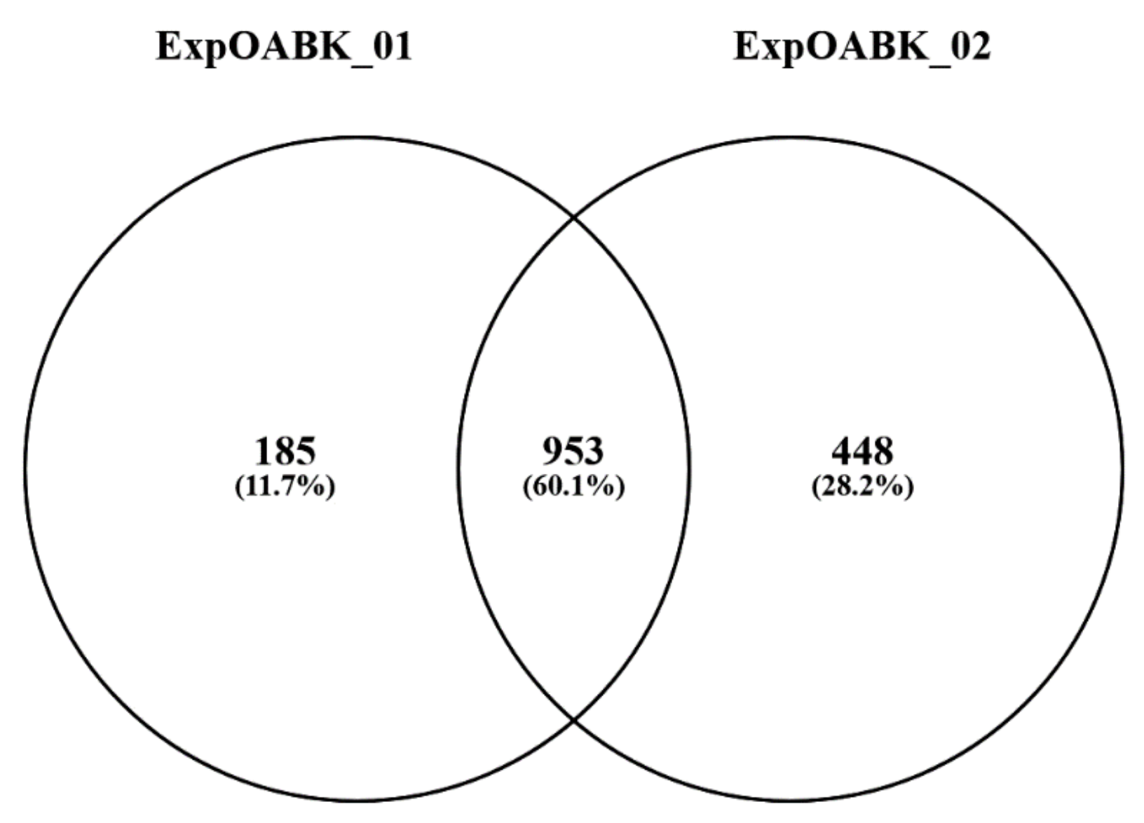
The proteome of urine collected from 8 OAB patients and 8 healthy subjects was compared using a combined liquid chromatography–mass spectrometry (LC-MS) relative quantitation of iTRAQ labeled peptides. The sample labeling yield rate with iTRAQ markers for both experiments was 99.7%. From each sample, an equal amount of total protein obtained in urine retentate, after filtration on >10 kDa cutoff filters, was used for the analysis. Qualitative results (protein lists) from two LC-MS experiments were combined, resulting in a dataset with all 953 proteins identified by at least two peptides (Figure 1). Within this dataset, protein identifications based on identical peptide sets were again grouped and each group was treated as a single protein cluster in further processing.
Qualitative proteins, that significantly differed between the OAB and control group in both experiments are presented in Table 2. Herein, vascular cell adhesion molecule-1 was the only protein found to have a q value <0.05 in both experiments. In addition, ratio and fold change were similar in both experiments. The complete list of differentiated proteins and full data is provided in separate Supplementary Files: first experiment: Full list ExpOABK_01.xlsx and second experiment: Full list ExpOABK_02.xlsx (Table S1 and Table S2, respectively). All identifications in both experiments are given in Table 3.
4. Discussion
There are several theories concerning the pathophysiology of OAB and UUI, including that it comes about urothelial changes (deteriorating urine–blood barrier function), myogenic alterations (bladder smooth muscle contractility and excitability), and neurogenic dysfunctions (increased or sensitized sensory mechanisms in the central nervous system) [14,15]. In the gastrointestinal tract, interstitial cells of Cajal (ICCs) act as pacemakers, driving peristaltic activity throughout the gut and also have a key role in the transmission of signals from nerves to smooth muscle. What is more, they were found to be able to mediate cholinergic signaling in the urinary bladder, thus, supporting the existence of a sensory transduction signaling system between urothelium, sensory neurons, and the underlying detrusor muscle fibers [16,17]. Thus, increased ICC number, along with gap-junction density in the bladder wall might be responsible for OAB symptoms occurrence [18,19]. Furthermore, a number of urinary inflammatory biomarkers have been found to be elevated in OAB patients, as well as in bladder pain syndrome/interstitial cystitis (BPS/IC), including monocyte chemotactic protein-1 (MCP-1), macrophage inflammatory protein (MIP-1β), tumor necrosis factor-α (TNF-α), granulocyte colony-stimulating factor (GC-SF), and epidermal growth factor receptor (EGFR) or eotaxin. In addition, altered synthesis of several proteoglycans, cell adhesion, and tight junction proteins might predispose to frequency and urgency symptoms manifestation [20]. Beyond the aforementioned, monocyte chemotactic protein-1 provokes mast cell activation and has chemotactic activity for monocytes. Moreover, MIP-1β released from mast cells and macrophages can bind heparin and has inflammatory and neutrophil chemokinetic properties, whereas eotaxin possesses selective chemotactic activity for eosinophils. Furthermore, chemokines activate target cells in the bladder, and thereby contribute to the inflammatory-induced changes in the bladder wall tissues [21,22]. In our work, vascular cell adhesion molecule-1 was the only protein that reached statistical significance, as a differentiating protein, in both experiments when assessing the proteomic constitution in OAB patients. Interestingly, this is consistent with the results published by Corcoran et al., where VCAM-1 and ICAM-1 were found to be the most possibly indicative cytokines for interstitial cystitis/painful bladder syndrome (IC/PBS) [3]. Overactive bladder syndrome and IC/PBS are considered to be separate pathological conditions, however, there is now significant scientific evidence that both are related to structural, synaptic, and complex signaling pathway changes that triggers altered bladder sensation [23,24].
Vascular cell adhesion molecule-1, a member of chemo-attractant cytokines family, is a protein that mediates the adhesion of the inflammatory cells to the endothelium and is responsible for the leukocyte–endothelial cell signal transduction. Thus, it may play an important role in the urothelium pathology involving changes in cell-to-cell integrity and the damaging of urothelial barrier function due to inflammatory processes [25].
In various animal model experiments, VCAM-1 blockade reduces the severity of inflammatory reaction in atopic dermatitis, multiple sclerosis, and Crohn’s disease, mostly by blocking T cell infiltration into the affected tissues [25,26]. Injection of anti-VCAM-1, blocking antibodies also inhibits eosinophil recruitment in asthma models in several species [27,28].
The limitations of our preliminary study are the small number of patients in each group, that only females were included, and that only duplicate analysis was done for each sample. Other issues include the absence of an orthogonal method for the validation of the significantly different levels of VCAM-1 between the two groups and a lack of parallel serum proteome analysis in order to ascertain whether the protein differences are found only in the urine. Studies on a larger group of patients accompanied by other parallel testing methods may provide further information on urinary bladder proteomics—which potentially might be supportive for novel diagnosis and treatment options.
5. Conclusions
Our working theory is that OAB might be one of several allergic or immunogenic conditions that involve inflammatory reactions driven by VCAM-1 and other proinflammatory cytokines. Preliminary results of this OAB proteomics study indicated that there are differences of proteome between OAB patients and healthy subjects that might suggest that autoimmune response is, at least partially involved in OAB onset.
Supplementary Materials
The following are available online at https://www.mdpi.com/2077-0383/9/5/1389/s1, Table S1: Full list ExpOABK_01, Table S2: Full list ExpOABK_02, File S1: Detailed material and methods.
Author Contributions
Conceptualization: T.R., K.F., and Ł.N.; methodology: K.F. and Ł.N.; validation: K.F. and Ł.N.; formal analysis K.F. and Ł.N.; investigation: K.F. and Ł.N.; resources: K.F., Ł.N., and A.Z.-S.; software: Ł.N.; writing—original draft preparation: K.F.; writing—review and editing: Ł.N., A.Z.-S., A.K., N.T., and T.R.; visualization: K.F. and N.T.; supervision: K.F. and T.R.; project administration: K.F. All authors have read and agreed to the published version of the manuscript.
Acknowledgments
This study was supported by the Medical University of Lublin grant DS 325.
Conflicts of Interest
The authors declare no conflict of interest.
References
- Haylen, B.T.; de Ridder, D.; Freeman, R.M.; Swift, S.E.; Berghmans, B.; Lee, J.; Monga, A.; Petri, E.; Rizk, D.E.; Sand, P.K.; et al. An International Urogynecological Association (IUGA)/International Continence Society (ICS) joint report on the terminology for female pelvic floor dysfunction. Neurourol. Urodyn. 2010, 29, 4–20. [Google Scholar] [PubMed]
- Mannikarottu, A.S.; Disanto, M.E.; Zderic, S.A.; Wein, A.J.; Chacko, S. Altered expression of thin filament-associated proteins in hypertrophied urinary bladder smooth muscle. Neurourol. Urodyn. 2006, 25, 78–88. [Google Scholar] [CrossRef] [PubMed]
- Corcoran, A.T.; Yoshimura, N.; Tyagi, V.; Jacobs, B.; Leng, W.; Tyagi, P. Mapping the cytokine profile of painful bladder syndrome/interstitial cystitis in human bladder and urine specimens. World J. Urol. 2013, 31, 241–246. [Google Scholar] [CrossRef] [PubMed] [Green Version]
- Julian, B.A.; Wittke, S.; Haubitz, M.; Zürbig, P.; Schiffer, E.; McGuire, B.M.; Wyatt, R.J.; Novak, J. Urinary biomarkers of IgA nephropathy and other IgA-associated renal diseases. World J. Urol. 2007, 25, 467–476. [Google Scholar] [CrossRef]
- Urinology Think Tank Writing Group. Urine: Waste product or biologically active tissue? Neurourol. Urodyn. 2018, 37, 1162–1168. [Google Scholar] [CrossRef]
- Mossa, A.H.; Shamout, S.; Cammisotto, P.; Campeau, L. Urinary metabolomics predict the severity of overactive bladder syndrome in an aging female population. Int. Urogynecol. J. 2019. [Google Scholar] [CrossRef]
- Fry, C.H.; Vaha bi, B. The Role of the Mucosa in Normal and Abnormal Bladder Function. Basic Clin. Pharmacol. Toxicol. 2016, 119 (Suppl. S3), 57–62. [Google Scholar] [CrossRef] [Green Version]
- Figueroa, J.D.; Koutros, S.; Colt, J.S.; Kogevinas, M.; Garcia-Closas, M.; Real, F.X.; Friesen, M.C.; Baris, D.; Stewart, P.; Schwenn, M.; et al. Modification of occupational exposures on bladder cancer risk by common genetic polymorphisms. J. Natl. Cancer Inst. 2015, 107, djv223. [Google Scholar] [CrossRef] [Green Version]
- Girard, B.M.; Malley, S.; May, V.; Vizzard, M.A. Effects of cyp-induced cystitis on growth factors and associated receptor expression in micturition pathways in mice with chronic overexpression of ngf in urothelium. J. Mol. Neurosci. 2016, 59, 531–543. [Google Scholar] [CrossRef] [Green Version]
- Wessel, D.; Fluegge, U.I. A method for the quantitative recovery of protein in dilute solution in the presence of detergents and lipids. Anal. Biochem. 1984, 138, 141–143. [Google Scholar] [CrossRef]
- Bakun, M.; Niemczyk, M.; Domanski, D.; Jazwiec, R.; Perzanowska, A.; Niemczyk, S.; Kistowski, M.; Fabijanska, A.; Borowiec, A.; Paczek, L.; et al. Urine proteome of autosomal dominant polycystic kidney disease patients. Clin. Proteomics 2012, 9, 13. [Google Scholar] [CrossRef] [Green Version]
- Malinowska, A.; Kistowski, M.; Bakun, M.; Rubel, T.; Tkaczyk, M.; Mierzejewska, J.; Dadlez, M. Diffprot—software for non-parametric statistical analysis of differential proteomics data. J. Proteomics 2012, 75, 4062–4073. [Google Scholar] [CrossRef]
- Perez-Riverol, Y.; Csordas, A.; Bai, J.; Bernal-Llinares, M.; Hewapathirana, S.; Kundu, D.J.; Inuganti, A.; Griss, J.; Mayer, G.; Eisenacher, M. The PRIDE database and related tools and resources in 2019: Improving support for quantification data. Nucleic Acids Res. 2019, 47, 442–450. [Google Scholar] [CrossRef] [PubMed]
- McCloskey, K.D. Interstitial cells in the urinary bladder—Localization and function. Neurourol. Urodyn. 2010, 29, 82–87. [Google Scholar] [CrossRef] [PubMed]
- Steers, W.D. Pathophysiology of overactive bladder and urge urinary incontinence. Rev. Urol. 2002, 4, 7–18. [Google Scholar]
- Mukerji, G.; Yiangou, Y.; Grogono, J.; Underwood, J.; Agarwal, S.K.; Khullar, V.; Anand, P. Localization of M2 and M3 muscarinic receptors in human bladder disorders and their clinical correlations. J. Urol. 2006, 176, 367–373. [Google Scholar] [CrossRef]
- De Groat, W.C. Highlights in basic autonomic neuroscience: Contribution of the urothelium to sensory mechanisms in the urinary bladder. Auton. Neurosci. 2013, 177, 67–71. [Google Scholar] [CrossRef] [Green Version]
- Ikeda, Y.; Fry, C.; Hayashi, F.; Stolz, D.; Griffiths, D.; Kanai, A. Role of gap junctions in spontaneous activity of the rat bladder. Am. J. Physiol. Renal Physiol. 2007, 293, 1018–1025. [Google Scholar] [CrossRef] [Green Version]
- Biers, S.M.; Reynard, J.M.; Doore, T.; Brading, A.F. The functional effects of a c-kit tyrosine inhibitor on guinea-pig and human detrusor. BJU Int. 2006, 97, 612–616. [Google Scholar] [CrossRef]
- Kuo, H.C. Potential urine and serum biomarkers for patients with bladder pain syndrome/interstitial cystitis. Int. J. Urol. 2014, 21, 34–41. [Google Scholar] [CrossRef]
- Bouchelouche, K.; Alvarez, S.; Andersen, L.; Nordling, J.; Horn, T.; Bouchelouche, P. Monocyte chemoattractant protein-1 production by human detrusor smooth muscle cells. J. Urol. 2004, 171, 462–466. [Google Scholar] [CrossRef] [PubMed]
- Lintomen, L.; Franchi, G.; Nowill, A.; Condino-Neto, A.; de Nucci, G.; Zanesco, A.; Antunes, E. Human eosinophil adhesion and degranulation stimulated with eotaxin and RANTES in vitro: Lack of interaction with nitric oxide. BMC Pulm. Med. 2008, 8, 13. [Google Scholar] [CrossRef] [PubMed]
- Furuta, A.; Yamamoto, T.; Suzuki, Y.; Gotoh, M.; Egawa, S.; Yoshimura, N. Comparison of inflammatory urine markers in patients with interstitial cystitis and overactive bladder. Int. Urogynecol. J. 2018, 29, 961–966. [Google Scholar] [CrossRef] [PubMed]
- Grundy, L.; Caldwell, A.; Brierley, S.M. Mechanisms underlying overactive bladder and interstitial cystitis/painful bladder syndrome. Front. Neurosci. 2018, 12, 931. [Google Scholar] [CrossRef] [Green Version]
- Cook-Mills, J.M.; Marchese, M.E.; Abdala-Valencia, H. Vascular cell adhesion molecule-1 expression and signaling during disease: Regulation by reactive oxygen species and antioxidants. Antioxid. Redox Signal. 2011, 15, 1607–1638. [Google Scholar] [CrossRef] [Green Version]
- Chen, L.; Lin, S.X.; Amin, S.; Overbergh, L.; Maggiolino, G.; Chan, L.S. VCAM-1 blockade delays disease onset, reduces disease severity and inflammatory cells in an atopic dermatitis model. Immunol. Cell Biol. 2010, 88, 334–342. [Google Scholar] [CrossRef] [Green Version]
- Chin, J.E.; Hatfield, C.A.; Winterrowd, G.E.; Brashler, J.R.; Vonderfecht, S.L.; Fidler, S.F.; Griffin, R.L.; Kolbasa, K.P.; Krzesicki, R.F.; Sly, L.M.; et al. Airway recruitment of leukocytes in mice is dependent on alpha4-integrins and vascular cell adhesion molecule-1. Am. J. Physiol. 1997, 272, 219–229. [Google Scholar] [CrossRef]
- Hakugawa, J.; Bae, S.J.; Tanaka, Y.; Katayama, I. The inhibitory effect of anti-adhesion molecule antibodies on eosinophil infiltration in cutaneous late phase response in Balb/c mice sensitized with ovalbumin (OVA). J. Dermatol. 1997, 24, 73–79. [Google Scholar] [CrossRef]
Figure 1.
Results of qualitative analysis—a Venn diagram representing the number of proteins identified by two or more peptides in two biological replicates of the iTRAQ experiment. Here, 953 proteins are common to both experiments. iTRAQ, isobaric tags for relative and absolute quantification.
Figure 1.
Results of qualitative analysis—a Venn diagram representing the number of proteins identified by two or more peptides in two biological replicates of the iTRAQ experiment. Here, 953 proteins are common to both experiments. iTRAQ, isobaric tags for relative and absolute quantification.

Table 1.
Demographic data of the study groups.
| Parameter | OAB Group (n = 8) | Control Group (n = 8) | p Value |
|---|---|---|---|
| Age (years) | 57.4 | 62.0 | 0.08 |
| BMI (kg/m2) | 24.1 | 25.2 | 0.69 |
| Parity (n) | 1.9 | 1.5 | 0.44 |
OAB, overactive bladder; BMI, body mass index.
Table 2.
Qualitative and quantitative results of the differential proteins in OAB vs. control group in both experiments (ExpOABK_01 and ExpOABK_02).
Table 2.
Qualitative and quantitative results of the differential proteins in OAB vs. control group in both experiments (ExpOABK_01 and ExpOABK_02).
| Experiment 1 (ExpOABK_01) | ||||||
| No. | Protein | q Value | Ratio | Fold Change | Peptides | Description |
| 1 | P25311 | 0.0002 | 1.46 | 1.46 | 40 | Zinc-alpha-2-glycoprotein |
| 2 | P12830 | 0.0002 | 1.45 | 1.45 | 28 | Cadherin-1 |
| 3 | P06396 | 0.0002 | 1.37 | 1.37 | 30 | Gelsolin |
| 4 | P02760 | 0.0009 | 1.15 | 1.15 | 50 | Protein AMBP |
| 5 | P41222 | 0.00391 | 1.21 | 1.21 | 53 | Prostaglandin-H2 D-isomerase |
| 6 | P08246 | 0.00397 | 0.43 | 2.35 | 4 | Neutrophil elastase |
| 7 | Q9Y279 | 0.00465 | 2.17 | 2.17 | 4 | V-set and immunoglobulin domain-containing protein 4 |
| 8 | P19320 | 0.00723 | 1.36 | 1.36 | 10 | Vascular cell adhesion molecule 1 |
| 9 | P0DOX8 | 0.06346 | 1.1 | 1.1 | 32 | Immunoglobulin lambda-1 light chain |
| 10 | Q5T013 | 0.02613 | 1.25 | 1.25 | 18 | Putative hydroxypyruvate isomerase |
| 11 | P01619 | 0.02754 | 1.19 | 1.19 | 9 | Immunoglobulin kappa variable 3–20 |
| Experiment 2 (ExpOABK_02) | ||||||
| No. | Protein | q Value | Ratio | Fold Change | Peptides | Description |
| 1 | P98160 | 0.00018 | 0.76 | 1.32 | 60 | Basement membrane-specific heparan sulfate proteoglycan core protein |
| 2 | Q9HCU0 | 0.00018 | 0.49 | 2.05 | 5 | Endosialin |
| 3 | P02787 | 0.00018 | 1.4 | 1.4 | 47 | Serotransferrin |
| 4 | P00738 | 0.00018 | 2.07 | 2.07 | 13 | Haptoglobin |
| 5 | P20742 | 0.00029 | 1.36 | 1.36 | 43 | Pregnancy zone protein |
| 6 | P0DOX7 | 0.00172 | 1.23 | 1.23 | 34 | Immunoglobulin kappa light chain |
| 7 | P06870 | 0.00652 | 0.53 | 1.87 | 7 | Kallikrein-1 |
| 8 | P05155 | 0.00704 | 0.75 | 1.34 | 38 | Plasma protease C1 inhibitor |
| 9 | P04083 | 0.00735 | 1.53 | 1.53 | 15 | Annexin A1 |
| 10 | O95460 | 0.01472 | 0.47 | 2.11 | 3 | Matrilin-4 |
| 11 | P02750 | 0.01886 | 1.37 | 1.37 | 25 | Leucine-rich alpha-2-glycoprotein |
| 12 | P36957 | 0.01934 | 0.34 | 2.97 | 2 | Dihydrolipoyllysine-residue succinyltransferase component of 2-oxoglutarate dehydrogenase complex. mitochondrial |
| 13 | P13647 | 0.01991 | 0.56 | 1.78 | 7 | Keratin. type II cytoskeletal 5 |
| 14 | P0DOX5 | 0.01992 | 1.33 | 1.33 | 11 | Immunoglobulin gamma-1 heavy chain |
| 15 | P19320 | 0.02104 | 1.24 | 1.24 | 12 | Vascular cell adhesion molecule 1 |
| 16 | P01861 | 0.02134 | 1.47 | 1.47 | 9 | Immunoglobulin heavy constant gamma 4 |
| 17 | Q99715 | 0.03727 | 0.8 | 1.26 | 27 | Collagen alpha-1(XII) chain |
| 18 | P22105 | 0.03768 | 0.78 | 1.28 | 38 | Tenascin-X |
The blod shows that only this protein was significantly changed in both experiments as mentioned in the text.
Table 3.
The number of identified peptides and proteins in both experiments.
| Sample | Total Queries | Queries | Peptides | Proteins |
|---|---|---|---|---|
| ExpOABK_01 | 1,388,534 | 19,605 | 5529 | 1138 |
| ExpOABK_02 | 1,434,226 | 22,496 | 7003 | 1401 |
© 2020 by the authors. Licensee MDPI, Basel, Switzerland. This article is an open access article distributed under the terms and conditions of the Creative Commons Attribution (CC BY) license (http://creativecommons.org/licenses/by/4.0/).
Share and Cite
MDPI and ACS Style
Futyma, K.; Nowakowski, Ł.; Ziętek-Strobl, A.; Kamińska, A.; Taoussi, N.; Rechberger, T. Urine Proteomic Study in OAB Patients—Preliminary Report. J. Clin. Med. 2020, 9, 1389. https://doi.org/10.3390/jcm9051389
AMA Style
Futyma K, Nowakowski Ł, Ziętek-Strobl A, Kamińska A, Taoussi N, Rechberger T. Urine Proteomic Study in OAB Patients—Preliminary Report. Journal of Clinical Medicine. 2020; 9(5):1389. https://doi.org/10.3390/jcm9051389
Chicago/Turabian StyleFutyma, Konrad, Łukasz Nowakowski, Alicja Ziętek-Strobl, Aleksandra Kamińska, Nadia Taoussi, and Tomasz Rechberger. 2020. "Urine Proteomic Study in OAB Patients—Preliminary Report" Journal of Clinical Medicine 9, no. 5: 1389. https://doi.org/10.3390/jcm9051389
Note that from the first issue of 2016, this journal uses article numbers instead of page numbers. See further details here.