Physical Frailty and Amyloid-β Deposits in the Brains of Older Adults with Cognitive Frailty


Abstract
:1. Introduction
2. Materials and Methods
2.1. Study Sample
2.2. Measurements
2.2.1. Frailty Definition
2.2.2. Functional Performance
2.2.3. Neuropsychological Battery
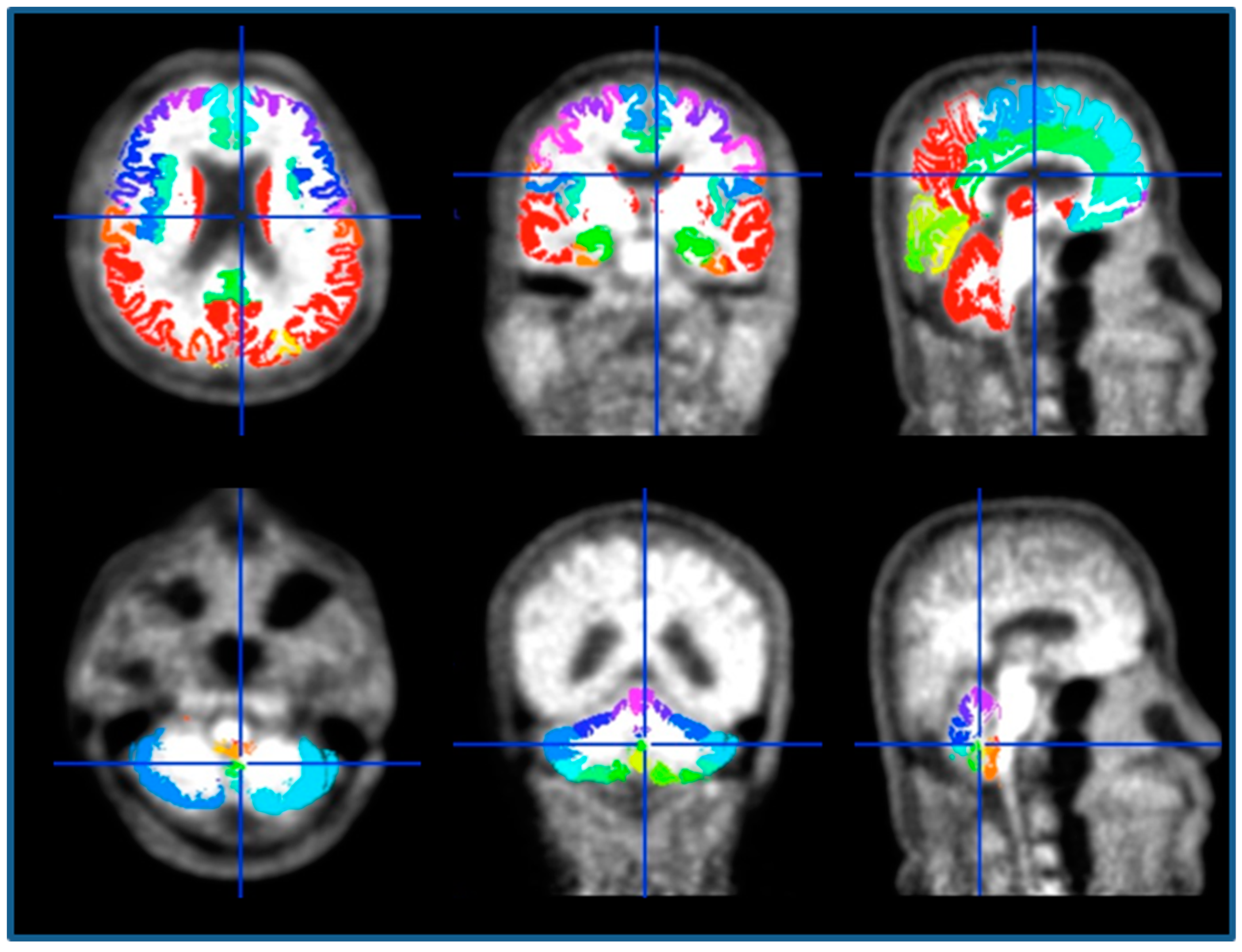
2.2.4. 11C-Pittsburgh Compound B (PiB)-PET Image Acquisition and Processing
2.3. Statistical Analyses
3. Results
3.1. Baseline Characteristics
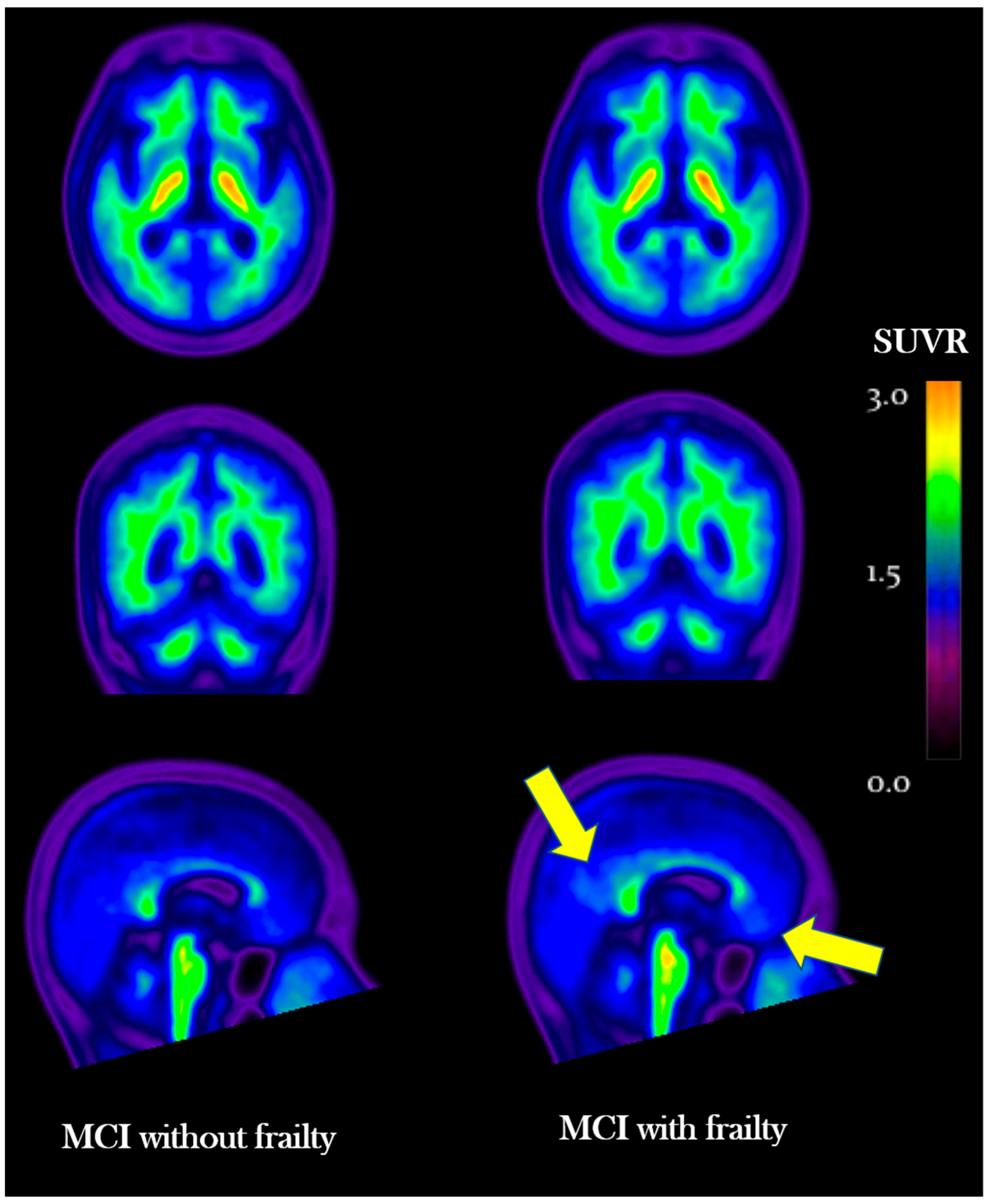
3.2. Quantitative and Visual Analysis Comparing SUVR between MCI and Cognitive Frailty Groups
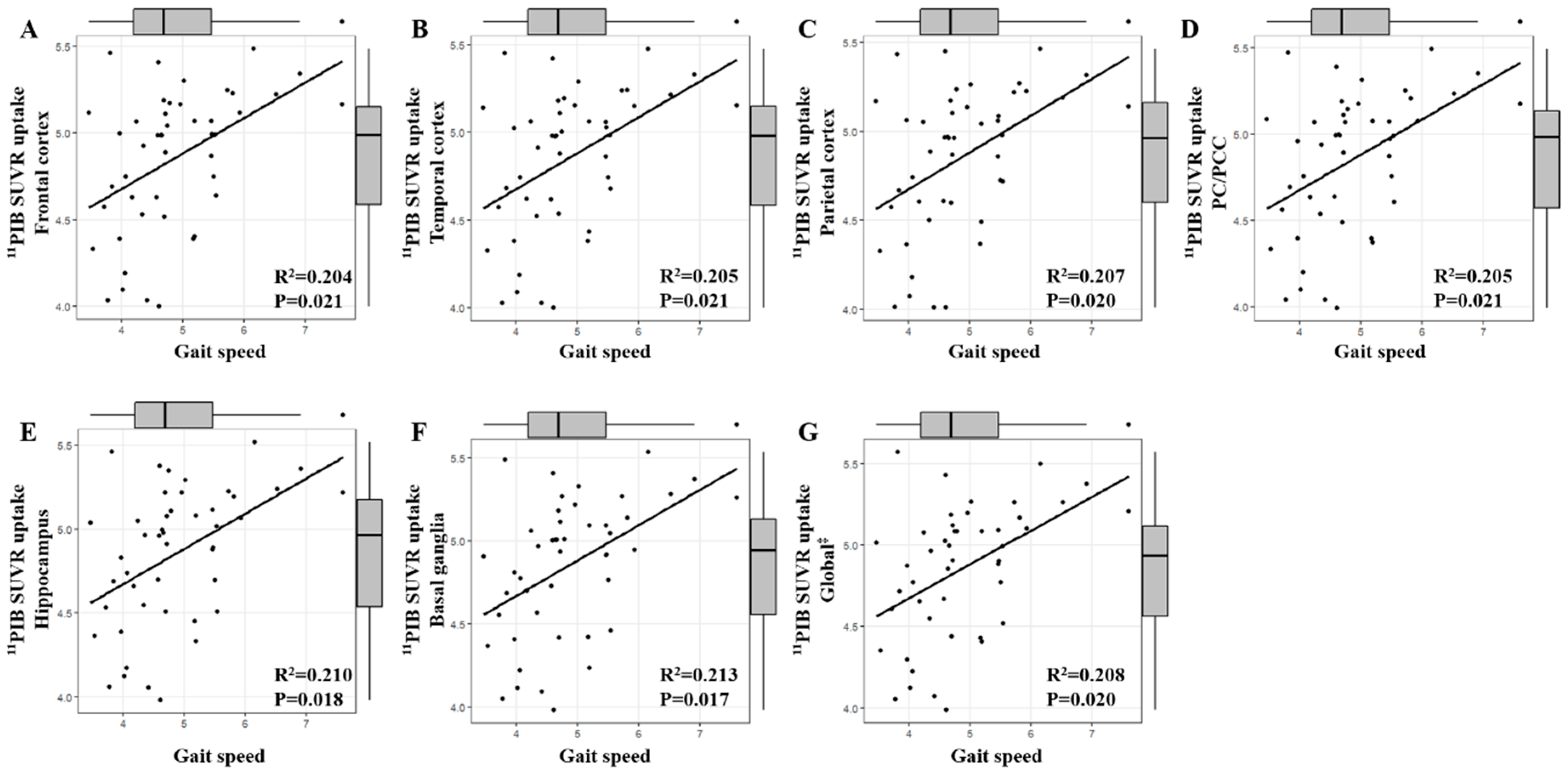
3.3. Association between SUVR and Measures of Physical Function by Brain Region
3.4. Association between SUVR and Physical Frailty by Brain Region
4. Discussion
5. Conclusions
Author Contributions
Funding
Conflicts of Interest
Appendix A

Appendix B

References
- Ruan, Q.; Yu, Z.; Chen, M.; Bao, Z.; Li, J.; He, W. Cognitive frailty, a novel target for the prevention of elderly dependency. Ageing Res. Rev. 2015, 20, 1–10. [Google Scholar] [CrossRef] [PubMed]
- Morley, J.E.; Vellas, B.; van Kan, G.A.; Anker, S.D.; Bauer, J.M.; Bernabei, R.; Cesari, M.; Chumlea, W.C.; Doehner, W.; Evans, J.; et al. Frailty consensus: A call to action. J. Am. Med. Dir. Assoc. 2013, 14, 392–397. [Google Scholar] [CrossRef] [PubMed]
- Kelaiditi, E.; Cesari, M.; Canevelli, M.; van Kan, G.A.; Ousset, P.J.; Gillette-Guyonnet, S.; Ritz, P.; Duveau, F.; Soto, M.E.; Provencher, V.; et al. Cognitive frailty: Rational and definition from an (i.A.N.A./i.A.G.G.) international consensus group. J. Nutr. Health Aging 2013, 17, 726–734. [Google Scholar] [CrossRef] [PubMed]
- Arts, M.H.; Collard, R.M.; Comijs, H.C.; Zuidersma, M.; De Rooij, S.E.; Naarding, P.; Oude Voshaar, R.C. Physical frailty and cognitive functioning in depressed older adults: Findings from the nesdo study. J. Am. Med. Dir. Assoc. 2016, 17, 36–43. [Google Scholar] [CrossRef] [PubMed]
- Brown, B.M.; Peiffer, J.J.; Taddei, K.; Lui, J.K.; Laws, S.M.; Gupta, V.B.; Taddei, T.; Ward, V.K.; Rodrigues, M.A.; Burnham, S.; et al. Physical activity and amyloid-beta plasma and brain levels: Results from the australian imaging, biomarkers and lifestyle study of ageing. Mol. Psychiatry 2013, 18, 875–881. [Google Scholar] [CrossRef] [PubMed]
- Schultz, S.A.; Boots, E.A.; Almeida, R.P.; Oh, J.M.; Einerson, J.; Korcarz, C.E.; Edwards, D.F.; Koscik, R.L.; Dowling, M.N.; Gallagher, C.L.; et al. Cardiorespiratory fitness attenuates the influence of amyloid on cognition. J. Int. Neuropsychol. Soc. 2015, 21, 841–850. [Google Scholar] [CrossRef] [PubMed]
- De Souto Barreto, P.; Andrieu, S.; Payoux, P.; Demougeot, L.; Rolland, Y.; Vellas, B. Multidomain Alzheimer Preventive Trial/Data Sharing Alzheimer Study Group: Physical activity and amyloid-beta brain levels in elderly adults with intact cognition and mild cognitive impairment. J. Am. Geriatr. Soc. 2015, 63, 1634–1639. [Google Scholar] [CrossRef] [PubMed]
- Del Campo, N.; Payoux, P.; Djilali, A.; Delrieu, J.; Hoogendijk, E.O.; Rolland, Y.; Cesari, M.; Weiner, M.W.; Andrieu, S.; Vellas, B.; et al. Relationship of regional brain beta-amyloid to gait speed. Neurology 2016, 86, 36–43. [Google Scholar] [CrossRef] [PubMed]
- Wennberg, A.M.V.; Savica, R.; Hagen, C.E.; Roberts, R.O.; Knopman, D.S.; Hollman, J.H.; Vemuri, P.; Jack, C.R., Jr.; Petersen, R.C.; Mielke, M.M. Cerebral amyloid deposition is associated with gait parameters in the mayo clinic study of aging. J. Am. Geriatr. Soc. 2017, 65, 792–799. [Google Scholar] [CrossRef] [PubMed]
- Nadkarni, N.K.; Perera, S.; Snitz, B.E.; Mathis, C.A.; Price, J.; Williamson, J.D.; DeKosky, S.T.; Klunk, W.E.; Lopez, O.L. Association of brain amyloid-beta with slow gait in elderly individuals without dementia: Influence of cognition and apolipoprotein E ε4 genotype. JAMA Neurol. 2017, 74, 82–90. [Google Scholar] [CrossRef] [PubMed]
- Shtifman, A.; Ward, C.W.; Laver, D.R.; Bannister, M.L.; Lopez, J.R.; Kitazawa, M.; LaFerla, F.M.; Ikemoto, N.; Querfurth, H.W. Amyloid-beta protein impairs Ca2+ release and contractility in skeletal muscle. Neurobiol. Aging 2010, 31, 2080–2090. [Google Scholar] [CrossRef] [PubMed]
- Fried, L.P.; Tangen, C.M.; Walston, J.; Newman, A.B.; Hirsch, C.; Gottdiener, J.; Seeman, T.; Tracy, R.; Kop, W.J.; Burke, G.; et al. Frailty in older adults: Evidence for a phenotype. J. Gerontol. A. Biol. Sci. Med. Sci. 2001, 56, M146–M156. [Google Scholar] [CrossRef] [PubMed]
- Collard, R.M.; Comijs, H.C.; Naarding, P.; Penninx, B.W.; Milaneschi, Y.; Ferrucci, L.; Oude Voshaar, R.C. Frailty as a predictor of the incidence and course of depressed mood. J. Am. Med. Dir. Assoc. 2015, 16, 509–514. [Google Scholar] [CrossRef] [PubMed]
- Jung, H.W.; Jang, I.Y.; Lee, Y.S.; Lee, C.K.; Cho, E.I.; Kang, W.Y.; Chae, J.H.; Lee, E.J.; Kim, D.H. Prevalence of frailty and aging-related health conditions in older koreans in rural communities: A cross-sectional analysis of the aging study of pyeongchang rural area. J. Korean Med. Sci. 2016, 31, 345–352. [Google Scholar] [CrossRef] [PubMed]
- Perera, S.; Mody, S.H.; Woodman, R.C.; Studenski, S.A. Meaningful change and responsiveness in common physical performance measures in older adults. J. Am. Geriatr. Soc. 2006, 54, 743–749. [Google Scholar] [CrossRef] [PubMed]
- Yoon, D.H.; Kang, D.; Kim, H.J.; Kim, J.S.; Song, H.S.; Song, W. Effect of elastic band-based high-speed power training on cognitive function, physical performance and muscle strength in older women with mild cognitive impairment. Geriatr. Gerontol. Int. 2017, 17, 765–772. [Google Scholar] [CrossRef] [PubMed]
- Jeong, S.K.; Cho, K.H.; Kim, J.M. The usefulness of the korean version of modified mini-mental state examination (k-mmmse) for dementia screening in community dwelling elderly people. BMC. Public Health 2004, 4, 31. [Google Scholar] [CrossRef] [PubMed] [Green Version]
- Park, S.; Park, S.E.; Kim, M.J.; Jung, H.Y.; Choi, J.S.; Park, K.H.; Kim, I.; Lee, J.Y. Development and validation of the pictorial cognitive screening inventory for illiterate people with dementia. Neuropsychiatr. Dis. Treat 2014, 10, 1837–1845. [Google Scholar] [PubMed]
- Tzourio-Mazoyer, N.; Landeau, B.; Papathanassiou, D.; Crivello, F.; Etard, O.; Delcroix, N.; Mazoyer, B.; Joliot, M. Automated anatomical labeling of activations in spm using a macroscopic anatomical parcellation of the mni mri single-subject brain. Neuroimage 2002, 15, 273–289. [Google Scholar] [CrossRef] [PubMed]
- Lopresti, B.J.; Klunk, W.E.; Mathis, C.A.; Hoge, J.A.; Ziolko, S.K.; Lu, X.; Meltzer, C.C.; Schimmel, K.; Tsopelas, N.D.; DeKosky, S.T.; et al. Simplified quantification of pittsburgh compound b amyloid imaging pet studies: A comparative analysis. J. Nucl. Med. 2005, 46, 1959–1972. [Google Scholar] [PubMed]
- Reiman, E.M.; Chen, K.; Liu, X.; Bandy, D.; Yu, M.; Lee, W.; Ayutyanont, N.; Keppler, J.; Reeder, S.A.; Langbaum, J.B.; et al. Fibrillar amyloid-beta burden in cognitively normal people at 3 levels of genetic risk for alzheimer’s disease. Proc. Natl. Acad. Sci. 2009, 106, 6820–6825. [Google Scholar] [CrossRef] [PubMed]
- Klunk, W.E.; Engler, H.; Nordberg, A.; Wang, Y.; Blomqvist, G.; Holt, D.P.; Bergstrom, M.; Savitcheva, I.; Huang, G.F.; Estrada, S.; et al. Imaging brain amyloid in alzheimer's disease with pittsburgh compound-b. Ann. Neurol. 2004, 55, 306–319. [Google Scholar] [CrossRef] [PubMed]
- Byun, B.H.; Kim, B.I.; Park, S.Y.; Ko, I.O.; Lee, K.C.; Kim, K.M.; Kim, Y.K.; Lee, J.Y.; Bu, S.H.; Kim, J.H.; et al. Head-to-head comparison of 11c-pib and 18f-fc119s for abeta imaging in healthy subjects, mild cognitive impairment patients, and alzheimer's disease patients. Medicine (Baltimore) 2017, 96, e6441. [Google Scholar] [CrossRef] [PubMed]
- Mathis, C.A.; Lopresti, B.J.; Klunk, W.E. Impact of amyloid imaging on drug development in alzheimer’s disease. Nucl. Med. Biol. 2007, 34, 809–822. [Google Scholar] [CrossRef] [PubMed]
- Cesari, M.; Kritchevsky, S.B.; Penninx, B.W.; Nicklas, B.J.; Simonsick, E.M.; Newman, A.B.; Tylavsky, F.A.; Brach, J.S.; Satterfield, S.; Bauer, D.C.; et al. Prognostic value of usual gait speed in well-functioning older people-results from the health, aging and body composition study. J. Am. Geriatr. Soc. 2005, 53, 1675–1680. [Google Scholar] [CrossRef] [PubMed]
- Houles, M.; Canevelli, M.; van Kan, G.A.; Ousset, P.J.; Cesari, M.; Vellas, B. Frailty and cognition. J. Frailty Aging 2012, 1, 56–63. [Google Scholar] [PubMed]


| Variable | Full Sample, n = 48 | Physical Frailty Status | p Value | |
|---|---|---|---|---|
| MCI + Robust, n = 21 (43.8%) | Cognitive Frailty, n = 27 (56.3%) | |||
| Demographics | ||||
| Age, mean (SD) | 75.1 (6.55) | 74.6 (5.65) | 75.5 (7.28) | 0.644 |
| Female, n (%) | 35 (73%) | 14 (67%) | 21 (78%) | 0.285 |
| Education, y, mean (SD) | 9.4 (4.20) | 9.1 (4.12) | 9.7 (4.33) | 0.665 |
| Frailty criteria, n (%) | ||||
| Slow gait velocity | 5 (10.2%) | 0 | 5 (19%) | 0.034 |
| Shrinking | 4 (8.2%) | 0 | 4 (15%) | 0.061 |
| Weakness | 13 (26.5%) | 0 | 13 (48%) | <0.001 |
| Exhaustion | 7 (14.3%) | 0 | 7 (26%) | 0.009 |
| Low activity level | 11 (22.4%) | 0 | 11 (41%) | <0.001 |
| Cognitive functioning | ||||
| MMSE (score), mean (SD) | 24.3 (2.31) | 24.7 (2.46) | 24.0 (2.19) | 0.285 |
| Cognitive impairment (MMSE <23), n (%) | 18 (36.7%) | 6 (27.3%) | 13 (48.1%) | 0.142 |
| CERAD-K | ||||
| Memory, mean (SD) | 25.0 (5.74) | 24.5 (5.97) | 25.4 (5.63) | 0.622 |
| Construction, mean (SD) | 9.7 (1.44) | 10.2 (1.33) | 9.3 (1.44) | 0.039 |
| Execution, mean (SD) | 13.3 (4.58) | 14.9 (4.59) | 12.1 (4.25) | 0.035 |
| Naming, mean (SD) | 10.0 (2.43) | 10.1 (2.37) | 9.9 (2.53) | 0.816 |
| Total score, mean (SD) | 58.0 (10.06) | 59.7 (10.63) | 56.6 (9.56) | 0.296 |
| MCI + Robust | Cognitive Frailty | p Value | |
|---|---|---|---|
| Frontal cortex | 1.28 ± 0.41 | 1.47 ± 0.54 | 0.371 |
| Temporal cortex | 1.24 ± 0.35 | 1.40 ± 0.50 | 0.433 |
| Parietal cortex | 1.27 ± 0.43 | 1.44 ± 0.54 | 0.438 |
| PC/PCC | 1.43 ± 0.46 | 1.63 ± 0.60 | 0.424 |
| Hippocampus | 1.22 ± 0.21 | 1.27 ± 0.16 | 0.330 |
| Basal ganglia | 1.37 ± 0.37 | 1.43 ± 0.35 | 0.560 |
| Global ‡ | 1.32 ± 0.39 | 1.41 ± 0.40 | 0.429 |
| Weight Loss | Exhaustion | Weakness | Slowness | Low Activity | ||||||
|---|---|---|---|---|---|---|---|---|---|---|
| β | p | β | p | β | p | β | p | β | p | |
| Frontal cortex | −0.149 | 0.312 | 0.072 | 0.627 | 0.367 | 0.010 | −0.033 | 0.821 | −0.023 | 0.877 |
| Temporal cortex | −0.138 | 0.350 | −0.010 | 0.345 | 0.377 | 0.008 | −0.003 | 0.986 | −0.020 | 0.895 |
| Parietal cortex | −0.179 | 0.223 | 0.076 | 0.609 | 0.328 | 0.023 | 0.000 | 0.997 | −0.035 | 0.811 |
| PC/PCC | −0.144 | 0.327 | 0.049 | 0.742 | 0.372 | 0.009 | 0.030 | 0.837 | −0.017 | 0.911 |
| Hippocampus | 0.018 | 0.905 | −0.086 | 0.563 | 0.377 | 0.008 | 0.030 | 0.841 | −0.010 | 0.946 |
| Basal ganglia | −0.104 | 0.482 | −0.047 | 0.753 | 0.374 | 0.009 | 0.011 | 0.943 | −0.030 | 0.842 |
| Global ‡ | −0.148 | 0.316 | 0.033 | 0.823 | 0.371 | 0.009 | 0.002 | 0.991 | −0.025 | 0.864 |
© 2018 by the authors. Licensee MDPI, Basel, Switzerland. This article is an open access article distributed under the terms and conditions of the Creative Commons Attribution (CC BY) license (http://creativecommons.org/licenses/by/4.0/).
Share and Cite
Yoon, D.H.; Lee, J.-Y.; Shin, S.A.; Kim, Y.K.; Song, W. Physical Frailty and Amyloid-β Deposits in the Brains of Older Adults with Cognitive Frailty. J. Clin. Med. 2018, 7, 169. https://doi.org/10.3390/jcm7070169
Yoon DH, Lee J-Y, Shin SA, Kim YK, Song W. Physical Frailty and Amyloid-β Deposits in the Brains of Older Adults with Cognitive Frailty. Journal of Clinical Medicine. 2018; 7(7):169. https://doi.org/10.3390/jcm7070169
Chicago/Turabian StyleYoon, Dong Hyun, Jun-Young Lee, Seong A Shin, Yu Kyeong Kim, and Wook Song. 2018. "Physical Frailty and Amyloid-β Deposits in the Brains of Older Adults with Cognitive Frailty" Journal of Clinical Medicine 7, no. 7: 169. https://doi.org/10.3390/jcm7070169