
Gastric Outlet Obstruction from Stomach-Containing Groin Hernias: Case Report and a Systematic Review
Abstract
:1. Introduction
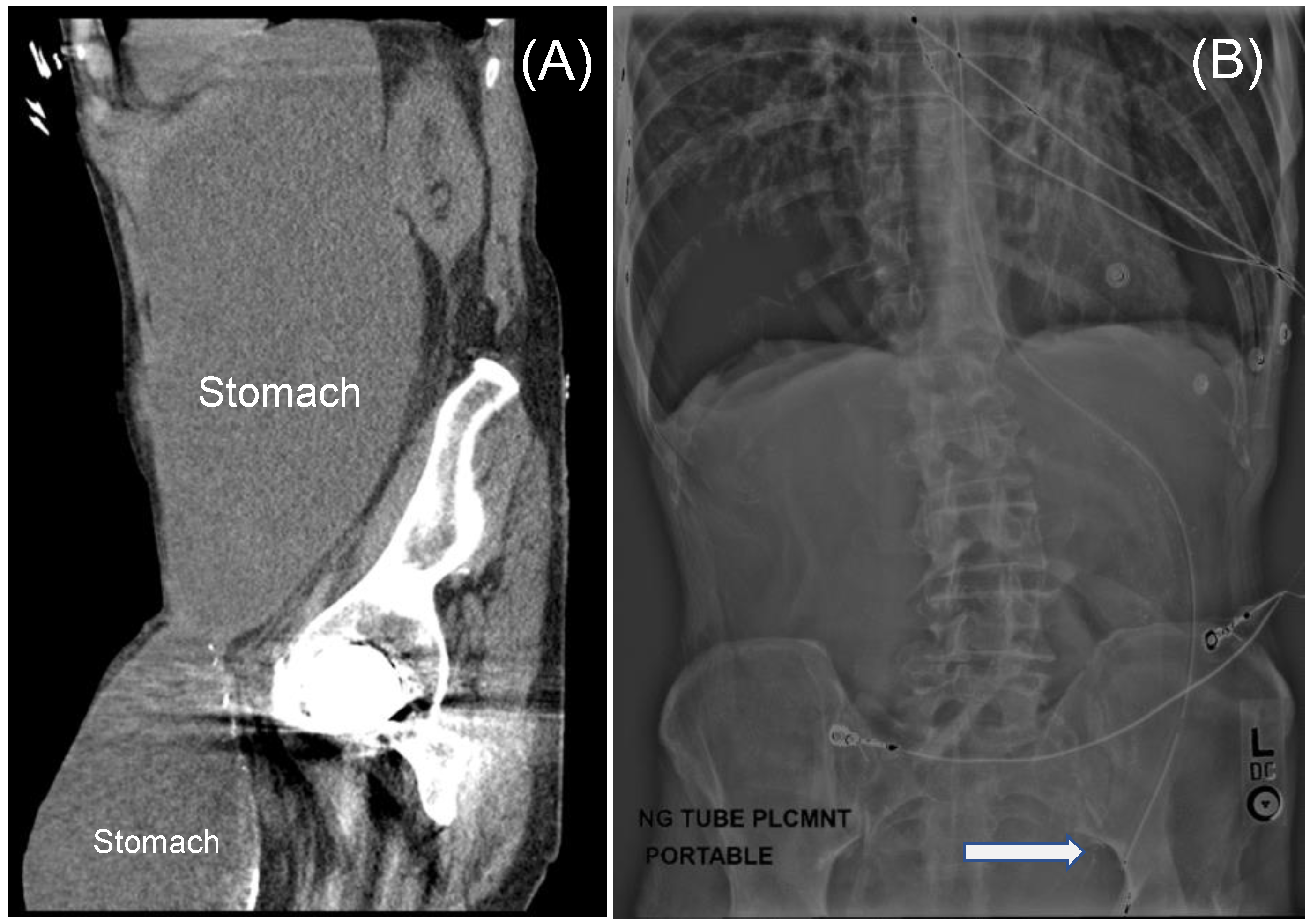
2. Case Presentation
Case Report
3. Review of the Literature
4. Results
4.1. Review of the Literature
4.1.1. History
4.1.2. Mechanism of Migration
4.1.3. Patient Demographics

| Characteristics | Inguinal (n = 85) | Femoral (n = 6) |
|---|---|---|
| Age [Years (SD) *] | 74.2 (13.0) | 62.0 (11.5) |
| Sex [male (%)] | 95.2 | 33.3 |
| Laterality [Left (%)] | 78.0 | 100.0 |
4.1.4. Management
4.1.5. Complications
4.1.6. Mortality
| n | Reference, Year | Age | Sex | Laterality | Clinical Presentation |
|---|---|---|---|---|---|
| 1 | Lallement, 1802 [21] | 64 | Male | NR | Abdominal pain/discomfort and vomiting |
| 2 | Yvan, 1830 [39] | NR | Male | NR | Vomiting |
| 3 | Febre, 1832 [40] | 73 | Male | Right | No symptoms |
| 4 | Fogt, 1884 [41] | 60 | Male | Left | Vomiting |
| 5 | Schmidt, 1885 [42] | 65 | Male | Left | Hematemesis and inguinal pain |
| 6 | Chiari, 1888 [43] | 74 | Male | Right | No symptoms |
| 7 | Lewin, 1893 [44] | 53 | Male | Left | Emesis and pain |
| 8 | Chevereau, 1894 [45] | 77 | Male | Left | Emesis and pain |
| 9 | Souligoux, 1896 [46] | NR | Male | Left | NR |
| 10 | Brunner, 1897 [30] | 28 | Male | NR | NR |
| 11 | Hilgeneriner, 1910 [47] | 52 | Female | Left | Pain and vomiting |
| 12 | Chambard, 1912 [19] | 62 | Male | Left | Vomiting, pain, and an incarcerated hernia |
| 13 | Rieder, 1915 [48] | 62 | Male | Left | Hematemesis and melena |
| 14 | Ahrens, 1920 [49] | 40 | Male | Right | Pain |
| 15 | Maag, 1920 [50] | 81 | Male | Left | No symptoms |
| 16 | Stokes, 1922 [51] | 42 | Male | Right | Vomiting and an incarcerated hernia |
| 17 | Elischer, 1923 [31] | 53 | Male | Left | Nausea and an incarcerated hernia |
| 18 | Elischer, 1923 [31] | 70 | Male | Left | Incarcerated hernia |
| 19 | Dressen, 1925 [52] | 62 | Male | Left | Vomiting, pain, and inguinal symptoms when eating |
| 20 | de Vernejoul, 1925 [32] | 57 | Female | Left | NR |
| 21 | Sicot, 1927 [33] | 59 | Male | Left | Pain, vomiting, and dyspepsia |
| 22 | Lipkin, 1928 [53] | 60 | Male | Left | Incarcerated hernia |
| 23 | Siegmund, 1929 [54] | NR | Male | Right | NR |
| 24 | Novaro, 1930 [34] | 53 | Male | Right | Vomiting, pain, and an irreducible hernia |
| 25 | Rodzevich, 1935 [55] | 54 | Male | Left | Vomiting and abdominal pain |
| 26 | Oakley, 1937 [56] | 81 | Male | Right | Abdominal and groin pain |
| 27 | Herrmann, 1937 [57] | 80 | Male | Left | Vomiting |
| 28 | Lemaitre, 1937 [58] | 51 | Male | Left | Dyspepsia |
| 29 | Lust, 1937 [59] | 62 | Male | Left | NR |
| 30 | Alexsandrovskiv, 1940 [60] | 73 | Male | Left | Incarcerated hernia |
| 31 | Feldman, 1943 [61] | 66 | Male | Right | No symptoms |
| 32 | Hartley, 1945 [62] | 67 | Male | Left | Dyspepsia |
| 33 | Simmons, 1949 [63] | 66 | Male | Left | Nausea, vomiting, and abdominal pain |
| 34 | Lewis, 1950 [64] | 69 | Male | Right | Occasional vomiting |
| 35 | Anger, 1952 [65] | 74 | Male | Left | Vague symptoms |
| 36 | Bernard, 1953 [66] | NR | NR | NR | NR |
| 37 | Meinterz, 1953 [67] | NR | NR | NR | NR |
| 38 | Davey, 1954 [25] | 61 | Male | Left | Vomiting with markedly distended and tense abdomen |
| 39 | Legrand, 1955 [68] | NR | NR | NR | NR |
| 40 | D’Eshougues, 1956 [69] | NR | NR | NR | NR |
| 41 | Allende, 1956 [70] | NR | NR | NR | NR |
| 42 | Kislenskii, 1959 [71] | NR | NR | Left | NR |
| 43 | Hagarty, 1959 [72] | NR | NR | NR | NR |
| 44 | Ship, 1960 [26] | 83 | Male | Left | Persistent nausea and vomiting |
| 45 | Herrera, 1960 [73] | NR | NR | NR | NR |
| 46 | Jackson, 1964 [74] | NR | NR | NR | Strangulation and perforation of the stomach in the inguinal canal |
| 47 | Falugiani, 1968 a | NR | NR | NR | NR |
| 48 | Gue, 1970 [75] | NR | NR | NR | NR |
| 49 | Soudek, 1975 [76] | NR | NR | NR | NR |
| 50 | Padmanabhan, 1976 [77] | 65 | Male | Left | NR |
| 51 | Nagendran, 1977 [78] | NR | NR | NR | NR |
| 52 | Rozencwajg, 1981 [79] | NR | NR | NR | NR |
| 53 | Udwadia, 1984 [80] | NR | NR | NR | Hematemesis |
| 54 | Quaranta, 1984 [81] | NR | NR | NR | NR |
| 55 | Resente,1986 [82] | NR | NR | NR | NR |
| 56 | Naraynsingh, 1987 [83] | 62 | Male | Left | Recurrent bouts of vomiting, recurrent GOO |
| 57 | Levy, 1987 [84] | 49 | Male | Left | Abdominal pain, nausea, and weight loss |
| 58 | Loizate, 1988 [85] | NR | NR | NR | Upper gastrointestinal tract hemorrhage |
| 59 | Broquet, 1992 [86] | 64 | Female | Bilateral | Perforation of gastric ulcer within the hernia sac |
| 60 | Diaz, 1997 [27] | NR | NR | NR | NR |
| 61 | Diaz, 1997 [27] | NR | NR | NR | NR |
| 62 | Walgenbach, 2001 [87] | 72 | Male | Left | A 6-h history of abdominal distension and pain |
| 63 | Birnbaum, 2011 [88] | 86 | Male | Right | Nausea and vomiting |
| 64 | Dogar, 2011 [89] | 65 | Male | Left | Irreducible groin bulge, abdominal pain, distention, darkish red vomitus, and obstipation |
| 65 | Kerschaever, 2012 [90] | 79 | Male | Left | Anorexia, vomiting, and abdominal distension |
| 66 | Ogul, 2013 [28] | 56 | Male | Left | Recurrent vomiting and bilateral incarcerated groin bulges |
| 67 | Ferdinand, 2013 [38] | 73 | Male | Right | Iron deficiency anemia and gastric volvulus |
| 68 | Fazekas, 2014 [37] | 85 | Male | Left | Three-day history of gastrointestinal obstructive symptoms |
| 69 | Creedon, 2014 [91] | 87 | Male | Left | Colicky abdominal pain for 48 h and vomiting |
| 70 | Patel, 2014 [92] | 85 | Male | Left | 3-day history of profuse vomiting and abdominal pain |
| 71 | Lajevardi, 2015 [35] | 83 | Male | Left | Four-day history of vomiting and constipation |
| 72 | Fitz, 2016 [14] | 46 | Male | Bilateral | Severe abdominal pain after dinner brought in by ambulance to the emergency department |
| 73 | Mora-Guzman, 2016 [93] | 79 | Male | Right | Abdominal pain and vomiting |
| 74 | Periz-Pueyo, 2016 [94] | 61 | Male | Left | Gastric necrosis secondary to an incarcerated inguinal hernia |
| 75 | Nugud, 2017 [95] | 67 | Male | Left | Bilious vomiting with abdominal pain |
| 76 | Sayad, 2019 [96] | 50 | Male | NR | Severe abdominal pain |
| 77 | Junge, 2019 [97] | 75 | Male | Left | Abdominal pain and nausea |
| 78 | Mehta, 2019 [98] | 75 | Male | Left | 5-day history of hematemesis |
| 79 | Heylen, 2020 [36] | 74 | Male | Left | Dark vomitus and generalized abdominal tenderness |
| 80 | Patel, 2021 a | 84 | Male | NR | Nausea, vomiting, constipation, GOO, peritonitis |
| 81 | Vinod, 2021 [99] | 49 | Male | Left | Acute abdominal pain with nausea and dysuria |
| 82 | Alexandre, 2022 [100] | 71 | Male | Left | Nausea, vomiting, constipation, and GOO |
| 83 | Grantham, 2022 [18] | 81 | Male | Lett | Coffee ground emesis |
| 84 | Abbakar, 2022 [101] | 84 | Male | Right | Double GOO, abdominal pain and vomiting |
| 85 | Huerta, 2023 a | 77 | Male | Left | Abdominal pain, nausea, vomiting, and GOO |
| Reference, Year | Age | Sex | Laterality | Clinical Presentation |
|---|---|---|---|---|
| Keller, 1885 [29] | 47 | Female | Left | Abdominal pain and vomiting |
| Spiegel, 1920 [102] | 55 | Female | Left | Gastric strangulation |
| Cave, 1948 [103] | 56 | Female | Left | Dyspepsia |
| Davey, 1954 [25] | 68 | Male | Left | No symptoms |
| Cade, 1984 [104] | 79 | Female | Left | Abdominal pain, emesis, and hematemesis |
| Natsis, 2008 [20] | 67 | Male | Left | Findings at autopsy |
5. Discussion
6. Conclusions
Author Contributions
Funding
Institutional Review Board Statement
Informed Consent Statement
Data Availability Statement
Conflicts of Interest
References
- Kingsnorth, A.; LeBlanc, K. Hernias: Inguinal and incisional. Lancet 2003, 362, 1561–1571. [Google Scholar] [CrossRef]
- Rutkow, I.M. Epidemiologic, economic, and sociologic aspects of hernia surgery in the United States in the 1990s. Surg. Clin. N. Am. 1998, 78, 941–951. [Google Scholar] [CrossRef]
- Isernia, R.M.; De Luca, G.M.; De Luca, A.; Franzoso, L.; Navazio, L.R.; Caruso, R.; Ferri, V.; Ielpo, B.; Giungato, S. Sliding ureteral inguinal hernia: An uncommon embryological trick. Case report and literature review. Int. J. Surg. Case Rep. 2022, 94, 107006. [Google Scholar] [CrossRef] [PubMed]
- Sugumar, K.; Gupta, M. Anatomy, Abdomen and Pelvis: Inguinal Ligament (Crural Ligament. Poupart Ligament). In StatPearls; StatPearls Publishing: Treasure Island, FL, USA, 2023. [Google Scholar]
- Ponka, J.L.; Brush, B.E. Sliding inguinal hernia in patients over 70 years of age. J. Am. Geriatr. Soc. 1978, 26, 68–73. [Google Scholar] [CrossRef]
- Huerta, S.; Fairbanks, T.; Cinat, M. Incarcerated vesicoinguinal hernia presenting with gross hematuria. J. Am. Coll. Surg. 2005, 201, 992–993. [Google Scholar] [CrossRef] [PubMed]
- Dunn, T.M.; Markgraf, W.H. Littre hernia—Incarcerated Meckel’s diverticulum. Am. J. Surg. 1962, 103, 144–145. [Google Scholar] [CrossRef]
- Michalinos, A.; Moris, D.; Vernadakis, S. Amyand’s hernia: A review. Am. J. Surg. 2014, 207, 989–995. [Google Scholar] [CrossRef] [PubMed]
- Goyal, S.; Shrivastva, M.; Verma, R.; Goyal, S. “Uncommon contents of inguinal hernial sac”: A surgical dilemma. Indian J. Surg. 2015, 77, 305–309. [Google Scholar] [CrossRef]
- Ganesaratnam, M. Maydl’s hernia: Report of a series of seven cases and review of the literature. Br. J. Surg. 1985, 72, 737–738. [Google Scholar] [CrossRef]
- Taveras, L.R.; Huerta, S. A case report of a de Garengeot hernia in a nonagenarian veteran. Int. J. Surg. Case Rep. 2017, 41, 301–303. [Google Scholar] [CrossRef]
- Sidiqi, M.M.; Menezes, G. Asymptomatic herniation of ureter in the routine inguinal hernia: A dangerous trap for general surgeons. Int. J. Surg. Case Rep. 2018, 49, 244–246. [Google Scholar] [CrossRef]
- Aldhafar, A.; Mohammed, A.; Alwabari, M.; Aldhafar, R. A Strangulated Right Inguinal Hernia Containing the Transverse Colon: An Unusual Case Report. Asian J. Case Rep. Surg. 2020, 5, 10–13. [Google Scholar]
- Fitz, E.; Chihara, R.; Stanton-Maxey, K.J. Gastric Perforation Associated with Bilateral Inguinal Hernias. J. Am. Coll. Surg. 2016, 222, e12–e13. [Google Scholar] [CrossRef]
- Tajti, J., Jr.; Pieler, J.; Abraham, S.; Simonka, Z.; Paszt, A.; Lazar, G. Incarcerated gallbladder in inguinal hernia: A case report and literature review. BMC Gastroenterol. 2020, 20, 425. [Google Scholar] [CrossRef] [PubMed]
- Chen, G.; Wang, X.; Zhao, Y.; Zhu, L.; Tang, D. Splenogonadal fusion: A case report and review of the literature. BMC Urol. 2021, 21, 16. [Google Scholar] [CrossRef] [PubMed]
- Arredondo Montero, J.; Guillen Redondo, P.; Antona, G.; Bronte Anaut, M. Newborn inguinal hernia containing uterus, ovary and Fallopian tube. An. Pediatría (Engl. Ed.) 2023, 98, 232–233. [Google Scholar] [CrossRef]
- Grantham, T.A.; Ramachandran, R.; Parvataneni, S.; Gaduputi, V. Stomach within a Large Inguinal Hernia. Cureus 2022, 14, e24783. [Google Scholar] [CrossRef] [PubMed]
- Chambard, M.E. La hernie inguinale de l’estomac. Rev. Gynec. d’Obst. 1912, 19, 61–79. [Google Scholar]
- Natsis, K.; Apostolidis, S.; Papadopoulou, A.; Vlasis, K.; Totlis, T.; Skandalakis, P. Gastric femoral hernia in a male cadaver with gastroptosis: Case report and review of the literature. Hernia 2008, 12, 205–208. [Google Scholar] [CrossRef]
- Lallement, L. Observation d’un entero-gastrocele. J. Med. Chir. Pharm. 1802, 1, 329. [Google Scholar]
- Ochoa-Hernandez, A.; Timmerman, C.; Ortiz, C.; Huertas, V.L.; Huerta, S. Emergent groin hernia repair at a County Hospital in Guatemala: Patient-related issues vs. health care system limitations. Hernia 2020, 24, 625–632. [Google Scholar] [CrossRef]
- McInnes, M.D.; Moher, D.; Thombs, B.D.; McGrath, T.A.; Bossuyt, P.M.; Clifford, T.; Cohen, J.F.; Deeks, J.J.; Gatsonis, C.; Hooft, L. Preferred reporting items for a systematic review and meta-analysis of diagnostic test accuracy studies: The PRISMA-DTA statement. JAMA 2018, 319, 388–396. [Google Scholar] [CrossRef]
- Heylen, J.; Campioni-Norman, D.; Lowcock, D.; Varatharajan, L.; Kostalas, M.; Irukulla, M.; Ratnasingham, K. Inguinoscrotal hernias containing stomach: Risk of emergency presentation. Ann. R. Coll. Surg. Engl. 2021, 103, 713–717. [Google Scholar] [CrossRef] [PubMed]
- Davey, W.; Strange, S. The stomach as a content of inguinal and femoral herniae. Br. J. Surg. 1954, 41, 651–658. [Google Scholar] [CrossRef]
- Ship, A.G.; Glick, A.H.; Goldenson, A.B. Incarceration of the stomach in an inguinal hernia: Report of a case. N. Engl. J. Med. 1960, 262, 78–80. [Google Scholar] [CrossRef] [PubMed]
- Diaz Oller, J.; Medina Dominguez, M.T.; Arcos Navarro, A.; Alia Diaz, J.J.; Asanza Llorente, J.A.; Numancia Andreu, M.B.; Nevado Infante, A.; Martos Torres, J.; Moya Vazquez, R.J. Stomach in inguinal-scrotal hernia. Rev. Esp. Enferm. Dig. 1997, 89, 325–328. [Google Scholar]
- Ogul, H.; Yuce, I.; Eren, S.; Kantarci, M. Bilateral inguinal hernia with uncommon content. Can. J. Gastroenterol. 2013, 27, 14. [Google Scholar] [CrossRef] [PubMed]
- Keller, O. Schenkelhernie mit Magen als Inhalt. Inag. Diss. Bern. 1885. [Google Scholar]
- Brunner, F. Kleinere Mittheilungen: Ein Fall von Hernia ventriculi inguinalis incarcerata. Zentralbl. Chir. 1897, 24, 919–921. [Google Scholar]
- Elisscher, E.V. Ibid 1923, 50, 1814.
- de Vernejoul, M.C.; de Luna, D. Sociétés de province: Société de Chirurgie de Marseille: Volumineuse hernie inguinale renfermant l’estomac et les annexes gauches. Pr. Med. 1925, 33, 208. [Google Scholar]
- Sicot, R. Contribu a l’Etude des Hernies inguinales de l’Etomc. These de Paris 1927. [Google Scholar]
- Novaro, N. Arch. Ital. Chir. 1920, 27, 483.
- Lajevardi, S.S.; Gundara, J.S.; Collins, S.A.; Samra, J.S. Acute Gastric Rupture in a Giant Inguinoscrotal Hernia. J. Gastrointest. Surg. 2015, 19, 2283–2285. [Google Scholar] [CrossRef] [PubMed]
- Heylen, J.; Campioni-Norman, D. Bilateral inguinoscrotal hernia with gastric contents and subsequent perforation: Lessons in operative management. Int. J. Surg. Case Rep. 2020, 77, 853–856. [Google Scholar] [CrossRef] [PubMed]
- Fazekas, B.; Frecker, P.; Francis, L.; Patel, K. Aspiration pneumonia as a complication of a rare type of hernia. Int. J. Surg. Case Rep. 2014, 5, 1061–1063. [Google Scholar] [CrossRef] [PubMed]
- Ferdinand, E.; Amir, R.; Manjunath, S. Serendipity of giant proportions. Clin. Gastroenterol. Hepatol. 2013, 11, A25–A26. [Google Scholar] [CrossRef]
- Yvan. Arch. Gen. Med. 1830, 22, 139.
- Febre, J.A. These de Paris 1832, 132.
- Fogt, L. Arztl. Zntelligenzbl 1884, 31, 285.
- Schmidt, M. Berl. Klin. Wschr. 1885, 22.
- Chiari, H. Prag. Med. Wschr. 1888, 13, 115.
- Lewin, T.F. Ann. Soc. Belge Chir. 1893, 1.
- Chevereau, P. Bull. Soc. Anat. Paris 1894, 8.
- Souligoux. Bull. Soc. Anat. Paris 1896, 10.
- Hilgenreiner, H. Siltene und bemerkenswerte Hernien. Beitr. Klin. Chir. 1910, 69, 333–430. [Google Scholar]
- Rieder, H. Fortschr. Riintgensrr. 1915, 23.
- Ahrens, P. Zbl. Chir. 1920, 47.
- Maag, O. Dtsh. Z. Chir. 1920, 152.
- Stokes, A.F. Med. F. Aust. 1922, 2.
- Dressen, J. Stomach in Inguinal Hernia. Edinb. Med. J. 1925, 32, 774. [Google Scholar]
- Lipkin, L.N. Odessky Med. J. 1928, 3.
- Siegmund, H. Hand Book de Spesillen Paht. Anat. U. Histo 1929, 4.
- Rodzevich, E.V. Vestn. Khir. Grekov. 1935, 41.
- Oakley, R.H. Strangulation of right inguinal hernia cotaining part of the stomach with simultaneus perforation of a gastric ulcer. Br. J. Surg. 1937, 25, 454–455. [Google Scholar] [CrossRef]
- Herrmann, J. Cas. Lek. Ces 1937, 76.
- Lemaitre, L. Bull. Soc. Radiol. Med. Paris 1937, 25.
- Lust, F.J. Herniation of the stomach into the scrotum. Am. J. Roentgenol. 1937, 37, 666–667. [Google Scholar]
- Alexsandrovskiy, D.G.; Degtyarev, I.G. Vrac. Dyelo 1940, 22.
- Feldman, J.J.M.; TIturrioz, T. Sem. Med. G. Aires 1943, 1.
- Hartley, J.B. Diverticulum of stomach found to enter left inguinal hernial sac. Br. J. Radiol. 1945, 18, 231–232. [Google Scholar] [CrossRef]
- Simmons, H. Perforation of stomach in scrotal hernia. Br. Med. J. 1949, 1, 808. [Google Scholar] [CrossRef] [PubMed]
- Lewis, T.F.; Ross, P.S. Inguinal herniation of stomach: Case report. Ohio State Med. J. 1950, 46, 987–988. [Google Scholar] [PubMed]
- Anger, H. Extreme Gastroenteroptose bei beiderseitben Scrotalhernien. Fortschr. Roentgenstr. 1952, 76. [Google Scholar] [CrossRef]
- Bernard, A. Inguino-scrotal hernia of the stomach. Arch. Mal. Appar. Dig. Mal. Nutr. 1953, 42, 1400–1403. [Google Scholar] [PubMed]
- Meinertz, O. Inguinal hernia with the stomach functioning as hernial sac content. Fortschr. Geb. Rontgenstr. 1953, 79, 400–401. [Google Scholar] [CrossRef] [PubMed]
- Legrand, J.; Tesson, A. Estomac et hernie scrotale: Présentation de clichés. J. Radiol. Electrol. 1955, 36, 246. [Google Scholar]
- Robert D’Eshougucs, J.; Miniconi, P.; Clerget, O.; Scotto, J.C. Une ptose gastrointestinale peu banale (hernie inguinale de l’estomac). Algérie Mèd. 1956, 60, 1043. [Google Scholar]
- Allende, C.I. Hernia escrotal de estómago. Bol. Trab. Soc. Cir. Buenos Aires 1956, 40, 548–551. [Google Scholar]
- Kislenskii, I.F. Left-sided scrotal inguinal hernia, containing the stomach, cecum, ascending & transverse colon. Khirurgiia 1959, 35, 99–100. [Google Scholar]
- Hagarty, G. The stomach in an inguinal hernia. Med. J. Aust. 1959, 46, 403. [Google Scholar] [CrossRef]
- Herrera, C.A. Large inguinoscrotal hernia containing the stomach. Prensa Med. Argent. 1960, 47, 2443. [Google Scholar]
- Jackson, F.C.; Brown, J.C.; Stiegel, D.D. Strangulation and perforation of the stomach in an inguinal hernia. Am. Surg. 1964, 30, 677–681. [Google Scholar] [PubMed]
- Gue, S. Spontaneous rupture of stomach, a rare complication of inguinal hernia. J. Br. Surg. 1970, 57, 154–155. [Google Scholar] [CrossRef] [PubMed]
- Soudek, K.; Súna, Z. Study of a rare ectopic localization ofthe stomach in a large scrotal hernial. Rozhl. Chir. 1975, 54, 336–337. [Google Scholar] [PubMed]
- Padmanabhan, A.I.; Lowenfels, A.B. Letter: An unusual hernia. JAMA 1976, 235, 2813. [Google Scholar] [CrossRef] [PubMed]
- Nagendran, T. Stomach contained in a giant scrotal hernia. Am. Surg. 1977, 43, 473–476. [Google Scholar] [PubMed]
- Rozencwajg, J. Stomach rupture in inguino-scrotal hernia. Acta Chir. Belg. 1981, 80, 159–160. [Google Scholar] [PubMed]
- Udwadia, T. Stomach strangulated in inguinal hernia presenting with hematemesis. Int. Surg. 1984, 69, 177–179. [Google Scholar]
- Quaranta, M.; Rivetti, R.; Cartia, Q. Rare case of inguino-scrotal hernia containing the stomach. Minerva Chir. 1984, 39, 1757–1759. [Google Scholar]
- Resente, F.; Corbetti, F.; Bettini, F. Inguinal hernia of the stomach. A case. Radiol. Med. 1986, 72, 597–598. [Google Scholar] [PubMed]
- Naraynsingh, V.; Sieunarine, K.; Raju, G. Recurrent gastric outlet obstruction due to an inguinal hernia. Postgrad. Med. J. 1987, 63, 905–906. [Google Scholar] [CrossRef] [PubMed]
- Levy, I.; Ebbs, S.R. Inguinal hernia: An unusual cause of dyspepsia. Br. J. Surg. 1987, 74, 363. [Google Scholar] [CrossRef] [PubMed]
- Loizate Totoricaguena, A.; Lamíquiz Vallejo, A. Stomach incarcerated in an inguinal hernia as a cause of upper digestive hemorrhage. Rev. Esp. Enferm. Apar. Dig. 1988, 74, 172–174. [Google Scholar] [PubMed]
- Prêtre, R.; Broquet, P.E.; Robert, J.; Rohner, A. Perforation of gastric ulcer in inguinal hernia. J. Chir. 1992, 129, 218–220. [Google Scholar]
- Walgenbach, K.J.; Lauschke, H.; Brunagel, G.; Hirner, A. An uncommon form of gastric rupture in giant scrotal hernia. Zentralbl Chir. 2001, 126, 1015–1017. [Google Scholar] [CrossRef] [PubMed]
- Birnbaum, D.J.; Grègoire, E.; Campan, P.; Hardwigsen, J.; Le Treut, Y.P. A large inguinoscrotal hernia with stomach content. ANZ J. Surg. 2011, 81, 86–87. [Google Scholar] [CrossRef]
- Dogar, M.; Chaudhary, A. Inguinal hernia containing stomach, transverse colon and small bowel. Pak. J. Med. Health Sci. 2011, 5. [Google Scholar]
- Kerschaever, I.; Poelmans, S.; Vankeirsbilck, J.; Vandewoude, M. Rare cause of gastric outlet obstruction: Incarcerated pylorus within an inguinal hernia. Ann. R. Coll. Surg. Engl. 2012, 94, e46–e48. [Google Scholar] [CrossRef]
- Creedon, L.; Peacock, O.; Singh, R.; Awan, A. Gastric outlet obstruction secondary to incarcerated pylorus in an inguinal hernia. Ann. R. Coll. Surg. Engl. 2014, 96, e26–e27. [Google Scholar] [CrossRef]
- Patel, K.; Francis, L.; Fazekas, B.; Panthagani, J. Unusual contents of a large incarcerated inguinal hernia. BMJ Case Rep. 2014. [Google Scholar] [CrossRef]
- Mora-Guzman, I.; Munoz de Nova, J.L.; di Martino, M.; Marin-Perez, E. Antro gástrico como contenido insólito de una hernia inguinal. Rev. Chil. Cir. 2016, 68, 281–282. [Google Scholar] [CrossRef]
- Pueyo Périz, E.M.; Andrés Pérez, S.; García Tejero, A.; García Fernández, C. Gastric necrosis secondary to inguinal incarceration. Cir. Esp. 2016, 94, 180. [Google Scholar] [CrossRef] [PubMed]
- Nugud, A.A.; Nugud, S. Long-standing asymptomatic Inguinoscrotal hernia. Sultan Qaboos Univ. Med. J. 2017, 17, 250–251. [Google Scholar] [CrossRef]
- Sayad, P.; Tan, A.Z. A case report of a gastric perforation in a giant inguinoscrotal hernia: A two-step approach. Int. J. Surg. Case Rep. 2019, 55, 174–178. [Google Scholar] [CrossRef] [PubMed]
- Junge, K.; Otto, J.; Oral, H. A Rare Cause of Gastric Outlet Stenosis—Scrotal Hernia. Dtsch. Ärzteblatt Int. 2019, 116, 507. [Google Scholar] [CrossRef] [PubMed]
- Mehta, T.; Weissman, S.; Vash, A.; Yim, D.; Serrano, O. Gastric Inguinoscrotal Hernia. ACG Case Rep. J. 2019, 6, e00187. [Google Scholar] [CrossRef] [PubMed]
- Vinod, V.C.; Younis, M.U. Gastric strangulation and perforation caused by a giant inguinal-scrotal hernia. Turk. J. Emerg. Med. 2021, 21, 122–124. [Google Scholar] [CrossRef] [PubMed]
- Alexandre, K.; Vandeveer, Z.; Barnwell, J.M.; Vandeveer, Z.T. A Rare Case of Left Inguinoscrotal Hernia Containing Stomach. Cureus 2022, 14, e30838. [Google Scholar] [CrossRef]
- Abbakar, M.; Ahmed, S.; Lim, M. EP-25 Double Gastric Outlet Obstruction Caused by an Incarcerated Right Inguinal Hernia, a Rare Presentation. Br. J. Surg. 2022, 109, znac245-006. [Google Scholar] [CrossRef]
- Spiegel, B. Einklemmung des Magens im Schenkelbruch. Zbl Chir. 1920, 16, 373–374. [Google Scholar]
- Cave, P. Stomach in a Femoral Hernia. Br. J. Radiol. 1948, 21, 143–145. [Google Scholar] [CrossRef]
- Cade, R.J.; Lane, R.H. Strangulated stomach within a femoral hernia. Postgrad. Med. J. 1984, 60, 376. [Google Scholar] [CrossRef]

Disclaimer/Publisher’s Note: The statements, opinions and data contained in all publications are solely those of the individual author(s) and contributor(s) and not of MDPI and/or the editor(s). MDPI and/or the editor(s) disclaim responsibility for any injury to people or property resulting from any ideas, methods, instructions or products referred to in the content. |
© 2023 by the authors. Licensee MDPI, Basel, Switzerland. This article is an open access article distributed under the terms and conditions of the Creative Commons Attribution (CC BY) license (https://creativecommons.org/licenses/by/4.0/).
Share and Cite
Favela, J.G.; Argo, M.B.; McAllister, J.; Waldrop, C.L.; Huerta, S. Gastric Outlet Obstruction from Stomach-Containing Groin Hernias: Case Report and a Systematic Review. J. Clin. Med. 2024, 13, 155. https://doi.org/10.3390/jcm13010155
Favela JG, Argo MB, McAllister J, Waldrop CL, Huerta S. Gastric Outlet Obstruction from Stomach-Containing Groin Hernias: Case Report and a Systematic Review. Journal of Clinical Medicine. 2024; 13(1):155. https://doi.org/10.3390/jcm13010155
Chicago/Turabian StyleFavela, Juan G., Madison B. Argo, Jared McAllister, Caitlyn L. Waldrop, and Sergio Huerta. 2024. "Gastric Outlet Obstruction from Stomach-Containing Groin Hernias: Case Report and a Systematic Review" Journal of Clinical Medicine 13, no. 1: 155. https://doi.org/10.3390/jcm13010155