
The Role of Intracoronary Imaging for the Management of Calcified Lesions
by
 ,
,
Stylianos Petousis
*,
Emmanouil Skalidis
,
Evangelos Zacharis
,
George Kochiadakis
and
Michalis Hamilos
* Cardiology Department, University Hospital of Heraklion, Voutes and Stavrakia, 71110 Heraklion, Crete, Greece
*
Authors to whom correspondence should be addressed.
J. Clin. Med. 2023, 12(14), 4622; https://doi.org/10.3390/jcm12144622
Submission received: 6 June 2023
/
Revised: 4 July 2023
/
Accepted: 9 July 2023
/
Published: 11 July 2023
(This article belongs to the Special Issue Coronary Angiography: Recent Advances in Cardiovascular Imaging)
Abstract
:Interventional cardiologists in everyday practice are often confronted with calcified coronary lesions indicated for percutaneous transluminal coronary angioplasty (PTCA). PTCA of calcified lesions is associated with diverse technical challenges resulting in suboptimal coronary stenting and adverse long-term clinical outcomes. Angiography itself offers limited information regarding coronary calcification, and the adjuvant use of intracoronary imaging such as intravascular ultrasound (IVUS) and Optical Coherence Tomography (OCT) can guide the treatment of calcified coronary lesions, optimizing the different stages of the procedure. This review offers a description of why, when, and how to use intracoronary imaging for PTCA of calcified coronary lesions in order to obtain the most favorable results. We used the PubMed and Google Scholar databases to search for relevant articles. Keywords were calcified coronary lesions, intracoronary imaging, IVUS, OCT, coronary calcium modification techniques, PTCA, and artificial intelligence in intracoronary imaging. A total of 192 articles were identified. Ninety-one were excluded because of repetitive or non-important information.
1. Introduction
Coronary calcification often coexists with atheromatous disease, sharing common predisposing factors such as arterial hypertension, diabetes mellitus, dyslipidemia, and smoking. Considering the aging of the general population and the increase in diseases such as diabetes mellitus and chronic renal failure, the prevalence of coronary artery calcification is expected to increase and become an even more frequent finding in catheterization laboratories. Percutaneous transluminal coronary angioplasty (PTCA) is a common therapeutic intervention, particularly in developed countries. Diverse anatomical and pathophysiological parameters can hinder the procedure, and coronary calcification certainly represents one of the major causes of short- and long-term negative outcomes. Moderate to severe coronary calcification is a common finding in lesions undergoing PTCA, varying from 18% to 30%, and poses significant technical difficulties in stent placement [1,2,3]. Inadequate calcified plaque preparation can lead to stent underexpansion, which is strongly correlated with stent thrombosis, stent restenosis, and target lesion failure [4,5,6]. Intracoronary imaging can provide diverse information about the extent and morphology of coronary calcification, aiding in the choice of the most adequate calcium modification strategy, evaluating its effect, and optimizing coronary stenting results.
Coronary calcification is strongly associated with male sex, Caucasian race, advanced age, and comorbidities such as diabetes mellitus, arterial hypertension, chronic kidney disease, dyslipidemia and smoking [7,8]. Most importantly, coronary artery calcification has a well-documented correlation with atheromatic plaque burden and the extent and complexity of atherosclerotic coronary artery disease (multivessel disease, multiple lesions, chronic total occlusions, and bifurcation lesions) [8,9]. This association has an interesting historical reference, as it appears in a manuscript of 1799, written by two physicians at the time [10].
2. Pathophysiology of Coronary Calcification
The mechanisms underlying coronary calcification are not fully understood. Coronary calcification follows the development of atherosclerotic plaque, and it seems that calcium phosphate deposits at sites of accumulation of debris derived from the apoptosis of smooth muscle cells. This is the main initial step of microcalcification formation [11] and macrophages have also a pivotal role [12]. Microcalcifications coalesce over time in larger formations, resulting in calcium speckles, fragments, sheets, plates, and calcified plaques [1]. Other mechanisms of the calcification process include the transformation of smooth muscle cells into osteoblast-like phenotypes under the effect of stress factors (inflammation, hypoxia, and flow disturbances), the loss of systematic calcification inhibitors, and disturbances in calcium-phosphate metabolic equilibrium [13,14]. Calcification alters the composition and architecture of vessels. The vessel becomes less compliant, and its mechanical and physiological properties are significantly affected [15]. Different coronary calcification patterns are observed. Calcium can be superficial in an intimal-luminal location, deeper in a medial adventitial position, or both. Spotty calcifications mostly correlate with unstable atherosclerotic plaques and acute coronary syndromes [16,17]. Nodular calcification, a particular form of calcification, is derived from the gradual fracture of calcium sheets, extending towards the vessel media or lumen, and is associated with acute coronary events, often causing fibrous cap rupture [1,18].
3. Clinical Implications
Coronary calcification is associated with several unfavorable issues during percutaneous treatment of coronary lesions. It causes difficulty in balloon crossability and dilation, but also in stent expansion, resulting in time-consuming interventions with excessive use of contrast and radiation. Severe complications can occur, such as balloon rupture, stent loss distortion or fracture, and sometimes coronary dissection or perforation because of the disparate forces applied on the coronary vessel wall from balloon dilation, impeded by calcium [2,8,19,20]. Damage to the stent polymer and altered drug kinetics have also been described [21]. Calcific plaques hamper adequate lesion preparation, predisposing to stent underexpansion and malapposition [22]. There is strong evidence that patients with calcified coronary stenosis who undergo PTCA have a worse prognosis with high percentages of major adverse cardiac events (MACEs), restenosis, and target lesion revascularization (TLR) [23,24,25] (Table 1).
Data from two large randomized controlled trials (ACUITY and HORIZONS-AMI) derived from patients with acute coronary syndromes who underwent PTCA showed that moderate-to-severe lesion calcification was a frequent finding (26.1% and 5.9%, respectively). These patients had a higher risk of stent thrombosis and TLR at one year [8]. Bourantas et al., in a metanalysis of seven clinical trials, reported that severe calcification was detected in 20% of patients. These patients underwent complete revascularization less frequently (48% vs. 55.6%) and had an increased mortality rate [2]. This finding was recently confirmed in a substudy of the SYNTAXES trial, which showed that heavy calcification in revascularized patients either with PTCA or CABG was associated with an increased mortality rate at 10 years [26]. Généreux et al. found that coronary artery calcification in patients who underwent PTCA was very common (30.8%) and strongly correlated with MACEs and bleeding complications [27]. Huisman et al., after analyzing the TWENTE and DUTCH PEERS trials, concluded that severe lesion calcification in patients with stable coronary disease which underwent PTCA was correlated with an increased risk of MACE at one year (target vessel failure 16.4% vs. 9.8%, cardiac death 4.4% vs. 1.5%, target vessel myocardial infarction 7.6% vs. 3.4%, and stent thrombosis 1.8% vs. 0.4%) [28]. Similarly, patients with acute coronary syndrome and severe coronary lesion calcification treated with PTCA showed high rates of target vessel revascularization during the years of the follow up period [29]. Copeland-Halperin et al. reported that, in a large cohort of patients who underwent PTCA with drug-eluting stents, moderate and severe calcification was associated with high rates of MACE at one year (death 5.5% for severely calcified lesions, 3.3% for moderate and 1.8% for mild/noncalcified lesions) [3]. Jinnouchi et al. found that in patients with heavy coronary calcification, treated with IVUS-guided PCI and rotational atherectomy, TLR at one year was significantly higher when calcified nodules were present [30]. In a pooled analysis of the ISAR-TEST 4 and 5 trials, MACE rates at 10 years follow up among patients who underwent PTCA proportionally increased according to the degree of calcification of the lesion’s calcification [31]. All of these studies consistently showed the negative impact of calcium in the coronaries on hard clinical outcomes.

Table 1.
Clinical studies regarding patients with calcified lesions treated with PCI.
| Author | Population | Main Results |
|---|---|---|
| Onuma Y et al. Catheter Cardiovasc Interv. 2010 [24] | 212 patients, 68 with moderately or severely calcified lesions | MACE rates in patients treated with everolimus eluting stents for calcified lesions were higher than in those for non-calcified lesions |
| Bourantas et al. Heart 2014 [2] | Data from 7 clinical trials, 6296 patients undergoing PCI, 20% of them had severe calcification | Patients with severe calcification undergoing PCI are less likely to receive complete revascularization and have a worse prognosis with increased mortality |
| Généreux P et al. JACC 2014 [8] | Data from 6855 patients with ACS who underwent PCI, pooled from 2 large-scale randomized controlled trials (ACUITY and HORIZONS-AMI) |
Moderate/severe lesion calcification was frequent in patients with NSTEMI and STEMI and it was strongly predictive of stent thrombosis and ischemic Target Lesion Revascularization at 1 year |
| Copeland-Halperin RS Catheter Cardiovasc Interv. 2018 [3] | Retrospective analysis of a large, multiethnic cohort of patients (12.445) undergoing PCI with new generation DES between 2009 and 2013 | Moderate to severe calcification was found in 10% and 8% of patients respectively, independently associated with adverse outcomes |
| Huisman J et al. Am Heart J. 2016 [28] | 1423 patients with stable angina/342 with severe calcification (analysis of the TWENTE and DUTCH PEERS randomized trials) | Severe calcification increases the risk of adverse cardiovascular events in patients with stable angina treated with newer generation DES |
| Kawashima H JACC Cardiovasc Interv. 2022 [26] | Substudy of the SYNTAXES study | At 10 years, the presence of a heavily calcified lesion was an independent predictor of mortality, with a similar prognosis following PCI or CABG |
| Huisman J et al. Journ of Cardiol 2017 [28] | 1779 ACS patients, 340 with severe target lesion calcification (metanalysis of the TWENTE and DUTCH PEERS randomized trials) | Patients with severe calcification had a higher Target Vessel Revascularization rate |
| Guedeney P et al. JACC Cardiov Interv 2020 [25] | Patient’s (19,833) data, pooled from 18 randomized trials evaluating DES; 6211 presented moderate or severe coronary calcification in angiography | PCI of moderate or severe coronary calcified lesions was associated with adverse outcomes at 5 years |
| Rheude T et al. Eurointervention 2023 [31] | 4953 patients with 6924 lesions included (analysis of the ISAR-TEST 4 and 5 randomised trials), moderate and severe calcification presented in 25.8% and 8.0%, respectively | At 10 years after PCI with new-gen DES, there was an increase in adverse events by severity of coronary calcification, independent of the different polymer-coating |
| Jinnouchi, H et al. J Atheroscl and Thromb 2022 [30] | 249 calcified lesions underwent intravascular ultrasound-guided PCI with rotational atherectomy (RA), 100 of them presented calcified nodules | In heavily calcified lesions treated with RA before PCI, calcified nodule was associated with high target lesion revascularization rate at one year |
Abbreviations: MACE: Major Adverse Cardiac Events; NSTEMI: Non ST segment Elevation Myocardial Infarction; STEMI: ST segment Elevation Myocardial Infarction; PCI: Percutaneous Coronary Intervention; CABG: Coronary Artery Bypass Graft; DES: Drug Eluting Stents; RA: Rotational Atherectomy.
4. Imaging of Coronary Calcified Lesions
Coronary angiography has low sensitivity but high specificity for calcium detection. Calcifications appear as dark linear formations along the contour of the coronary artery during fluoroscopy. Coronary calcification can be mild when no radiopacities are visible on coronary angiography, or moderate when radiopacities can be detected during cardiac cycle motions and before contrast medium injection. In cases of severe calcification, these formations can be identified in both borders of the vessel wall before contrast injection, while the vessel lumen can have a “foggy’’ appearance on angiography. Despite its high specificity, angiography alone offers only a limited assessment of calcified lesions, as it cannot provide complete information regarding calcium’s depth, circumferential extent (arc) and precise lesion length.
Intracoronary imaging using IVUS and/or OCT (Table 2) can provide a more accurate estimation of calcified lesions, helping the operator choose appropriate calcium modification tools and guide stenting when PTCA is indicated. IVUS uses highly penetrable ultrasound waves, giving operators sequential cross-sectional images of the coronary vessel, permitting vessel, lumen, and lesion length measurements. Conventional IVUS systems have a spatial resolution of approximately 100 μm and a tissue depth penetration of 4–8 mm. Newer systems of 45 MHZ (Refinity, Philips) or even 60 MHz (Opticross HD, Boston Scientific, Acist HDi IVUS system) offer a better spatial resolution of up to 22 μm. In studies examining post-mortem coronary histologic findings, IVUS has high sensitivity and specificity for detecting calcified lesions [32]. The diagnostic value of IVUS compared to coronary angiography alone was highlighted in the pioneering work of Mintz et al. [33], which was confirmed in later studies [34]. However, IVUS has lower sensitivity in the presence of microcalcifications (because of its relatively low resolution) and fibrous plaques [35,36]. Calcium typically appears as hyperechoic bright areas with acoustic “shadowing’’ as calcium reflects echo waves, making unfeasible deeper imaging of the calcified lesion or vessel’s wall, especially in cases of heavily calcified plaques. This phenomenon impedes the use of IVUS to properly estimate calcium thickness. An indirect method for thickness evaluation is the presence of posterior reverberations which indicate a calcium thickness of <0.5 mm, while a complete echo shadowing indicates a thicker lesion of >1 mm [37]. IVUS detects deep calcium (into the deep media and adventitia) owing to its ultrasound properties. Calcified lesion location can be characterized as superficial when its outer margin is positioned close to the intima-lumen interface, deep when it is found deeper in the media-adventitia, or both when it occupies all vessel layers. IVUS can provide an accurate estimation of calcium circumferential extension (arc) and length. Calcified nodules can be identified with IVUS as convex or irregular formations, with severe acoustic “shadowing”, often protruding into the lumen of the vessel [37] (relative IVUS images are shown in Figure 1).
OCT is based on infrared spectrum light to obtain high-resolution images of 10–20 μm but with a limited depth of tissue penetration of 1–2 mm. Although OCT is much more sensitive than angiography, it is less sensitive than IVUS in detecting calcified lesions [38]. In contrast, infrared light penetrates calcium, allowing visualization beyond the arterial wall. Calcium appears as low-intensity signal areas with well-delineated borders, which permits accurate measurements of calcium area, thickness, and volume, in addition to its circumferential extension (arc) and length (OCT images are shown in Figure 2). Moreover, it has a better ability to detect microcalcifications than IVUS because of its much better resolution capability [39,40,41]. There are some limitations of OCT. The low penetration properties of OCT limit the estimation of deep calcium, whereas the interposition of lipid or necrotic content attenuates light penetration, rendering calcium evaluation inaccurate [41]. In addition, OCT cannot be used in large-diameter vessels (owing to its limited penetration depth) and in the presence of aortic ostial lesions, as it presents technical limitations [42], where IVUS has an obvious advantage [43]. Calcified nodules appear as nodular formations of various dimensions and locations [44], whereas disruption of the fibrous cap is correlated with acute coronary syndromes and cardiac death [45,46]. OCT offers the unique potential of distinguishing between the two main morphologies of calcified nodules: eruptive and protruding [47,48]. The eruptive form has an irregular surface appearance, often coexisting with disruption of the intimal fibrous cap and presence of thrombus. This pattern of calcified nodules, despite better stent expansion results than the protruding form, is correlated with worse clinical outcomes in terms of cardiac death and target lesion failure [47,48]. On the other hand, the protruding form is associated more often with stent underexpansion, as it affects vessel compliance [47].
Recently, the FDA approved high-frequency (HF) OCT (Gentuity, Nipro Corporation, Osaka, Japan). HF-OCT uses a lower profile catheter (60% compared to conventional OCT), resulting in better crossability through tightly calcified lesions. Moreover, it is capable of achieving a faster pullback registration. Both IVUS and OCT, in addition to evaluating vessel diameter, lesion length, and lumen area, can provide important information related to calcified lesion characteristics, guiding lesion preparation, stent implantation, and assessment of post-PTCA results. Because both IVUS and OCT imaging technologies have strengths and weaknesses, new hybrid systems combining these two methods have been developed (Novasight Hybrid System, Conavi Medical Inc., Toronto, ON, Canada and Dual Sensor system, TERUMO, Tokyo, Japan). Although coronary imaging with near-infrared spectroscopy (NIRS) is used to estimate the intraplaque lipid content, hybrid forms of IVUS/NIRS and OCT/NIRS can contribute to global plaque characterization, identification of vulnerable plaques, and optimization of stenting [49].
Earlier IVUS studies showed that coronary calcification impedes stent expansion, particularly when calcium is eccentric and thick [50], suggesting the need for lesion modification. Even after inflation of balloons at high pressures, moderate to severe calcified lesions estimated with IVUS negatively affect stent expansion in proportion to the calcium arc [51]. Similar problems with stent expansion were observed when OCT was used, as reported by Kobayashi et al. [22]. The extent of calcium has a negative impact on optimal stent expansion, and accordingly, on PCI success. Interventional cardiologists must consider diverse parameters of calcified lesions, such as calcium arc, area, thickness, length, and location, to perform an uncomplicated, successful PTCA. There is evidence that the presence of a larger arc and smaller thickness of calcium, estimated by OCT, increases the probability of fracture formation after the application of rotational atherectomy and/or balloon inflation [52,53]. Fujino et al. developed an OCT-based calcium scoring system, analyzing parameters from a retrospective study to predict stent underexpansion [54]. It was found that calcified lesions with a score of 4 (maximum calcium arc of >1800 (2 points), length > 5 mm (1 point), and maximum thickness of >0.5 mm (1 point), a score of 4 points predicted stent underexpansion.
A similar IVUS-based scoring system that identifies calcified lesions at risk for stent underexpansion was proposed by Zhang et al. based on a retrospective observational study [55]. In their score calculation, they included 360° of superficial calcium (1 point), superficial calcium arc >270° longer than 5 mm (1 point), presence of a calcified nodule (1 point), and vessel diameter smaller than 3.5 mm (1 point). Calcified lesions with a score ≥2 have an increased risk of stent underexpansion. It seems that when an IVUS-guided PCI strategy is adopted in lesions with moderate-to-severe calcification, it is associated with favorable clinical outcomes, similar to PCI in lesions with no or mild calcification [56]. Finally, calcified lesions amenable to PTCA in which calcium cannot be detected by angiography and are visible only with IVUS/OCT have a low risk of stent underexpansion [37].
Detailed estimation and stratification of calcium using intravascular imaging helps to select the most appropriate calcium modification tools and even evaluate their effects.
5. Tools and Techniques for Calcified Coronary Lesions Treatment
5.1. Non-Compliant Balloons
Non-compliant balloons are used very often and are more effective and safer than semi-compliant balloons, as they dilate lesions more uniformly and at higher inflation pressures. They can be used successfully in mild or mild to moderate calcified lesions, while in severe calcification, they dilate the lesion asymmetrically at high pressures, causing complications (balloon rupture, coronary dissection, or perforation) [57]. Nevertheless, they should be used after lithotripsy balloon or rotational atherectomy to optimize and confirm appropriate lesion preparation before stent implantation [58].
5.2. Super High-Pressure Balloons
The OPN high-pressure balloon (SIS Medical AG, Winterthur, Switzerland) has a structure of two layers of non-compliant balloons with an inflation capacity of 30 to 45 atm, without significant increases in balloon diameter. This balloon can be effective in eccentric and concentric calcified lesions, particularly in undilatable lesions. Attention must be paid to the presence of calcified nodules where vessel rupture can occur after balloon inflation at very high pressures. Additionally, the OPN balloon can be used for post-dilatation for stent underexpansion when conventional non-compliant balloons fail to achieve adequate expansions [59].
5.3. Cutting Balloons
Cutting balloons have microblades placed on their surfaces, causing controlled longitudinal incisions during inflation. Thus, they can modify fibrous or calcific plaques. Their use is limited because of their difficult crossability and deliverability; however, this has improved in the latest edition. They appear to be more effective for calcium modification than scoring balloons when intravascular imaging is used [60].
5.4. Scoring Balloons
Scoring balloons have a better crossability and deliverability profile than cutting balloons, causing less vessel wall trauma and lower rates of coronary dissections. They apply focal, uniform forces to the atherosclerotic plaque surface through scoring elements mounted on their surface. Some small studies have shown promising results of scoring balloons on their efficacy in calcified lesions [61,62,63] and more results from ongoing studies are expected.
5.5. Lithotripsy Balloon
Intracoronary lithotripsy (Shockwave Medical, Santa Clara, CA, USA) uses a semi-compliant balloon advanced over a workhorse support guidewire containing small emitters that generate sparks that produce expanding vapor bubbles in the balloon, resulting in bursts of acoustic waves that crack both superficial and deep calcium. This therapy is very effective for both superficial and deep calcium, has no effect on soft tissues, and ameliorates vessel compliance [64]. Subsequent dilation of the calcified lesion with a non-compliant balloon is often performed before stent implantation [65]. With this method, inflation of balloons at very high pressures can be avoided by sparing vessels from traumatic lesions. Intracoronary lithotripsy is effective in both eccentric and concentric calcified lesions and calcified nodules [66,67], which have been previously identified with intravascular imaging. Lithotripsy balloons are commonly used to treat stent underexpansion in calcified lesions [68,69,70] when balloon-based strategies usually fail. Recent studies have shown that this method can be used safely with optimal results [71,72,73], although long-term clinical data are missing, and concerns about damage to stent polymers exist [74]. In most of the cases, the use of intravascular imaging for calcium evaluation before stent implantation could probably have prevented the use of intravascular lithotripsy as a “bail out’’ therapy for stent underexpansion.
5.6. Rotational Atherectomy
Rotational atherectomy is an effective tool for the treatment of severely calcified coronary lesions with PTCA. This technology (Boston Scientific, Marlborough, Massachusets) uses a diamond-covered burr advanced on a specialized wire of 0.009 inch (Rotawire Floppy or Extra support), rotating at very high speeds of 140,000 to 200,000 rpm, resulting in ablation of the calcified and fibrotic material of the atherosclerotic plaque, sparing soft tissue. In this way, the lumen diameter of the vessel increases and becomes smoother, while calcium cracks or “fissures’’ can be detected using intravascular imaging [75,76]. This allows for more effective balloon inflation and successful stent placement. The technique is accompanied by a higher incidence of complications compared to conventional PTCA, including coronary dissection, vessel perforation, slow-flow/no-reflow phenomenon, atrioventricular block, rotawire fracture, and burr entrapment [77]. IVUS findings such as longer lesion length, maximum number of reverberations, and greater arc of calcification at the MLA may predict slow flow after RA [78]. There is evidence that OCT-guided rotational atherectomy results in better stent expansion than IVUS-guided rotational atherectomy [79,80]. Rotational atherectomy is preferred in cases of balloon uncrossable, serial, or long calcified lesions ideally with superficial and concentric pattern of calcium in intracoronary imaging.
5.7. Orbital Atherectomy
This new tool for the treatment of severely calcified lesions (Cardiovascular Systems, Inc. Diamond 360 Minnesota YSA) consists of a single-sized, full diamond coated crown of 1.25 mm that is mounted eccentrically, rotating at high speeds (80.000 or 120.000 rpm mode) in an elliptical trajectory, selectively ablating rigid calcified tissue. Compared with rotational atherectomy, the depth of ablation increases with the rotational speed of the crown. Moreover, it acts on rigid tissue in both forward and backward directions, reducing crown entrapment [81,82]. Intravascular imaging using OCT reveals deeper and more extensive modification of calcified lesions, resulting in better stent apposition [76] in comparison with rotational atherectomy.
5.8. Excimer Laser Atherectomy
Excimer Laser Coronary Atherectomy (ELCA) uses pulses of high-energy ultraviolet light, and its action is mediated by the synergistic effects of photochemical, photomechanical, and photothermal mechanisms [83,84]. This method can be applied in combination with rotational atherectomy (RAZER technique) [85]. Although there is some positive evidence for the effectiveness of this technology [86,87,88], data on its use are limited. It must be noted that the presence of severe calcification limits its efficacy [89], and most importantly, the availability of the method is limited.
6. Use of Imaging to Guide the Treatment of Calcified Lesions
It is widely accepted that intravascular imaging using IVUS or OCT plays a pivotal role in the assessment of calcified coronary lesions and identification of high-risk characteristics for stent underexpansion [40,43,90]. Despite the existence of position papers and a recent consensus document [58,91,92], there are no dedicated guidelines regarding the guidance of calcium modification tools according to the intravascular imaging findings. Diagnostic and therapeutic approaches depend mainly on the availability of the devices and local expertise. Published knowledge, diverse proposed algorithms, and shared experience provide useful information regarding the appropriate use of intravascular imaging for calcium management.
In cases of angiographically mild-to-moderate calcification, the lesion can be initially prepared with non-compliant balloon inflations, ensuring its full expansion in at least two different projections. If the non-compliant balloon is not fully expanded or a “waist’’ phenomenon is noted, intravascular imaging with IVUS or OCT can be considered to further estimate calcium [92] (as it may have been underestimated) or directly escalate the calcium modification technique. If the aforementioned high-risk criteria are not met on intracoronary imaging, further lesion preparation using cutting, scoring, or super-high-pressure balloons can be performed. Generally, balloon-based techniques can adequately treat mild-to-moderately calcified coronary lesions. Super high-pressure balloons can be effective when NC balloons fail and in the case of underexpanded stents.
In cases of angiographically moderate to severe calcification or ambiguous appearance, intravascular imaging can be of crucial importance [43,56,90,91,92] before any intervention to accurately assess calcium length, arc, and depth. OCT has an obvious advantage over IVUS for accurate estimation of calcium thickness and volume. If high-risk characteristics are detected with either IVUS or OCT, lesion modification using more aggressive techniques such as rotational/orbital atherectomy or intravascular lithotripsy is obligatory. Furthermore, intravascular imaging can help choose the most adequate burr size and IVL balloon diameter according to vessel dimensions and provide information regarding stent size and length prior to implantation. Mechanical atherectomy strategies are mostly used in balloon uncrossable stenoses, in long diffuse calcified lesions, in the presence of calcified nodules, or when calcium distribution is eccentric. Although rotational atherectomy effectively ablates superficial calcium, it has no effect on deep calcium. In such cases, rotational atherectomy can be used in combination with intravascular lithotripsy if there are few focused locations of deep calcium along a long lesion. Orbital atherectomy can offer advantages for long lesions with concomitant superficial and deep calcium localizations, as it can provide a more profound ablation effect, increasing the rotational speed of the crown. Furthermore, the same crown can be used equally for coronary segments of different diameters. In cases of tortuous vessels or steep angulations, atherectomy should be avoided as it can cause severe complications (wire fracture, burr entrapment, coronary dissection, and perforation). IVL is preferred in cases of focused calcified lesions, preferably those with concentric and deep calcium distributions. IVL is also effective in large-diameter vessels and bifurcation lesions, particularly in the presence of a large side branch when keeping a second guidewire in the jeopardized side branch is of critical importance. An emerging, although off-label use of IVL is stent underexpansion due to underlying inadequately modified calcium, a situation which could be probably avoided if proper use of intracoronary imaging had been previously performed.
After applying the selected calcium modification technique, intravascular imaging can provide information on its effectiveness. Calcium fractures and luminal enlargement indicate better vessel compliance and predict adequate stent expansion [52,93]. If the result obtained is unsatisfactory, an additional modifying technique should be considered (e.g., adding IVL in focused lesions with thick calcium after RA). After stent placement, intracoronary imaging can provide additional data, such as geographical miss, stent expansion, apposition, and minimum stent cross-sectional area. Complications, such as dissection, intramural hematoma, thrombus, and stent fracture, can also be identified using intravascular imaging. Such complications are often observed, particularly after a complex treatment strategy is used for calcified lesions. The use of intracoronary imaging for the treatment of calcified lesions is shown in Figure 3 and a relative algorithm is shown in Figure 4.
7. Future Implications
Data on the use of intracoronary imaging for the choice of the most appropriate calcium modification tool for guiding PTCA are scarce, the field is rapidly evolving, and related studies are needed. In most cases, the use of conventional balloons is the default practice, whereas ablation techniques and lithotripsy are used as bailout therapy. One reason for this is the high cost of intracoronary imaging and modification tools. On the other hand, the impact of underexpanded stents and periprocedural complications due to inadequate calcium assessment and treatment also increases the cost of treatment for such complex lesions.
Coronary calcification can have variable patterns and its arc, depth, and thickness are not always the same. The choice of the most suitable modification technique is difficult, and the effect that it will provide is not always predictable. Recently, a series of studies on the use of artificial intelligence in the field of intracoronary imaging have emerged. Deep learning algorithms use convolutional neural networks specifically designed and trained to analyze a large number of images. This allows the evaluation of large amounts of data derived from IVUS or OCT for different vessel segments in a few seconds. In the evolving field of artificial intelligence, several deep learning models for intracoronary imaging have already been developed. Such models can provide a plethora of information, helping the operator in different stages of the PTCA. Cho H. et al. developed an IVUS-based deep learning algorithm for quick and accurate coronary plaque characterization, oriented to the identification of high-risk plaque features, quantifying both calcium extent and fibroatheroma [94]. A promising IVUS-based deep learning method was proposed by Nishi et al., which provides automatic measurements of the lumen vessel and stent area, facilitating stent implantation and optimization [95]. Min et al. developed an IVUS deep learning system that elaborates data as stent length, diameter and dilation pressure, balloon diameter and max inflation pressure, predicting preprocedural stent underexpansion with high accuracy (94%) [96]. Other researchers have proposed OCT-based, automated, deep learning combined algorithms for atheromatic plaque characterization by detecting fibrolipidic and fibrocalcific components [97,98]. Accordingly, appropriately trained convolutional neural networks, by obtaining multiple OCT images, can automatically detect specific characteristics of calcified lesions (length, arc, and thickness) by obtaining multiple OCT images [99]. Similar methods of artificial intelligence after analyses of OCT images can provide calcium risk scores, facilitating the modification strategy of the lesion and stent placement [100], which can predict stent underexpansion and malapposition in calcified lesions undergoing PTCA [101].
In the future, evolved forms of intravascular imaging will be used based on IVUS and OCT technologies, probably in hybrid forms, with higher resolution properties and lower profiles. Artificial intelligence will accurately indicate the most appropriate calcium modification technique or a combination of these, guiding stent implantation and predicting PTCA results. A synergistic integration of clinical information (patient characteristics, comorbidities, and clinical syndromes) elaborated by specialized deep learning algorithms will provide an individualized estimation of the patient’s clinical outcome.
8. Conclusions
The number of patients with calcified coronary lesions is expected to grow in the future due to the aging of the general population and the increase in relative comorbidities in developed countries. PTCA in such lesions is associated with high rates of peri-interventional complications and procedural failures. Intravascular imaging using IVUS or OCT can be used pre-procedurally, indicating the most adequate calcium modification techniques and guiding stent implantation, peri-procedurally evaluating the modification result, and post-stent deployment to optimize the implantation and check for complications. More studies are needed in this field, while newer devices, techniques, and artificial intelligence tools are expected to play pivotal roles in the effective and uncomplicated treatment of calcified lesions.
Funding
This research received no external funding.
Institutional Review Board Statement
Not applicable.
Informed Consent Statement
Not applicable.
Data Availability Statement
No new data are created.
Conflicts of Interest
The authors declare no conflict of interest.
References
- Madhavan, M.V.; Tarigopula, M.; Mintz, G.S.; Maehara, A.; Stone, G.W.; Généreux, P. Coronary Artery Calcification: Pathogenesis and Prognostic Implications. J. Am. Coll. Cardiol. 2014, 63, 1703–1714. [Google Scholar] [CrossRef] [PubMed] [Green Version]
- Bourantas, C.V.; Zhang, Y.-J.; Garg, S.; Iqbal, J.; Valgimigli, M.; Windecker, S.; Mohr, F.W.; Silber, S.; de Vries, T.; Onuma, Y.; et al. Prognostic Implications of Coronary Calcification in Patients with Obstructive Coronary Artery Disease Treated by Percutaneous Coronary Intervention: A Patient-Level Pooled Analysis of 7 Contemporary Stent Trials. Heart 2014, 100, 1158–1164. [Google Scholar] [CrossRef] [Green Version]
- Copeland-Halperin, R.S.; Baber, U.; Aquino, M.; Rajamanickam, A.; Roy, S.; Hasan, C.; Barman, N.; Kovacic, J.C.; Moreno, P.; Krishnan, P.; et al. Prevalence, Correlates, and Impact of Coronary Calcification on Adverse Events Following PCI with Newer-Generation DES: Findings from a Large Multiethnic Registry. Catheter. Cardiovasc. Interv. 2018, 91, 859–866. [Google Scholar] [CrossRef] [PubMed]
- Kaneda, H.; Ma, J.; Morita, T. Stent Underexpansion in Early Drug-Eluting Stent Thrombosis. JACC Cardiovasc. Interv. 2009, 2, 807. [Google Scholar] [CrossRef] [PubMed] [Green Version]
- Kang, S.-J.; Mintz, G.S.; Park, D.-W.; Lee, S.-W.; Kim, Y.-H.; Whan Lee, C.; Han, K.-H.; Kim, J.-J.; Park, S.-W.; Park, S.-J. Mechanisms of In-Stent Restenosis after Drug-Eluting Stent Implantation: Intravascular Ultrasound Analysis. Circ. Cardiovasc. Interv. 2011, 4, 9–14. [Google Scholar] [CrossRef] [Green Version]
- Lee, M.S.; Shah, N. The Impact and Pathophysiologic Consequences of Coronary Artery Calcium Deposition in Percutaneous Coronary Interventions. J. Invasive Cardiol. 2016, 28, 160–167. [Google Scholar]
- Andrews, J.; Psaltis, P.J.; Bartolo, B.A.D.; Nicholls, S.J.; Puri, R. Coronary Arterial Calcification: A Review of Mechanisms, Promoters and Imaging. Trends Cardiovasc. Med. 2018, 28, 491–501. [Google Scholar] [CrossRef]
- Généreux, P.; Madhavan, M.V.; Mintz, G.S.; Maehara, A.; Palmerini, T.; Lasalle, L.; Xu, K.; McAndrew, T.; Kirtane, A.; Lansky, A.J.; et al. Ischemic Outcomes after Coronary Intervention of Calcified Vessels in Acute Coronary Syndromes. Pooled Analysis from the HORIZONS-AMI (Harmonizing Outcomes with Revascularization and Stents in Acute Myocardial Infarction) and ACUITY (Acute Catheterization and Urgent Intervention Triage Strategy) TRIALS. J. Am. Coll. Cardiol. 2014, 63, 1845–1854. [Google Scholar] [CrossRef] [Green Version]
- Sangiorgi, G.; Rumberger, J.A.; Severson, A.; Edwards, W.D.; Gregoire, J.; Fitzpatrick, L.A.; Schwartz, R.S. Arterial Calcification and Not Lumen Stenosis Is Highly Correlated with Atherosclerotic Plaque Burden in Humans: A Histologic Study of 723 Coronary Artery Segments Using Non decalcifying Methodology. J. Am. Coll. Cardiol. 1998, 31, 126–133. [Google Scholar] [CrossRef] [Green Version]
- Parry, C.H. An Inquiry into the Symptoms and Causes of the Syncope Anginosa, Commonly Called Angina Pectoris, Illustrated by Dissections. Ann. Med. 1800, 5, 53–78. [Google Scholar]
- Kapustin, A.N.; Shanahan, C.M. Calcium Regulation of Vascular Smooth Muscle Cell-Derived Matrix Vesicles. Trends Cardiovasc. Med. 2012, 22, 133–137. [Google Scholar] [CrossRef] [PubMed]
- New, S.E.P.; Goettsch, C.; Aikawa, M.; Marchini, J.F.; Shibasaki, M.; Yabusaki, K.; Libby, P.; Shanahan, C.M.; Croce, K.; Aikawa, E. Macrophage-Derived Matrix Vesicles: An Alternative Novel Mechanism for Microcalcification in Atherosclerotic Plaques. Circ. Res. 2013, 113, 72–77. [Google Scholar] [CrossRef] [PubMed]
- Johnson, R.C.; Leopold, J.A.; Loscalzo, J. Vascular Calcification: Pathobiological Mechanisms and Clinical Implications. Circ. Res. 2006, 99, 1044–1059. [Google Scholar] [CrossRef] [PubMed]
- Nakahara, T.; Narula, J.; Strauss, H.W. Calcification and Inflammation in Atherosclerosis: Which Is the Chicken, and Which Is the Egg? J. Am. Coll. Cardiol. 2016, 67, 79–80. [Google Scholar] [CrossRef] [PubMed] [Green Version]
- Alfonso, F.; Macaya, C.; Goicolea, J.; Hernandez, R.; Segovia, J.; Zamorano, J.; Bañuelos, C.; Zarco, P. Determinants of Coronary Compliance in Patients with Coronary Artery Disease: An Intravascular Ultrasound Study. J. Am. Coll. Cardiol. 1994, 23, 879–884. [Google Scholar] [CrossRef] [Green Version]
- Motoyama, S.; Kondo, T.; Sarai, M.; Sugiura, A.; Harigaya, H.; Sato, T.; Inoue, K.; Okumura, M.; Ishii, J.; Anno, H.; et al. Multislice Computed Tomographic Characteristics of Coronary Lesions in Acute Coronary Syndromes. J. Am. Coll. Cardiol. 2007, 50, 319–326. [Google Scholar] [CrossRef] [Green Version]
- Mizukoshi, M.; Kubo, T.; Takarada, S.; Kitabata, H.; Ino, Y.; Tanimoto, T.; Komukai, K.; Tanaka, A.; Imanishi, T.; Akasaka, T. Coronary Superficial and Spotty Calcium Deposits in Culprit Coronary Lesions of Acute Coronary Syndrome as Determined by Optical Coherence Tomography. Am. J. Cardiol. 2013, 112, 34–40. [Google Scholar] [CrossRef]
- Virmani, R.; Kolodgie, F.D.; Burke, A.P.; Farb, A.; Schwartz, S.M. Lessons from Sudden Coronary Death: A Comprehensive Morphological Classification Scheme for Atherosclerotic Lesions. Arter. Thromb. Vasc. Biol. 2000, 20, 1262–1275. [Google Scholar] [CrossRef] [Green Version]
- Brilakis, E.S.; Best, P.J.M.; Elesber, A.A.; Barsness, G.W.; Lennon, R.J.; Holmes, D.R.; Rihal, C.S.; Garratt, K.N. Incidence, Retrieval Methods, and Outcomes of Stent Loss during Percutaneous Coronary Intervention: A Large Single-Center Experience. Catheter. Cardiovasc. Interv. 2005, 66, 333–340. [Google Scholar] [CrossRef]
- Hendry, C.; Fraser, D.; Eichhofer, J.; Mamas, M.A.; Fath-Ordoubadi, F.; El-Omar, M.; Williams, P. Coronary Perforation in the Drug-Eluting Stent Era: Incidence, Risk Factors, Management and Outcome: The UK Experience. EuroIntervention 2012, 8, 79–86. [Google Scholar] [CrossRef]
- Kuriyama, N.; Kobayashi, Y.; Yamaguchi, M.; Shibata, Y. Usefulness of Rotational Atherectomy in Preventing Polymer Damage of Everolimus-Eluting Stent in Calcified Coronary Artery. JACC Cardiovasc. Interv. 2011, 4, 588–589. [Google Scholar] [CrossRef] [PubMed] [Green Version]
- Kobayashi, Y.; Okura, H.; Kume, T.; Yamada, R.; Kobayashi, Y.; Fukuhara, K.; Koyama, T.; Nezuo, S.; Neishi, Y.; Hayashida, A.; et al. Impact of Target Lesion Coronary Calcification on Stent Expansion. Circ. J. 2014, 78, 2209–2214. [Google Scholar] [CrossRef] [PubMed] [Green Version]
- Kawaguchi, R.; Tsurugaya, H.; Hoshizaki, H.; Toyama, T.; Oshima, S.; Taniguchi, K. Impact of Lesion Calcification on Clinical and Angiographic Outcome after Sirolimus-Eluting Stent Implantation in Real-World Patients. Cardiovasc. Revasc. Med. 2008, 9, 2–8. [Google Scholar] [CrossRef] [PubMed]
- Onuma, Y.; Tanimoto, S.; Ruygrok, P.; Neuzner, J.; Piek, J.J.; Seth, A.; Schofer, J.J.; Richardt, G.; Wiemer, M.; Carrié, D.; et al. Efficacy of Everolimus Eluting Stent Implantation in Patients with Calcified Coronary Culprit Lesions: Two-Year Angiographic and Three-Year Clinical Results from the SPIRIT II Study. Catheter. Cardiovasc. Interv. 2010, 76, 634–642. [Google Scholar] [CrossRef]
- Guedeney, P.; Claessen, B.E.; Mehran, R.; Mintz, G.S.; Liu, M.; Sorrentino, S.; Giustino, G.; Farhan, S.; Leon, M.B.; Serruys, P.W.; et al. Coronary Calcification and Long-Term Outcomes According to Drug-Eluting Stent Generation. JACC Cardiovasc. Interv. 2020, 13, 1417–1428. [Google Scholar] [CrossRef]
- Kawashima, H.; Serruys, P.W.; Hara, H.; Ono, M.; Gao, C.; Wang, R.; Garg, S.; Sharif, F.; de Winter, R.J.; Mack, M.J.; et al. 10-Year All-Cause Mortality Following Percutaneous or Surgical Revascularization in Patients with Heavy Calcification. JACC Cardiovasc. Interv. 2022, 15, 193–204. [Google Scholar] [CrossRef]
- Généreux, P.; Redfors, B.; Witzenbichler, B.; Arsenault, M.-P.; Weisz, G.; Stuckey, T.D.; Rinaldi, M.J.; Neumann, F.-J.; Christopher Metzger, D.; Henry, T.D.; et al. Two-Year Outcomes after Percutaneous Coronary Intervention of Calcified Lesions with Drug-Eluting Stents. Int. J. Cardiol. 2017, 231, 61–67. [Google Scholar] [CrossRef]
- Huisman, J.; van der Heijden, L.C.; Kok, M.M.; Danse, P.W.; Jessurun, G.A.J.; Stoel, M.G.; van Houwelingen, K.G.; Löwik, M.M.; Hautvast, R.W.M.; IJzerman, M.J.; et al. Impact of Severe Lesion Calcification on Clinical Outcome of Patients with Stable Angina, Treated with Newer Generation Permanent Polymer-Coated Drug-Eluting Stents: A Patient-Level Pooled Analysis from TWENTE and DUTCH PEERS (TWENTE II). Am. Heart J. 2016, 175, 121–129. [Google Scholar] [CrossRef]
- Huisman, J.; van der Heijden, L.C.; Kok, M.M.; Louwerenburg, J.H.W.; Danse, P.W.; Jessurun, G.A.J.; de Man, F.H.A.F.; Löwik, M.M.; Linssen, G.C.M.; IJzerman, M.J.; et al. Two-Year Outcome after Treatment of Severely Calcified Lesions with Newer-Generation Drug-Eluting Stents in Acute Coronary Syndromes: A Patient-Level Pooled Analysis from TWENTE and DUTCH PEERS. J. Cardiol. 2017, 69, 660–665. [Google Scholar] [CrossRef] [Green Version]
- Jinnouchi, H.; Sakakura, K.; Taniguchi, Y.; Tsukui, T.; Watanabe, Y.; Yamamoto, K.; Seguchi, M.; Wada, H.; Fujita, H. Clinical Outcomes and Unique Restenosis of Calcified Nodule in Heavily Calcified Coronary Artery. J. Atheroscler. Thromb. 2022, 30, 649–662. [Google Scholar] [CrossRef]
- Rheude, T.; Koch, T.; Joner, M.; Lenz, T.; Xhepa, E.; Wiebe, J.; Coughlan, J.J.; Aytekin, A.; Cassese, S.; Laugwitz, K.-L.; et al. Ten-Year Clinical Outcomes of Drug-Eluting Stents with Different Polymer Coating Strategies by Degree of Coronary Calcification: A Pooled Analysis of the ISAR-TEST 4 and 5 Randomised Trials. EuroIntervention 2023, 18, 1188–1196. [Google Scholar] [CrossRef] [PubMed]
- Friedrich, G.J.; Moes, N.Y.; Mühlberger, V.A.; Gabl, C.; Mikuz, G.; Hausmann, D.; Fitzgerald, P.J.; Yock, P.G. Detection of Intralesional Calcium by Intracoronary Ultrasound Depends on the Histologic Pattern. Am. Heart J. 1994, 128, 435–441. [Google Scholar] [CrossRef] [PubMed]
- Mintz, G.S.; Popma, J.J.; Pichard, A.D.; Kent, K.M.; Satler, L.F.; Chuang, Y.C.; Ditrano, C.J.; Leon, M.B. Patterns of Calcification in Coronary Artery Disease. A Statistical Analysis of Intravascular Ultrasound and Coronary Angiography in 1155 Lesions. Circulation 1995, 91, 1959–1965. [Google Scholar] [CrossRef] [PubMed]
- Wang, X.; Matsumura, M.; Mintz, G.S.; Lee, T.; Zhang, W.; Cao, Y.; Fujino, A.; Lin, Y.; Usui, E.; Kanaji, Y.; et al. In Vivo Calcium Detection by Comparing Optical Coherence Tomography, Intravascular Ultrasound, and Angiography. JACC Cardiovasc. Imaging 2017, 10, 869–879. [Google Scholar] [CrossRef]
- Pu, J.; Mintz, G.S.; Biro, S.; Lee, J.-B.; Sum, S.T.; Madden, S.P.; Burke, A.P.; Zhang, P.; He, B.; Goldstein, J.A.; et al. Insights into Echo-Attenuated Plaques, Echolucent Plaques, and Plaques with Spotty Calcification: Novel Findings from Comparisons among Intravascular Ultrasound, near-Infrared Spectroscopy, and Pathological Histology in 2294 Human Coronary Artery Segments. J. Am. Coll. Cardiol. 2014, 63, 2220–2233. [Google Scholar] [CrossRef] [PubMed]
- Kim, S.-W.; Mintz, G.S.; Lee, W.-S.; Cho, J.H.; Hong, S.A.; Kwon, J.E.; Lee, T.J.; Park, E.S.; Park, K.S.; Hong, J.H.; et al. DICOM-Based Intravascular Ultrasound Signal Intensity Analysis: An Echoplaque Medical Imaging Bench Study. Coron. Artery Dis. 2014, 25, 236–241. [Google Scholar] [CrossRef]
- Mintz, G.S. Intravascular Imaging of Coronary Calcification and Its Clinical Implications. JACC Cardiovasc. Imaging 2015, 8, 461–471. [Google Scholar] [CrossRef]
- Ijichi, T.; Nakazawa, G.; Torii, S.; Nakano, M.; Yoshikawa, A.; Morino, Y.; Ikari, Y. Evaluation of Coronary Arterial Calcification—Ex-Vivo Assessment by Optical Frequency Domain Imaging. Atherosclerosis 2015, 243, 242–247. [Google Scholar] [CrossRef]
- Tearney, G.J.; Regar, E.; Akasaka, T.; Adriaenssens, T.; Barlis, P.; Bezerra, H.G.; Bouma, B.; Bruining, N.; Cho, J.; Chowdhary, S.; et al. Consensus Standards for Acquisition, Measurement, and Reporting of Intravascular Optical Coherence Tomography Studies: A Report from the International Working Group for Intravascular Optical Coherence Tomography Standardization and Validation. J. Am. Coll. Cardiol. 2012, 59, 1058–1072. [Google Scholar] [CrossRef] [Green Version]
- Räber, L.; Mintz, G.S.; Koskinas, K.C.; Johnson, T.W.; Holm, N.R.; Onuma, Y.; Radu, M.D.; Joner, M.; Yu, B.; Jia, H.; et al. Clinical Use of Intracoronary Imaging. Part 1: Guidance and Optimization of Coronary Interventions. An Expert Consensus Document of the European Association of Percutaneous Cardiovascular Interventions. EuroIntervention 2018, 14, 656–677. [Google Scholar] [CrossRef]
- Mehanna, E.; Bezerra, H.G.; Prabhu, D.; Brandt, E.; Chamié, D.; Yamamoto, H.; Attizzani, G.F.; Tahara, S.; Van Ditzhuijzen, N.; Fujino, Y.; et al. Volumetric Characterization of Human Coronary Calcification by Frequency-Domain Optical Coherence Tomography. Circ. J. 2013, 77, 2334–2340. [Google Scholar] [CrossRef] [PubMed] [Green Version]
- Prati, F.; Regar, E.; Mintz, G.S.; Arbustini, E.; Di Mario, C.; Jang, I.K.; Akasaka, T.; Costa, M.; Guagliumi, G.; Grube, E.; et al. Expert review document on methodology, terminology, and clinical applications of optical coherence tomography: Physical principles, methodology of image acquisition, and clinical application for assessment of coronary arteries and atherosclerosis. Eur. Heart J. 2010, 31, 401–415. [Google Scholar] [CrossRef] [PubMed]
- Truesdell, A.G.; Alasnag, M.A.; Kaul, P.; Rab, S.T.; Riley, R.F.; Young, M.N.; Batchelor, W.B.; Maehara, A.; Welt, F.G.; Kirtane, A.J. Intravascular Imaging During Percutaneous Coronary Intervention. J. Am. Coll. Cardiol. 2023, 81, 590–605. [Google Scholar] [CrossRef] [PubMed]
- Lee, T.; Mintz, G.S.; Matsumura, M.; Zhang, W.; Cao, Y.; Usui, E.; Kanaji, Y.; Murai, T.; Yonetsu, T.; Kakuta, T.; et al. Prevalence, Predictors, and Clinical Presentation of a Calcified Nodule as Assessed by Optical Coherence Tomography. JACC Cardiovasc. Imaging 2017, 10, 883–891. [Google Scholar] [CrossRef]
- Prati, F.; Gatto, L.; Fabbiocchi, F.; Vergallo, R.; Paoletti, G.; Ruscica, G.; Marco, V.; Romagnoli, E.; Boi, A.; Fineschi, M.; et al. Clinical Outcomes of Calcified Nodules Detected by Optical Coherence Tomography: A Sub-Analysis of the CLIMA Study. EuroIntervention 2020, 16, 380–386. [Google Scholar] [CrossRef]
- Jia, H.; Abtahian, F.; Aguirre, A.D.; Lee, S.; Chia, S.; Lowe, H.; Kato, K.; Yonetsu, T.; Vergallo, R.; Hu, S.; et al. In Vivo Diagnosis of Plaque Erosion and Calcified Nodule in Patients with Acute Coronary Syndrome by Intravascular Optical Coherence Tomography. J. Am. Coll. Cardiol. 2013, 62, 1748–1758. [Google Scholar] [CrossRef] [Green Version]
- Sato, T.; Matsumura, M.; Yamamoto, K.; Shlofmitz, E.; Moses, J.W.; Khalique, O.K.; Thomas, S.V.; Tsoulios, A.; Cohen, D.J.; Mintz, G.S.; et al. Impact of Eruptive vs. Noneruptive Calcified Nodule Morphology on Acute and Long-Term Outcomes After Stenting. JACC Cardiovasc. Interv. 2023, 16, 1024–1035. [Google Scholar] [CrossRef]
- Torii, S.; Sato, Y.; Otsuka, F.; Kolodgie, F.D.; Jinnouchi, H.; Sakamoto, A.; Park, J.; Yahagi, K.; Sakakura, K.; Cornelissen, A.; et al. Eruptive Calcified Nodules as a Potential Mechanism of Acute Coronary Thrombosis and Sudden Death. J. Am. Coll. Cardiol. 2021, 77, 1599–1611. [Google Scholar] [CrossRef]
- Gurgoglione, F.L.; Denegri, A.; Russo, M.; Calvieri, C.; Benatti, G.; Niccoli, G. Intracoronary Imaging of Coronary Atherosclerotic Plaque: From Assessment of Pathophysiological Mechanisms to Therapeutic Implication. Int. J. Mol. Sci. 2023, 24, 5155. [Google Scholar] [CrossRef]
- Hoffmann, R.; Mintz, G.S.; Popma, J.J.; Satler, L.F.; Kent, K.M.; Pichard, A.D.; Leon, M.B. Treatment of Calcified Coronary Lesions with Palmaz-Schatz Stents. An Intravascular Ultrasound Study. Eur. Heart J. 1998, 19, 1224–1231. [Google Scholar] [CrossRef] [Green Version]
- Vavuranakis, M.; Toutouzas, K.; Stefanadis, C.; Chrisohou, C.; Markou, D.; Toutouzas, P. Stent Deployment in Calcified Lesions: Can We Overcome Calcific Restraint with High-Pressure Balloon Inflations? Catheter. Cardiovasc. Interv. 2001, 52, 164–172. [Google Scholar] [CrossRef] [PubMed]
- Maejima, N.; Hibi, K.; Saka, K.; Akiyama, E.; Konishi, M.; Endo, M.; Iwahashi, N.; Tsukahara, K.; Kosuge, M.; Ebina, T.; et al. Relationship Between Thickness of Calcium on Optical Coherence Tomography and Crack Formation After Balloon Dilatation in Calcified Plaque Requiring Rotational Atherectomy. Circ. J. 2016, 80, 1413–1419. [Google Scholar] [CrossRef] [PubMed] [Green Version]
- Fujino, A.; Mintz, G.S.; Lee, T.; Hoshino, M.; Usui, E.; Kanaji, Y.; Murai, T.; Yonetsu, T.; Matsumura, M.; Ali, Z.A.; et al. Predictors of Calcium Fracture Derived from Balloon Angioplasty and Its Effect on Stent Expansion Assessed by Optical Coherence Tomography. JACC Cardiovasc. Interv. 2018, 11, 1015–1017. [Google Scholar] [CrossRef] [PubMed]
- Fujino, A.; Mintz, G.S.; Matsumura, M.; Lee, T.; Kim, S.-Y.; Hoshino, M.; Usui, E.; Yonetsu, T.; Haag, E.S.; Shlofmitz, R.A.; et al. A New Optical Coherence Tomography-Based Calcium Scoring System to Predict Stent Underexpansion. EuroIntervention 2018, 13, e2182–e2189. [Google Scholar] [CrossRef] [PubMed]
- Zhang, M.; Matsumura, M.; Usui, E.; Noguchi, M.; Fujimura, T.; Fall, K.N.; Zhang, Z.; Nazif, T.M.; Parikh, S.A.; Rabbani, L.E.; et al. Intravascular Ultrasound-Derived Calcium Score to Predict Stent Expansion in Severely Calcified Lesions. Circ. Cardiovasc. Interv. 2021, 14, e010296. [Google Scholar] [CrossRef]
- Doan, K.-H.; Liu, T.-L.; Yun, W.-S.; Kim, Y.-S.; Yun, K.H.; Oh, S.K.; Park, J.-P.; Rhew, J.Y.; Lee, S.-R. Intravascular Ultrasound Guided Intervention in Calcified Coronary Lesions Showed Good Clinical Outcomes during One Year Follow-Up. J. Clin. Med. 2023, 12, 4073. [Google Scholar] [CrossRef]
- Secco, G.G.; Buettner, A.; Parisi, R.; Pistis, G.; Vercellino, M.; Audo, A.; Kambis, M.; Garbo, R.; Porto, I.; Tarantini, G.; et al. Clinical Experience with Very High-Pressure Dilatation for Resistant Coronary Lesions. Cardiovasc. Revasc. Med. 2019, 20, 1083–1087. [Google Scholar] [CrossRef]
- Riley, R.F.; Henry, T.D.; Mahmud, E.; Kirtane, A.J.; Brilakis, E.S.; Goyal, A.; Grines, C.L.; Lombardi, W.L.; Maran, A.; Rab, T.; et al. SCAI Position Statement on Optimal Percutaneous Coronary Interventional Therapy for Complex Coronary Artery Disease. Catheter. Cardiovasc. Interv. 2020, 96, 346–362. [Google Scholar] [CrossRef]
- Secco, G.G.; Ghione, M.; Mattesini, A.; Dall’Ara, G.; Ghilencea, L.; Kilickesmez, K.; De Luca, G.; Fattori, R.; Parisi, R.; Marino, P.N.; et al. Very high-pressure dilatation for undilatable coronary lesions: Indications and results with a new dedicated balloon. EuroIntervention 2016, 12, 359–365. [Google Scholar] [CrossRef] [Green Version]
- Matsukawa, R.; Kozai, T.; Tokutome, M.; Nakashima, R.; Nishimura, R.; Matsumoto, S.; Katsuki, M.; Masuda, S.; Meno, H. Plaque Modification Using a Cutting Balloon Is More Effective for Stenting of Heavily Calcified Lesion than Other Scoring Balloons. Cardiovasc. Interv. Ther. 2019, 34, 325–334. [Google Scholar] [CrossRef]
- Takano, M.; Yamamoto, M.; Murakami, D.; Takano, H.; Asai, K.; Yasutake, M.; Seino, Y.; Mizuno, K. Optical Coherence Tomography after New Scoring Balloon Angioplasty for In-Stent Restenosis and de Novo Coronary Lesions. Int. J. Cardiol. 2010, 141, e51–e53. [Google Scholar] [CrossRef] [PubMed]
- Otsuka, Y.; Koyama, T.; Imoto, Y.; Katsuki, Y.; Kawahara, M.; Nakamura, K.; Kodama, S.; Noguchi, H.; Iwasaki, K. Prolonged Inflation Technique Using a Scoring Balloon for Severe Calcified Lesion. Int. Heart J. 2017, 58, 982–987. [Google Scholar] [CrossRef] [PubMed] [Green Version]
- Kawase, Y.; Saito, N.; Watanabe, S.; Bao, B.; Yamamoto, E.; Watanabe, H.; Higami, H.; Matsuo, H.; Ueno, K.; Kimura, T. Utility of a Scoring Balloon for a Severely Calcified Lesion: Bench Test and Finite Element Analysis. Cardiovasc. Interv. Ther. 2014, 29, 134–139. [Google Scholar] [CrossRef] [PubMed]
- Brinton, T.J.; Ali, Z.A.; Hill, J.M.; Meredith, I.T.; Maehara, A.; Illindala, U.; Lansky, A.; Götberg, M.; Van Mieghem, N.M.; Whitbourn, R.; et al. Feasibility of Shockwave Coronary Intravascular Lithotripsy for the Treatment of Calcified Coronary Stenoses. Circulation 2019, 139, 834–836. [Google Scholar] [CrossRef]
- Honton, B.; Monsegu, J. Best Practice in Intravascular Lithotripsy. Interv. Cardiol. 2022, 17, e02. [Google Scholar] [CrossRef]
- Hill, J.M.; Kereiakes, D.J.; Shlofmitz, R.A.; Klein, A.J.; Riley, R.F.; Price, M.J.; Herrmann, H.C.; Bachinsky, W.; Waksman, R.; Stone, G.W.; et al. Intravascular Lithotripsy for Treatment of Severely Calcified Coronary Artery Disease. J. Am. Coll. Cardiol. 2020, 76, 2635–2646. [Google Scholar] [CrossRef]
- Ali, Z.A.; Nef, H.; Escaned, J.; Werner, N.; Banning, A.P.; Hill, J.M.; De Bruyne, B.; Montorfano, M.; Lefevre, T.; Stone, G.W.; et al. Safety and Effectiveness of Coronary Intravascular Lithotripsy for Treatment of Severely Calcified Coronary Stenoses: The Disrupt CAD II Study. Circ. Cardiovasc. Interv. 2019, 12, e008434. [Google Scholar] [CrossRef]
- Tovar Forero, M.N.; Wilschut, J.; Van Mieghem, N.M.; Daemen, J. Coronary Lithoplasty: A Novel Treatment for Stent Underexpansion. Eur. Heart J. 2019, 40, 221. [Google Scholar] [CrossRef]
- Watkins, S.; Good, R.; Hill, J.; Brinton, T.J.; Oldroyd, K.G. Intravascular Lithotripsy to Treat a Severely Underexpanded Coronary Stent. EuroIntervention 2019, 15, 124–125. [Google Scholar] [CrossRef]
- Ali, Z.A.; McEntegart, M.; Hill, J.M.; Spratt, J.C. Intravascular Lithotripsy for Treatment of Stent Underexpansion Secondary to Severe Coronary Calcification. Eur. Heart J. 2020, 41, 485–486. [Google Scholar] [CrossRef]
- Yeoh, J.; Cottens, D.; Cosgrove, C.; Mallek, K.; Strange, J.; Anderson, R.; Wilson, S.; Hanratty, C.; Walsh, S.; McEntegart, M.; et al. Management of Stent Underexpansion Using Intravascular Lithotripsy-Defining the Utility of a Novel Device. Catheter. Cardiovasc. Interv. 2021, 97, 22–29. [Google Scholar] [CrossRef]
- Ielasi, A.; Moscarella, E.; Testa, L.; Gioffrè, G.; Morabito, G.; Cortese, B.; Colangelo, S.; Tomai, F.; Arioli, F.; Maioli, M.; et al. IntravaScular Lithotripsy for the Management of UndILatable Coronary StEnt: The SMILE Registry. Cardiovasc. Revasc. Med. 2020, 21, 1555–1559. [Google Scholar] [CrossRef]
- Tovar Forero, M.N.; Sardella, G.; Salvi, N.; Cortese, B.; di Palma, G.; Werner, N.; Aksoy, A.; Escaned, J.; Salazar, C.H.; Gonzalo, N.; et al. Coronary Lithotripsy for the Treatment of Underexpanded Stents: The International & Multicentre CRUNCH Registry. EuroIntervention 2022, 18, 574–581. [Google Scholar] [CrossRef]
- Achim, A.; Alampi, C.; Krivoshei, L.; Leibundgut, G. In Vitro Effect of Intravascular Lithotripsy on the Polymer of a Drug-Eluting Stent. EuroIntervention 2022, 18, e333–e334. [Google Scholar] [CrossRef]
- Sakakura, K.; Yamamoto, K.; Taniguchi, Y.; Tsurumaki, Y.; Momomura, S.-I.; Fujita, H. Intravascular Ultrasound Enhances the Safety of Rotational Atherectomy. Cardiovasc. Revasc. Med. 2018, 19, 286–291. [Google Scholar] [CrossRef]
- Kini, A.S.; Vengrenyuk, Y.; Pena, J.; Motoyama, S.; Feig, J.E.; Meelu, O.A.; Rajamanickam, A.; Bhat, A.M.; Panwar, S.; Baber, U.; et al. Optical Coherence Tomography Assessment of the Mechanistic Effects of Rotational and Orbital Atherectomy in Severely Calcified Coronary Lesions. Catheter. Cardiovasc. Interv. 2015, 86, 1024–1032. [Google Scholar] [CrossRef]
- Sakakura, K.; Inohara, T.; Kohsaka, S.; Amano, T.; Uemura, S.; Ishii, H.; Kadota, K.; Nakamura, M.; Funayama, H.; Fujita, H.; et al. Incidence and Determinants of Complications in Rotational Atherectomy: Insights from the National Clinical Data (J-PCI Registry). Circ. Cardiovasc. Interv. 2016, 9, e004278. [Google Scholar] [CrossRef]
- Jinnouchi, H.; Sakakura, K.; Taniguchi, Y.; Tsukui, T.; Watanabe, Y.; Yamamoto, K.; Seguchi, M.; Wada, H.; Fujita, H. Intravascular Ultrasound-Factors Associated with Slow Flow Following Rotational Atherectomy in Heavily Calcified Coronary Artery. Sci. Rep. 2022, 12, 5674. [Google Scholar] [CrossRef]
- Kobayashi, N.; Ito, Y.; Yamawaki, M.; Araki, M.; Obokata, M.; Sakamoto, Y.; Mori, S.; Tsutsumi, M.; Honda, Y.; Makino, K.; et al. Optical Coherence Tomography-Guided versus Intravascular Ultrasound-Guided Rotational Atherectomy in Patients with Calcified Coronary Lesions. EuroIntervention 2020, 16, e313–e321. [Google Scholar] [CrossRef]
- Teng, W.; Li, Q.; Ma, Y.; Cao, C.; Liu, J.; Zhao, H.; Lu, M.; Hou, C.; Wang, W. Comparison of Optical Coherence Tomography-Guided and Intravascular Ultrasound-Guided Rotational Atherectomy for Calcified Coronary Lesions. BMC Cardiovasc. Disord. 2021, 21, 290. [Google Scholar] [CrossRef]
- Shlofmitz, E.; Martinsen, B.J.; Lee, M.; Rao, S.V.; Généreux, P.; Higgins, J.; Chambers, J.W.; Kirtane, A.J.; Brilakis, E.S.; Kandzari, D.E.; et al. Orbital Atherectomy for the Treatment of Severely Calcified Coronary Lesions: Evidence, Technique, and Best Practices. Expert Rev. Med. Devices 2017, 14, 867–879. [Google Scholar] [CrossRef]
- Shlofmitz, E.; Shlofmitz, R.; Lee, M.S. Orbital Atherectomy: A Comprehensive Review. Interv. Cardiol. Clin. 2019, 8, 161–171. [Google Scholar] [CrossRef]
- Goldberg, S.L.; Colombo, A.; Akiyama, T. Stent Under-Expansion Refractory to Balloon Dilatation: A Novel Solution with Excimer Laser. J. Invasive Cardiol. 1998, 10, 269–273. [Google Scholar]
- Mohandes, M.; Rojas, S.; Moreno, C.; Fernández, F.; Fuertes, M.; Guarinos, J. Excimer Laser in Percutaneous Coronary Intervention of Device Uncrossable Chronic Total and Functional Occlusions. Cardiovasc. Revasc. Med. 2020, 21, 657–660. [Google Scholar] [CrossRef]
- Fernandez, J.P.; Hobson, A.R.; McKenzie, D.; Shah, N.; Sinha, M.K.; Wells, T.A.; Levy, T.M.; Swallow, R.A.; Talwar, S.; O’Kane, P.D. Beyond the Balloon: Excimer Coronary Laser Atherectomy Used Alone or in Combination with Rotational Atherectomy in the Treatment of Chronic Total Occlusions, Non-Crossable and Non-Expansible Coronary Lesions. EuroIntervention 2013, 9, 243–250. [Google Scholar] [CrossRef]
- Bilodeau, L.; Fretz, E.B.; Taeymans, Y.; Koolen, J.; Taylor, K.; Hilton, D.J. Novel Use of a High-Energy Excimer Laser Catheter for Calcified and Complex Coronary Artery Lesions. Catheter. Cardiovasc. Interv. 2004, 62, 155–161. [Google Scholar] [CrossRef]
- Lee, T.; Shlofmitz, R.A.; Song, L.; Tsiamtsiouris, T.; Pappas, T.; Madrid, A.; Jeremias, A.; Haag, E.S.; Ali, Z.A.; Moses, J.W.; et al. The Effectiveness of Excimer Laser Angioplasty to Treat Coronary In-Stent Restenosis with Peri-Stent Calcium as Assessed by Optical Coherence Tomography. EuroIntervention 2019, 15, e279–e288. [Google Scholar] [CrossRef] [Green Version]
- Latib, A.; Takagi, K.; Chizzola, G.; Tobis, J.; Ambrosini, V.; Niccoli, G.; Sardella, G.; DiSalvo, M.E.; Armigliato, P.; Valgimigli, M.; et al. Excimer Laser LEsion Modification to Expand Non-Dilatable Stents: The ELLEMENT Registry. Cardiovasc. Revasc. Med. 2014, 15, 8–12. [Google Scholar] [CrossRef] [Green Version]
- Ojeda, S.; Azzalini, L.; Suárez de Lezo, J.; Johal, G.S.; González, R.; Barman, N.; Hidalgo, F.; Bellera, N.; Dangas, G.; Jurado-Román, A.; et al. Excimer Laser Coronary Atherectomy for Uncrossable Coronary Lesions. A Multicenter Registry. Catheter. Cardiovasc. Interv. 2021, 98, 1241–1249. [Google Scholar] [CrossRef]
- De Maria, G.L.; Scarsini, R.; Banning, A.P. Management of Calcific Coronary Artery Lesions: Is It Time to Change Our Interventional Therapeutic Approach? JACC Cardiovasc. Interv. 2019, 12, 1465–1478. [Google Scholar] [CrossRef]
- Barbato, E.; Gallinoro, E.; Abdel-Wahab, M.; Andreini, D.; Carrié, D.; Di Mario, C.; Dudek, D.; Escaned, J.; Fajadet, J.; Guagliumi, G.; et al. Management Strategies for Heavily Calcified Coronary Stenoses: An EAPCI Clinical Consensus Statement in Collaboration with the EURO4C-PCR Group. Eur. Heart J. 2023, ehad342. [Google Scholar] [CrossRef] [PubMed]
- Jurado-Román, A.; Gómez-Menchero, A.; Gonzalo, N.; Martín-Moreiras, J.; Ocaranza, R.; Ojeda, S.; Palazuelos, J.; Rodríguez-Leor, O.; Salinas, P.; Vaquerizo, B.; et al. Plaque Modification Techniques to Treat Calcified Coronary Lesions. Position Paper from the ACI-SEC. RECICE 2023, 46, 9672. [Google Scholar] [CrossRef]
- Kubo, T.; Shimamura, K.; Ino, Y.; Yamaguchi, T.; Matsuo, Y.; Shiono, Y.; Taruya, A.; Nishiguchi, T.; Shimokado, A.; Teraguchi, I.; et al. Superficial Calcium Fracture After PCI as Assessed by OCT. JACC Cardiovasc. Imaging 2015, 8, 1228–1229. [Google Scholar] [CrossRef] [Green Version]
- Cho, H.; Kang, S.-J.; Min, H.-S.; Lee, J.-G.; Kim, W.-J.; Kang, S.H.; Kang, D.-Y.; Lee, P.H.; Ahn, J.-M.; Park, D.-W.; et al. Intravascular Ultrasound-Based Deep Learning for Plaque Characterization in Coronary Artery Disease. Atherosclerosis 2021, 324, 69–75. [Google Scholar] [CrossRef]
- Nishi, T.; Yamashita, R.; Imura, S.; Tateishi, K.; Kitahara, H.; Kobayashi, Y.; Yock, P.G.; Fitzgerald, P.J.; Honda, Y. Deep Learning-Based Intravascular Ultrasound Segmentation for the Assessment of Coronary Artery Disease. Int. J. Cardiol. 2021, 333, 55–59. [Google Scholar] [CrossRef]
- Min, H.-S.; Ryu, D.; Kang, S.-J.; Lee, J.-G.; Yoo, J.H.; Cho, H.; Kang, D.-Y.; Lee, P.H.; Ahn, J.-M.; Park, D.-W.; et al. Prediction of Coronary Stent Underexpansion by Pre-Procedural Intravascular Ultrasound-Based Deep Learning. JACC Cardiovasc. Interv. 2021, 14, 1021–1029. [Google Scholar] [CrossRef] [PubMed]
- Lee, J.; Kolluru, C.; Gharaibeh, Y.; Prabhu, D.; Zimin, V.N.; Bezerra, H.; Wilson, D. Automatic A-Line Coronary Plaque Classification Using Combined Deep Learning and Textural Features in Intravascular OCT Images. Proc. SPIE Int. Soc. Opt. Eng. 2020, 11315, 1131513. [Google Scholar] [CrossRef] [PubMed]
- Chu, M.; Jia, H.; Gutiérrez-Chico, J.L.; Maehara, A.; Ali, Z.A.; Zeng, X.; He, L.; Zhao, C.; Matsumura, M.; Wu, P.; et al. Artificial intelligence and optical coherence tomography for the automatic characterisation of human atherosclerotic plaques. EuroIntervention 2021, 17, 41–50. [Google Scholar] [CrossRef]
- Lee, J.; Gharaibeh, Y.; Kolluru, C.; Zimin, V.N.; Dallan, L.A.P.; Kim, J.N.; Bezerra, H.G.; Wilson, D.L. Segmentation of Coronary Calcified Plaque in Intravascular OCT Images Using a Two-Step Deep Learning Approach. IEEE Access 2020, 8, 225581–225593. [Google Scholar] [CrossRef]
- Gharaibeh, Y.; Prabhu, D.; Kolluru, C.; Lee, J.; Zimin, V.; Bezerra, H.; Wilson, D. Coronary Calcification Segmentation in Intravascular OCT Images Using Deep Learning: Application to Calcification Scoring. J. Med. Imaging 2019, 6, 045002. [Google Scholar] [CrossRef] [Green Version]
- Gharaibeh, Y.; Lee, J.; Prabhu, D.; Dong, P.; Zimin, V.N.; Dallan, L.A.; Bezerra, H.; Gu, L.; Wilson, D. Co-Registration of Pre- and Post-Stent Intravascular OCT Images for Validation of Finite Element Model Simulation of Stent Expansion. Proc. SPIE Int. Soc. Opt. Eng. 2020, 11317, 1131717. [Google Scholar] [CrossRef]
Figure 1.
IVUS images during PTCA of calcified lesions. Legend: (A). Calcified nodule. (Β). Concentric calcium of 270 degrees. (C). Presence of calcium fractures after IVL treatment (white arrows). (D). IVUS after stent implantation. Abbreviations. IVUS: IntraVascular UltraSound, PTCA: Percutaneous Transluminal Coronary Angioplasty.
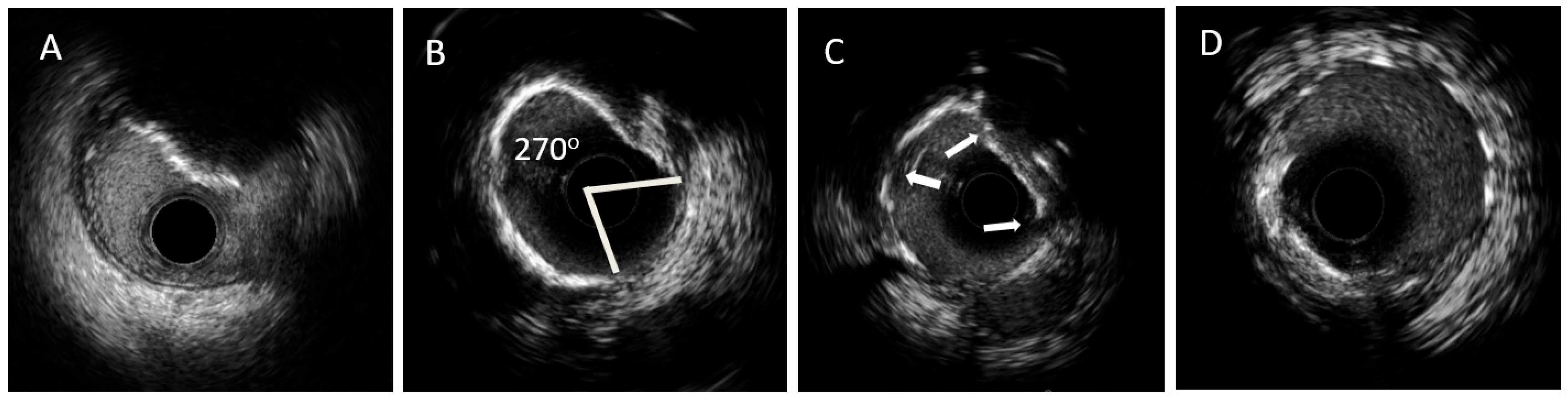
Figure 2.
OCT images of coronary calcified lesions. Legend: (A). Concentric calcium appearance. (B). Calcium fractures. (C). Calcified nodules. (D). Eccentric calcium appearance. Abbreviation: OCT: Optical Coherence Tomography (Images are courtesy of Abbott, Diegem, Belgium).
Figure 2.
OCT images of coronary calcified lesions. Legend: (A). Concentric calcium appearance. (B). Calcium fractures. (C). Calcified nodules. (D). Eccentric calcium appearance. Abbreviation: OCT: Optical Coherence Tomography (Images are courtesy of Abbott, Diegem, Belgium).

Figure 3.
Use of intracoronary imaging for the treatment of PCI indicated calcified lesions.

Figure 4.
Algorithm of the use of intracoronary imaging in calcified lesions with indication for PCI. Abbreviations: PCI: Percutaneous coronary intervention, ICI: Intracoronary imaging, IVUS: IntraVascular UltraSound, OCT: Optical Coherence Tomography, NC: Non-compliant. Leggend: Asterisk * refers to intracoronary imaging high risk criteria as follows: IVUS high risk criteria: 1. Superficial calcium of >270° (longer than 5 mm), 2. 360° superficial calcium, 3. Calcified nodule, 4. Vessel diameter ≤ 3.5 mm, OCT high risk criteria: 1. Calcium max angle > 180°, 2. Calcium max thickness > 0.5 mm, 3. Calcium length > 5 mm.

Table 2.
IVUS and OCT for the imaging of calcified lesions.
| IVUS | OCT | |
|---|---|---|
| Resolution | Conventional IVUS 100 μm, HD-IVUS up to 22 μm | High, 10–20 μm |
| Calcium detection | Very high sensitivity and specificity | High sensitivity and specificity |
| Calcium appearance | Bright hyperechoic signal (echodense) with posterior ‘’acoustic shadow’’ | Low signal intensity areas with well defined borders |
| Microcalcifications | Not detected | Detected |
| Superficial calcium | Detected | Detected |
| Deep calcium | Detected but can be hidden from superficial calcium | Limited deep calcium localization |
| Calcium thickness | Cannot be measured directly (acoustic shadowing) | Can be measured |
| Calcium arc | Can be measured | Can be measured |
| Calcium length | Can be measured | Can be measured |
| Calcium area volume | Cannot be measured | Can be measured |
| Ostial lesions | Can be assessed | Cannot be assessed |
| Calcified nodules | Can be identified | Can be identified |
| Firocalcific plaque | Possible assessment | Difficult assessment |
| Assessment of calcium modification technique’s effect (fractures, fissures) | Possible | Possible |
| Stent guidance lesion length, vessel diameter, landing zone | Feasible | Feasible |
| Estimation of calcium in large caliber/aneurysmatic vessels | Feasible | Difficult/not possible (low penetration depth) |
| Estimation of calcium in aortic-ostial lesions | Feasible | Difficult |
| Identification of complications dissection, hematoma, thrombus stent underexpansion/malapposition | Feasible | Feasible Better for malapposition assessment |
| Criteria of calcium severity and risk of stent underexpansion indicating the need for lesion modification | Superficial calcium angle of >270° (longer than 5 mm) 360° angle of superficial calcium Calcified nodule Vessel diameter ≤ 3.5 mm | Calcium max angle > 180° Calcium max thickness > 0.5 mm Calcium length > 5 mm |
Abbreviations: IVUS: IntraVascular UltraSound; HD: High Definition; OCT: Optical Coherence Tomography.
Disclaimer/Publisher’s Note: The statements, opinions and data contained in all publications are solely those of the individual author(s) and contributor(s) and not of MDPI and/or the editor(s). MDPI and/or the editor(s) disclaim responsibility for any injury to people or property resulting from any ideas, methods, instructions or products referred to in the content. |
© 2023 by the authors. Licensee MDPI, Basel, Switzerland. This article is an open access article distributed under the terms and conditions of the Creative Commons Attribution (CC BY) license (https://creativecommons.org/licenses/by/4.0/).
Share and Cite
MDPI and ACS Style
Petousis, S.; Skalidis, E.; Zacharis, E.; Kochiadakis, G.; Hamilos, M. The Role of Intracoronary Imaging for the Management of Calcified Lesions. J. Clin. Med. 2023, 12, 4622. https://doi.org/10.3390/jcm12144622
AMA Style
Petousis S, Skalidis E, Zacharis E, Kochiadakis G, Hamilos M. The Role of Intracoronary Imaging for the Management of Calcified Lesions. Journal of Clinical Medicine. 2023; 12(14):4622. https://doi.org/10.3390/jcm12144622
Chicago/Turabian StylePetousis, Stylianos, Emmanouil Skalidis, Evangelos Zacharis, George Kochiadakis, and Michalis Hamilos. 2023. "The Role of Intracoronary Imaging for the Management of Calcified Lesions" Journal of Clinical Medicine 12, no. 14: 4622. https://doi.org/10.3390/jcm12144622
Note that from the first issue of 2016, this journal uses article numbers instead of page numbers. See further details here.