
Induced Dipoles and Possible Modulation of Wireless Effects in Implanted Electrodes. Effects of Implanting Insulated Electrodes on an Animal Test to Screen Antidepressant Activity

 , , , and
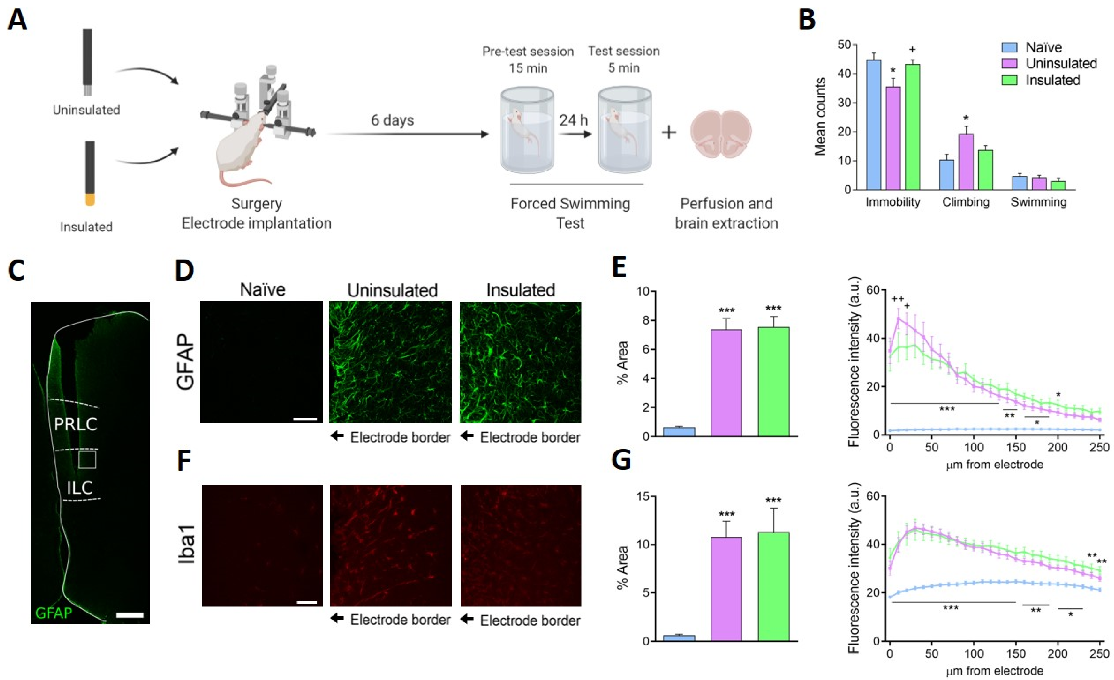
Abstract
:1. Introduction
2. Materials and Methods
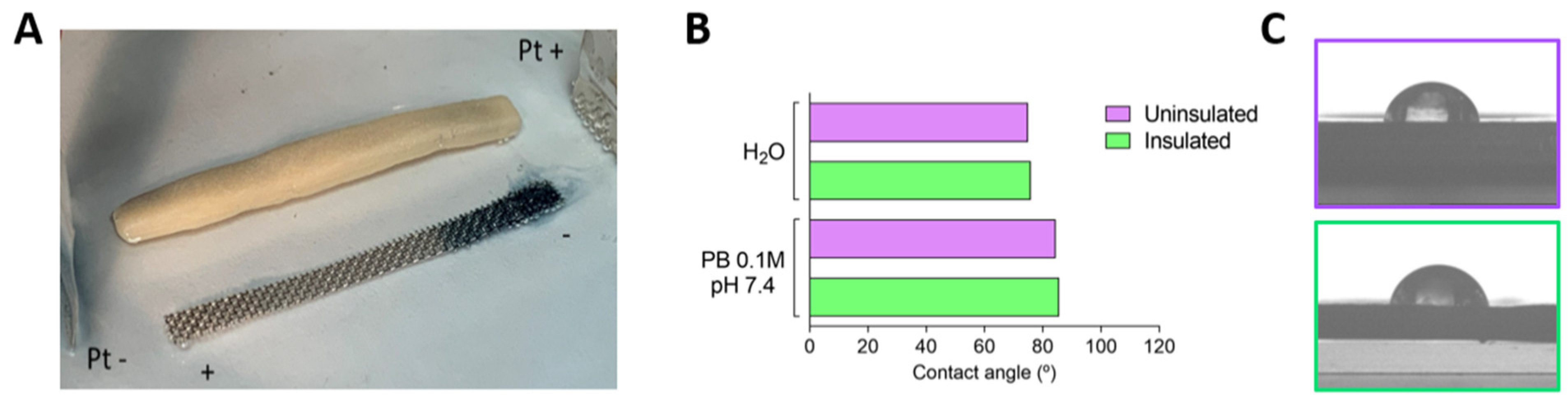
2.1. Materials Implanted and Characterization
2.2. Induced Dipoles and Bipolar Electrochemistry Visualization
2.3. Animals
2.4. Experimental Design
2.5. Surgery
2.6. mFST
2.7. Immunohistochemistry
2.8. Statistical Analysis
3. Results
3.1. Visualization of Bipolar Electrochemistry Effects Due to Induced Dipoles in Unwired Implants
3.2. Antidepressant-Like Effect
3.3. Glial Activation in the ILC
4. Discussion
5. Conclusions
Author Contributions
Funding
Institutional Review Board Statement
Acknowledgments
Conflicts of Interest
References
- Fenoy, A.J.; Simpson, R.K., Jr. Risks of common complications in deep brain stimulation surgery: Management and avoidance. J. Neurosurg. 2014, 120, 132–139. [Google Scholar] [CrossRef] [PubMed]
- Kenney, C.; Simpson, R.; Hunter, C.; Ondo, W.; Almaguer, M.; Davidson, A.; Jankovic, J. Short-term and long-term safety of deep brain stimulation in the treatment of movement disorders. J. Neurosurg. 2007, 106, 621–625. [Google Scholar] [CrossRef] [PubMed] [Green Version]
- Krack, P.; Volkmann, J.; Tinkhauser, G.; Deuschl, G. Deep Brain Stimulation in Movement Disorders: From Experimental Surgery to Evidence-Based Therapy. Mov. Disord. 2019, 34, 1795–1810. [Google Scholar] [CrossRef]
- Clair, A.H.; Haynes, W.; Mallet, L. Recent advances in deep brain stimulation in psychiatric disorders. F1000Res 2018, 7, 699. [Google Scholar] [CrossRef] [PubMed] [Green Version]
- Lee, D.J.; Lozano, C.S.; Dallapiazza, R.F.; Lozano, A.M. Current and future directions of deep brain stimulation for neurological and psychiatric disorders. J. Neurosurg. 2019, 131, 333–342. [Google Scholar] [CrossRef] [Green Version]
- Lozano, A.M.; Lipsman, N.; Bergman, H.; Brown, P.; Chabardes, S.; Chang, J.W.; Matthews, K.; McIntyre, C.C.; Schlaepfer, T.E.; Schulder, M.; et al. Deep brain stimulation: Current challenges and future directions. Nat. Rev. Neurol. 2019, 15, 148–160. [Google Scholar] [CrossRef]
- McCaig, C.D.; Rajnicek, A.M. Electrical fields, nerve growth and nerve regeneration. Exp. Physiol. 1991, 76, 473–494. [Google Scholar] [CrossRef] [Green Version]
- Casquero-Veiga, M.; Bueno-Fernandez, C.; Romero-Miguel, D.; Lamanna-Rama, N.; Nacher, J.; Desco, M.; Soto-Montenegro, M.L. Exploratory study of the long-term footprint of deep brain stimulation on brain metabolism and neuroplasticity in an animal model of obesity. Sci. Rep. 2021, 11, 5580. [Google Scholar] [CrossRef] [PubMed]
- Chakravarty, M.M.; Hamani, C.; Martinez-Canabal, A.; Ellegood, J.; Laliberte, C.; Nobrega, J.N.; Sankar, T.; Lozano, A.M.; Frankland, P.W.; Lerch, J.P. Deep brain stimulation of the ventromedial prefrontal cortex causes reorganization of neuronal processes and vasculature. Neuroimage 2016, 125, 422–427. [Google Scholar] [CrossRef]
- Jimenez-Sanchez, L.; Linge, R.; Campa, L.; Valdizan, E.M.; Pazos, A.; Diaz, A.; Adell, A. Behavioral, neurochemical and molecular changes after acute deep brain stimulation of the infralimbic prefrontal cortex. Neuropharmacology 2016, 108, 91–102. [Google Scholar] [CrossRef]
- Shen, K.Z.; Zhu, Z.T.; Munhall, A.; Johnson, S.W. Synaptic plasticity in rat subthalamic nucleus induced by high-frequency stimulation. Synapse 2003, 50, 314–319. [Google Scholar] [CrossRef]
- Tisch, S.; Rothwell, J.C.; Bhatia, K.P.; Quinn, N.; Zrinzo, L.; Jahanshahi, M.; Ashkan, K.; Hariz, M.; Limousin, P. Pallidal stimulation modifies after-effects of paired associative stimulation on motor cortex excitability in primary generalised dystonia. Exp. Neurol. 2007, 206, 80–85. [Google Scholar] [CrossRef]
- Veerakumar, A.; Challis, C.; Gupta, P.; Da, J.; Upadhyay, A.; Beck, S.G.; Berton, O. Antidepressant-like effects of cortical deep brain stimulation coincide with pro-neuroplastic adaptations of serotonin systems. Biol. Psychiatry 2014, 76, 203–212. [Google Scholar] [CrossRef] [Green Version]
- Cersosimo, M.G.; Raina, G.B.; Benarroch, E.E.; Piedimonte, F.; Aleman, G.G.; Micheli, F.E. Micro lesion effect of the globus pallidus internus and outcome with deep brain stimulation in patients with Parkinson disease and dystonia. Mov. Disord. 2009, 24, 1488–1493. [Google Scholar] [CrossRef] [PubMed]
- Hodaie, M.; Wennberg, R.A.; Dostrovsky, J.O.; Lozano, A.M. Chronic anterior thalamus stimulation for intractable epilepsy. Epilepsia 2002, 43, 603–608. [Google Scholar] [CrossRef] [PubMed]
- Lozano, A.M.; Mayberg, H.S.; Giacobbe, P.; Hamani, C.; Craddock, R.C.; Kennedy, S.H. Subcallosal cingulate gyrus deep brain stimulation for treatment-resistant depression. Biol. Psychiatry 2008, 64, 461–467. [Google Scholar] [CrossRef]
- Mann, J.M.; Foote, K.D.; Garvan, C.W.; Fernandez, H.H.; Jacobson, C.E.; Rodriguez, R.L.; Haq, I.U.; Siddiqui, M.S.; Malaty, I.A.; Morishita, T.; et al. Brain penetration effects of microelectrodes and DBS leads in STN or GPi. J. Neurol. Neurosurg. Psychiatry 2009, 80, 794–797. [Google Scholar] [CrossRef]
- Mayberg, H.S.; Lozano, A.M.; Voon, V.; McNeely, H.E.; Seminowicz, D.; Hamani, C.; Schwalb, J.M.; Kennedy, S.H. Deep brain stimulation for treatment-resistant depression. Neuron 2005, 45, 651–660. [Google Scholar] [CrossRef] [Green Version]
- Morishita, T.; Foote, K.D.; Wu, S.S.; Jacobson, C.E.; Rodriguez, R.L.; Haq, I.U.; Siddiqui, M.S.; Malaty, I.A.; Hass, C.J.; Okun, M.S. Brain penetration effects of microelectrodes and deep brain stimulation leads in ventral intermediate nucleus stimulation for essential tremor. J. Neurosurg. 2010, 112, 491–496. [Google Scholar] [CrossRef]
- Tykocki, T.; Nauman, P.; Koziara, H.; Mandat, T. Microlesion effect as a predictor of the effectiveness of subthalamic deep brain stimulation for Parkinson’s disease. Stereotact Funct. Neurosurg. 2013, 91, 12–17. [Google Scholar] [CrossRef] [PubMed]
- Perez-Caballero, L.; Perez-Egea, R.; Romero-Grimaldi, C.; Puigdemont, D.; Molet, J.; Caso, J.R.; Mico, J.A.; Perez, V.; Leza, J.C.; Berrocoso, E. Early responses to deep brain stimulation in depression are modulated by anti-inflammatory drugs. Mol. Psychiatry 2014, 19, 607–614. [Google Scholar] [CrossRef]
- Perez-Caballero, L.; Soto-Montenegro, M.L.; Hidalgo-Figueroa, M.; Mico, J.A.; Desco, M.; Berrocoso, E. Deep brain stimulation electrode insertion and depression: Patterns of activity and modulation by analgesics. Brain Stimul. 2018, 11, 1348–1355. [Google Scholar] [CrossRef]
- Rajnicek, A.M.; Zhao, Z.; Moral-Vico, J.; Cruz, A.M.; McCaig, C.D.; Casan-Pastor, N. Controlling Nerve Growth with an Electric Field Induced Indirectly in Transparent Conductive Substrate Materials. Adv. Health Mater. 2018, 7, e1800473. [Google Scholar] [CrossRef]
- Fuentes-Rodriguez, L.; Abad, L.; Simonelli, L.; Tonti, D.; Casañ-Pastor, N. Iridium oxide redox gradient material: Operando X Ray absorption of Ir gradient oxidation states during IrOx bipolar electrochemistry. J. Phys. Chem. C 2021, 125, 16629–16642. [Google Scholar] [CrossRef]
- Fuentes-Rodriguez, L.; Abad, L.; Pujades, E.; Tonti, D.; Casañ-Pastor, N. Induced dipoles and bipolar electrochemistry effects on electrolyte resistance. A macroscopic model experiment using immersed metal pieces. Electrochim. Acta 2021, submitted. [Google Scholar]
- Paxinos, G.; Watson, C. The Rat Brain in Stereotaxic Coordinates, 6th ed.; Academic Press: London, UK, 2009. [Google Scholar]
- Detke, M.J.; Rickels, M.; Lucki, I. Active behaviors in the rat forced swimming test differentially produced by serotonergic and noradrenergic antidepressants. Psychopharmacology 1995, 121, 66–72. [Google Scholar] [CrossRef] [PubMed]
- Carceller, H.; Guirado, R.; Ripolles-Campos, E.; Teruel-Marti, V.; Nacher, J. Perineuronal Nets Regulate the Inhibitory Perisomatic Input onto Parvalbumin Interneurons and gamma Activity in the Prefrontal Cortex. J. Neurosci. 2020, 40, 5008–5018. [Google Scholar] [CrossRef] [PubMed]
- Schindelin, J.; Arganda-Carreras, I.; Frise, E.; Kaynig, V.; Longair, M.; Pietzsch, T.; Preibisch, S.; Rueden, C.; Saalfeld, S.; Schmid, B.; et al. Fiji: An open-source platform for biological-image analysis. Nat. Methods 2012, 9, 676–682. [Google Scholar] [CrossRef] [Green Version]
- Guirado, R.; Carceller, H.; Castillo-Gomez, E.; Castren, E.; Nacher, J. Automated analysis of images for molecular quantification in immunohistochemistry. Heliyon 2018, 4, e00669. [Google Scholar] [CrossRef] [PubMed]
- Abad, L.; Rajnicek, A.; Casañ-Pastor, N. Electric Field Gradients and Bipolar Electrochemistry effects on Neural Growth. A finite element study on inmersed electroactive conducting electrode materials. Electrochim. Acta 2019, 317, 102–111. [Google Scholar] [CrossRef]
- Salatino, J.W.; Ludwig, K.A.; Kozai, T.D.Y.; Purcell, E.K. Glial responses to implanted electrodes in the brain. Nat. Biomed. Eng. 2017, 1, 862–877. [Google Scholar] [CrossRef]
- Tawfik, V.L.; Chang, S.Y.; Hitti, F.L.; Roberts, D.W.; Leiter, J.C.; Jovanovic, S.; Lee, K.H. Deep brain stimulation results in local glutamate and adenosine release: Investigation into the role of astrocytes. Neurosurgery 2010, 67, 367–375. [Google Scholar] [CrossRef] [PubMed] [Green Version]
- Lichtenstein, M.P.; Carretero, N.M.; Perez, E.; Pulido-Salgado, M.; Moral-Vico, J.; Sola, C.; Casan-Pastor, N.; Sunol, C. Biosafety assessment of conducting nanostructured materials by using co-cultures of neurons and astrocytes. Neurotoxicology 2018, 68, 115–125. [Google Scholar] [CrossRef] [PubMed]
- Lichtenstein, M.P.; Pérez, E.; Ballesteros, L.; Suñol, C.; Casañ-Pastor, N. Short term electrostimulation enhancing neural repair in vitro using large charge capacity intercalation electrodes. App. Mater. Today 2017, 6, 29–43. [Google Scholar] [CrossRef] [Green Version]


Publisher’s Note: MDPI stays neutral with regard to jurisdictional claims in published maps and institutional affiliations. |
© 2021 by the authors. Licensee MDPI, Basel, Switzerland. This article is an open access article distributed under the terms and conditions of the Creative Commons Attribution (CC BY) license (https://creativecommons.org/licenses/by/4.0/).
Share and Cite
Perez-Caballero, L.; Carceller, H.; Nacher, J.; Teruel-Marti, V.; Pujades, E.; Casañ-Pastor, N.; Berrocoso, E. Induced Dipoles and Possible Modulation of Wireless Effects in Implanted Electrodes. Effects of Implanting Insulated Electrodes on an Animal Test to Screen Antidepressant Activity. J. Clin. Med. 2021, 10, 4003. https://doi.org/10.3390/jcm10174003
Perez-Caballero L, Carceller H, Nacher J, Teruel-Marti V, Pujades E, Casañ-Pastor N, Berrocoso E. Induced Dipoles and Possible Modulation of Wireless Effects in Implanted Electrodes. Effects of Implanting Insulated Electrodes on an Animal Test to Screen Antidepressant Activity. Journal of Clinical Medicine. 2021; 10(17):4003. https://doi.org/10.3390/jcm10174003
Chicago/Turabian StylePerez-Caballero, Laura, Hector Carceller, Juan Nacher, Vicent Teruel-Marti, Eulalia Pujades, Nieves Casañ-Pastor, and Esther Berrocoso. 2021. "Induced Dipoles and Possible Modulation of Wireless Effects in Implanted Electrodes. Effects of Implanting Insulated Electrodes on an Animal Test to Screen Antidepressant Activity" Journal of Clinical Medicine 10, no. 17: 4003. https://doi.org/10.3390/jcm10174003