The Health-Benefits and Phytochemical Profile of Salvia apiana and Salvia farinacea var. Victoria Blue Decoctions

 , , ,
, , ,  and
and
Abstract
:1. Introduction
2. Materials and Methods
2.1. Chemicals
2.2. Plant Material
2.3. Preparation of Extracts
2.4. Identification and Quantification of Phenolic Compounds
2.5. Antioxidant Activity
2.5.1. DPPH● Scavenging Test
2.5.2. Ferric Reducing Power Assay
2.5.3. Thiobarbituric Acid Reactive Substances (TBARS)
2.5.4. β-Carotene Bleaching Assay
2.6. Anti-Inflammatory Activity
2.7. Cytotoxic Effect in Four Human Tumor Cell Lines
2.8. Cytotoxic Effect in Non-Tumor Liver Cells
2.9. Antimicrobial Activity
2.10. Statistical Analysis
3. Results and Discussion
3.1. Antioxidant Activity
3.2. Anti-Inflammatory Aactivity
3.3. Cytotoxic Activity
3.4. Antibacterial Activity
3.5. Characterization of the Extracts
4. Conclusions
Author Contributions
Funding
Acknowledgments
Conflicts of Interest
References
- Lin, D.; Xiao, M.; Zhao, J.; Li, Z.; Xing, B.; Li, X.; Kong, M.; Li, L.; Zhang, Q.; Liu, Y.; et al. An overview of plant phenolic compounds and their importance in human nutrition and management of type 2 diabetes. Molecules 2016, 21, 1374. [Google Scholar] [CrossRef] [PubMed]
- Guillaumet-Adkins, A.; Yañez, Y.; Peris-Diaz, M.D.; Calabria, I.; Palanca-Ballester, C.; Sandoval, J. Epigenetics and Oxidative Stress in Aging. Oxid. Med. Cell. Longev. 2017, 14, 17643–17663. [Google Scholar] [CrossRef] [PubMed]
- Li, A.N.; Li, S.; Zhang, Y.J.; Xu, X.R.; Chen, Y.M.; Li, H.B. Resources and biological activities of natural polyphenols. Nutrients 2014, 6, 6020–6047. [Google Scholar] [CrossRef] [PubMed]
- Brewer, M.S. Natural Antioxidants: Sources, Compounds, Mechanisms of Action, and Potential Applications. Compr. Rev. Food Sci. Food Saf. 2011, 10, 221–247. [Google Scholar] [CrossRef]
- Walker, J.B.; Sytsma, K.J.; Treutlein, J.; Wink, M. Salvia (Lamiaceae) is not monophyletic: Implications for the systematics, radiation and ecological specializations. Am. J. Bot. 2004, 91, 1115–1125. [Google Scholar] [CrossRef]
- Jeshvaghani, Z.A.; Rahimmalek, M.; Talebi, M.; Goli, S.A.H. Comparison of total phenolic content and antioxidant activity in different Salvia species using three model systems. Ind. Crops Prod. 2015, 77, 409–414. [Google Scholar] [CrossRef]
- Alimpić, A.; Knežević, A.; Milutinović, M.; Stević, T.; Šavikin, K.; Stajić, M.; Marković, S.; Marin, P.D.; Matevski, V.; Duletić-Laušević, S. Biological activities and chemical composition of Salvia amplexicaulis Lam. extracts. Ind. Crops Prod. 2017, 105, 1–9. [Google Scholar] [CrossRef]
- Jassbi, A.R.; Zare, S.; Firuzi, O.; Xiao, J. Bioactive phytochemicals from shoots and roots of Salvia species. Phytochem. Rev. 2016, 15, 829–867. [Google Scholar] [CrossRef]
- Sytar, O.; Bruckova, K.; Hunkova, E.; Zivcak, M.; Konate, K.; Brestic, M. The application of multiplex fluorimetric sensor for the analysis of flavonoids content in the medicinal herbs family Asteraceae, Lamiaceae, Rosaceae. Biol. Res. 2015, 48, 5. [Google Scholar] [CrossRef]
- Kumar, S.; Pandey, A.K. Chemistry and biological activities of flavonoids: An overview. Sci. World J. 2013, 2013. [Google Scholar] [CrossRef]
- Turner, B.L. Recension of Salvia sect. farinaceae (lamiaceae). Phytologia 2008, 90, 163–175. [Google Scholar]
- Zhang, X.; Sawhney, V.K.; Davis, A.R. Annular floral nectary with oil-producing trichomes in Salvia farinacea (lamiaceae): Anatomy, histochemistry, ultrastructure, and significance. Am. J. Bot. 2014, 101, 1849–1867. [Google Scholar] [CrossRef] [PubMed]
- Giffen, J.E.; Lesiak, A.D.; Dane, A.J.; Cody, B.; Musah, R.A. Rapid Species-level Identification of Salvias by Chemometric Processing of Ambient Ionisation Mass Spectrometry-derived Chemical Profiles. Phytochem. Anal. 2017, 28, 16–26. [Google Scholar] [CrossRef] [PubMed]
- Ali, A.; Tabanca, N.; Demirci, B.; Blythe, E.K. Chemical Composition and Biological Activity of Four Salvia Essential Oils and Individual Compounds against Two Species of Mosquitoes. J. Agric. Food Chem. 2015, 63, 447–456. [Google Scholar] [CrossRef] [PubMed]
- Ott, D.; Hühn, P.; Claßen-bockhoff, R. Salvia apiana—A carpenter bee flower? Flora 2016, 221, 82–91. [Google Scholar] [CrossRef]
- Borek, T.T.; Hochrien, J.M.; Irwin, A.N. Composition of the essential oil of white sage, Salvia apiana. Flavour Fragr. J. 2006, 21, 571–572. [Google Scholar] [CrossRef]
- Srivedavyasasri, R.; Hayes, T.; Ross, S.A. Phytochemical and biological evaluation of Salvia apiana. Nat. Prod. Res. 2017, 31, 2058–2061. [Google Scholar] [CrossRef]
- Luis, J.G.; Lahlou, E.H.; Andros, L.S.; Islands, C. Hassananes: C23 terpenoids with a new type of skeleton from Salvia apiana Jeps. Tetrahedron 1996, 52, 12309–12312. [Google Scholar] [CrossRef]
- Saeed, M.E.M.; Meyer, M.; Hussein, A.; Efferth, T. Cytotoxicity of South-African medicinal plants towards sensitive and multidrug-resistant cancer cells. J. Ethnopharmacol. 2016, 186, 209–223. [Google Scholar] [CrossRef]
- Ferreira, F.M.; Dinis, L.T.; Azedo, P.; Galhano, C.I.C.; Simões, A.; Cardoso, S.M.; Domingues, M.R.M.; Pereira, O.R.; Palmeira, C.M.; Peixoto, F.P. Antioxidant capacity and toxicological evaluation of Pterospartum tridentatum flower extracts. CYTA-J. Food 2012, 10, 92–102. [Google Scholar] [CrossRef]
- Afonso, A.F.; Pereira, O.R.; Neto, R.T.; Silva, A.M.S.; Cardoso, S.M. Health-promoting effects of Thymus herba-barona, Thymus pseudolanuginosus, and Thymus caespititius decoctions. Int. J. Mol. Sci. 2017, 18, 1879. [Google Scholar] [CrossRef] [PubMed]
- Afonso, A.F.; Pereira, O.R.; Cardoso, S.M. Metabolites and Biological Activities of Thymus zygis, Thymus pulegioides, and Thymus fragrantissimus Grown under Organic Cultivation. Molecules 2018, 23, 1514. [Google Scholar] [CrossRef] [PubMed]
- Catarino, M.D.; Silva, A.M.S.; Saraiva, S.C.; Sobral, A.J.F.N.; Cardoso, S.M. Characterization of phenolic constituents and evaluation of antioxidant properties of leaves and stems of Eriocephalus africanus. Arab. J. Chem. 2018, 11, 62–69. [Google Scholar] [CrossRef]
- Martins, N.; Barros, L.; Santos-Buelga, C.; Henriques, M.; Silva, S.; Ferreira, I.C.F.R. Evaluation of bioactive properties and phenolic compounds in different extracts prepared from Salvia officinalis L. Food Chem. 2015, 170, 378–385. [Google Scholar] [CrossRef] [PubMed]
- Barros, L.; Ferreira, M.J.; Queirós, B.; Ferreira, I.C.F.R.; Baptista, P. Total phenols, ascorbic acid, β-carotene and lycopene in Portuguese wild edible mushrooms and their antioxidant activities. Food Chem. 2007, 103, 413–419. [Google Scholar] [CrossRef]
- Souza, A.H.P.; Corrêa, R.C.G.; Barros, L.; Calhelha, R.C.; Santos-buelga, C.; Peralta, R.M.; Bracht, A.; Matsushita, M.; Ferreira, I.C.F.R. Phytochemicals and bioactive properties of Ilex paraguariensis: An in-vitro comparative study between the whole plant, leaves and stems. Food Res. Int. 2015, 78, 286–294. [Google Scholar] [CrossRef] [PubMed]
- Andrews, J.M. Determination of minimum inhibitory concentrations. J. Antimicrob. Chemother. 2001, 48, 5–16. [Google Scholar] [CrossRef] [Green Version]
- Shami, A.M.M.; Philip, K.; Muniandy, S. Synergy of antibacterial and antioxidant activities from crude extracts and peptides of selected plant mixture. BMC Complement. Altern. Med. 2013, 13, 1–11. [Google Scholar] [CrossRef]
- Piluzza, G.; Bullitta, S. Correlations between phenolic content and antioxidant properties in twenty-four plant species of traditional ethnoveterinary use in the Mediterranean area Correlations between phenolic content and antioxidant properties in twenty-four plant species of tra. Pharm. Biol. 2011, 49, 240–247. [Google Scholar] [CrossRef]
- Koşar, M.; Göger, F.; Hüsnü Can Başer, K. In vitro antioxidant properties and phenolic composition of Salvia halophila Hedge from Turkey. Food Chem. 2011, 129, 374–379. [Google Scholar] [CrossRef]
- Kostic, M.; Petrovic, M.B.; Jevtovic, T.; Jovic, M.; Petrovic, A.; Slavoljub, Ž. Anti-inflammatory effect of the Salvia sclarea L. ethanolic extract on lipopolysaccharide-induced periodontitis in rats. J. Ethnopharmacol. 2017, 199, 52–59. [Google Scholar] [CrossRef] [PubMed]
- Azab, A.; Nassar, A.; Azab, A.N. Anti-inflammatory activity of natural products. Molecules 2016, 21, 1321. [Google Scholar] [CrossRef] [PubMed]
- Ravipati, A.S.; Zhang, L.; Koyyalamudi, S.R.; Jeong, S.C.; Reddy, N.; Bartlett, J.; Smith, P.T.; Shanmugam, K.; Münch, G.; Wu, M.J.; et al. Antioxidant and anti-inflammatory activities of selected Chinese medicinal plants and their relation with antioxidant content. BMC Complement. Altern. Med. 2012, 12, 173. [Google Scholar] [CrossRef] [PubMed]
- Jang, H.H.; Cho, S.Y.; Kim, M.J.; Kim, J.B.; Lee, S.H.; Lee, M.Y.; Lee, Y.M. Anti-nflammatory effects of Salvia plebeia R. Br extract in vitro and in ovalbumin-induced mouse model. Biol. Res. 2016, 49, 41. [Google Scholar] [CrossRef] [PubMed]
- Firuzi, O.; Miri, R.; Asadollahi, M.; Eslami, S.; Jassbi, A.R. Cytotoxic, Antioxidant and Antimicrobial Activities and Phenolic Contents of Eleven Salvia Species from Iran. Iran J. Pharm. Res. 2013, 12, 801–810. [Google Scholar] [PubMed]
- Jiang, Y.; Zhang, L.; Rupasinghe, H.P.V. Antiproliferative effects of extracts from Salvia officinalis L. and Saliva miltiorrhiza Bunge on hepatocellular carcinoma cells. Biomed. Pharmacother. 2017, 85, 57–67. [Google Scholar] [CrossRef]
- Shahneh, F.Z.; Baradaran, B.; Orangi, M.; Zamani, F. In vitro Cytotoxic Activity of Four Plants Used in Persian Traditional Medicine. Adv. Farm. Bull. 2013, 3, 453–455. [Google Scholar] [Green Version]
- Córdova-guerrero, I.; Aragon-martinez, O.H.; Díaz-rubio, L. Actividad antibacteriana y antifúngica de un extracto de Salvia apiana frente a microorganismos de importancia clínica. Rev. Argent. Microbiol. 2016, 48, 217–221. [Google Scholar] [CrossRef]
- Ibrahim, T.A. Chemical composition and biological activity of extracts from Salvia bicolor desf. growing in Egypt. Molecules 2012, 17, 11315–11334. [Google Scholar] [CrossRef]
- Pereira, O.R.; Catarino, M.D.; Afonso, A.F.; Silva, A.M.S.; Cardoso, S.M. Salvia elegans, Salvia greggii and Salvia officinalis Decoctions: Antioxidant Activities and Inhibition of Carbohydrate and Lipid Metabolic Enzymes. Molecules 2018, 23, 3169. [Google Scholar] [CrossRef]
- Abreu, M.E.; Maren, M. Phenolic diterpene and α-tocopherol contents in leaf extracts of 60 Salvia. J. Sci. Food Agric. 2008, 88, 2648–2653. [Google Scholar] [CrossRef]
- Zimmermann, B.F.; Walch, S.G.; Tinzoh, L.N.; Stühlinger, W.; Lachenmeier, D.W. Rapid UHPLC determination of polyphenols in aqueous infusions of Salvia officinalis L. (sage tea). J. Chromatogr. B Analyt. Technol. Biomed. Life Sci. 2011, 879, 2459–2464. [Google Scholar] [CrossRef] [PubMed]
- Cvetkovikj, I.; Stefkov, G.; Acevska, J.; Stanoeva, J.P.; Karapandzova, M.; Stefova, M.; Dimitrovska, A.; Kulevanova, S. Polyphenolic characterization and chromatographic methods for fast assessment of culinary Salvia species from South East Europe. J. Chromatogr. A 2013, 1282, 38–45. [Google Scholar] [CrossRef] [PubMed]
- Milevskaya, V.V.; Temerdashev, Z.A.; Butyl, T.S.; Kiseleva, N.V. Determination of Phenolic Compounds in Medicinal Plants from the Lamiaceae Family. J. Anal. Chem. 2017, 72, 342–348. [Google Scholar] [CrossRef]
- González, M.A. Aromatic Abietane Diterpenoids: Their Biological Activity and Synthesis. Nat. Prod. Rep. 2013, 32, 684–704. [Google Scholar] [CrossRef] [PubMed]
- Ghorbani, A.; Esmaeilizadeh, M. Pharmacological properties of Salvia officinalis and its components. J. Tradit. Complement. Med. 2017, 7, 433–440. [Google Scholar] [CrossRef] [PubMed]
- Ninomiya, K.; Matsuda, H.; Shimoda, H.; Nishida, N.; Kasajima, N.; Yoshino, T.; Morikawa, T.; Yoshikawa, M. Carnosic acid, a new class of lipid absorption inhibitor from sage. Bioorg. Med. Chem. Lett. 2004, 14, 1943–1946. [Google Scholar] [CrossRef]
- Johnson, J.J. Carnosol: A promising anti-cancer and anti-inflammatory agent. Cancer Lett. 2011, 305, 1–7. [Google Scholar] [CrossRef] [Green Version]


| Assay | S. apiana | S. farinacea var. Victoria Blue | Standard | ||
|---|---|---|---|---|---|
| AA | BHA | Trolox | |||
| DPPH• | 13.3 ± 1.1 a | 17.4 ± 5.5 a | 6.7 ± 0.7 b | ||
| Ferric reducing power | 55.0 ± 5.6 a | 59.9 ± 3.6 a | 16.1 ± 2.0 b | ||
| TBARS | 2.79 ± 0.1 a | 42.2 ± 0.6 b | 23.0 ± 1.0 c | ||
| β-carotene bleaching inhibition | 41.2 ± 1.6 a | 153.5 ± 2.2 b | 41.7 ± 0.3 a | ||
| Effect | S. apiana | S. farinacea var. Victoria Blue | Standard |
|---|---|---|---|
| Anti-inflammatory activity | Dexamethasone | ||
| NO● production | 49.9 ± 2.5 a | 80.8 ± 0.4 b | 16.0 ± 1.0 c |
| Cytotoxicity to tumor cell lines | Ellipticine | ||
| HepG2 (hepatocellular carcinoma) | 40.9 ± 3.3 a | 87.4 ± 5.4 b | 1.0 ± 0.2 c |
| HeLa (cervical carcinoma) | 57.3 ± 5.1 a | 77.8 ± 3.5 b | 2.0 ± 0.1 c |
| MCF-7 (breast carcinoma) | 60.2 ± 4.2 a | 59.8 ± 0.1 a | 1.0 ± 0.04 b |
| NCI-H460 (non-small cell lung cancer) | 245.7 ± 6.3 a | 279.5 ± 10.1 b | 1.0 ± 0.1 c |
| Cytotoxicity to non-tumor cells | |||
| PLP2 growth inhibition | 361.7 ± 5.3 a | 335.4 ± 8.0 b | 3.0 ± 1.0 c |
| Bacteria | S. apiana | S. farinacea var. Victoria Blue | Nisin | |||
|---|---|---|---|---|---|---|
| MIC | MBC | MIC | MBC | MIC | MBC | |
| Gram-positive | ||||||
| S. epidermidis | 0.34 | 0.69 | 8.50 | 8.50 | <0.63 | <0.63 |
| S. aureus | 0.69 | 0.69 | 1.06 | 2.12 | <0.63 | <0.63 |
| Gram-negative | ||||||
| S. typhimurium | 2.75 | 2.75 | >8.5 | >8.5 | 0.5 | 0.5 |
| E. coli | 2.75 | 2.75 | 8.5 | 8.5 | 0.5 | 1.0 |
| P. aeruginosa | 2.75 | 2.75 | >8.5 | >8.5 | 1.0 | 1.0 |
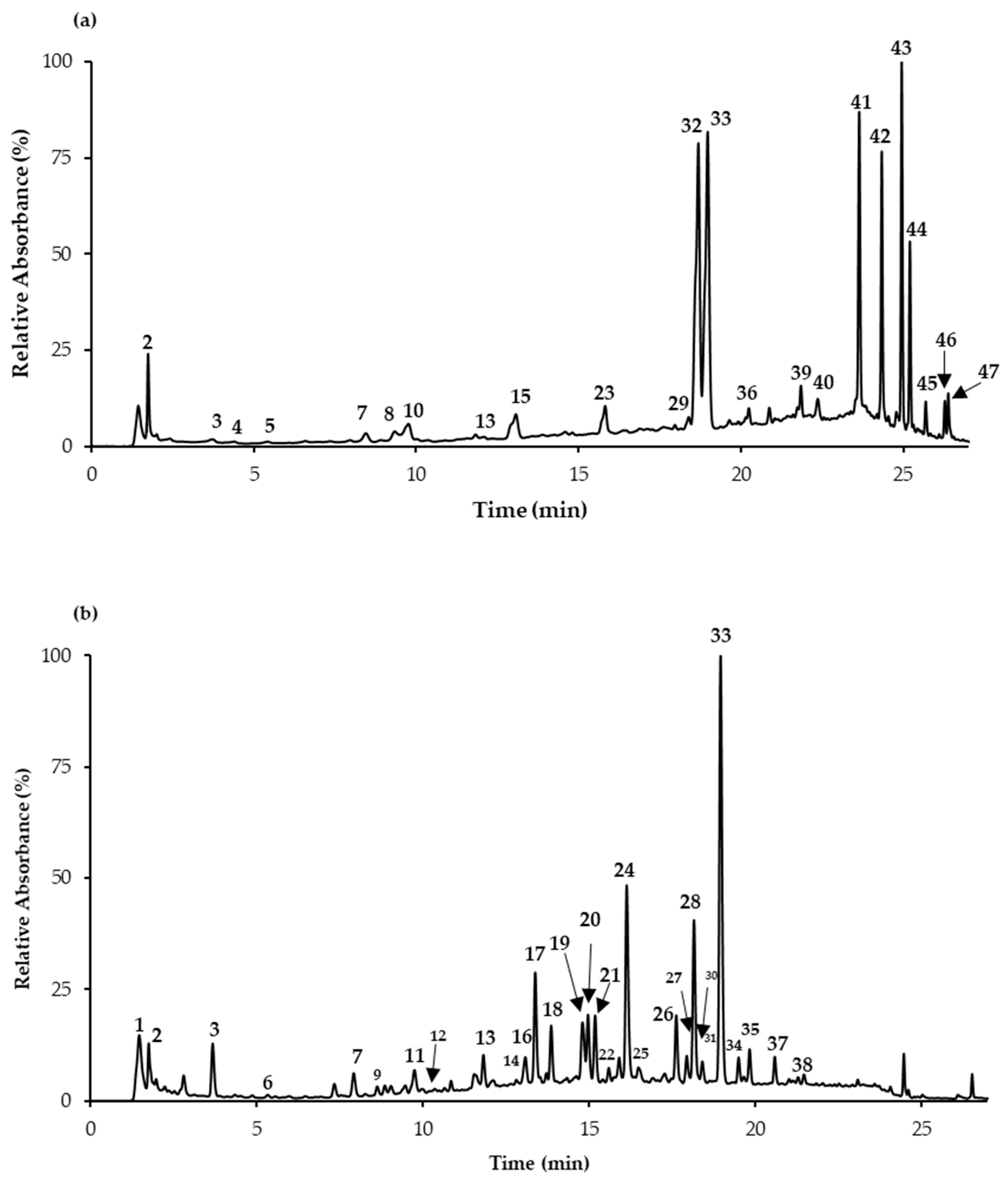
| NF | Rt | UVmax (nm) | [H–M]− | MS/MS Fragments (m/z) | Probable Compound | S. apiana * | S. farinacea Var. Victoria Blue * |
|---|---|---|---|---|---|---|---|
| 1 | 1.5 | 275 | 149 | 103, 87, 131, 59 | DimethylBA | - | 5.9 ± 0.1 |
| 2 | 1.7 | 205 | 191 | 111, 173 | Quinic Ac | 5.0 ± 0.3 | 0.4 ± 0.01 |
| 3 | 3.6 | 280 | 197 | 179, 73, 153 | Danshensu | D | D |
| 4 | 4.4 | 261, 289 | 153 | 109 | Protoc Ac | D | - |
| 5 | 5.1 | 290sh, 324 | 353 | 191, 179, 135 | cis 3-O-CQA | D | - |
| 6 | 5.4 | 294sh, 322 | 353 | 191, 179, 135 | trans 3-O-CQA | - | D |
| 7 | 7.9 | 309 | 337 | 163 | Coum Quinic Ac | D | 0.3 ± 0.03 |
| 8 | 8.3 | 313 | 295 | 163 | p-Coum Ac Pent | 0.4 ± 0.04 | - |
| 9 | 8.8 | 290sh, 325 | 353 | 191 | trans 5-O-CQA | - | 0.6 ± 0.03 |
| 10 | 9.5 | 290sh, 325 | 353 | 173, 179, 191 | 4-O-CQA | 5.5 ± 0.1 | - |
| 11 | 9.7 | 290sh, 323 | 179 | 135 | Caffeic Ac | - | 0.8 ± 0.0 |
| 12 | 9.8 | 314 | 325 | 265, 235, 163 | Coum Hex | - | 0.3 ± 0.0 |
| 13 | 11.8 | 311 | 337 | 191, 163 | Coum Quinic Ac | D | 0.2 ± 0.0 |
| 14 | 12.8 | 287sh, 324 | 367 | 173, 191 | Fer Quinic Ac | - | D |
| 15 | 13.0 | 309 | 225 | 207, 181, 165, 163 | Coum Ac Der | 1.8 ± 0.1 | - |
| 16 | 13.1 | 291sh, 311 | 637 | 351, 285, 193 | Ferulic Ac Der | - | 0.5 ± 0.0 |
| 17 | 13.5 | 274 | 571 | 527, 483, 439, 373 | YA E (isom1) | - | 8.4 ± 0.01 |
| 18 | 13.9 | 256, 267, 345 | 447 | 327, 357 | Lut-C-Hex | - | 3.2 ± 0.02 |
| 19 | 14.7 | 274 | 571 | 527, 509, 553, 483, 285 | YA E (isom2) | - | 4.5 ± 0.2 |
| 20 | 15.0 | 235, 277 | 539 | 297, 359, 377, 279, 315 | YA D/isomer | - | 3.9 ± 0.3 |
| 21 | 15.2 | 268, 336 | 431 | 311, 341, 269 | Api-C-Hex | - | 5.9 ± 0.6 |
| 22 | 15.6 | 285, 315 | 555 | 409, 391, 537, 511, 365 | SA K | - | D |
| 23 | 15.8 | 255, 350 | 463 | 301 | Querc-O-Hex | 14.6 ± 0.3 | - |
| 593 | 285 | Lut Rut | D | - | |||
| 24 | 16.1 | 255, 266, 345 | 461 | 285 | Lut-7-O-GlcA | - | 15.8 ± 0.02 |
| 25 | 16.5 | 274 | 571 | 527, 409 | YA E (isom3) | - | 1.0 ± 0.08 |
| 26 | 17.6 | 268, 336 | 575 | 431, 341, 311, 513, 413 | Api Hex HMG | - | 6.3 ± 0.02 |
| 27 | 17.9 | 283 | 719 | 359, 539, 521, 341 | Sag Ac (isom1) | D | 2.1 ± 0.08 |
| 28 | 18.1 | 269, 329 | 431 | 269 | Api-O-Hex | - | 16.7 ± 0.05 |
| 29 | 18.3 | 238, 341 | 607 | 299, 284 | Chrys-O-Rut | D | - |
| 30 | 18.4 | 267, 337 | 445 | 269, 175 | Api-O-GlcA | - | 2.2 ± 0.01 |
| 31 | 18.6 | 270, 291, 326sh | 717 | 555, 519, 475, 357 | SA B (isom1) | - | D |
| 32 | 18.7 | 284, 330sh | 609 | 301 | Hesperidin | 41.3 ± 2.2 | - |
| 33 | 19.0 | 290sh, 328 | 359 | 161, 179, 197, 223 | RA | 56.8 ± 0.6 | 17.8 ± 0.1 |
| 34 | 19.5 | 285sh, 305 | 537 | 493, 295 | CaffRA (isom1) | - | 1.0 ± 0.04 |
| 35 | 19.8 | 278 | 719 | 521, 341, 359 | Sag Ac (isom2) | - | 2.2 ± 0.02 |
| 36 | 20.2 | 290sh, 333 | 537 | 493, 359, 375 | CaffRA/ SA I (isom2) | D | - |
| 37 | 20.6 | 267, 336 | 517 | 269, 473 | Api malonyl Hex | - | 2.5 ± 0.03 |
| 38 | 21.5 | 287sh, 320 | 373 | 179, 161, 135, 197, 355, 329 | Methyl Rosmarinate | - | 0.6 ± 0.02 |
| 39 | 21.8 | 290 | 491 | 163, 329, 119 | Coumaric Ac Der | 0.5 ± 0.01 | - |
| 40 | 22.4 | 281, 330sh | 717 | 537, 357 | SA B (isom2) | 6.6 ± 0.4 | - |
| 41 | 23.7 | 199, 229, 287 | 361 | 299, 269, 281, 213, 343 | Sageone Der | 174.1 ± 14.1 | - |
| 42 | 24.3 | 275, 333sh | 313 | 298, 283, 269 | Cirsimaritin | 25.9 ± 0.6 | - |
| 43 | 25.0 | 207, 237sh, 285 | 345 | 301, 271, 283 | Rosmanol | 192.4 ± 17.1 | - |
| 44 | 25.2 | 286 | 347 | 303, 273 | Hydroxycarnosic Ac | 69.7 ± 11.2 | - |
| 45 | 25.7 | 286 | 329 | 285 | Carnosol | 17.3 ± 0.7 | - |
| 46 | 26.3 | 262 | 331 | 287 | Carnosic Ac | 14.3 ± 0.7 | - |
| 47 | 26.4 | 277 | 301 | 271, 283 | Tetrahydrohydroxyrosmariquinone | 17.4 ± 0.2 | - |
| Total | 643.3 ± 18.9 | 102.1 ± 0.7 |
© 2019 by the authors. Licensee MDPI, Basel, Switzerland. This article is an open access article distributed under the terms and conditions of the Creative Commons Attribution (CC BY) license (http://creativecommons.org/licenses/by/4.0/).
Share and Cite
Afonso, A.F.; Pereira, O.R.; Fernandes, Â.S.F.; Calhelha, R.C.; Silva, A.M.S.; Ferreira, I.C.F.R.; Cardoso, S.M. The Health-Benefits and Phytochemical Profile of Salvia apiana and Salvia farinacea var. Victoria Blue Decoctions. Antioxidants 2019, 8, 241. https://doi.org/10.3390/antiox8080241
Afonso AF, Pereira OR, Fernandes ÂSF, Calhelha RC, Silva AMS, Ferreira ICFR, Cardoso SM. The Health-Benefits and Phytochemical Profile of Salvia apiana and Salvia farinacea var. Victoria Blue Decoctions. Antioxidants. 2019; 8(8):241. https://doi.org/10.3390/antiox8080241
Chicago/Turabian StyleAfonso, Andrea F., Olívia R. Pereira, Ângela S. F. Fernandes, Ricardo C. Calhelha, Artur M. S. Silva, Isabel C.F.R. Ferreira, and Susana M. Cardoso. 2019. "The Health-Benefits and Phytochemical Profile of Salvia apiana and Salvia farinacea var. Victoria Blue Decoctions" Antioxidants 8, no. 8: 241. https://doi.org/10.3390/antiox8080241