
Effects of Tannic Acid on Lipid and Protein Oxidation, Color, and Volatiles of Raw and Cooked Chicken Breast Meat during Storage


Abstract
:
1. Introduction
2. Materials and Methods
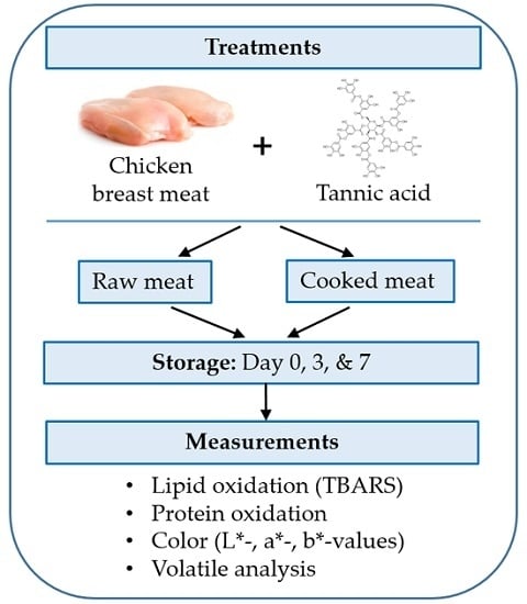
2.1. Sample Preparation
2.2. 2-Thiobarbituric Acid-Reactive Substances (TBARS) Measurement
2.3. Color Measurement
2.4. Volatile Analysis
2.5. Protein Oxidation (Total Carbonyl)
2.6. Statistical Analysis
3. Results and Discussion
3.1. Lipid Oxidation
3.2. Protein Oxidation
3.3. Color Values
3.4. Volatiles Production
4. Conclusions
Acknowledgments
Author Contributions
Conflicts of Interest
References
- Solomakos, N.; Govaris, A.; Koidis, P.; Botsoglou, N. The antimicrobial effect of thyme essential oil, nisin, and their combination against Listeria monocytogenes in minced beef during refrigerated storage. Food Microbiol. 2008, 25, 120–127. [Google Scholar] [CrossRef] [PubMed]
- Naveena, B.M.; Sen, A.R.; Vaithiyanathan, S.; Babji, Y.; Kondaiah, N. Comparative efficacy of pomegranate juice, pomegranate rind power extract and BHT as antioxidants in cooked chicken patties. Meat Sci. 2008, 80, 1304–1308. [Google Scholar] [CrossRef] [PubMed]
- Abdel-Hamied, A.A.; Nassar, A.G.; El-Badary, N. Investigation on antioxidant and antibacterial activities of some natural extracts. World J. Food Sci. 2009, 4, 1–7. [Google Scholar]
- Karre, L.; Lopez, K.; Getty, J.K. Natural antioxidants in meat and poultry products. Meat Sci. 2013, 94, 220–227. [Google Scholar] [CrossRef] [PubMed]
- Liu, Q.; Lanari, M.C.; Schaefer, D.M. A review of dietary vitamin E supplementation for improvement of beef quality. J. Anim. Sci. 1995, 73, 3131–3140. [Google Scholar] [PubMed]
- Mancini, R.A.; Hunt, M.C. Current research in meat color. Meat Sci. 2005, 71, 100–121. [Google Scholar] [CrossRef] [PubMed]
- Bekhit, A.E.D.; Faustman, C. Metmyoglobin reducing activity. Meat Sci. 2005, 71, 407–439. [Google Scholar] [CrossRef] [PubMed]
- Nam, K.C.; Ko, K.Y.; Min, B.R.; Ismail, H.; Lee, E.J.; Cordray, J.; Ahn, D.U. Effect of oleoresin-tocopherol combinations on lipid oxidation, off-odor, and color of irradiated raw and cooked pork patties. Meat Sci. 2007, 75, 61–70. [Google Scholar] [CrossRef] [PubMed]
- Min, B.; Ahn, D.U. Mechanism of lipid peroxidation in meat and meat products—A review. Food Sci. Biotechnol. 2005, 14, 152–163. [Google Scholar]
- Hall, G. Interactions between products of lipid oxidation and proteins. Food Sci. Technol. Today 1987, 1, 155–158. [Google Scholar]
- Gray, J.I.; Gomaa, E.A.; Buckley, D.J. Oxidative quality and shelf life of meats. Meat Sci. 1996, 43, 111–123. [Google Scholar] [CrossRef]
- Ahn, D.U.; Nam, K.C.; Lee, E.J. Lipid oxidation and flavor. In Applied Muscle Biology and Meat Science; Du, M., McCormick, R., Eds.; CRC Press: New York, NY, USA, 2009; pp. 227–246. [Google Scholar]
- Sebranek, J.G.; Sewalt, V.J.; Robbins, K.L.; Houser, T.A. Comparison of a natural rosemary extract and BHA/BHT for relative antioxidant effectiveness in pork sausage. Meat Sci. 2005, 69, 289–296. [Google Scholar] [CrossRef] [PubMed]
- Rojas, M.C.; Brewer, M.S. Effect of natural antioxidants on oxidative stability of frozen, vacuum-packaged beef and pork. J. Food Qual. 2008, 31, 173–188. [Google Scholar] [CrossRef]
- Carpenter, R.; O’Grady, M.N.; O’Callaghan, Y.C.; O’Brien, N.M.; Kerry, J.P. Evaluation of the antioxidant potential of grape seed and bearberry extract in raw and cooked pork. Meat Sci. 2007, 76, 604–610. [Google Scholar] [CrossRef] [PubMed]
- Brannan, R.G. Effect of grape seed extract on physicochemical properties of ground, salted, chicken thigh meat during refrigerated storage at different relative humidity levels. J. Food Sci. 2008, 73, C36–C40. [Google Scholar] [CrossRef] [PubMed]
- Lee, E.J.; Ahn, D.U. Quality characteristics of irradiated turkey breast rolls formulated with plum extract. Meat Sci. 2005, 71, 300–305. [Google Scholar] [CrossRef] [PubMed]
- Salminen, J.P.; Karonen, M.; Sinkkonen, J. Chemical ecology of tannins: recent developments in tannin chemistry reveal new structures and structure-activity patterns. Chem. Eur. J. 2011, 17, 2806–2816. [Google Scholar] [CrossRef] [PubMed]
- Hagerman, A.E. Hydrolyzable tannin structural chemistry. In The Tannin Handbook; Miami University: Oxford, OH, USA, 2011; pp. 1–8. [Google Scholar]
- Maqsood, S.; Benjakul, S. Preventive effect of tannic acid in combination with modified atmospheric packaging on the quality losses of the refrigerated ground beef. Food Control 2010, 21, 1282–1290. [Google Scholar] [CrossRef]
- Bate-smith, E.C.; Swain, T. Flavonoid compounds. In Comparative Biochemistry; Florkin, M., Mason, H.S., Eds.; Academic Press: New York, NY, USA, 1962; pp. 755–809. [Google Scholar]
- Okuda, T.; Ito, H. Tannins of constant structure in medicinal and food plants-hydrolyzable tannins and polyphenols. Molecules 2011, 16, 2191–2217. [Google Scholar] [CrossRef]
- Chung, K.T.; Wong, T.Y.; Wei, C.I.; Hang, Y.W.; Lin, Y. Tannin and human health: A review. Food Sci. Nutr. 1998, 38, 421–464. [Google Scholar] [CrossRef] [PubMed]
- Lopes, G.; Schulman, H.; Hermes-Lima, M. Polyphenol tannic acid inhibits hydroxyl radical formation from Fenton reaction by complexing ferrous ions. Biochim. Biophys. Acta 1999, 1472, 142–152. [Google Scholar] [CrossRef]
- Gulcin, I.; Huyut, Z.; Elmastas, M.; Aboul-Enein, H.Y. Radical scavenging and antioxidant activity of tannic acid. Arabian J. Chem. 2010, 3, 43–53. [Google Scholar] [CrossRef]
- Maqsood, S.; Benjakul, S. Comparative studies of four different phenolic compounds on in vitro antioxidant activity and the preventive effect on lipid oxidation of fish oil emulsion and fish mince. Food Chem. 2010, 119, 123–132. [Google Scholar] [CrossRef]
- Maqsood, S.; Benjakul, S. Synergistic effect of tannic acid and modified atmospheric packaging on the prevention of lipid oxidation and quality losses of refrigerated striped catfish slices. Food Chem. 2010, 121, 29–38. [Google Scholar] [CrossRef]
- Food and Drug Administration (FDA). Direct Food Substances Affirmed as Generally Recognized as Safe, Code of Federal Regulation. Available online: https://www.accessdata.fda.gov/scripts/cdrh/cfdocs/cfcfr/CFRSearch.cfm?fr=184.1097 (accessed on 28 April 2016).
- United States Department of Agriculture (USDA). Guidelines for Establishing and Operating Poultry Processing Plants; USDA-ARS: Washington, DC, USA, 1982.
- Ahn, D.U.; Olson, D.G.; Jo, C.; Chen, X.; Wu, C.; Lee, J.I. Effect of muscle type, packaging, and irradiation on lipid oxidation, volatile production and color in raw pork patties. Meat Sci. 1998, 49, 27–39. [Google Scholar] [CrossRef]
- AMSA (American Meat Science Association). Guideline for meat color evaluation. In Proceedings of the 44th Reciprocal Meat Conference, Manhattan, KS, USA, 9–12 June 1991; National Live Stock and Meat Board: Chicago, USA, 1992; pp. 1–17. [Google Scholar]
- Ahn, D.U.; Nam, K.C.; Du, M.; Jo, C. Volatile production in irradiated normal, pale soft exudative (PSE) and dark firm dry (DFD) pork under different packaging and storage conditions. Meat Sci. 2001, 57, 419–426. [Google Scholar] [CrossRef]
- Lund, M.N.; Hviid, M.S.; Claudi-Magnussen, C.; Skibsted, L.H. Effects of dietary soybean oil on lipid and protein oxidation in pork patties during chill storage. Meat Sci. 2008, 79, 727–733. [Google Scholar] [CrossRef] [PubMed]
- Levine, R.L.; Williams, J.A.; Stadtman, E.R.; Shacter, E. Carbonyl assays for determination of oxidatively modified proteins. Methods Enzymol. 1994, 233, 346–357. [Google Scholar] [PubMed]
- SAS Institute Inc. Cary, NC: SAS Institute Inc. Base SAS® 9.3; SAS Institute Inc.: Cary, NC, USA, 2012. [Google Scholar]
- Ahn, D.U.; Wolfe, F.H.; Sim, J.S.; Kim, D.H. Packaging cooked turkey meat patties while hot reduced lipid oxidation. J. Food Sci. 1992, 57, 1075–1077. [Google Scholar] [CrossRef]
- Nieto, G.; Jongberg, S.; Anderson, M.L.; Skibsted, L.H. Thiol oxidation and protein cross-link formation during chill storage of pork patties added essential oil of oregano, rosemary, or garlic. Meat Sci. 2013, 95, 177–184. [Google Scholar] [CrossRef] [PubMed]
- Xiong, Y.L. Protein oxidation and implications for muscle foods quality. In Antioxidants in Muscle Foods; Decker, E.A., Faustman, C., Lopez-Bote, C.J., Eds.; Wiley: New York, NY, USA, 2000; pp. 85–111. [Google Scholar]
- Xiao, S.; Zhang, W.G.; Lee, E.J.; Ma, C.W.; Ahn, D.U. Effect of diet, packaging, and irradiation on protein oxidation, lipid oxidation, and color of raw broiler thigh meat during refrigerated storage. Poultry Sci. 2011, 90, 1348–1357. [Google Scholar] [CrossRef] [PubMed]
- Estevez, M. Protein carbonyls in meat system: A review. Meat Sci. 2011, 89, 259–279. [Google Scholar] [CrossRef] [PubMed]
- Estevez, M.; Ventanas, S.; Cava, R. Protein oxidation in frankfurters with increasing levels of added rosemary essential oil: Effect on color and texture deterioration. J. Food Sci. 2005, 70, 427–432. [Google Scholar] [CrossRef]
- Sun, W.Q.; Zhang, Y.J.; Zhou, G.H.; Xu, X.L.; Peng, Z.Q. Effect of apple polyphenol on oxidative stability of sliced cooked cured beef and pork hams during chilled storage. J. Muscle Food 2010, 21, 722–737. [Google Scholar] [CrossRef]
- South, P.K.; Miller, D.D. Iron binding by tannic acid: effect of selected ligands. Food Chem. 1998, 63, 167–172. [Google Scholar] [CrossRef]
- Sandusky, C.L.; Heath, J.L. Sensory and instrument-measured ground chicken meat color. Poultry Sci. 1998, 77, 481–486. [Google Scholar] [CrossRef]
- Hood, D.E.; Riordan, E.B. Discoloration in pre-packaged beef measurement by reflectance spectrophotometry and shopper discrimination. J. Food Technol. 1973, 8, 333–343. [Google Scholar] [CrossRef]
- Luciano, G.; Monahan, F.J.; Vasta, V.; Biond, L.; Lanza, M.; Priolo, A. Dietary tannins improve lamb color stability. Meat Sci. 2009, 81, 120–125. [Google Scholar] [CrossRef] [PubMed]
- Shahidi, F.; Yun, J.; Rubin, L.J.; Wood, D.F. The hexanal content as an indicator of oxidative stability and flavor acceptability in cooked ground pork. J. Can. Inst. Food Sci. Technol. 1987, 20, 104–106. [Google Scholar] [CrossRef]
- Ahn, D.U.; Jo, C.; Olson, D.G.; Nam, K.C. Quality characteristics of pork patties irradiated and stored in different packaging and storage conditions. Meat Sci. 2000, 56, 203–209. [Google Scholar] [CrossRef]
- Nam, K.C.; Ahn, D.U. Use of double-packaging and antioxidant combinations to improve color, lipid oxidation, and volatiles of irradiated raw and cooked turkey breast patties. Poultry Sci. 2003, 82, 850–857. [Google Scholar] [CrossRef]

| Time | Control | 2.5 ppm | 5 ppm | 10 ppm | 5 ppm | SEM |
|---|---|---|---|---|---|---|
| (None) | Tannic | Tannic | Tannic | BHA | ||
| Raw meat | TBARS (mg MDA/kg meat) | |||||
| Day 0 | 0.14 a,z | 0.13 a,z | 0.13 a,y | 0.12 a,x | 0.13 a,y | 0.01 |
| Day 3 | 0.18 a,y | 0.17 a,y | 0.15 ab,x | 0.13 b,x | 0.18 a,x | 0.01 |
| Day 7 | 0.34 a,x | 0.33 a,x | 0.17 bc,x | 0.14 c,x | 0.19 b,x | 0.01 |
| SEM | 0.00 | 0.01 | 0.01 | 0.01 | 0.02 | |
| Cooked meat | TBARS (mg MDA/kg meat) | |||||
| Day 0 | 0.19 a,z | 0.15 b,y | 0.13 b,z | 0.11 bc,z | 0.14 b,y | 0.01 |
| Day 3 | 1.42 a,y | 1.08 b,x | 0.57 c,y | 0.28 d,y | 0.97 b,x | 0.06 |
| Day 7 | 2.23 a,x | 1.26 b,x | 0.79 c,x | 0.34 d,x | 0.99 c,x | 0.05 |
| SEM | 0.06 | 0.10 | 0.03 | 0.01 | 0.03 | |
| Time | Control | 2.5 ppm | 5 ppm | 10 ppm | 5 ppm | SEM |
|---|---|---|---|---|---|---|
| (None) | Tannic | Tannic | Tannic | BHA | ||
| Raw meat | Carbonyl content (nmole/mg protein) | |||||
| Day 0 | 0.54 a,y | 0.54 a,y | 0.52 a,y | 0.52 a,x | 0.53 a,y | 0.03 |
| Day 3 | 0.72 a,x | 0.72 a,x | 0.70 a,x | 0.64 a,x | 0.71 a,x | 0.06 |
| Day 7 | 0.82 a,x | 0.81 a,x | 0.77 a,x | 0.66 b,x | 0.77 a,x | 0.02 |
| SEM | 0.04 | 0.04 | 0.03 | 0.04 | 0.03 | |
| Cooked meat | Carbonyl content (nmole/mg protein) | |||||
| Day 0 | 0.58 a,z | 0.57 a,y | 0.46 a,x | 0.45 a,y | 0.47 a,y | 0.07 |
| Day 3 | 1.21 a,y | 1.13 a,xy | 0.62 b,x | 0.59 b,xy | 0.80 b,x | 0.05 |
| Day 7 | 2.01 a,x | 1.38 b,x | 0.64 c,x | 0.60 c,x | 0.82 c,x | 0.10 |
| SEM | 0.03 | 0.14 | 0.08 | 0.04 | 0.03 | |
| Time | Control | 2.5 ppm | 5 ppm | 10 ppm | 5 ppm | SEM |
|---|---|---|---|---|---|---|
| (None) | Tannic | Tannic | Tannic | BHA | ||
| L*-value | ||||||
| Day 0 | 64.52 a,x | 64.28 a,x | 64.32 a,x | 64.40 a,x | 64.32 a,x | 0.17 |
| Day 3 | 63.87 a,x | 63.74 a,x | 63.63 a,y | 64.04 a,x | 63.64 a,y | 0.14 |
| Day 7 | 61.44 b,y | 62.45 ab,y | 62.58 a,z | 63.27 a,y | 62.44 ab,z | 0.24 |
| SEM | 0.29 | 0.16 | 0.14 | 0.18 | 0.08 | |
| a*-value | ||||||
| Day 0 | 8.53 a,x | 8.45 a,x | 8.43 a,x | 8.41 a,x | 8.43 a,x | 0.14 |
| Day 3 | 6.50 b,y | 6.42 b,y | 6.71 ab,y | 7.00 a,y | 6.40 b,y | 0.10 |
| Day 7 | 6.09 b,z | 6.07 b,y | 6.43 ab,y | 6.91 a,y | 6.35 ab,y | 0.15 |
| SEM | 0.09 | 0.20 | 0.09 | 0.12 | 0.13 | |
| b*-value | ||||||
| Day 0 | 20.09 b,x | 20.14 b,x | 19.63 b,x | 21.33 a,x | 20.21 b,x | 0.15 |
| Day 3 | 19.03 c,y | 19.02 c,y | 19.75 bc,x | 21.41 a,x | 20.43 ab,x | 0.27 |
| Day 7 | 20.34 ab,x | 20.26 ab,x | 19.88 b,x | 20.81 a,x | 20.53 ab,x | 0.20 |
| SEM | 0.19 | 0.18 | 0.29 | 0.21 | 0.16 | |
| Compounds | Control | 2.5 ppm | 5 ppm | 10 ppm | 5 ppm | SEM |
|---|---|---|---|---|---|---|
| (None) | Tannic | Tannic | Tannic | BHA | ||
| Total ion counts × 104 | ||||||
| Pentane | 238 a | 240 a | 0 b | 0 b | 0 b | 38 |
| 2-Propanone | 5267 a | 3407 | 3325 a | 4171 a | 1923 a | 1057 |
| Ethanol | 8757 a | 8805 a | 8302 a | 8483 a | 3557 b | 830 |
| 2-Propanol | 810 a | 551 a | 564 a | 494 a | 779 a | 142 |
| Hexane | 331 b | 277 b | 144 bc | 24 c | 830 a | 50 |
| Heptane | 56 ab | 50 b | 0 c | 0 c | 85 a | 7 |
| Pentanal | 566 a | 69 b | 0 b | 0 b | 0 b | 51 |
| Octane | 274 a | 148 ab | 143 ab | 54 b | 162 ab | 31 |
| Hexanal | 1761 a | 1447 a | 78 b | 0 b | 162 b | 174 |
| Compounds | Control | 2.5 ppm | 5 ppm | 10 ppm | 5 ppm | SEM |
|---|---|---|---|---|---|---|
| (None) | Tannic | Tannic | Tannic | BHA | ||
| Total ion counts × 104 | ||||||
| Pentane | 912 a | 766 a | 0 b | 0 b | 0 b | 50 |
| Propanal | 1459 a | 981 a | 0 b | 0 b | 0 b | 152 |
| 2-Propanone | 3792 b | 2283 b | 5796 a | 5638 a | 2674 b | 403 |
| Ethanol | 5466 a | 6959 a | 4546 ab | 6540 a | 2845 b | 576 |
| 2-Propanol | 4826 b | 8903 a | 8552 a | 4842 b | 5671 b | 461 |
| Heptane | 410 a | 303 a | 0 b | 0 b | 0 b | 31 |
| Pentanal | 3916 a | 1732 b | 0 c | 0 c | 110 c | 115 |
| Octane | 171 a | 60 b | 61 b | 0 b | 149 a | 17 |
| Hexanal | 11755 a | 9757 a | 257 b | 0 b | 924 b | 559 |
| Heptanal | 64 a | 56 a | 0 b | 0 b | 0 b | 3 |
| Nonanal | 77 a | 47 b | 0 c | 0 c | 0 c | 4 |
| Compounds | Control | 2.5 ppm | 5 ppm | 10 ppm | 5 ppm | SEM |
|---|---|---|---|---|---|---|
| (None) | Tannic | Tannic | Tannic | BHA | ||
| Total ion counts × 104 | ||||||
| Pentane | 1195 b | 2349 a | 0 c | 0 c | 0 c | 101 |
| Propanal | 350 a | 310 a | 0 b | 0 b | 0 b | 19 |
| 2-Propanone | 6506 a | 4420 ab | 3682 ab | 3348 b | 3873 ab | 662 |
| 2-Propanol | 9122 a | 1354 b | 1435 b | 1438 b | 1364 b | 249 |
| 1-Propanol | 2625 a | 1228 b | 1377 b | 1440 b | 1322 b | 172 |
| Heptane | 302 a | 144 ab | 0 b | 0 b | 0 b | 42 |
| Pentanal | 3849 a | 4148 a | 0 b | 0 b | 126 b | 225 |
| Octane | 776 a | 407 b | 56 bc | 0 c | 200 bc | 83 |
| Hexanal | 67351 a | 51368 b | 621 c | 0 c | 1328 c | 537 |
| Heptanal | 252 a | 223 a | 0 b | 0 b | 0 b | 8 |
| Nonanal | 101 a | 76 ab | 0 b | 0 b | 0 b | 18 |
© 2016 by the authors; licensee MDPI, Basel, Switzerland. This article is an open access article distributed under the terms and conditions of the Creative Commons Attribution (CC-BY) license (http://creativecommons.org/licenses/by/4.0/).
Share and Cite
Al-Hijazeen, M.; Lee, E.J.; Mendonca, A.; Ahn, D.U. Effects of Tannic Acid on Lipid and Protein Oxidation, Color, and Volatiles of Raw and Cooked Chicken Breast Meat during Storage. Antioxidants 2016, 5, 19. https://doi.org/10.3390/antiox5020019
Al-Hijazeen M, Lee EJ, Mendonca A, Ahn DU. Effects of Tannic Acid on Lipid and Protein Oxidation, Color, and Volatiles of Raw and Cooked Chicken Breast Meat during Storage. Antioxidants. 2016; 5(2):19. https://doi.org/10.3390/antiox5020019
Chicago/Turabian StyleAl-Hijazeen, Marwan, Eun Joo Lee, Aubrey Mendonca, and Dong Uk Ahn. 2016. "Effects of Tannic Acid on Lipid and Protein Oxidation, Color, and Volatiles of Raw and Cooked Chicken Breast Meat during Storage" Antioxidants 5, no. 2: 19. https://doi.org/10.3390/antiox5020019
APA StyleAl-Hijazeen, M., Lee, E. J., Mendonca, A., & Ahn, D. U. (2016). Effects of Tannic Acid on Lipid and Protein Oxidation, Color, and Volatiles of Raw and Cooked Chicken Breast Meat during Storage. Antioxidants, 5(2), 19. https://doi.org/10.3390/antiox5020019