A Deep Learning approach for Diagnosis of Mild Cognitive Impairment Based on MRI Images

Abstract
:1. Introduction
2. Materials
2.1. Database
2.2. Convolutional Neural Network
3. Methodology
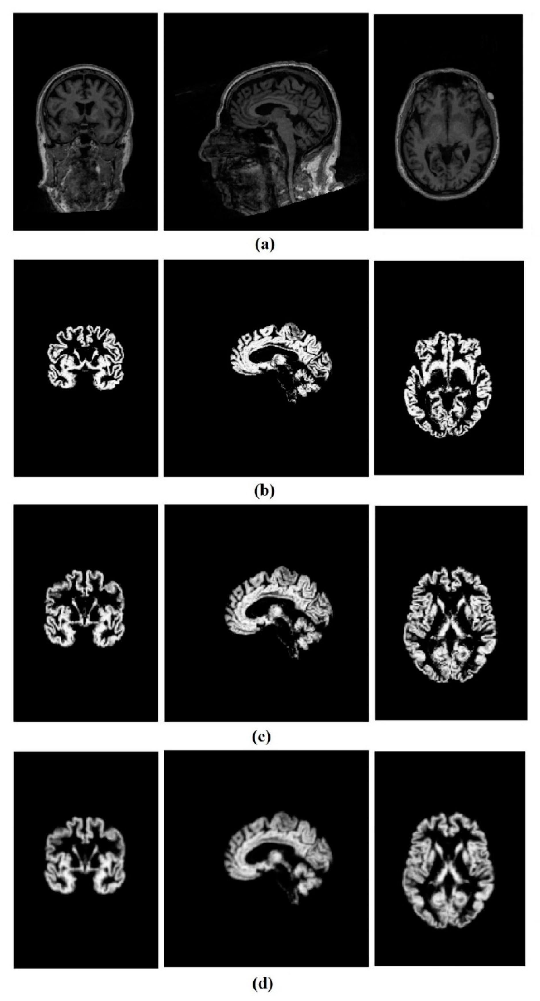
3.1. Image Preprocessing
3.1.1. Segmentation
3.1.2. Normalization
3.1.3. Smoothing
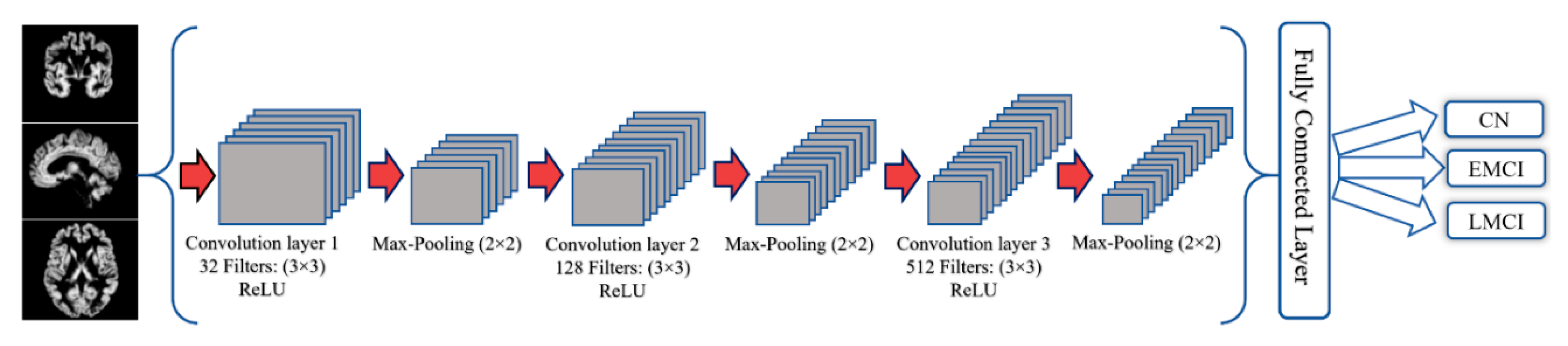
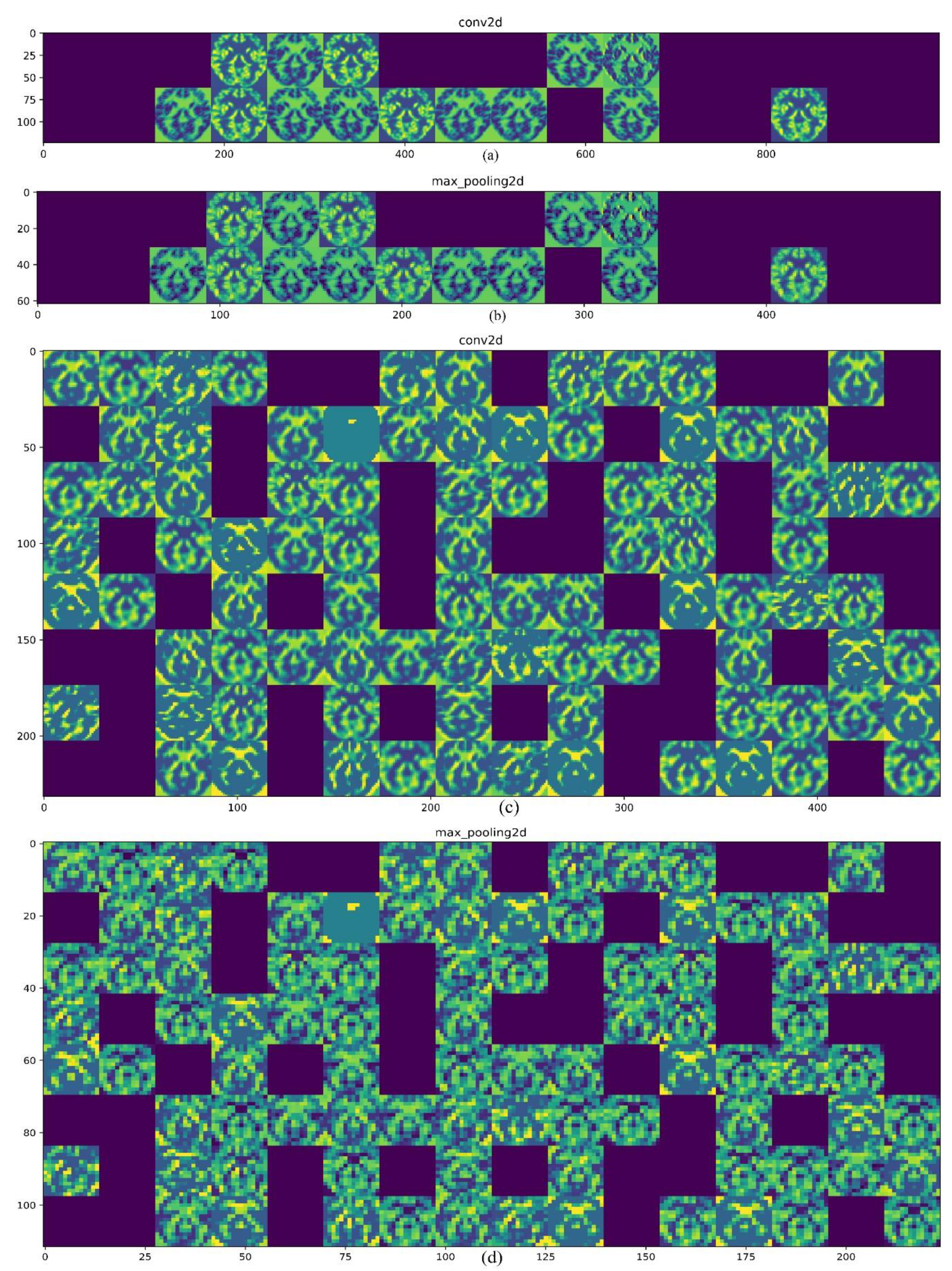
3.2. CNN architecture
4. Results
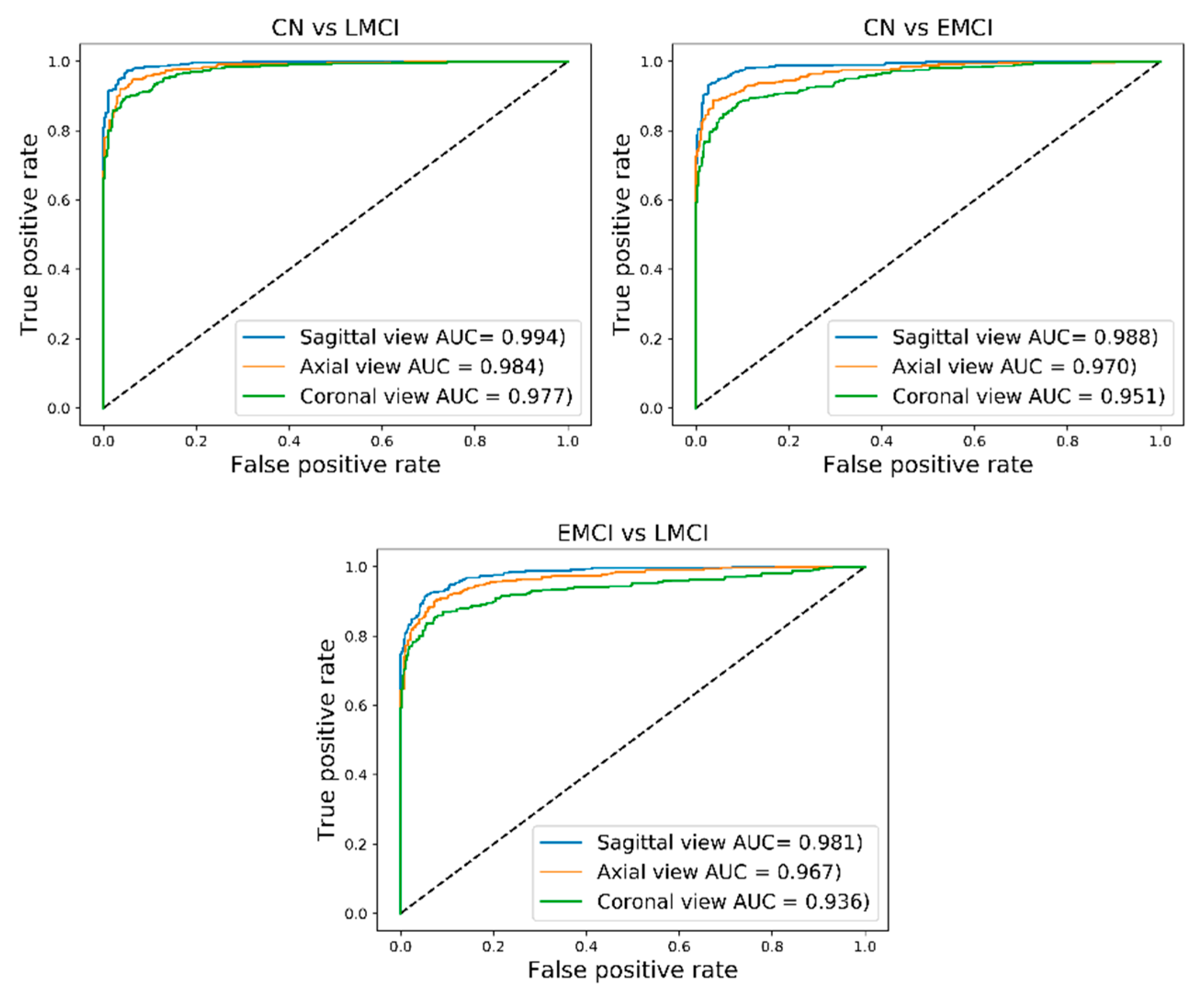
4.1. Classification of CN and LMCI
4.2. Classification of CN and EMCI
4.3. Classification of EMCI and LMCI
5. Discussion
6. Conclusions
Author Contributions
Funding
Conflicts of Interest
References
- Kantarci, K.; Weigand, S.; Przybelski, S.; Shiung, M.; Whitwell, J.L.; Negash, S.; Knopman, D.S.; Boeve, B.F.; O’Brien, P.; Petersen, R.C. Risk of dementia in MCI: Combined effect of cerebrovascular disease, volumetric MRI, and 1H MRS. Neurology 2009, 72, 1519–1525. [Google Scholar] [CrossRef] [PubMed] [Green Version]
- Mitchell, A.J.; Shiri-Feshki, M. Rate of progression of mild cognitive impairment to dementia—Meta-analysis of 41 robust inception cohort studies. Acta Psychiatr. Scand. 2009, 119, 252–265. [Google Scholar] [CrossRef] [PubMed]
- Rountree, S.; Waring, S.; Chan, W.; Lupo, P.; Darby, E.; Doody, R. Importance of subtle amnestic and nonamnestic deficits in mild cognitive impairment: Prognosis and conversion to dementia. Dement. Geriatr. Cogn. Disord. 2007, 24, 476–482. [Google Scholar] [CrossRef] [PubMed]
- Boyle, P.; Wilson, R.; Aggarwal, N.; Tang, Y.; Bennett, D. Mild cognitive impairment Risk of Alzheimer disease and rate of cognitive decline. Neurology 2006, 67, 441–445. [Google Scholar] [CrossRef] [PubMed]
- DeCarli, C. Mild cognitive impairment: Prevalence, prognosis, aetiology and treatment. Lancet Neurol. 2003, 2, 15–21. [Google Scholar] [CrossRef]
- Kochanek, K.D.; Murphy, S.L.; Xu, J.; Tejada-Vera, B. Deaths: Final data for 2014. Natl. Vital. Stat. Rep. 2016, 65, 1–122. [Google Scholar] [PubMed]
- Association, A.S. 2018 Alzheimer’s disease facts and figures. Alzheimer Dement. 2018, 14, 367–429. [Google Scholar]
- Hebert, L.E.; Weuve, J.; Scherr, P.A.; Evans, D.A. Alzheimer disease in the United States (2010–2050) estimated using the 2010 census. Neurology 2013, 80, 1778–1783. [Google Scholar] [CrossRef] [PubMed]
- Gaugler, J.; James, B.; Johnson, T.; Marin, A.; Weuve, J. 2019 Alzheimer’s disease facts and figures. Alzheimer Dement. J. Alzheimer Assoc. 2019, 15, 321–387. [Google Scholar] [CrossRef]
- Aisen, P.S.; Petersen, R.C.; Donohue, M.C.; Gamst, A.; Raman, R.; Thomas, R.G.; Walter, S.; Trojanowski, J.Q.; Shaw, L.M.; Beckett, L.A. Clinical Core of the Alzheimer’s Disease Neuroimaging Initiative: Progress and plans. Alzheimer Dement. 2010, 6, 239–246. [Google Scholar] [CrossRef]
- Johnson, K.A.; Fox, N.C.; Sperling, R.A.; Klunk, W.E. Brain imaging in Alzheimer disease. Cold Spring Harb. Perspect. Med. 2012, 2, a006213. [Google Scholar] [CrossRef] [PubMed]
- McGeer, P.L. Brain imaging in Alzheimer’s disease. Br. Med. Bull. 1986, 42, 24–28. [Google Scholar] [CrossRef] [PubMed]
- Fox, N.C.; Schott, J.M. Imaging cerebral atrophy: Normal ageing to Alzheimer’s disease. Lancet 2004, 363, 392–394. [Google Scholar] [CrossRef]
- Tabatabaei-Jafari, H.; Shaw, M.E.; Cherbuin, N. Cerebral atrophy in mild cognitive impairment: A systematic review with meta-analysis. Alzheimer Dement. Diagn. Assess. Dis. Monit. 2015, 1, 487–504. [Google Scholar] [CrossRef] [PubMed]
- Nestor, S.M.; Rupsingh, R.; Borrie, M.; Smith, M.; Accomazzi, V.; Wells, J.L.; Fogarty, J.; Bartha, R.; Initiative, A.D.N. Ventricular enlargement as a possible measure of Alzheimer’s disease progression validated using the Alzheimer’s disease neuroimaging initiative database. Brain 2008, 131, 2443–2454. [Google Scholar] [CrossRef] [PubMed]
- Prados, F.; Cardoso, M.J.; Leung, K.K.; Cash, D.M.; Modat, M.; Fox, N.C.; Wheeler-Kingshott, C.A.; Ourselin, S.; Initiative, A.D.N. Measuring brain atrophy with a generalized formulation of the boundary shift integral. Neurobiol. Aging 2015, 36, S81–S90. [Google Scholar] [CrossRef]
- Henneman, W.; Sluimer, J.; Barnes, J.; Van Der Flier, W.; Sluimer, I.; Fox, N.; Scheltens, P.; Vrenken, H.; Barkhof, F. Hippocampal atrophy rates in Alzheimer disease: Added value over whole brain volume measures. Neurology 2009, 72, 999–1007. [Google Scholar] [CrossRef] [Green Version]
- Wang, Y.; West, J.D.; Flashman, L.A.; Wishart, H.A.; Santulli, R.B.; Rabin, L.A.; Pare, N.; Arfanakis, K.; Saykin, A.J. Selective changes in white matter integrity in MCI and older adults with cognitive complaints. Biochim. Biophys. Acta Mol. Basis Dis. 2012, 1822, 423–430. [Google Scholar] [CrossRef] [Green Version]
- Zhang, H.; Sachdev, P.S.; Wen, W.; Kochan, N.A.; Crawford, J.D.; Brodaty, H.; Slavin, M.J.; Reppermund, S.; Draper, B.; Zhu, W. Gray matter atrophy patterns of mild cognitive impairment subtypes. J. Neurol. Sci. 2012, 315, 26–32. [Google Scholar] [CrossRef]
- Popp, J.; Wolfsgruber, S.; Heuser, I.; Peters, O.; Hüll, M.; Schröder, J.; Möller, H.-J.; Lewczuk, P.; Schneider, A.; Jahn, H. Cerebrospinal fluid cortisol and clinical disease progression in MCI and dementia of Alzheimer’s type. Neurobiol. Aging 2015, 36, 601–607. [Google Scholar] [CrossRef]
- Zhang, Y.; Schuff, N.; Camacho, M.; Chao, L.L.; Fletcher, T.P.; Yaffe, K.; Woolley, S.C.; Madison, C.; Rosen, H.J.; Miller, B.L. MRI markers for mild cognitive impairment: Comparisons between white matter integrity and gray matter volume measurements. PLoS ONE 2013, 8, e66367. [Google Scholar] [CrossRef] [PubMed]
- Grundman, M.; Sencakova, D.; Jack, C.R.; Petersen, R.C.; Kim, H.T.; Schultz, A.; Weiner, M.F.; DeCarli, C.; DeKosky, S.T.; Van Dyck, C. Brain MRI hippocampal volume and prediction of clinical status in a mild cognitive impairment trial. J. Mol. Neurosci. 2002, 19, 23–27. [Google Scholar] [CrossRef] [PubMed]
- Loewenstein, D.A.; Acevedo, A.; Potter, E.; Schinka, J.A.; Raj, A.; Greig, M.T.; Agron, J.; Barker, W.W.; Wu, Y.; Small, B. Severity of medial temporal atrophy and amnestic mild cognitive impairment: Selecting type and number of memory tests. Am. J. Geriatr. Psychiatry 2009, 17, 1050–1058. [Google Scholar] [CrossRef] [PubMed]
- Chetelat, G.; Desgranges, B.; De La Sayette, V.; Viader, F.; Eustache, F.; Baron, J.-C. Mapping gray matter loss with voxel-based morphometry in mild cognitive impairment. Neuroreport 2002, 13, 1939–1943. [Google Scholar] [CrossRef] [PubMed]
- Guo, X.; Wang, Z.; Li, K.; Li, Z.; Qi, Z.; Jin, Z.; Yao, L.; Chen, K. Voxel-based assessment of gray and white matter volumes in Alzheimer’s disease. Neurosci. Lett. 2010, 468, 146–150. [Google Scholar] [CrossRef]
- Davatzikos, C.; Bhatt, P.; Shaw, L.M.; Batmanghelich, K.N.; Trojanowski, J.Q. Prediction of MCI to AD conversion, via MRI, CSF biomarkers, and pattern classification. Neurobiol. Aging 2011, 32, 2322.e19–2322.e27. [Google Scholar] [CrossRef] [PubMed] [Green Version]
- Friston, K.J.; Holmes, A.P.; Worsley, K.J.; Poline, J.P.; Frith, C.D.; Frackowiak, R.S. Statistical parametric maps in functional imaging: A general linear approach. Hum. Brain Mapp. 1994, 2, 189–210. [Google Scholar] [CrossRef]
- Gorji, H.; Haddadnia, J. A novel method for early diagnosis of Alzheimer’s disease based on pseudo Zernike moment from structural MRI. Neuroscience 2015, 305, 361–371. [Google Scholar] [CrossRef]
- Liu, S.; Liu, S.; Cai, W.; Pujol, S.; Kikinis, R.; Feng, D. Early diagnosis of Alzheimer’s disease with deep learning. In Proceedings of the 2014 IEEE 11th International Symposium on Biomedical Imaging (ISBI), Beijing, China, 29 April–2 May 2014; pp. 1015–1018. [Google Scholar]
- Sarraf, S.; Tofighi, G. Classification of alzheimer’s disease using fmri data and deep learning convolutional neural networks. arXiv 2016, arXiv:1603.08631. [Google Scholar]
- Ramírez, J.; Górriz, J.; Segovia, F.; Chaves, R.; Salas-Gonzalez, D.; López, M.; Álvarez, I.; Padilla, P. Computer aided diagnosis system for the Alzheimer’s disease based on partial least squares and random forest SPECT image classification. Neurosci. Lett. 2010, 472, 99–103. [Google Scholar] [CrossRef]
- Traore, B.B.; Kamsu-Foguem, B.; Tangara, F. Deep convolution neural network for image recognition. Ecol. Inform. 2018, 48, 257–268. [Google Scholar] [CrossRef] [Green Version]
- Krizhevsky, A.; Sutskever, I.; Hinton, G.E. Imagenet classification with deep convolutional neural networks. In Proceedings of the Advances in Neural Information Processing Systems, Lake Tahoe, CA, USA, 3 December 2012; pp. 1097–1105. [Google Scholar]
- Ronneberger, O.; Fischer, P.; Brox, T. U-net: Convolutional networks for biomedical image segmentation. In Lecture Notes in Computer Science, Proceedings of the International Conference on Medical Image Computing and Computer-Assisted Intervention, Munich, Germany, 5–9 October 2015; Springer: Cham, Switzerland, 2015; pp. 234–241. [Google Scholar]
- LeCun, Y.; Bengio, Y.; Hinton, G. Deep learning. Nature 2015, 521, 436. [Google Scholar] [CrossRef] [PubMed]
- Karpathy, A.; Toderici, G.; Shetty, S.; Leung, T.; Sukthankar, R.; Fei-Fei, L. Large-scale video classification with convolutional neural networks. In Proceedings of the IEEE Conference on Computer Vision and Pattern Recognition, Columbus, OH, USA, 24–27 June 2014; pp. 1725–1732. [Google Scholar]
- Acharya, U.R.; Oh, S.L.; Hagiwara, Y.; Tan, J.H.; Adeli, H.; Subha, D.P. Automated EEG-based screening of depression using deep convolutional neural network. Comput. Methods Programs Biomed. 2018, 161, 103–113. [Google Scholar] [CrossRef] [PubMed]
- Spasov, S.E.; Passamonti, L.; Duggento, A.; Liò, P.; Toschi, N. A Multi-modal Convolutional Neural Network Framework for the Prediction of Alzheimer’s Disease. In Proceedings of the 2018 40th Annual International Conference of the IEEE Engineering in Medicine and Biology Society (EMBC), Honolulu, HI, USA, 18–21 July 2018; pp. 1271–1274. [Google Scholar]
- Payan, A.; Montana, G. Predicting Alzheimer’s disease: A neuroimaging study with 3D convolutional neural networks. arXiv 2015, arXiv:1502.02506. [Google Scholar]
- Maas, A.L.; Hannun, A.Y.; Ng, A.Y. Rectifier nonlinearities improve neural network acoustic models. In Proceedings of the Proc. Icml, Atlanta, GA, USA, 16–21 June 2013; p. 3. [Google Scholar]
- Jacobs, H.I.; van Boxtel, M.P.; Gronenschild, E.H.; Uylings, H.B.; Jolles, J.; Verhey, F.R. Decreased gray matter diffusivity: A potential early Alzheimer’s disease biomarker? Alzheimer Dement. 2013, 9, 93–97. [Google Scholar] [CrossRef] [PubMed]
- Glorot, X.; Bengio, Y. Understanding the difficulty of training deep feedforward neural networks. In Proceedings of the Thirteenth International Conference on Artificial Intelligence and Statistics, Sardinia, Italy, 13–15 May 2010; pp. 249–256. [Google Scholar]
- Kingma, D.P.; Ba, J. Adam: A method for stochastic optimization. arXiv 2014, arXiv:1412.6980. [Google Scholar]
- Wang, H.; Shen, Y.; Wang, S.; Xiao, T.; Deng, L.; Wang, X.; Zhao, X. Ensemble of 3D densely connected convolutional network for diagnosis of mild cognitive impairment and Alzheimer’s disease. Neurocomputing 2019, 333, 145–156. [Google Scholar] [CrossRef]
- Ju, R.; Hu, C.; Zhou, P.; Li, Q. Early diagnosis of Alzheimer’s disease based on resting-state brain networks and deep learning. IEEE ACM Trans. Comput. Biol. Bioinform. (TCBB) 2019, 16, 244–257. [Google Scholar] [CrossRef]
- Huang, Y.; Xu, J.; Zhou, Y.; Tong, T.; Zhuang, X. Diagnosis of Alzheimer’s Disease via Multi-modality 3D Convolutional Neural Network. arXiv 2019, arXiv:1902.09904. [Google Scholar] [CrossRef]
- Jessen, F.; Wolfsgruber, S.; Wiese, B.; Bickel, H.; Mösch, E.; Kaduszkiewicz, H.; Pentzek, M.; Riedel-Heller, S.G.; Luck, T.; Fuchs, A. AD dementia risk in late MCI, in early MCI, and in subjective memory impairment. Alzheimer Dement. 2014, 10, 76–83. [Google Scholar] [CrossRef]
- Hinton, G.E.; Srivastava, N.; Krizhevsky, A.; Sutskever, I.; Salakhutdinov, R.R. Improving neural networks by preventing co-adaptation of feature detectors. arXiv 2012, arXiv:1207.0580. [Google Scholar]
- Ortiz, A.; Munilla, J.; Gorriz, J.M.; Ramirez, J. Ensembles of deep learning architectures for the early diagnosis of the Alzheimer’s disease. Int. J. Neural Syst. 2016, 26, 1650025. [Google Scholar] [CrossRef] [PubMed]
- Suk, H.-I.; Shen, D. Deep learning-based feature representation for AD/MCI classification. In Lecture Notes in Computer Science, Proceedings of the International Conference on Medical Image Computing and Computer—Assisted Intervention, Nagoya, Japan, 22–26 September 2013; Springer: Berlin/Heidelberg, Germany, 2013; pp. 583–590. [Google Scholar]
- Li, F.; Tran, L.; Thung, K.-H.; Ji, S.; Shen, D.; Li, J. Robust deep learning for improved classification of AD/MCI patients. In Lecture Notes in Computer Science, Proceedings of the International Workshop on Machine Learning in Medical Imaging, Boston, MA, USA, 14 September 2014; Springer: Cham, Switzerland, 2014; pp. 240–247. [Google Scholar]
- Korolev, S.; Safiullin, A.; Belyaev, M.; Dodonova, Y. Residual and plain convolutional neural networks for 3D brain MRI classification. In Proceedings of the 2017 IEEE 14th International Symposium on Biomedical Imaging (ISBI 2017), Melbourne, VIC, Australia, 18–21 April 2017; pp. 835–838. [Google Scholar]
- Cabrera-León, Y.; Báez, P.G.; Ruiz-Alzola, J.; Suárez-Araujo, C.P. Classification of Mild Cognitive Impairment Stages Using Machine Learning Methods. In Proceedings of the 2018 IEEE 22nd International Conference on Intelligent Engineering Systems (INES), Las Palmas de Gran Canaria, Spain, 21–23 June 2018; pp. 000067–000072. [Google Scholar]
- Singh, S.; Srivastava, A.; Mi, L.; Caselli, R.J.; Chen, K.; Goradia, D.; Reiman, E.M.; Wang, Y. Deep-learning-based classification of FDG-PET data for Alzheimer’s disease categories. In Proceedings of the 13th International Conference on Medical Information Processing and Analysis, San Andres Island, Colombia, 17 November 2017; p. 105720. [Google Scholar]


| CN (N = 200; 112 F/88 M) | EMCI (N = 200;93 F/107 M) | LMCI (N = 200; 84 F/116 M) | ||||
|---|---|---|---|---|---|---|
| Mean | SD | Mean | SD | Mean | SD | |
| Age | 74.2 | 6.1 | 68.2 | 6.9 | 71.1 | 7.2 |
| MMSE | 28.8 | 1.3 | 28.4 | 1.2 | 27.3 | 1.8 |
| MRI Views | Sensitivity (%) | Specificity (%) | Accuracy (%) | F-Score (%) | AUC (%) | |
|---|---|---|---|---|---|---|
| CN vs. LMCI | Sagittal | 91.70 | 97.96 | 94.54 | 94.84 | 99.40 |
| Axial | 90.02 | 97.01 | 93.18 | 93.53 | 98.40 | |
| Coronal | 90.28 | 93.30 | 91.65 | 92.19 | 97.70 | |
| CN vs. EMCI | Sagittal | 90.46 | 98.19 | 93.96 | 94.25 | 98.80 |
| Axial | 90.63 | 91.42 | 90.99 | 91.65 | 97.00 | |
| Coronal | 88.60 | 89.95 | 89.21 | 89.96 | 95.10 | |
| EMCI vs. LMCI | Sagittal | 91.48 | 94.82 | 93.00 | 93.46 | 98.10 |
| Axial | 87.01 | 94.57 | 90.45 | 90.86 | 96.70 | |
| Coronal | 85.44 | 92.07 | 88.45 | 88.98 | 93.60 |
© 2019 by the authors. Licensee MDPI, Basel, Switzerland. This article is an open access article distributed under the terms and conditions of the Creative Commons Attribution (CC BY) license (http://creativecommons.org/licenses/by/4.0/).
Share and Cite
Taheri Gorji, H.; Kaabouch, N. A Deep Learning approach for Diagnosis of Mild Cognitive Impairment Based on MRI Images. Brain Sci. 2019, 9, 217. https://doi.org/10.3390/brainsci9090217
Taheri Gorji H, Kaabouch N. A Deep Learning approach for Diagnosis of Mild Cognitive Impairment Based on MRI Images. Brain Sciences. 2019; 9(9):217. https://doi.org/10.3390/brainsci9090217
Chicago/Turabian StyleTaheri Gorji, Hamed, and Naima Kaabouch. 2019. "A Deep Learning approach for Diagnosis of Mild Cognitive Impairment Based on MRI Images" Brain Sciences 9, no. 9: 217. https://doi.org/10.3390/brainsci9090217