Dry Bio-Decontamination Process in Reduced-Pressure O2 Plasma

Department of Environmental Science and Engineering, School of Energy and Power Engineering, Xi’an Jiaotong University, Xi’an 710049, China
*
Author to whom correspondence should be addressed.
Appl. Sci. 2019, 9(9), 1933; https://doi.org/10.3390/app9091933
Submission received: 21 March 2019
/
Revised: 22 April 2019
/
Accepted: 7 May 2019
/
Published: 10 May 2019
(This article belongs to the Special Issue Plasma Technology for Biomedical Applications)
Abstract
:Featured Application
The specific findings of this research will deep our understanding for the mechanisms of bio-decontamination from material surfaces by plasma technology.
Abstract
The main objective of this work was to fully understand the bio-decontamination process in a reduced-pressure oxygen plasma. Gram-negative Escherichia coli species was chosen as the target microorganism in this test. The comparison of decontamination efficacy between plasma total and UV radiation individually under various treatment parameters and tests of DNA agarose electrophoresis were made to evaluate the inactivation effect of UV radiation. The quantity of protein leakage and the concentration of malondialdehyde (MDA), which are markers of the end products of lipid peroxidation, in bacterial suspension after treatment were determined to estimate the contribution of both charged particles and free radicals for bacterial death. In addition, a scanning electronic microscope was used to visualize the plasma effect on microorganisms. The results showed that the essential action of the oxygen plasma on Escherichia coli is believed to be attributed to the fast and intense etching on cell membrane by electrons and ions. Attacks on polyunsaturation fatty acid (PUFA) in the cell membrane by oxygen free radicals and the destruction of the DNA in the cell by UV radiation are accessorial during an effective decontamination process.
1. Introduction
The elimination of disease-causing agents from the surfaces of equipment, which can sometimes be challenging to fulfill without using toxic materials or high temperatures, is an absolutely necessary requirement in many fields. Historically, many different approaches have been used to inactivate pathogens. Two widely used inactivation methods, especially in the medical field, are autoclaving and exposure to gases such as ethylene oxide (EtO). Though effective, both methods suffer from drawbacks such as exposure to extremely high temperatures (>1000 °C) in the case of autoclaves, and toxic exposure in the case of EtO. Another concern with the latter is the long aeration process which, importantly, creates a serious threat for both personnel and the environment [1]. For these reasons, it is extremely important to develop a new bio-decontamination or disinfection technology that is safe and easy to apply. In recent years, one of the most serious current alternatives to gaseous bio-decontamination is the use of gas-discharge plasma as a sterilizing agent. Plasma-based bio-decontamination techniques do not suffer from the problems of traditional techniques. Adequate processes are efficient, do affect only slightly the bulk material, are environmentally sound, do not produce toxic by-products, and are fast and cost-effective. Therefore, this kind of method is regarded as a green technique and even as the most promising bio-decontamination technique [2,3,4].
During the plasma process, micro-organisms are exposed to reactive species, which are produced by applying electromagnetic fields to a gas or gas mixture. But so far, all the mechanisms which may be responsible for the treatment, such as the interaction of UV radiation with the DNA of cells, the removal of the material of cells by reactive species and so on [5,6,7,8,9,10], are only presumed on the basis of the apparent reactive results in plasma. There is no thorough understanding about the respective contribution of different reactive species for microbial inactivation. That is, the present conclusions about plasma bio-decontamination mechanisms, although various, have less persuasion. As we know, being a bio-decontamination technique, those with higher stability and security than other ones must be paid attention. It is absolutely necessary to first investigate the bio-decontamination mechanisms of gas-discharge plasma.
Generally speaking, during plasma bio-decontamination, the complex reactive species can be divided into three categories: charged species (including electrons and ions), free radicals, and UV radiation. The last, UV, is the only one can be separated from other species by a special fitting due to its penetrability. From current research [7,8,11,12,13], we can see that UV radiation reacts with DNA to injure the micro-organism; the charged particles remove the material (i.e., etching) of cells, resulting in protein leakage; and the activated free radicals initiate the lipid peroxidation by attacking polyunsaturation fatty acid (PUFA) in the cell membrane. In accordance with these reasons, in this paper, the comparison of germicidal efficacy under various treatment conditions between plasma and UV radiation and the test of DNA state are combined to determine the decontamination effect of UV radiation firstly; then the observation of bacterial ultrastructure and the quantity of protein leakage as well as the yield of malondialdehyde (MDA), being the marker of the end products of the lipid peroxidation, are measured to ascertain the efficiency of the other two kinds reactive species. Reduced-pressure oxygen plasma was applied to perform in this experiment and Gram-negative Escherichia coli (E. coli) 8099 was used as a test strain.
2. Materials and Methods
2.1. Materials
E. coli 8099 slant lawn incubated at 37 °C for 24 h was oscillated and eluted by phosphate buffer solution (PBS), then the eluent was diluted to form certain concentration suspensions of bacilli. This suspension was taken 0.01 mL to spread uniformly on 15 × 15 mm glass sheet and dried naturally at room temperature.
2.2. O2 Plasma Treatment
Figure 1 shows the schematic diagram of experimental apparatus. An RF generator was the type of SY-500 W, used whose power was 13.56 MHz and whose output power could be adjusted in succession in order to match the SP-II matcher. The reaction chamber was a Pyrex glass tube (length 150 mm, diameter 45 mm), initiated inductively coupled discharge. In addition, a vacuum pump was used to insure the reduced pressure in reactor.
In the first test, some samples were directly exposed to plasma discharge zone to obtain the decontamination effects of E. coli by oxygen plasma in consideration of different operation parameters, such as gas flow—20–100 cm3·min−1, pressure—45 Pa, RF power—20–100 W and treatment time—20–120 s. In the second test, at the above-mentioned conditions, other samples were treated under the filter of lithium fluoride (LiF), which achieved the separation of UV and other active species, to determine the relative contribution of UV radiation to microbial inactivation. All factors of plasma generated reactive species (e.g., electrons, ion, free radicals and UV radiation) play roles in microbial inactivation in the former test, whereas in the latter test, microbial inactivation was performed with the action of UV radiation from plasma discharge with wavelength λ ≥ 120 nm [14].
2.3. Viable Cell Counts and Decontamination Effect
Based on the living conditions of E. coli, the viable cell counts of teat samples was determined by cell culture methods, which was slightly modified from Hertwig et al. [15]. Simply, treated samples were withdrawn from the reactor immediately after the above-mentioned treatment. Then, eluting bacilli from control or treated samples with phosphate buffer solution (PBS) obtained the suspensions of bacilli, which were transferred to nutrient-containing Petri dish and incubated for 48 h at 37 °C prior to determining the bacilli number of colony forming units (CFU). The decontamination effect (E) of E. coli by oxygen plasma and UV radiation individual was determined by Equation (1) below [16,17,18,19],
where N0 and Nt represent the number of CFU of control and treated surviving bacilli, respectively.
2.4. Determination of Protein Leakage Quantity
The dye binding method was used as the detection of protein leakage in the cells after plasma treatment. The preparation of bacilli suspensions was described in Section 2.2. Then, the suspensions were placed in a beaker containing 5 mL of PBS, shaken for 10 min, and centrifuged at 3000 r/min for 15 min. We took 1.0 mL of the supernatant to mix with 5.0 mL of Coomassie Brilliant Blue dye, which was placed for 5 min at room temperature. The absorbency of the mixed solution was measured at the wavelength of 595 nm, meanwhile, obtained absorbance value was placed in the regression equation of standard curve of bovine serum albumin to obtain the quantity of protein leakage of cells after treatment [17,18,20].
2.5. Determination of Malondialdehyde (MDA) Production
Due to the condensation reaction of malondialdehyde (MDA) and thibabituric acid (TBA, obtained from Nanjing Jiancheng biological engineering institute) formed a red product with a maximum absorption peak at 532 nm, the determination of MDA production after treatment was obtained by colorimetry. The bacterial suspension was obtained by the same method as mentioned above. The MDA concentration was calculated by Equation (2) below [12,20,21],
where N is the concentration of MDA (mol·L-1), Am is determination tube absorbency, Amo is determination blank tube absorbency, As is standard tube absorbency, Aso is s standard blank tube absorbency, n is the sample diluted multiple before test.
2.6. DNA Measurement
The DNA measurement of samples cells was extracted by distilling reagent of Genome DNA (TIANGEN Biological Technology Co., Ltd., Beijing, China), and measured by ECP3000 agarose electrophoresis apparatus (Six-one Instrument Co., Ltd., Beijing, China) [22]. Electrophoresis buffer was 0.5 × TBE, which pH was maintained at 7.5–7.8.
2.7. Scanning Electron Microscopy
The suspension of bacilli before and after inactivation was taken at 0.03 mL to spread uniformly on glass sheet to form folium with 1 cm diameter, and then dried naturally at room temperature. The ultrastructure of bacilli was observed using scanning electron microscopy (SEM) in a JEOL instrument (Model JSM–6700F, Tokyo, Japan) after coating the dry specimens with gold. The magnification of 10000× was used.
2.8. Statistical Analysis
In Section 2.3, Section 2.4, Section 2.5 and Section 2.6 the determination of decontamination effect, protein leakage quantity, MDA production and DNA for samples were carried out in triplicate to ensure the repeatability. The data were expressed as the mean ± standard deviation (SD) in Figures 2, 4, and 6. The significant of differences among data were statistically analyzed by one-way ANOVA test using SPSS software (version 20.0), choosing p < 0.05 as statistically significant.
3. Results
3.1. Decontamination Effect of UV Radiation in Oxygen Plasma
The decontamination efficiency of oxygen plasma total and UV radiation individual (excluded the charge particles and free radicals generated by plasma discharge using the LiF) was investigated at the same operation parameters, respectively, from E as functions of plasma RF power, treatment time and gas flow rate. The samples were positioned at the center of the induction coil in the Pyrex glass tube of the reactor. Figure 2 shows the comparison of both the decontamination effects of oxygen plasma total and UV radiation individually. From Figure 2, for one thing, it could be clearly seen that the E of oxygen plasma total showed a strong dependence on the conditions mentioned above, and the effective bio-decontamination was carried out when oxygen plasma discharge under the conditions of 100 W, 60 s and 40 cm3·min−1, where the E was largest, up to 3.42.
In addition, the decontamination rate of E. coli by oxygen plasma as a function of O2 flow rate increased firstly (p < 0.05) and subsequently decreased (p < 0.05) with the increase of gas flow and was optimal at a flow rate of 40 cm3·min−1. This phenomenon was due to the inactivation effect of E. coli depending on the average energy, number, and residence time of reactive species in the discharge zone. Reactive species had higher average energy and longer residence time in discharge zone at low flux than high flux, however, the number of species were limited, as opposed to high flow rate, which directly affected the performance of plasma on microbial inactivation. For another thing, the E of UV radiation individual changed only within a narrow range, from 0.4 to 1.5, in the whole process no matter how many changes these conditions underwent. These values indicate that during effective bio-decontamination, the contribution of UV radiation to decontaminating features of the oxygen plasma is less. This conclusion is also confirmed by the following.
It is known that UV radiation kills bacilli through destroying the DNA of cells. So, we investigated the state of DNA after oxygen plasma treatment in order to further ascertain the effect of UV radiation on E. coli. Figure 3 displays the DNA agarose electrophoresis of E. coil after three treatment conditions such as control, UV radiation individual and plasma total treatment at 100 W power, 60 s treatment time and 40 cm3·min−1 oxygen flow rate. As far as we know, if the skeleton of DNA is destroyed to generate small DNA fragments or light molecular DNA when exposes E. coli to UV radiation or plasma [22,23], which will produce tail phenomena during the DNA agarose electrophoresis [24]. But the diagrams in Figure 3 (2), which were treated by UV radiation individually, indicate that there are only fewer tail phenomena compared to oxygen plasma total Figure 3 (3). This result makes sure again that UV radiation in oxygen plasma slightly acts on bacilli during bio-decontamination process.
3.2. Decontamination Effect of Charged Species in Oxygen Plasma
Reactive species strongly bombard the cell surface promoting surface etching. Meanwhile, the living cell cannot quickly repair, resulting in rapid destruction for some microbials in many cases [5]. In addition, charged particles may converge on the surface of cell membranes and cause electrostatic stress when the force is greater than the tensile strength of membrane itself, which will cause damage to cell materials [7,25]. Those action acts on cell materials leading to cell membrane ruptures and leakage of cellular contents, and finally leads the bacterial death [7,11,12]. Therefore, the protein leakage quantity of sample cells was chosen as an index to reflect the inactivation effect of E. coli by plasma [24]. Figure 4 shows the tendency of protein leakage quantity in suspensions of bacilli after plasma exposure under different conditions. Compared with the curves of the total decontamination efficiency of oxygen plasma in Figure 2, it can be found that both the changed trends are almost accordant, that is, the action of charged species on the bacilli plays a major role in the inactivation of E. coli.
This conclusion also can be confirmed further by observing the ultrastructure of bacilli with SEM, as shown in Figure 5. Clearly, the untreated E. coli is look like a pole (Figure 5a), whereas after treatment at 100 W power, 60 s treatment time and 40 cm3·min−1 oxygen flow (Figure 5b), bacilli is swollen, incompact and shows obvious ‘thawing’. The content of cell leaks completely. This is just due to the intense damage action on cell membrane by the charged particles with high energy.
3.3. Decontamination Effect of Free Radicals in Oxygen Plasma
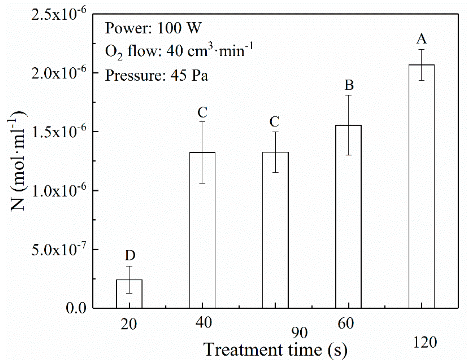
Acting on PUFA in cell membrane by oxygen radicals, specifically highly reactive oxygen atom and hydroxyl radicals, can produce MDA, which is marker as one of the end products of the lipid peroxidation. MDA can destroy the whole structure and functional properties of cell membrane. With the increase of MDA concentration, the content of unsaturated fatty acids in cell membrane reduces, electrical potential of membrane changes and finally, the liquidity of cell membrane is lost [26,27]. So, the changes of MDA concentration in suspensions of bacilli after treatment, as shown in Figure 6, can be used to analyze the interaction between oxygen radicals and cell of bacilli [26,27]. From Figure 6, the MDA concentration has an apparent increase (p < 0.05) in a short time (<40 s), and then, little changes occur up to 60 s. Subsequently, it increases gradually again. Also Compared with the curves of the total decontamination efficiency of oxygen plasma in Figure 2, one can find that when treatment time is lower than 40 s, the oxygen radicals in plasma play a major role at a certain extent in the E. coli killing process, whereas when the reaction goes on, the contribution of oxygen radicals for inactivation is weakened, whose possible reason is the restraint by intense etching action.
4. Conclusions
In this study, a thorough understanding about the respective contribution of different reactive species for the decontamination of E. coli from surfaces in reduced-pressure oxygen plasma was carried out as the following steps: comparing the decontamination efficacy between total plasma and individual UV radiation, measuring the state of DNA and the ultrastructure of bacilli as well as the concentration of protein leakage and MDA production in bacterial suspension after plasma treatment. With the results, the essential effectiveness on E. coli of the oxygen plasma was believed to be attributed to the intense etching action of electrons and ions on the bacilli materials. Attacking PUFA in the cell membrane by oxygen radicals plays a major role at a certain extent only during the initial inactivation, and then is restrained by the effect of charged particles with the reaction underway. The function of UV radiation is assistant in the whole process, which results in only the slight damage and rupture of DNA.
Author Contributions
Conceptualization, H.L.; methodology, H.L.; validation, X.F., J.X. and X.M.; formal analysis, X.F.; investigation, X.M.; resources, C.H.; data curation, J.X.; writing—original draft preparation, H.L.; supervision, C.H.; project administration, H.L.
Funding
This work was financially supported by the National Natural Science Foundation of China (No. 31871889), the Natural Science Basic Research Plan in Shaanxi Province of China (No. 2017JM5067), the Fundamental Research Funds for the Central Universities (No. xjj2015132).
Acknowledgments
We also wish to thank Professor Jierong Chen for her support of this work.
Conflicts of Interest
The authors declare no conflict of interest. The funders had no role in the design of the study; in the collection, analyses, or interpretation of data; in the writing of the manuscript, or in the decision to publish the results.
References
- Moisan, M.; Barbeau, J.; Moreau, S.; Pelletier, J.; Tabrizian, M.; Yahia, L. Low-temperature sterilization using gas plasmas: a review of the experiments and an analysis of the inactivation mechanisms. Int. J. Pharm. 2001, 226, 1–21. [Google Scholar] [CrossRef]
- Lee, T.; Puligundla, P.; Mok, C. Inactivation of foodborne pathogens on the surfaces of different packaging materials using low-pressure air plasma. Food Control. 2015, 51, 149–155. [Google Scholar] [CrossRef]
- Jiang, C.; Schaudinn, C.; Jaramillo, D.E.; Webster, P.; Costerton, J.W. In vitro antimicrobial effect of a cold plasma jet against Enterococcus faecalis biofilms. ISRN Dent. 2012, 2012. [Google Scholar] [CrossRef] [PubMed]
- Shi, X.M.; Zhang, G.J.; Yuan, Y.K.; Ma, Y.; Xu, G.M.; Yang, Y. Research on the inactivation effect of low-temperature plasma on Candida albicans. IEEE Trans. Plasma Sci. 2008, 36, 498–503. [Google Scholar]
- Misra, N.N.; Tiwari, B.K.; Raghavarao, K.S.M.S.; Cullen, P.J. Nonthermal plasma inactivation of food-borne pathogens. Food Eng. Rev. 2011, 3, 159–170. [Google Scholar] [CrossRef]
- Phan, K.T.K.; Phan, H.T.; Brennan, C.S.; Phimolsiripol, Y. Nonthermal plasma for pesticide and microbial elimination on fruits and vegetables: An overview. Int. J. Food Sci. Tech. 2017, 52, 2127–2137. [Google Scholar] [CrossRef]
- Bourke, P.; Zuizina, D.; Han, L.; Cullen, P.J.; Gilmore, B.F. Microbiological interactions with cold plasma. J. Appl. Microbiol. 2017, 123, 308–324. [Google Scholar] [CrossRef]
- Surowsky, B.; Schlüter, O.; Knorr, D. Interactions of non-thermal atmospheric pressure plasma with solid and liquid food systems: A review. Food Eng. Rev. 2015, 7, 82–108. [Google Scholar] [CrossRef]
- Niemira, B.A. Cold plasma decontamination of foods. Annu. Rev. Food Sci. Technol. 2012, 3, 125–142. [Google Scholar] [CrossRef]
- Lacombe, A.; Niemira, B.A.; Gurtler, J.B.; Fan, X.; Sites, J.; Boyd, G.; Chen, H. Atmospheric cold plasma inactivation of aerobic microorganisms on blueberries and effects on quality attributes. Food Microbiol. 2015, 46, 479–484. [Google Scholar] [CrossRef] [PubMed]
- Dobrynin, D.; Fridman, G.; Friedman, G.; Fridman, A. Physical and biological mechanisms of direct plasma interaction with living tissue. New J. Phys. 2009, 11, 115020. [Google Scholar] [CrossRef] [Green Version]
- Joshi, S.G.; Cooper, M.; Yost, A.; Paff, M.; Ercan, U.K.; Fridman, G.; Friedman, G.; Fridman, A.; Brooks, A.D. Nonthermal dielectric-barrier discharge plasma-induced inactivation involves oxidative DNA damage and membrane lipid peroxidation in Escherichia coli. Antimicrob. Agents Chemother. 2011, 55, 1053–1062. [Google Scholar] [CrossRef] [PubMed]
- Laroussi, M.; Leipold, F. Evaluation of the roles of reactive species, heat, and UV radiation in the inactivation of bacterial cells by air plasmas at atmospheric pressure. Int. J. Mass Spectrom. 2004, 233, 81–86. [Google Scholar] [CrossRef]
- Liu, H.X.; Chen, J.R. Analysis of surface sterilization and properties of medical poly(tetrafluoroethylene) in remote argon plasma. IEEE Trans. Plasma Sci. 2008, 36, 230–236. [Google Scholar] [CrossRef]
- Hertwig, C.; Reineke, K.; Ehlbeck, J.; Knorr, D.; Schlüter, O. Decontamination of whole black pepper using different cold atmospheric pressure plasma applications. Food Control. 2015, 55, 221–229. [Google Scholar] [CrossRef]
- Halfmann, H.; Bibinov, N.; Wunderlich, J.; Awakowicz, P.A. Double inductively coupled plasma for sterilization of medical devices. J. Phys. D. Appl. Phys. 2007, 40, 41–45. [Google Scholar] [CrossRef]
- Hu, M.; Chen, J.R. Inactivation of Escherichia coli and properties of medical poly (vinyl chloride) in remote-oxygen plasma. Appl. Surf. Sci. 2009, 255, 5690–5697. [Google Scholar]
- Yang, L.Q.; Chen, J.R.; Gao, J.L. Low temperature argon plasma sterilization effect on pseudomonas aeruginosa and its mechanisms. J. Electrostat. 2009, 67, 646–651. [Google Scholar] [CrossRef]
- Lerouge, S.; Wertheimer, M.R.; Marchand, R.; Tabrizian, M.; Yahia, L. Effect of gas composition on spore mortality and etching during low-pressure plasma sterilization. J. Biomed. Mater. Res. 2000, 51, 128–135. [Google Scholar] [CrossRef]
- Liu, H.X.; Chen, J.R.; Yang, L.Q.; Zhou, Y. Long-distance oxygen plasma sterilization: Effects and mechanisms. Appl. Surf. Sci. 2008, 254, 1815–1821. [Google Scholar]
- Perez, J.M.; Arenas, F.A.; Pradenas, G.A.; Sandoval, J.M.; Vasquez, C.C. Escherichia coli yqhd exhibits aldehyde reductase activity and protects from the harmful effect of lipid peroxidation-derived aldehydes. J. Biol. Chem. 2008, 283, 7346–7353. [Google Scholar] [CrossRef]
- Sambrook, J.; Russell, D.W. Condensed Protocols from Molecular Cloning: A Laboratory Manual; Translated by Huang, P.T.; Science Press: Beijing, China, 2002; pp. 387–396. [Google Scholar]
- Mogul, R.; Bol’shakov, A.A.; Chan, S.L.; Stevens, R.M.; Khare, B.N.; Meyyappan, M.; Trent, J.D. Impact of low-temperature plasmas on deinococcus radiodurans and biomolecules. Biotechnol. Prog. 2003, 19, 776–783. [Google Scholar] [CrossRef] [PubMed]
- Gu, C.Y.; Xue, G.B.; Ju, X.C. The study on bactericidal mechanism of plasmas ozone against E. coli on the surface. Mod. Prevent. Med. 2004, 31, 33–35. [Google Scholar]
- Arana, I.; Santorum, P.; Muela, A.; Barcina, I. Chlorination and ozonation of wastewater: comparative analysis of efficacy through the effect on Escherichia coli membranes. J. Appl. Microbial. 1999, 86, 883–888. [Google Scholar] [CrossRef]
- Pang, Z.J.; Zhou, M.; Chen, Y. Study Method of Free Radical Medicine; Sanitation Press: Beijing, China, 2000; 62p. [Google Scholar]
- Köse, K.; Yazici, C.; Cambay, N.; Aşcioğlu, O.; Doğan, P. Lipid peroxidation and erythrocyte antioxidant enzymes in patients with Behcet’s disease. Tohoku J. Exp. Med. 2002, 197, 9–16. [Google Scholar] [CrossRef] [PubMed]
Figure 1.
Schematic structure of plasma reactor for bio-decontamination from surface.

Figure 2.
Comparison of the decontamination effect between oxygen plasma total and UV radiation individual under different treatment conditions (a) Time 60 s, O2 flow 40 cm3·min−1, Pressure 45 Pa; (b) Power 100 W, O2 flow 40 cm3·min−1, Pressure 45 Pa; (c) Power 100 W, Time 60 s, Pressure 45 Pa. Capital letters A–E are distinguished from a–d to represent statistical significance in direct exposure.
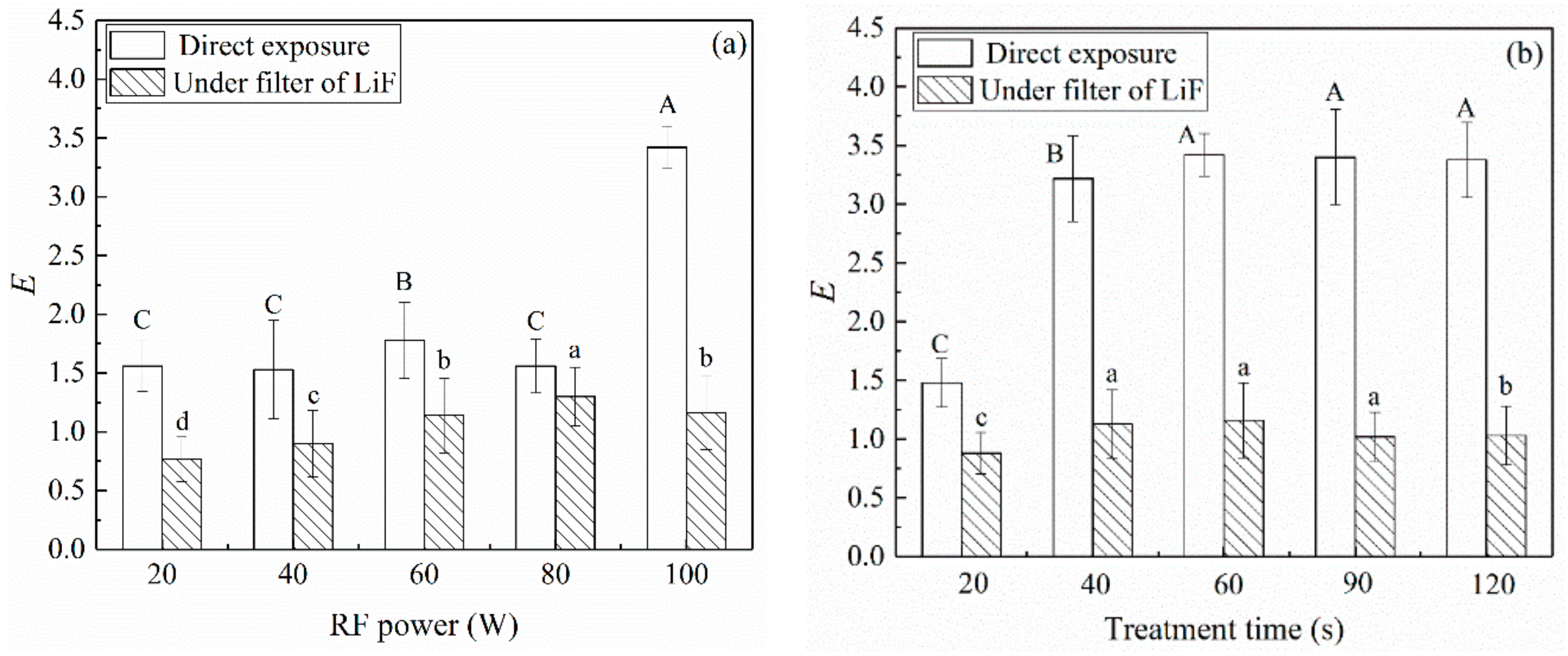
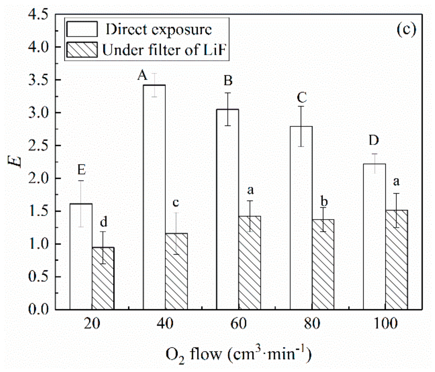
Figure 3.
DNA agarose electrophoresis of E. coil (1) control; (2) UV radiation individual generated by plasma discharge; (3) plasma total (under 100 W power, 60 s time, 40 cm3·min−1 gas flow rate and 45 Pa pressure).
Figure 3.
DNA agarose electrophoresis of E. coil (1) control; (2) UV radiation individual generated by plasma discharge; (3) plasma total (under 100 W power, 60 s time, 40 cm3·min−1 gas flow rate and 45 Pa pressure).
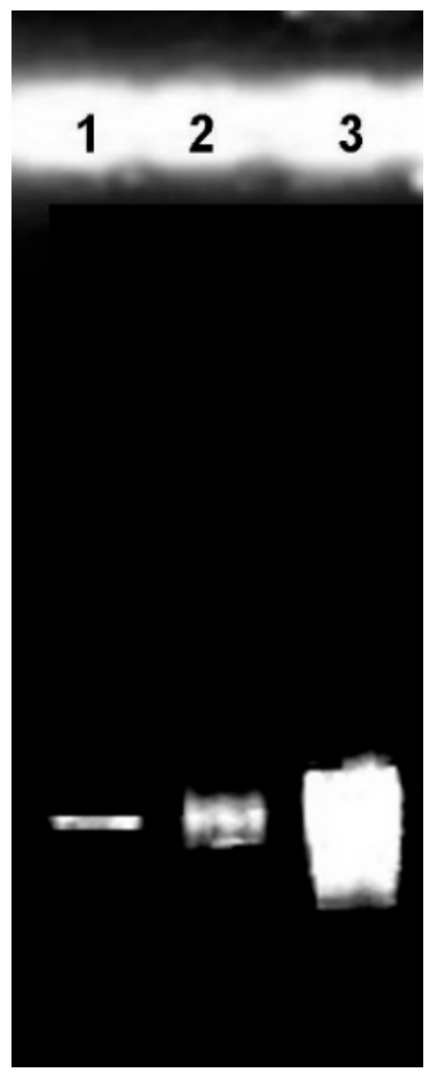
Figure 4.
Quantity of protein leakage under different treatment conditions. (a) Time 60 s, O2 flow 40 cm3·min−1, Pressure 45 Pa; (b) Power 100 W, O2 flow 40 cm3·min−1, Pressure 45 Pa; (c) Power 100 W, Time 60 s, Pressure 45 Pa. Capital letters A–E represent statistical significance.
Figure 4.
Quantity of protein leakage under different treatment conditions. (a) Time 60 s, O2 flow 40 cm3·min−1, Pressure 45 Pa; (b) Power 100 W, O2 flow 40 cm3·min−1, Pressure 45 Pa; (c) Power 100 W, Time 60 s, Pressure 45 Pa. Capital letters A–E represent statistical significance.
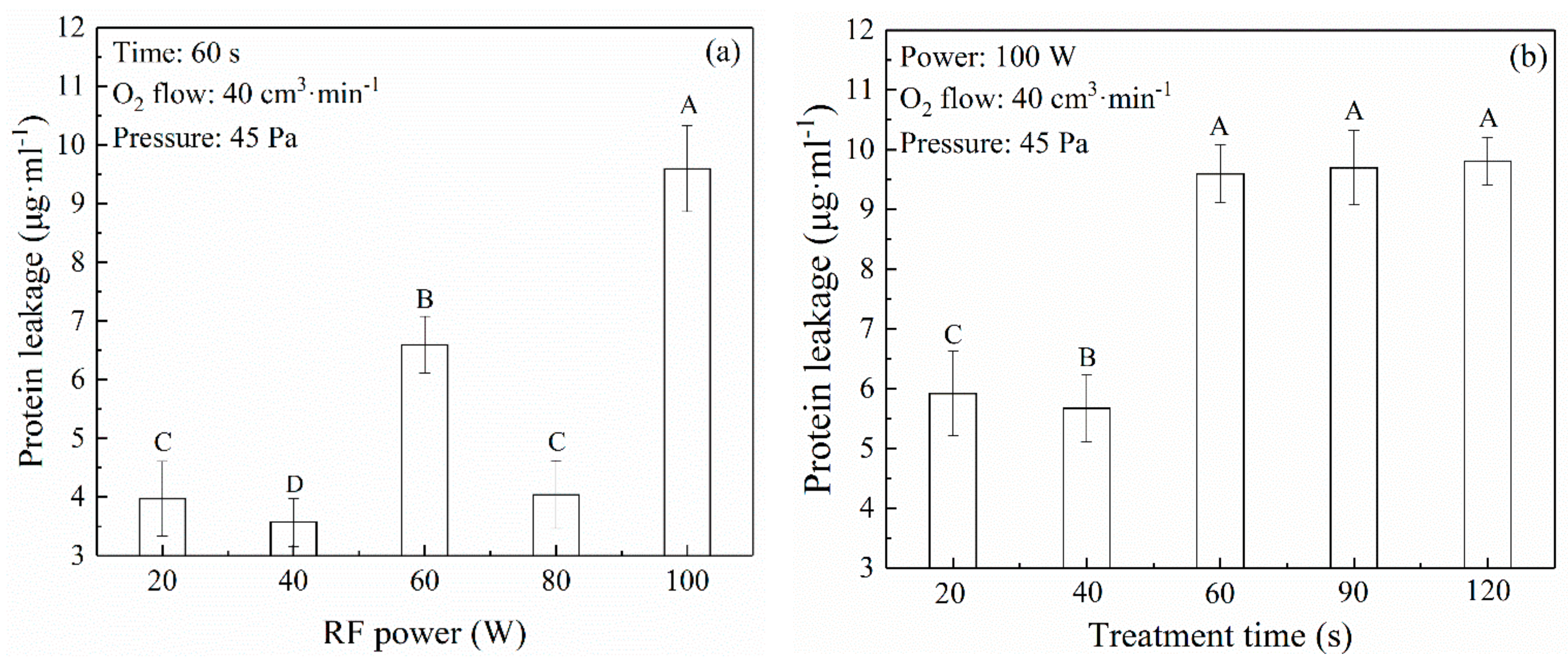

Figure 5.
SEM pictures of E. coil (a) control; (b) treated under 100 W power, 60 s time, 40 cm3·min−1 gas flow rate and 45 Pa pressure.
Figure 5.
SEM pictures of E. coil (a) control; (b) treated under 100 W power, 60 s time, 40 cm3·min−1 gas flow rate and 45 Pa pressure.

Figure 6.
Changes of malondialdehyde (MDA) concentration with increasing the plasma treatment time.
© 2019 by the authors. Licensee MDPI, Basel, Switzerland. This article is an open access article distributed under the terms and conditions of the Creative Commons Attribution (CC BY) license (http://creativecommons.org/licenses/by/4.0/).
Share and Cite
MDPI and ACS Style
Liu, H.; Feng, X.; Ma, X.; Xie, J.; He, C. Dry Bio-Decontamination Process in Reduced-Pressure O2 Plasma. Appl. Sci. 2019, 9, 1933. https://doi.org/10.3390/app9091933
AMA Style
Liu H, Feng X, Ma X, Xie J, He C. Dry Bio-Decontamination Process in Reduced-Pressure O2 Plasma. Applied Sciences. 2019; 9(9):1933. https://doi.org/10.3390/app9091933
Chicago/Turabian StyleLiu, Hongxia, Xinxin Feng, Xin Ma, Jinzhuo Xie, and Chi He. 2019. "Dry Bio-Decontamination Process in Reduced-Pressure O2 Plasma" Applied Sciences 9, no. 9: 1933. https://doi.org/10.3390/app9091933
Note that from the first issue of 2016, this journal uses article numbers instead of page numbers. See further details here.