Fresh Frozen Allogeneic Bone Block in Maxillary Sinus Floor Elevation: Histomorphometric Analysis of a Bone Specimen Retrieved 15 Years after Grafting Procedure

 ,
,  , , ,
, , ,  and
and 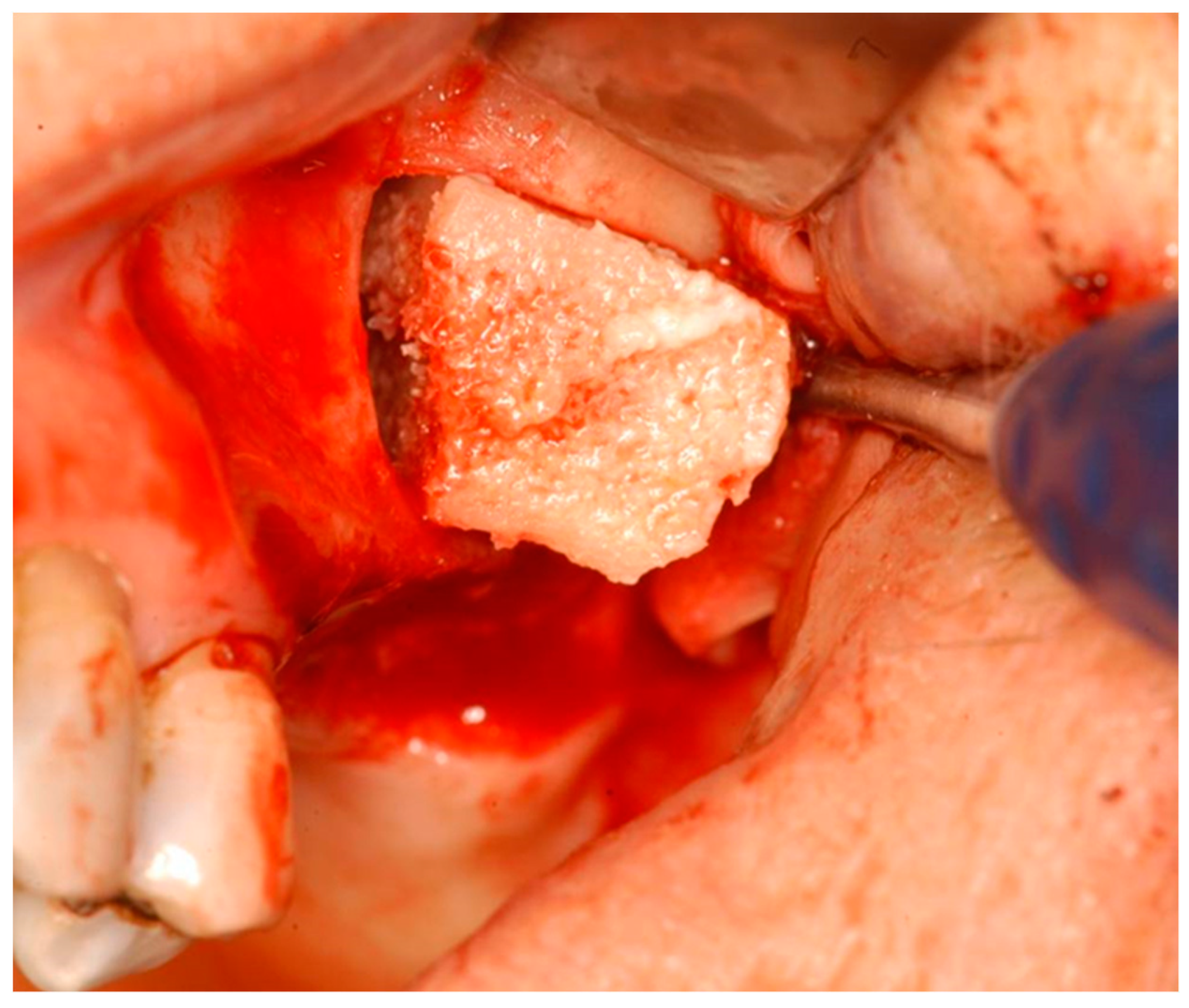
Abstract
:1. Introduction
2. Materials and Methods
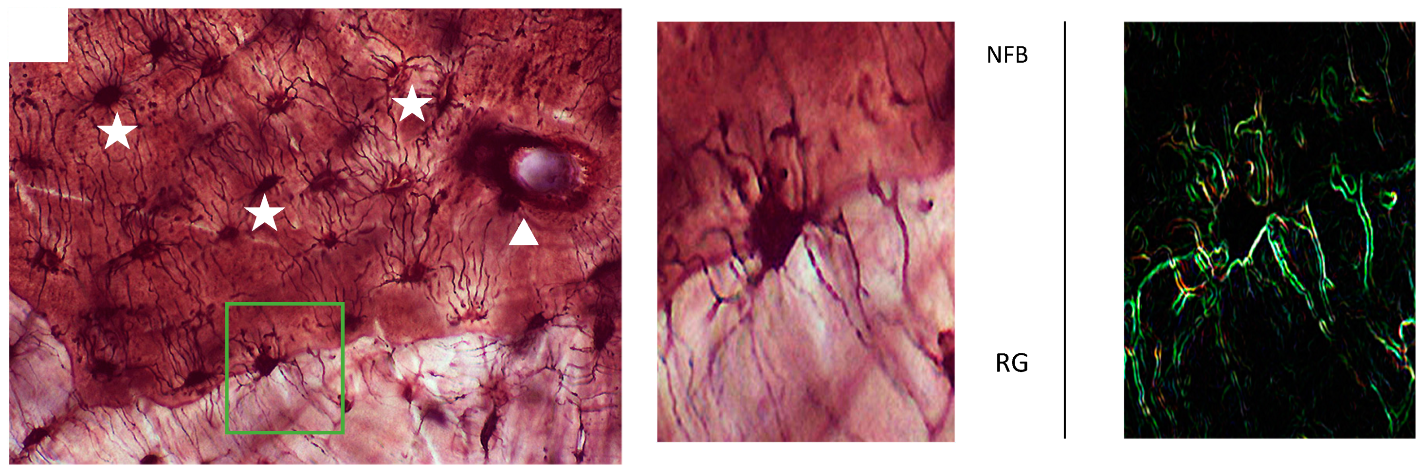
2.1. Specimen Processing and Histologic Analysis
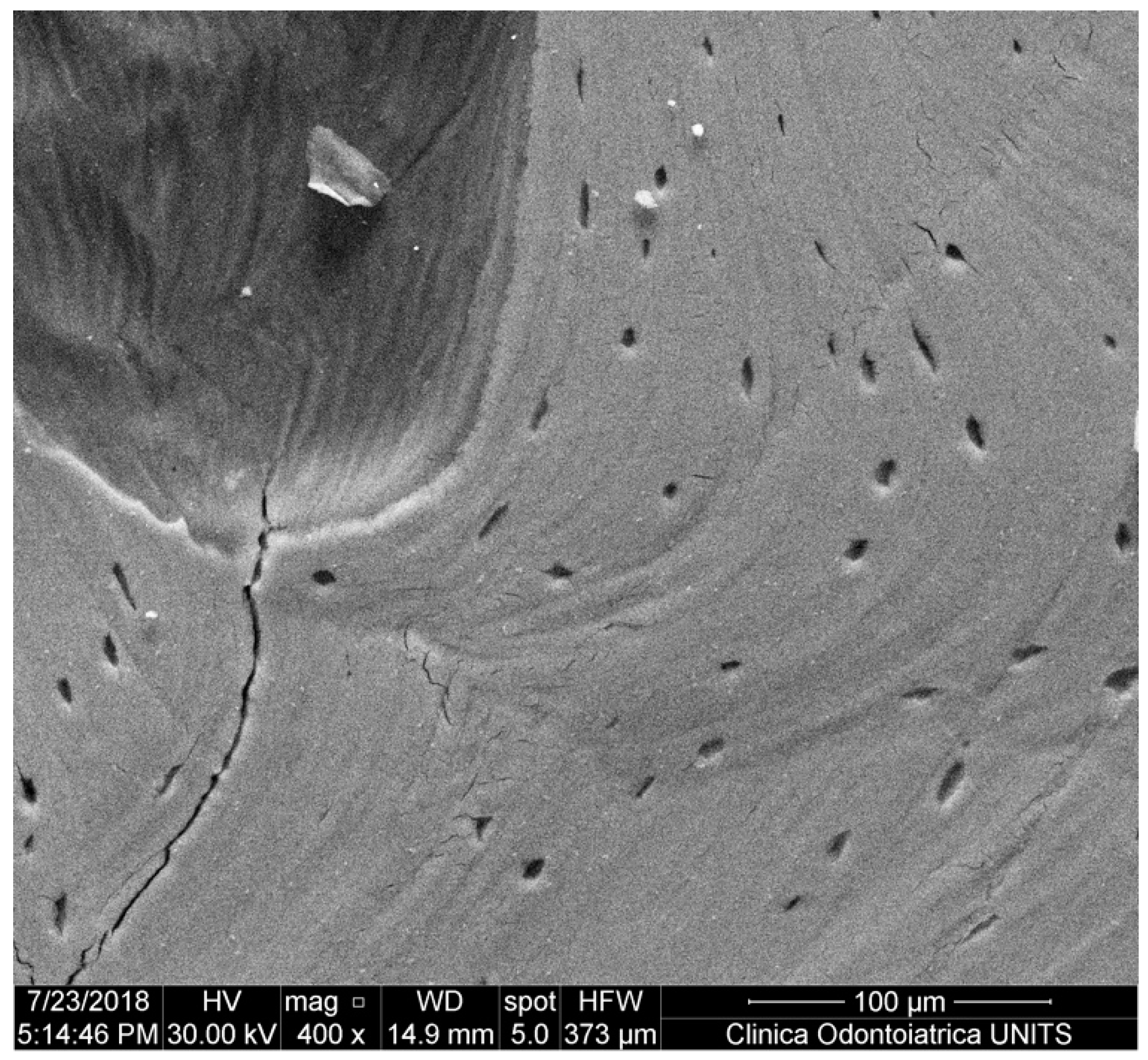
2.2. Scanning Electron Microscopy (SEM)
3. Results
4. Discussion
5. Conclusions
Author Contributions
Funding
Conflicts of Interest
References
- Boyne, P.J.; James, R.A. Grafting of the maxillary sinus floor with autogenous marrow and bone. J. Oral Surg. 1980, 38, 613–616. [Google Scholar] [PubMed]
- Tatum, H., Jr. Maxillary and sinus implant reconstructions. Dent. Clin. N. Am. 1986, 30, 207–229. [Google Scholar]
- Stacchi, C.; Vercellotti, T.; Toschetti, A.; Speroni, S.; Salgarello, S.; Di Lenarda, R. Intraoperative complications during sinus floor elevation using two different ultrasonic approaches: A two-center, randomized, controlled clinical trial. Clin. Implant Dent. Relat. Res. 2015, 17 (Suppl. 1), e117–e125. [Google Scholar] [CrossRef] [PubMed]
- Stacchi, C.; Andolsek, F.; Berton, F.; Perinetti, G.; Navarra, C.O.; Di Lenarda, R. Intraoperative Complications During Sinus Floor Elevation with Lateral Approach: A Systematic Review. Int. J. Oral Maxillofac. Implants 2017, 32, e107–e118. [Google Scholar] [CrossRef] [PubMed]
- Stacchi, C.; Lombardi, T.; Cusimano, P.; Berton, F.; Lauritano, F.; Cervino, G.; Di Lenarda, R.; Cicciù, M. Bone Scrapers Versus Piezoelectric Surgery in the Lateral Antrostomy for Sinus Floor Elevation. J. Craniofac. Surg. 2017, 28, 1191–1196. [Google Scholar] [CrossRef]
- Stacchi, C.; Lombardi, T.; Ottonelli, R.; Berton, F.; Perinetti, G.; Traini, T. New bone formation after transcrestal sinus floor elevation was influenced by sinus cavity dimensions: A prospective histologic and histomorphometric study. Clin. Oral Implants Res. 2018, 29, 465–479. [Google Scholar] [CrossRef]
- Thoma, D.S.; Cha, J.K.; Jung, U.W. Treatment concepts for the posterior maxilla and mandible: Short implants versus long implants in augmented bone. J. Periodontal Implant Sci. 2017, 47, 2–12. [Google Scholar] [CrossRef] [PubMed]
- Ravidà, A.; Wang, I.C.; Barootchi, S.; Askar, H.; Tavelli, L.; Gargallo-Albiol, J.; Wang, H.L. Meta-analysis of randomized clinical trials comparing clinical and patient-reported outcomes between extra-short (≤6 mm) and longer (≥10 mm) implants. J. Clin. Periodontol. 2019, 46, 118–142. [Google Scholar] [CrossRef] [PubMed]
- Stacchi, C.; Orsini, G.; Di Iorio, D.; Breschi, L.; Di Lenarda, R. Clinical, histologic, and histomorphometric analyses of regenerated bone in maxillary sinus augmentation using fresh frozen human bone allografts. J. Periodontol. 2008, 79, 1789–1796. [Google Scholar] [CrossRef] [PubMed]
- Laino, L.; Troiano, G.; Giannatempo, G.; Graziani, U.; Ciavarella, D.; Dioguardi, M.; Lo Muzio, L.; Lauritano, F.; Cicciù, M. Sinus Lift Augmentation by Using Calcium Sulphate. A Retrospective 12 Months Radiographic Evaluation Over 25 Treated Italian Patients. Open Dent. J. 2015, 9, 414–419. [Google Scholar] [CrossRef] [PubMed]
- Starch-Jensen, T.; Schou, S. Maxillary Sinus Membrane Elevation with Simultaneous Installation of Implants Without the Use of a Graft Material: A Systematic Review. Implant Dent. 2017, 26, 621–633. [Google Scholar] [CrossRef]
- Starch-Jensen, T.; Mordenfeld, A.; Becktor, J.P.; Jensen, S.S. Maxillary Sinus Floor Augmentation with Synthetic Bone Substitutes Compared with Other Grafting Materials: A Systematic Review and Meta-analysis. Implant Dent. 2018, 27, 363–374. [Google Scholar] [CrossRef]
- Turco, G.; Porrelli, D.; Marsich, E.; Vecchies, F.; Lombardi, T.; Stacchi, C.; Di Lenarda, R. Three-Dimensional Bone Substitutes for Oral and Maxillofacial Surgery: Biological and Structural Characterization. J. Funct. Biomater. 2018, 9, 62. [Google Scholar] [CrossRef]
- Cicciù, M.; Cervino, G.; Herford, A.S.; Famà, F.; Bramanti, E.; Fiorillo, L.; Lauritano, F.; Sambataro, S.; Troiano, G.; Laino, L. Facial Bone Reconstruction Using both Marine or Non-Marine Bone Substitutes: Evaluation of Current Outcomes in a Systematic Literature Review. Mar. Drugs 2018, 16, 27. [Google Scholar] [CrossRef]
- Busenlechner, D.; Huber, C.D.; Vasak, C.; Dobsak, A.; Gruber, R.; Watzek, G. Sinus augmentation analysis revised: The gradient of graft consolidation. Clin. Oral Implants Res. 2009, 20, 1078–1083. [Google Scholar] [CrossRef]
- Avila, G.; Wang, H.L.; Galindo-Moreno, P.; Misch, C.E.; Bagramian, R.A.; Rudek, I.; Benavides, E.; Moreno-Riestra, I.; Braun, T.; Neiva, R. The influence of the bucco-palatal distance on sinus augmentation outcomes. J. Periodontol. 2010, 81, 1041–1050. [Google Scholar] [CrossRef]
- Soardi, C.M.; Spinato, S.; Zaffe, D.; Wang, H.L. Atrophic maxillary floor augmentation by mineralized human bone allograft in sinuses of different size: An histologic and histomorphometric analysis. Clin. Oral Implants Res. 2011, 22, 560–566. [Google Scholar] [CrossRef]
- Spinato, S.; Bernardello, F.; Galindo-Moreno, P.; Zaffe, D. Maxillary sinus augmentation by crestal access: A retrospective study on cavity size and outcome correlation. Clin. Oral Implants Res. 2015, 26, 1375–1382. [Google Scholar] [CrossRef]
- Monje, A.; Catena, A.; Monje, F.; Gonzalez-García, R.; Galindo-Moreno, P.; Suarez, F.; Wang, H.L. Maxillary sinus lateral wall thickness and morphologic patterns in the atrophic posterior maxilla. J. Periodontol. 2014, 85, 676–682. [Google Scholar] [CrossRef]
- Lombardi, T.; Stacchi, C.; Berton, F.; Traini, T.; Torelli, L.; Di Lenarda, R. Influence of Maxillary Sinus Width on New Bone Formation After Transcrestal Sinus Floor Elevation: A Proof-of-Concept Prospective Cohort Study. Implant Dent. 2017, 26, 209–216. [Google Scholar] [CrossRef]
- Pardiñas López, S.; Froum, S.; Khouly, I. Histomorphometric Analysis of a Biopsy Harvested 10 Years after Maxillary Sinus Augmentation with Anorganic Bovine Bone Matrix and Plasma Rich in Growth Factors: A Case Report. Implant Dent. 2015, 24, 480–486. [Google Scholar] [CrossRef]
- Ayna, M.; Açil, Y.; Gulses, A. Fate of a Bovine-Derived Xenograft in Maxillary Sinus Floor Elevation after 14 Years: Histologic and Radiologic Analysis. Int. J. Periodontics Restorative Dent. 2015, 35, 541–547. [Google Scholar] [CrossRef] [PubMed]
- Mordenfeld, A.; Hallman, M.; Johansson, C.B.; Albrektsson, T. Histological and histomorphometrical analyses of biopsies harvested 11 years after maxillary sinus floor augmentation with deproteinized bovine and autogenous bone. Clin. Oral Implants Res. 2010, 21, 961–970. [Google Scholar] [CrossRef]
- Galindo-Moreno, P.; Hernández-Cortés, P.; Mesa, F.; Carranza, N.; Juodzbalys, G.; Aguilar, M.; O’Valle, F. Slow resorption of anorganic bovine bone by osteoclasts in maxillary sinus augmentation. Clin. Implant Dent. Relat. Res. 2013, 15, 858–866. [Google Scholar] [CrossRef]
- Di Stefano, D.A.; Gastaldi, G.; Vinci, R.; Polizzi, E.M.; Cinci, L.; Pieri, L.; Gherlone, E. Bone Formation Following Sinus Augmentation with an Equine-Derived Bone Graft: A Retrospective Histologic and Histomorphometric Study with 36-Month Follow-up. Int. J. Oral Maxillofac. Implants 2016, 31, 406–412. [Google Scholar] [CrossRef]
- Sentineri, R.; Lombardi, T.; Berton, F.; Stacchi, C. Laurell-Gottlow suture modified by Sentineri for tight closure of a wound with a single line of sutures. Br. J. Oral Maxillofac. Surg. 2016, 54, e18–e19. [Google Scholar] [CrossRef] [PubMed]
- Lebeau, A.; Muthmann, H.; Sendelhofert, A.; Diebold, J.; Löhrs, U. Histochemistry and Immunohistochemistry on Bone Marrow Biopsies: A Rapid Procedure for Methyl Methacrylate Pathology. Res. Pract. 1995, 191, 121–129. [Google Scholar] [CrossRef]
- Cervino, G.; Fiorillo, L.; Spagnuolo, G.; Bramanti, E.; Laino, L.; Lauritano, F.; Cicciù, M. Interface Between MTA and Dental Bonding Agents: Scanning Electron Microscope Evaluation. J. Int. Soc. Prev. Community Dent. 2017, 7, 64–68. [Google Scholar] [CrossRef] [PubMed] [Green Version]
- Carinci, F.; Brunelli, G.; Zollino, I.; Franco, M.; Viscioni, A.; Rigo, L.; Guidi, R.; Strohmenger, L. Mandibles grafted with fresh-frozen bone: An evaluation of implant outcome. Implant Dent. 2009, 18, 86–95. [Google Scholar] [CrossRef]
- Orsini, G.; Stacchi, C.; Visintini, E.; Di Iorio, D.; Putignano, A.; Breschi, L.; Di Lenarda, R. Clinical and histologic evaluation of fresh frozen human bone grafts for horizontal reconstruction of maxillary alveolar ridges. Int. J. Periodontics Restor. Dent. 2011, 31, 535–544. [Google Scholar]
- Silva, E.R.; Ferraz, E.P.; Neto, E.C.; Chaushu, G.; Chaushu, L.; Xavier, S.P. Volumetric Stability of Fresh Frozen Bone Blocks in Atrophic Posterior Mandible Augmentation. J. Oral Implantol. 2017, 43, 25–32. [Google Scholar] [CrossRef]
- Dias, R.R.; Sehn, F.P.; de Santana Santos, T.; Silva, E.R.; Chaushu, G.; Xavier, S.P. Corticocancellous fresh-frozen allograft bone blocks for augmenting atrophied posterior mandibles in humans. Clin. Oral Implants Res. 2016, 27, 39–46. [Google Scholar] [CrossRef] [PubMed]
- Chen, S.Y.; Liu, Z.H.; Lu, Y.; Hu, Y.J. Application of autologous bone fresh-frozen and re-implantation in treatment of cranio-maxillofacial bone tumors: Report of 4 cases. Shanghai Kou Qiang Yi Xue 2008, 17, 545–547. [Google Scholar]
- Albanese, M.; Procacci, P.; Sancassani, G.; Nocini, P.F. Fresh-frozen human bone graft to repair defect after mandibular giant follicular cyst removal: A case report. Cell Tissue Bank. 2012, 13, 305–313. [Google Scholar] [CrossRef] [PubMed]
- Bucci, T.; Nocini, P.F. Functional Reconstruction of Nonsegmental Mandibular Defect with Fresh Frozen Bone Graft and Delayed Implants Placement. J. Craniofac. Surg. 2017, 28, 810–812. [Google Scholar] [CrossRef] [PubMed]
- Rancitelli, D.; Borgonovo, A.E.; Cicciù, M.; Re, D.; Rizza, F.; Frigo, A.C.; Maiorana, C. Maxillary Sinus Septa and Anatomic Correlation with the Schneiderian Membrane. J. Craniofac. Surg. 2015, 26, 1394–1398. [Google Scholar] [CrossRef]
- Acocella, A.; Bertolai, R.; Nissan, J.; Sacco, R. Clinical, histological and histomorphometrical study of maxillary sinus augmentation using cortico-cancellous fresh frozen bone chips. J. Craniomaxillofac. Surg. 2011, 39, 192–199. [Google Scholar] [CrossRef] [PubMed]
- Viscioni, A.; Franco, M.; Paolin, A.; Cogliati, E.; Callegari, M.; Zollino, I.; Sollazzo, V.; Carinci, F. Effectiveness of fresh frozen and cryopreserved homologue iliac crest grafts used in sinus lifting: A comparative study. Cell Tissue Bank. 2011, 12, 263–271. [Google Scholar] [CrossRef]
- De Castilho, T.R.; Tortamano, P.; Marotti, J.; de Andrade, J.C., Jr.; Chilvarquer, I.; Ximenez, M.E.; Alves, M.T. Clinical and Histomorphometric Evaluation of Fresh Frozen Bone Allograft in Sinus Lift Surgery. Clin. Implant Dent. Relat. Res. 2016, 18, 40–50. [Google Scholar] [CrossRef]
- Xavier, S.P.; Dias, R.R.; Sehn, F.P.; Kahn, A.; Chaushu, L.; Chaushu, G. Maxillary sinus grafting with autograft vs. fresh frozen allograft: A split-mouth histomorphometric study. Clin. Oral Implants Res. 2015, 26, 1080–1085. [Google Scholar] [CrossRef]
- Eastlund, T. Infectious disease transmission through cell, tissue, and organ transplantation: Reducing the risk through donor selection. Cell Transplant 1995, 4, 455–477. [Google Scholar] [CrossRef] [PubMed]
- Lo Giudice, R.; Puleio, F.; Rizzo, D.; Alibrandi, A.; Lo Giudice, G.; Centofanti, A.; Fiorillo, L.; Di Mauro, D.; Nicita, F. Comparative Investigation of Cutting Devices on Bone Blocks: An SEM Morphological Analysis. Appl. Sci. 2019, 9, 351. [Google Scholar] [CrossRef]
- Aho, A.J.; Eskola, J.; Ekfors, T.; Manner, I.; Kouri, T.; Hollmen, T. Immune responses and clinical outcome of massive human osteoarticular allografts. Clin. Orthop. Relat. Res. 1998, 346, 196–206. [Google Scholar] [CrossRef]
- Spin Neto, R.; Felipe Leite, C.; Pereira, L.A.; Marcantonio, E.; Marcantonio, E., Jr. Is peripheral blood cell balanced altered by the use of fresh frozen bone block allografts in lateral maxillary ridge augmentation? Clin. Implant Dent. Relat. Res. 2013, 15, 262–270. [Google Scholar] [CrossRef] [PubMed]
- Deluiz, D.; Santos Oliveira, L.; Ramôa Pires, F.; Reiner, T.; Armada, L.; Nunes, M.A.; Muniz Barretto Tinoco, E. Incorporation and Remodeling of Bone Block Allografts in the Maxillary Reconstruction: A Randomized Clinical Trial. Clin. Implant Dent. Relat. Res. 2017, 19, 180–194. [Google Scholar] [CrossRef]
- Danesh-Sani, S.A.; Engebretson, S.P.; Janal, M.N. Histomorphometric results of different grafting materials and effect of healing time on bone maturation after sinus floor augmentation: A systematic review and meta-analysis. J. Periodontal Res. 2017, 52, 301–312. [Google Scholar] [CrossRef] [PubMed]
- Klijn, R.J.; Meijer, G.J.; Bronkhorst, E.M.; Jansen, J.A. A meta-analysis of histomorphometric results and graft healing time of various biomaterials compared to autologous bone used as sinus floor augmentation material in humans. Tissue Eng. Part B Rev. 2010, 16, 493–507. [Google Scholar] [CrossRef]
- Shanbhag, S.; Shanbhag, V.; Stavropoulos, A. Volume changes of maxillary sinus augmentations over time: A systematic review. Int. J. Oral Maxillofac. Implants 2014, 29, 881–892. [Google Scholar] [CrossRef]
- Sohn, D.S.; Bae, M.S.; Choi, B.J.; An, K.M.; Shin, H.I. Efficacy of demineralized bone matrix paste for maxillary sinus augmentation: A histologic and clinical study in humans. Oral Surg. Oral Med. Oral Pathol. Oral Radiol. Endod. 2009, 108, e30–e35. [Google Scholar] [CrossRef]
- Schwartz, Z.; Mellonig, J.T.; Carnes, D.L., Jr.; de la Fontaine, J.; Cochran, D.L.; Dean, D.D.; Boyan, B.D. Ability of commercial demineralized freeze-dried bone allograft to induce new bone formation. J. Periodontol. 1996, 67, 918–926. [Google Scholar] [CrossRef]
- Maridati, P.; Stoffella, E.; Speroni, S.; Cicciu, M.; Maiorana, C. Alveolar antral artery isolation during sinus lift procedure with the double window technique. Open Dent. J. 2014, 8, 95–103. [Google Scholar] [CrossRef] [PubMed]
- Albert, A.; Leemrijse, T.; Druez, V.; Delloye, C.; Cornu, O. Are bone autografts still necessary in 2006? A three-year retrospective study of bone grafting. Acta Orthop. Belg. 2006, 72, 734–740. [Google Scholar] [PubMed]
- Bernardi, S.; Mummolo, S.; Ciavarelli, L.M.; Li Vigni, M.; Continenza, M.A.; Marzo, G. Cone beam computed tomography investigation of the antral artery anastomosis in a population of Central Italy. Folia Morphol. 2016, 75, 149–153. [Google Scholar] [CrossRef] [PubMed]
- Varela-Centelles, P.; Loira-Gago, M.; Gonzalez-Mosquera, A.; Seoane-Romero, J.M.; Garcia-Martin, J.M.; Seoane, J. Distance of the alveolar antral artery from the alveolar crest. Related factors and surgical considerations in sinus floor elevation. Med. Oral Patol. Oral Cir. Bucal 2016, 21, e758–e765. [Google Scholar] [CrossRef] [PubMed]
- Favato, M.N.; Vidigal, B.C.; Cosso, M.G.; Manzi, F.R.; Shibli, J.A.; Zenóbio, E.G. Impact of human maxillary sinus volume on grafts dimensional changes used in maxillary sinus augmentation: A multislice tomographic study. Clin. Oral Implants Res. 2015, 26, 1450–1455. [Google Scholar] [CrossRef] [PubMed]
- Poli, P.P.; Beretta, M.; Cicciù, M.; Maiorana, C. Alveolar ridge augmentation with titanium mesh. A retrospective clinical study. Open Dent. J. 2014, 8, 148–158. [Google Scholar] [CrossRef]
- Herford, A.S.; Cicciù, M.; Eftimie, L.F.; Miller, M.; Signorino, F.; Famà, F.; Cervino, G.; Lo Giudice, G.; Bramanti, E.; Lauritano, F.; et al. rhBMP-2 applied as support of distraction osteogenesis: A split-mouth histological study over nonhuman primates mandibles. Int. J. Clin. Exp. Med. 2016, 9, 17187–17194. [Google Scholar]
- Bramanti, E.; Matacena, G.; Cecchetti, F.; Arcuri, C.; Cicciù, M. Oral health-related quality of life in partially edentulous patients before and after implant therapy: A 2-year longitudinal study. Oral Implantol. 2013, 6, 37–42. [Google Scholar] [CrossRef]
- Stähli, A.; Strauss, F.J.; Gruber, R. The use of platelet-rich plasma to enhance the outcomes of implant therapy: A systematic review. Clin. Oral Implants Res. 2018, 29 (Suppl. 18), 20–36. [Google Scholar] [CrossRef]
- Lo Giudice, G.; Iannello, G.; Terranova, A.; Lo Giudice, R.; Pantaleo, G.; Cicciù, M. Transcrestal Sinus Lift Procedure Approaching Atrophic Maxillary Ridge: A 60-Month Clinical and Radiological Follow-Up Evaluation. Int. J. Dent. 2015, 2015, 261652. [Google Scholar] [CrossRef]
- Abdalla, R.I.B.; Alqutaibi, A.Y.; Kaddah, A. Does the adjunctive use of platelet-rich plasma to bone graft during sinus augmentation reduce implant failure and complication? Systematic review and meta-analysis. Quintessence Int. 2018, 49, 139–146. [Google Scholar] [CrossRef] [PubMed]
- Matarese, G.; Ramaglia, L.; Fiorillo, L.; Cervino, G.; Lauritano, F.; Isola, G. Implantology and periodontal disease: The panacea to problem solving? Open Dent. J. 2017, 11, 460–465. [Google Scholar] [CrossRef] [PubMed]
- Fiorillo, L.; Cervino, G.; Herford, A.S.; Lauritano, F.; D’Amico, C.; Lo Giudice, R.; Laino, L.; Troiano, G.; Crimi, S.; Cicciù, M. Interferon Crevicular Fluid Profile and Correlation with Periodontal Disease and Wound Healing: A Systemic Review of Recent Data. Int. J. Mol. Sci. 2018, 19, 1908. [Google Scholar] [CrossRef] [PubMed]


© 2019 by the authors. Licensee MDPI, Basel, Switzerland. This article is an open access article distributed under the terms and conditions of the Creative Commons Attribution (CC BY) license (http://creativecommons.org/licenses/by/4.0/).
Share and Cite
Stacchi, C.; Berton, F.; Fiorillo, L.; Nicolin, V.; Lombardi, T.; Cicciù, M.; Di Lenarda, R. Fresh Frozen Allogeneic Bone Block in Maxillary Sinus Floor Elevation: Histomorphometric Analysis of a Bone Specimen Retrieved 15 Years after Grafting Procedure. Appl. Sci. 2019, 9, 1119. https://doi.org/10.3390/app9061119
Stacchi C, Berton F, Fiorillo L, Nicolin V, Lombardi T, Cicciù M, Di Lenarda R. Fresh Frozen Allogeneic Bone Block in Maxillary Sinus Floor Elevation: Histomorphometric Analysis of a Bone Specimen Retrieved 15 Years after Grafting Procedure. Applied Sciences. 2019; 9(6):1119. https://doi.org/10.3390/app9061119
Chicago/Turabian StyleStacchi, Claudio, Federico Berton, Luca Fiorillo, Vanessa Nicolin, Teresa Lombardi, Marco Cicciù, and Roberto Di Lenarda. 2019. "Fresh Frozen Allogeneic Bone Block in Maxillary Sinus Floor Elevation: Histomorphometric Analysis of a Bone Specimen Retrieved 15 Years after Grafting Procedure" Applied Sciences 9, no. 6: 1119. https://doi.org/10.3390/app9061119