Review

Bone Tissue Engineering Using Human Cells: A Comprehensive Review on Recent Trends, Current Prospects, and Recommendations

1
Department of Modern Sciences and Technologies, School of Medicine, Mashhad University of Medical Sciences, Mashhad 917794-8564, Iran
2
Bioengineering Research Group, Nanotechnology and Advanced Materials Department, Materials and Energy Research Center (MERC), Tehran 14155-4777, Iran
3
Cellular and Molecular Research Center, Iran University of Medical Sciences, Tehran, Iran
4
Department of Tissue Engineering & Regenerative Medicine, Faculty of Advanced Technologies in Medicine, Iran University of Medical Sciences (IUMS), Tehran 144961-4535, Iran
5
Department of Medical Genetics, School of Medicine, Tehran University of Medical Sciences, Tehran 14155-6447, Iran
6
Medical Genetics Network (MeGeNe), Universal Scientific Education and Research Network (USERN), Tehran, Iran
7
Department of Biomaterials Science, School of Dentistry, Dankook University, Cheonan 31116, Korea
8
Institute of Tissue Regeneration Engineering (ITREN), Dankook University, Cheonan 31116, Korea
9
Department of Nanobiomedical Science & BK21 PLUS NBM Global Research Center for Regenerative Medicine Research Center, Dankook University, Cheonan 31116, Korea
10
Institute of Materials Physics and Engineering, Applied Science and Technology Department, Politecnico di Torino, Corso Duca degli Abruzzi 24, 10129 Torino, Italy
*
Authors to whom correspondence should be addressed.
Appl. Sci. 2019, 9(1), 174; https://doi.org/10.3390/app9010174
Submission received: 27 October 2018 / Revised: 19 December 2018 / Accepted: 20 December 2018 / Published: 5 January 2019
(This article belongs to the Section Applied Biosciences and Bioengineering)

Abstract

:
The use of proper cells for bone tissue engineering remains a major challenge worldwide. Cells play a pivotal role in the repair and regeneration of the bone tissue in vitro and in vivo. Currently, a large number of differentiated (somatic) and undifferentiated (stem) cells have been used for bone reconstruction alone or in combination with different biomaterials and constructs (e.g., scaffolds). Although the results of the cell transplantation without any supporting or adjuvant material have been very effective with regard to bone healing. Recent advances in bone scaffolding are now becoming new players affecting the osteogenic potential of cells. In the present study, we have critically reviewed all the currently used cell sources for bone reconstruction and discussed the new horizons that are opening up in the context of cell-based bone tissue engineering strategies.

Graphical Abstract

1. Introduction

Bone tissue lesions are among the most common injuries of the human body, which affect the quality of millions of people worldwide [1]. It has been estimated that the prevalence of bone lesions requiring treatments will double during a period between 2012 and 2020 due to an aging population, traumas, congenital genetic abnormalities, and obesity [2]. As such, the need for bone replacement increases dramatically worldwide and becomes a key scientific, socio-economic, and clinical challenge. Recently, the use of tissue engineering (TE)-based strategies has appealed greatly for healing the damaged bone tissue. The fundamentals of TE include three principles including cells, biomaterials, and growth factors, which are necessary for generating the regenerated functional tissue [3]. Most of the strategies applied for BTE are based on bone formation through intramembranous ossification (IO), which commonly leads to poor vascularization and limited-area bone regeneration. Therefore, bone regeneration via a cartilage-mediated process similar to endochondral ossification (EO) has recently attracted greater attention in BTE, especially for long bones reconstruction, to overcome the limitations of the IO approach [4,5].
As one of the main building blocks of TE field, cells have attracted increasing attention for in vitro and in vivo bone regeneration due to their substantial role in the acceleration of the healing process. For example, cells are able to improve the healing process by secreting numerous cytokines including VEGF, HGF, bFGF, and IL-6 [6,7]. Moreover, cells are involved in the secretion of the extracellular matrix (ECM), which provides necessary physical scaffolding for the cellular constituents as well as initiates essential biochemical and biomechanical cues required for tissue morphogenesis, differentiation, and homeostasis [8]. There are some well-recognized and accepted criteria to use in cells for in vivo bone repair and regeneration including ease availability, abundance, a lack of the induced host immune response, and incorporate the proper potential for osteogenic differentiation [9,10]. Currently, various types of both differentiated (e.g. osteoblasts) and undifferentiated cells (embryonic, fetal, and adult stem cells) are used for treating damaged bone tissue. It was previously well documented that the nature and commitment of the transplanted cells significantly control the type of ossification in vivo [11,12,13]. For example, Tortelli et al. showed that the activation of an endochondral ossification process happened when they transplanted mesenchymal stem cells (MSCs)-seeded porous ceramic scaffolds in immunocompromised mice, while transplantation of osteoblasts (OB)-seeded scaffolds led to an intramembranous ossification [14].
Appropriate cell types are also used with a “probing function” to test the in vitro biocompatibility of biomaterials and scaffolds for possible use in TE approaches. In fact, correct strategies for the selection of implantable constructs for bone healing rely on the initial evaluation of biomaterials using different bone cells, i.e., osteoblasts and osteoclasts [15]. The cell fate in vivo will be determined by many biomaterial-related features, such as surface texture (e.g. micro-roughness and nano-roughness [16,17]), the size of particles [18], dissolution kinetics, and release of ionic species that may elicit specific biochemical and genetic responses in cells and tissues [19,20,21,22,23].
It is suggested that the biomedical scientists take measures on the cell source used for BTE strategies to get better outcomes. These considerations can be summarized as follows: (1) the choice between the use of autologous or allogeneic cells, (2) the availability of the desired tissue for cells and subsequent harvesting procedure, (3) donor site morbidity, (4) the efficiency of the cell isolation procedure and final yield of harvested cells, (5) the osteogenic and angiogenic potential of cells, (6) the proliferation rate of cells, (7) the homogeneity of the isolated cells, (8) the phenotype stability and cell safety after implantation, (9) the possibility of immunogenicity, and (10) the development of quality control measures for the generation of cells and grafts [24]. Prior to the use of in vivo experiments, the harvested cells are usually expanded in tissue culture treated (TC-treated) dishes under appropriate cell culture conditions. This work results in a significant increase in the number of cells desired, which may be useful for selection and their enrichment [25] (Figure 1).
In the present study, we have reviewed and summarized a large number of previously reported studies in which different somatic and stem cells (Table 1) were used for bone tissue engineering (BTE) strategies both in vitro and in vivo. For this aim, we collected the relevant articles from the databases of Scopus, Web of Science (ISI), and PubMed. In addition, we present and discuss the general information of mammalian cells including their history, isolation procedures, properties, advantages, and disadvantages regarding BTE. The focus of the present study is to show the usability of each cell type for bone reconstructive strategies and their pros and cons based on the evidence of the comparison in vitro and in vivo osteogenic potential. The conclusion on choosing the best cell source as an ideal candidate for bone tissue repair and regeneration is tough because of the various conditions applied in experiments.

2. Differentiated Cells

With respect to BTE strategies, a couple of differentiated cells (i.e., osteoblasts and osteoclasts) were used in both in vitro and in vivo experiments. One of the proposed applications of this type of cells is related to in vitro evaluation of osteogenesis. However, it is possible to use them as cell sources (i.e., auto-graft) for in vivo applications.

2.1. Osteoblasts

2.1.1. General Information

Within the nano-microstructure and microstructure of bone, predominant types of the cell population include osteoblasts, osteoclasts, and osteocytes. Other cell types like endothelial and chondrocyte cells have an essential role in the mineralization process. All these cell types contribute to physiological, mechanical, and structural functions in bone [27]. Osteoblast lineage cells, including osteo-progenitors, osteoblasts, and osteocytes, arise from pluripotent mesenchymal progenitor cells [28]. In the bone, pluripotent progenitor cells undergo several major developmental stages. The commitment of the pro-osteoblasts to osteoblastic lineage is regulated by a set of genes including c-fos, c-myc, Ets-1, Runx2, matrix associated genes, osteoblast-specific genes, and bone remodeling genes such as Dlx5 and osterix [29,30,31]. Mature osteoblasts progressively express specific markers including type I collagen, alkaline phosphatase (ALP), osteonectin (ON), and osteocalcin (OCN) [32]. Osteoblasts are bone-forming cells present throughout vertebrate life, but their function is uppermost during the embryonic period and growth [33]. Some mature osteoblasts may die by apoptosis while some undergo quiescent flatten lining cells, or are surrounded in the ECM as osteocytes expressing the Dmp1 Marker [34]. The steps involved in osteoblast differentiation are illustrated in Figure 2.
Osteoblasts were initially characterized based on their anatomical position as a continuous cell layer that appeared on the surface part of the bone (cortical bone). Within their niche, osteoblasts are commonly adjacent to endothelial cells. The close proximity of osteoblasts to the surface part of the bone tissue suggests that they may have an important role in the bone resorption and regeneration process [36]. Since the discovery of osteoblast cells, evidence has accumulated that they are the main contributors for the production of ECM under the regulation of systematic hormones such as the parathyroid hormone (PTH) and estrogens [37,38]. Osteoblasts are mainly involved in the secretion of collagenous and non-collagenous products in bone matrix, which can act as a pattern for the mineral deposition [27].

2.1.2. Application in BTE

These fully differentiated cells have been applied for both in vitro and in vivo studies related to bone reconstruction. The cyto-compatibility (e.g., cytotoxicity and genotoxicity) of various materials in any format (powder, granule, and scaffold) is usually evaluated using these cells [39,40,41]. In addition, the osteogenic potential of different substances is carried out on osteoblasts in vitro [42].
Apart from in vitro tests, the use of primary pre-differentiated osteoblastic cells is recommended for clinical applications regarding bone repair and regeneration since they can immediately produce a bone matrix, which would accelerate the healing process. These cells are usually obtained from bone chips using enzymatic extraction and then are cultured in vitro for scaling up [43,44]. However, the restricted proliferation in vitro is considered the main obstacle regarding in vivo use of this type of cells [45]. Therefore, there are very few studies about the direct use of pre-differentiated osteoblastic cells for in vivo applications as compared to stem cells [46]. For instance, Gentleman et al. reported the different quality of bone produced by different types of cells [47] and showed that osteoblasts and adult stem cells (i.e., MSCs) produced biomimetic tissue with many of the hallmarks of native bone, while embryonic stem cells could not present this performance.

2.2. Osteoclasts

2.2.1. General Information

Osteoclasts are multinucleate cells that have the ability to degrade mineralized bone tissue and are vital for calcium homeostasis, bone remodeling, and repair. Activated osteoclasts flattened and attached to the bone surface with αVβ3 integrin. The ruffled edge of osteoclasts prepares a wide surface area of the specified membrane through which hydrolytic materials are released for natural biodegradation [48].
Osteoclasts originate from myeloid lineage cells, and their differentiation is provided by numerous osteotrophic molecules such as simvastatin (Sim) or PTH, 1,25(OH)2 vitamin D3, and IL-11, which coordinate osteoclast development via specific receptors on osteoblasts and stromal cells. Osteoclast genesis needs cell-cell interaction and communication by osteotropic molecules through specific cell membrane proteins. It has been well documented that the interaction between pro-osteoclasts and osteoblasts is mediated by RANK, which is an osteoclast precursor specific marker and RANKL on the surface of osteoblasts. This interaction leads to the differentiation of pre-osteoclasts to osteoclasts [49,50]. Degradation of the mineralized bone matrix is the main function of this cell. This includes dissolution of crystalline hydroxyapatite and proteolytic digestion of the collagenous bone matrix. The cleavage of hydroxyapatite occurs by the production of HCl. The organic bone matrix is solubilized by several proteolytic enzymes such as lysosomal cysteine proteinases and matrix metalloproteinases [51,52].

2.2.2. Application in BTE

Ideally, the materials implanted in the body should be completely degraded within a defined period. This degradation depends on both chemical dissolution (physicochemical degradation) and resorption (cellular degradation by osteoclasts) [53]. Therefore, the main use of osteoclasts is the functional evaluation of the biomaterials designed for bone regeneration applications. On this matter, Tortelli et al. investigated the interaction between bone cells (osteoblasts and osteoclasts) and a 3D resorbable porous ceramic scaffold based on silicon-stabilized tri-calcium phosphate (Skelite®) to compare it to traditional 2D cultures [54]. In another study, Karpov et al. evaluated sol-gel BGs regarding their ability to support both osteoblast and osteoclast formation from human bone marrow cells [55]. The authors identified the response of osteoclasts for this biomaterial by morphology and positive staining for tartrate-resistant acid phosphatase (TRAP) (Figure 3). Midha et al. reported that bioactive sol-gel glass foams can be remodeled by using osteoclasts, as shown by the presence of resorption pits on the scaffold struts in vitro [56]. Regarding the importance of using osteoclasts in BTE strategies, Han and Zhang clarified that the absence of osteoclasts in the experiments leads to abnormalities of in vitro bone formation, such as disorganized matrix and defective mineralization as well as reduced osteoblast population and activity [57].

3. Undifferentiated Cells (Stem Cells)

The use of stem cells in bone tissue repair and regeneration applications has become increasingly important due to their supreme properties in terms of the bone healing process. Different types of stem cells (embryonic, fetal, and adult) have been considered as suitable cell sources for bone regeneration. All the stem cells show well-defined characteristics including the potential of self-renewal and the ability to differentiate into specialized cells [59]. In the following sections, we comprehensively discuss the application of all the stem cells in BTE.

3.1. Embryonic Stem Cells (ESCs)

3.1.1. General Information

The first reports regarding the successful isolation of ESCs from the inner cell mass (ICM) of blastocysts were related to the experiments performed by two groups coordinated by Evans and Martin in 1981 [60,61]. In addition to the mouse, scientists have also succeeded in the isolation of ESCs from other species including non-human primates (like the rhesus monkey) and humans [62,63]. Although there are ethical issues about the isolation of ESCs in the case of humans [64], new approaches such as in vitro fertilization (IVF) and nuclear cloning have been developed to remove these problems [65]. Under the defined conditions, these cells can differentiate into all the three germ lines (Figure 4).
Generally, culturing ESCs requires a notable commitment for time and resources, which makes it an overwhelming process [66]. However, it is necessary to culture these cells in the most optimal conditions because the following applications would depend on the starting sources [67]. Previously, the use of mouse embryonic fibroblasts (MEFs) [68] or human feeders [69] as a feeder layer was the most common method to culture ESCs since they provide a large number of growth factors required for the robust growth of all types of ESCs. However, there are some difficulties related to MEFs including their complexity, non-defined components, and xenogeneic origin with respect to the culture of human ESCs (hESCs) [70]. Therefore, the use of the human-originated feeder layers was considered to overcome the issues associated with the MEFs. In this regard, human fetal and adult fibroblast feeders have been applied to long-term undifferentiated growth of hESCs [69]. Other adult human cells have also been used to culture and expand hESCs including the fallopian tube, the foreskin, marrow-derived stromal cells, and the uterine endometrium [71]. In total, feeder cultures are not suitable for clinical application because of the release of undefined factors into cultures. Therefore, feeder-free systems with a defined medium were developed for culturing ESCs.

3.1.2. Application in BTE

Human ESCs are a suitable source for bone repair and regeneration applications since they can differentiate to unlimited numbers of all specialized lineages of the healthy bone [73]. The possibility of derivation of osteoblasts from hESCs has been under the influence of ascorbic acid, β-glycerophosphate, and dexamethasone [74]. In addition, it is possible to generate osteogenic cell lineages from ESCs without the need to prepare embryonic bodies [75]. The potential of these cells regarding the endochondral bone regeneration in vivo has also been shown in a study conducted by Jukes et al. in 2008 [76]. However, the risk of tumor formation (tumorigenicity) in the recipients is the main drawback regarding the use of hESCs for the clinical setting. Thus, the differentiation of them into progenitors is suggested as an alternative to overcome the problem mentioned above. In this regard, De Peppo et al. produced hESCs-derived mesodermal progenitors (hES-MPs) and compared their osteogenic potential to BM-MSCs [77]. They found that transcriptional modifications occurring during hES-MP derivation lead to a gene expression profile, which is highly similar to that of hMSCs. The authors concluded that the hES-MPs with the immune privileged characteristics (low expression of HLA class II) and increased proliferation properties could serve as a valuable cell source for tissue engineering application. In another study, Taiani et al. produced murine ESCs-derived osteoblasts for using in impaired fracture healing in the mouse [78]. For this aim, they loaded the prepared cells in a 3D collagen I gel and implanted the cells into the ovariectomized fractures. The authors observed the stem cell-derived osteoblasts at the fracture site after four weeks of the implantation through in situ hybridization histochemistry. The in vivo micro-computed tomography (μCT) imaging and histology analyses confirmed that ES cell-derived osteoblasts loaded on collagen I matrix can improve bone formation and restore fracture healing in vivo.

3.2. Induced Pluripotent Stem Cells (iPSCs)

3.2.1. General Information

The iPSCs are a new type of pluripotent stem cells that are generated by reprogramming technology. Yamanaka et al. introduced this approach by developing the iPSCs using the insertion of four specific genes encoding transcription factors (Oct4, Sox2, cMyc, and Klf4) into the adult cells. The lack of ethical issues is considered as the most important advantage of these cells over the embryonic cells. Moreover, the risk of immune rejection is solved since autologous cells could be used to generate transplants. However, there are some concerns about their possibility of tumor tissue formation in vivo.
The iPSCs technology is considered as a new promise for scientists who work on novel therapies for damaged or diseased tissues. The iPSCs can be successfully expanded through the same culture protocols developed for the ESCs, which may maintain their morphology, molecular signature, and differentiation potential [79,80,81]. Under the appropriate condition, the iPSCs reprogrammed are able to differentiate into other cell types. For example, different functional stem cells such as hepatocytes [82], neural cells [83], and osteogenic cells [84] have been derived from iPSCs. In this regard, greater attention was paid toward the use of these cells in bone tissue repair and regeneration. As an illustration, Phillips et al. in 2014 reported a successful differentiation of iPSCs toward the bone tissue both in vitro and in vivo [85].

3.2.2. Applications in BTE

Compared to other cell types, the use of iPSCs for BTE applications opens a new horizon. In order to differentiate iPSCs toward osteogenic lineages, the use of protocols applied for ESCs was suggested since both cell types show similar properties [86]. One of the ways proposed for preparing iPSCs with a commitment towards osteogenic lineage is genetic manipulations. On this matter, Tashiro et al. transduced an exogenous Runx2 gene into mouse iPSCs via adenoviral vectors [87]. This gene transduction resulted in a higher production of ALP and calcium levels in the transduced cells as compared to controls. This study is considered as one of the first experiments regarding the production of iPSC-derived osteoblasts. As a novel approach, the use of some specific anti-miRNA was reported as an effective way of improving osteogenic differentiation. For instance, Okamoto et al. showed that the transfection of anti-miRNAs of 124a, 181a, 10a, 10b, 9-3p, and 19b into mouse iPSCs significantly promote the osteoblastic differentiation [88].
Culturing iPSCs in the osteogenic medium is another proposed approach for obtaining osteogenic lineages. In this regard, Kao et al. cultured iPS cells in an osteogenic medium containing low-glucose DMEM supplemented with 15% FBS, 50 μg/mL ascorbate-2-phosphate, 10 nmol/L dexamethasone, and 10 mmol/L β-glycerophosphate for two weeks [89]. They used resveratrol to promote differentiation of iPSCs into osteocyte-like cells, protect the iPSC-derived osteocyte-like cells from glucocorticoid-induced oxidative damage, and reduce the risk of tumorigenicity of iPSCs.
Even though the production of iPSC-derived osteoblast-like cells is now a common process, the in vitro and in vivo stability of the generated cell’s phenotype is even more significant. This is a very important issue when iPSCs are used in in-vivo experiments. In this regard, Bilousova et al. reported the expression of osteoblast markers (e.g., RUNX2) in the implanted mouse iPSC-derived osteoblasts together with Gelfoam sponges at 12 weeks of post-implantation [90]. Their results proved the effectiveness of appropriate scaffolds in improving the osteogenic differentiation of iPSCs and bone formation in vivo.
It has been documented that BM-MSCs have a limited differentiation potential due to their senescence in vitro. To address this problem, the use of iPSCs-derived MSCs (iMSCs) was suggested since this source provides an unlimited source of MSCs. For example, Sheyn et al. differentiated human iPSCs toward functional MSCs for use in bone repair and regeneration [91]. In order to direct differentiation of iPSCs towards MSCs, the authors exposed embryoid bodies to TGF-β for a short period. They could prepare two types of iMSCs including early and late iMSCs. The in vitro results revealed that, even though both cell types maintained their multi-differentiation potential, the early iMSCs showed higher osteogenic activity as compared to other cells (the late iMSCs and BM-MSCs). Moreover, the in vivo results indicated that the ectopic administration of the early iMSCs overexpressing BMP6 resulted in a substantial bone formation in comparison to the other groups. The authors concluded that iMSCs could exhibit a self-renewal property without tumorigenic activity and considered them as very good candidates for bone regeneration.

3.3. Fetal Stem Cells

In pregnancy, two fetal membranes, i.e., the amnion and the chorion, surround the human fetus (Figure 5). The amnion is a thin layer that closely covers the human embryo. This membrane is filled with the amniotic fluid (AF), which serves as a protective environment for the developing embryo or fetus. The chorion is the outermost fetal membrane, which exists during pregnancy between the developing fetus and mother. The fetal tissue possesses stem cells in its structures, which are remarkably different from embryonic and adult stem cells [92,93]. These cells have been defined as a distinct type of stem cells with their own properties [94]. Human fetal stem cells are described as cells from the end of the two months of conception until birth, which is isolated from the fetal tissues and have the capacity to preserve clonality, high proliferative ability, and plasticity [92]. Prior studies have noted the superiority of fetal stem cells compared to embryonic and adult stem cells in terms of safety due to the low risk of teratomas forming and transmitting infection [95]. The growing body of evidence suggests significant heterogeneity within a population of fetal stem cells, which depends on the tissue of origin [96,97]. Fetal stem cells have been identified in umbilical cord blood (UCB-MSCs, UCB-HSCs, EPC) [98,99], Wharton’s jelly (UCMSCs), amniotic fluid (AF-HSCs, AF-MSCs, and AFSCs), amniotic membrane (AECs, AMSC, and ADSCs), placenta (CMSCs and CTCs), and even maternal circulation (PAPCs) [100,101,102,103,104]. Fetal-derived stem cells have been shown to exhibit high proliferative ability to resemble embryonic stem cells [105].

3.3.1. Amniotic Fluid-Derived Stem Cells (AFSCs)

3.3.1.1. General Information

The human amniotic fluid (hAF) is a clear, yellow fluid, which has important roles for the growing fetus including facilitating the exchange of nutrients, water, and biochemical products between the mother and the fetus as well as protection of the fetus. As previously well documented, this liquid is composed of embryonic and fetal cells derived from all the three germ layers (ectoderm, mesoderm, and endoderm). Hence, hAF is currently used as a suitable source of human stem cells that may be used in different biomedical fields. Actually, the first report on the presence of progenitor cells as small, nucleated round cells (hematopoietic progenitor cells) in the hAF was published in 1993 [107]. It was later reported that there is also multilineage non-hematopoietic cells in the hAF [108]. This liquid as a novel source of MSCs with therapeutic capability was introduced to the biomedical sciences in 2003 [109]. These cells were initially named human amniotic fluid stem cells (hAFSCs) [98]. To isolate and culture these cells, about 2 mL of amniotic fluid is sufficient. In a cell culture condition, they are able to double up to 250 times without any detectable loss of chromosomal telomere length [98]. Like other MSCs, they have the potential for self-renewal and multi-lineage differentiation. The differentiation of hAFSCs into the cells from the three embryonic layers including hepatocytes (endoderm) [98], osteoblasts (mesoderm) [98], and neurocytes (ectoderm) [110] has been previously confirmed. The presence of AFSCs in the other mammals such as mice [110], rats [111], rabbits [112], sheep [113], and pigs [114] was also reported.

3.3.1.2. Applications in BTE

One of the first reports on the osteogenic potential of AFSCs is related to the original work of De Coppi et al. in 2007 [98]. The authors observed the differentiation of these cells into functional osteoblasts (mesoderm) after culturing in an osteogenic medium in vitro, which produced mineralized calcium as well as the expression and secretion of ALP (a surface marker of osteoblasts). In order to provide a proof of concept, the authors subcutaneously implanted this type of cell with or without 3D printed alginate/collagen scaffolds into outbred athymic nude (nu/nu) mice. The in vivo results revealed a highly mineralized tissue in the animals that received the cell-seeded constructs, while no sign of mineralization was observed in the animals grafted by unseeded scaffolds. Rodrigues et al., in a comparative study, evaluated the behavior of hAFSCs and BMSCs in different culture systems (2D and 3D) to determine which cell type is more suitable for BTE applications [115]. Their results showed that both cells differentiate into the osteogenic cell lineage in the 2D culturing environment, which was confirmed by the production of a mineralized ECM. However, enhanced mineralization of the ECM was observed in AFSCs when compared to BMSCs. In contrast, the less mineralized ECM was observed in the AFSC scaffolds in comparison to BMSCs constructs, when cultured in the 3D system (starch and poly (e-caprolactone) (SPCL) scaffolds). The assessment of expression patterns of bone-related markers (e.g., ALP activity and RUNX-2 expression) and different timing of differentiation revealed that cell origin and the culture environment have remarkable effects on the differentiation process into the osteogenic lineage. With respect to some priorities such as higher proliferation, the authors stated that AFSCs could be a good alternative to BMSCs for BTE applications.

3.3.2. Amniotic Membrane Stem Cells (AMSCs)

3.3.2.1. General Information

Human amniotic membrane (hAM) or amnion as the innermost layer of the fetal membranes is an avascular stromal matrix. This membrane has been widely used in tissue engineering based-approaches for healing different tissues including eyes [116], skin [117], teeth [118], and the cardiovascular system [119]. Additionally, the hAM is clinically used as a tissue with therapeutic possibilities for repairing different burns [120], leg ulcers [121], oral and maxillofacial disorders [122], and stroke [123]. Histologically, the amniotic membrane is composed of five distinct layers including an epithelial cell monolayer, an acellular basement membrane layer, a compact layer, a mesenchymal cell layer, and a spongy layer [124]. HAM as a stem cell source benefits from several advantages such as a good yield of cells as well as not needing invasive or expensive procedures [125]. Moreover, the use of AM-derived stem cells for TE applications does not face ethical issues. Human amniotic epithelial cells (HAECs) and human amniotic mesenchymal stromal cells (HAMSCs) are the main stem cell types isolated and characterized from the hAM. It has been well documented that both of the cells possess stemness characteristics including the ability to self-renew [126], immunomodulation [127], and differentiation into cell lineages of different germ layers [128]. The isolation procedure of these cells is fairly easy and is performed via previously proposed methods [106,129,130,131].
Recently, the use of HAMSCs together with a scaffold for tissue engineering strategies has grown with interest [132]. HAMSCs were first introduced by In’t Anker et al. as cells with the potential of osteogenic and adipogenic differentiation [93]. According to the International Society for Cellular Therapy, these cells are formally named amniotic membrane-human mesenchymal stromal cells (AM-hMSCs) [133]. In a cell culture condition, AM-hMSCs display a spindle-like shape morphology and are able to attach to tissue culture plastic [134]. In addition to human, the presence of these cells has been reported in other mammals such as cat [135], horse [136], and bovine [137].

3.3.2.2. Applications in BTE

In order to evaluate the osteogenic differentiation potential of HAM-derived cells, Dıaz-Prado et al. examined the presence of the bone-related markers in the cultured cells [138]. Their results revealed that both cell populations have similar surface expression profiles of mesenchymal progenitors (CD90, CD44, CD73, CD166, CD105, and CD29). However, the differentiation potential of hAMSCs was much greater than hAECs based on the results of Alizarin Red staining and qRT-PCR (osteopontin (OP) and ALP). Other studies also confirmed the osteogenic potential of these cells. For example, Jiawen et al. showed the in vitro and in vivo potential of hAECs regarding bone regeneration [139]. They assessed an in vitro osteogenic differentiation process of the cells via biochemical staining, qRT-PCR, and immunofluorescence staining. The in vivo osteogenesis activity of hAECs was evaluated by implantation of the cells alone or in combination with β-tricalcium phosphate (β-TCP) scaffolds. The in vitro results showed up-regulation of bone-related genes (RUNX2, osterix (Osx), ALP, collagen I, and OP) with positive biochemical staining for osteoblasts. Moreover, the up-regulation of TGF-β was observed, which is related to the epithelial-mesenchymal transformation process involved in the osteogenic differentiation of hAECs. The in vivo implantation of the hAECs-seeded scaffolds led to enhanced bone reconstruction as well as reduced the early host immune response to the implanted scaffolds.
To show the osteogenic potential hAMSCs, Rodríguez-Fuentes et al. in 2013 tried to differentiate hAMSCs into osteoblastic-like cells by using co-incubation with bovine bone matrix Nukbone® (NKB) [140]. The authors prepared the NKB disks and placed them in cell culture plates in three different conditions including (1) positive control (AM-hMSC seeded on polystyrene disk with H-DMEM supplemented with ascorbic acid, dexamethasone, and β-glycerophosphate), (2) negative control (AM-hMSC cultured on polystyrene disk in H-DMEM), and (3) AM-hMSC with H-DMEM seeded on the NKB disk as experimental group. The obtained results showed more visible signs (over-expression of bone-related genes) of osteogenic differentiation of the cells cultured on the NKB disk when compared to other groups.
In a comparative study, Si et al. investigated the osteogenic potential of three different types of stem cells including hAFMSCs, hAECs, and hBMSCs both in vitro and in vivo [141]. The obtained results revealed that there were significant differences among the cells in terms of cell morphology and proliferation and the immuno-phenotypical profile. The authors found that, although hAECs exhibited a confirmed osteoblastic differentiation capacity, hAFMSCs and hBMSCs showed a higher osteoblastic phenotype. All the cells were seeded onto β-TCP scaffolds and were subcutaneously implanted into the nude mice to further investigate the osteogenic capacity. The immuno-histochemical analysis of the samples confirmed the up-regulation of the osteogenic markers (OP and BGLAP) in all three groups (the β-TCP scaffolds seeded with hAECs, hAFMSCs, and hBMSCs) as compared to the controls, which indicates the in vivo osteogenic potential of all three cell sources. However, there was a different expression profile of genes involved in ossification among the cell types since hAFMSCs and hBMSCs show a higher osteoblastic phenotype as compared to hAECs.

3.3.3. Chorion Mesenchymal Stem Cells (Ch-MSCs)

3.3.3.1. General Information

In humans and other mammals, the chorion is the outermost fetal membrane around the embryo, which exists during pregnancy between the fetus and mother. This tissue is comprised of two layers including an outer sheet (formed by the trophoblast) and an inner sheet (formed by the somatic mesoderm). The somatic mesoderm contains fibroblast-like cells called chorion stromal cells (CSCs). These cells show characteristics of MSCs under appropriate cell culture conditions [142,143]. They have very close features with human AMCs due to their strong similarity of origin. The differentiation potential of Ch-MSCs into osteogenic, chondrogenic, and adipogenic lineages is significantly higher than MSCs derived from other sources including AM-MSCs [144]. Similar to other fetal-originated stem cells, the isolation of Ch-MSCs faces minimal ethical and legal barriers. The isolation protocol of these cells from early-term, mid-term, and end-term gestational fetal membrane includes a simple procedure in which maternal decidua was mechanically removed and followed by enzymatic digestion of trophoblast layers and reticular stromal layers with collagenase [93,106,129,131].

3.3.3.2. Applications in BTE

The biological characteristics of Ch-MSCs have been evaluated by Gonzalez et al. to show the differentiation, immunosuppressive, and angiogenic capacity of these cells [145]. Their results revealed that these cells could express the osteogenic markers after specific inductions. In addition, these cells showed an excellent immune privilege and superior angiogenic activity, which makes them ideal candidates for TE strategies. These data have also been confirmed in a recently published study, which shows the ability of Ch-MSCs to differentiate toward osteogenic lineages [146]. Moreover, Kusuma et al. evaluated the ectopic bone formation by Ch-MSCs in comparison with decidua-derived MSCs (DMSCs) [147]. The in vitro experiments showed that both types of stem cells are capable of osteogenic differentiation. The in vivo results showed that the subcutaneous implantation of bromo-deoxyuridine (BrdU)-labeled CMSCs and DMSCs together with hydroxyapatite/tricalcium phosphate particles could form the ectopic bone at eight-weeks post-transplantation in SCID mice. The authors concluded that these cells could be considered proper alternatives for BTE strategies.

3.4. Adult Stem Cells

These stem cells are tissue-resident units, which provide homeostasis and regeneration after any damage to the human body. In the damaged tissues, adult stem cells are found in the non-proliferating state in which their cell cycle is reversibly arrested (known as quiescence). In this state, their stemness is preserved by preventing precocious differentiation, which results in keeping a pool of undifferentiated cells. These tissue-specific cells have been identified in the adult tissues of bone marrow, skin, muscle, nervous system, etc. [148]. Similar to all post-natal cells, mesenchymal stem cells present in the umbilical cords of newborn babies are recognized as adult stem cells. In the following sections, we present the well-documented sources of adult stem cells used for bone reconstruction strategies and discuss their potential for bone tissue repair and regeneration.

3.4.1. Bone Marrow Stem Cells (BM-MSCs)

3.4.1.1. General Information

Bone marrow (BM) is a flexible spongy tissue inside the large bones where new blood cells are produced. Two types of BM including red marrow and yellow marrow have been identified. Red bone marrow is responsible for the production of red blood cells, platelets, and most white blood cells, while the rest of white blood cells develop in yellow marrow [149].
Two major types of stem cells can be found in BM including hemopoietic (which can produce blood cells) and stromal cells (which can produce fat, cartilage, and bone). Bone marrow stromal stem cells (BMSSCs) are postnatal stem cells, which are also known as BM-MSCs. BM-MSCs were initially identified and characterized from bone marrow by Friedenstein et al. in the 1980s through an in vitro culture and transplantation in laboratory animals [150,151,152]. These cells comprise only 0.01% of the total nucleated cells in the bone marrow. Therefore, they are usually concentrated to use in the next steps of experiments. Human BM-MSCs are routinely harvested by a simple method, i.e., aspiration from the iliac crest in humans [153]. However, the isolation procedure of these cells is different in other animals. For example, the aspiration of tibia and femur has been standardized to isolate BM-MSCs from mouse and rabbit laboratory animals [154,155]. BM-MSCs as the most universal source of MSCs are identified through some criteria including the formation of clonogenic adherent cell clusters, fibroblastic morphology, and the potential to differentiate into all the three lineages including ectoderm [156,157], mesoderm [158,159], and endoderm [160,161]. To characterize BM-MSCs, a number of positive and negative surface antigens have been recommended by the International Society for Cellular Therapy [162]. These surface markers are used to purify BM-MSCs by fluorescence-activated cell sorting (FACS) as well as the magnetic-activated cell sorting (MACS). In addition, the use of Percoll gradient centrifugation and adherence to tissue-culture treated plastics are proposed as more simple techniques to purify BM-MSCs [163]. BM-MSCs can be used as undifferentiated or fully differentiated cells for TE applications. Undifferentiated forms of BM-MSCs are obtained through the culture in defined serum-free media or pre-screened fetal bovine serum. Fully differentiated cells can be obtained through physical, chemical, and mechanical approaches exerted on BM-MSCs [164,165,166]. These cells are used for orthopedic applications in vitro and in vivo. The reasons are summarized as the rapid proliferation, induction of osteogenesis, angiogenesis, and easy differentiation into osteogenic lineages [167,168]. As a result, more research has been conducted to treat various bone disorders using BM-MSCs or BM-MSCs-seeded constructs [169].

3.4.1.2. Applications in BTE

Similar to other stem cells, this type of cells can undergo osteogenic differentiation in vitro when cultured in the presence of dexamethasone, inorganic phosphate, and ascorbic acid [170,171]. The differentiation of BM-MSCs toward osteoblastic lineage cells is regulated through various systemic hormones (e.g., estrogens) and local growth factors. The bone morphogenetic protein (BMP) family, the transforming growth factor-beta (TGF-β), and the fibroblast growth factor-2 (FGF-2) are the most well-defined factors regarding the osteogenic differentiation [172].
The therapeutic effects of BM-MSCs are associated with the secretion of large amounts of different bioactive molecules (e.g., FGF-2, Ang-1, and G-CSF), which provide a regenerative microenvironment with a trophic activity for the damaged bone tissue [173]. In addition, BM-MSCs homing to the sites of bone tissue injury is considered another factor that enhances their therapeutic effect [174].
It has been reported that only a subset of high-proliferating single colony-derived BM-MSC clones (60%) is able to form ectopic bone tissue after in vivo transplantation into immunocompromised mice [175,176]. However, there are a large number of successful bone healing procedures using these cells (direct injection or in combination with 3-D scaffolds) in both calvarial and long bones in different animal models [177,178,179].
Due to their supreme properties, BM-MSCs have been used in preclinical and clinical models for repair and regeneration of the bone [180,181]. With the aim of enhancing the total-body bone mineral content and subsequent osteogenesis in children with imperfect osteogenesis, Horwitz and colleagues conducted the first clinical trials with BM-MSCs [182]. Since then, a large number of studies have been performed on these cells for treating patients who suffer from osteoarthritis and bone defects [183].

3.4.2. Adipose Tissue-Derived Mesenchymal Stem Cells (ASCs)

3.4.2.1. General Information

The origin of adipose tissue (AT) is the mesodermal layer of the embryo and develops during pre-natal and post-natal growth. Histologically, two types of adipose tissue were identified including white adipose tissue (WAT) and brown adipose tissue (BAT). There are some morphological and functional differences between the two AT types. Compared to BAT, WAT is more abundant in the human body [184]. One of the first reports on the cell isolation from AT belongs to Rodbell et al. in the early 1960s [185]. Cellular components in AT contain adipocytes and a heterogeneous set of cell populations, i.e., the stromal vascular fraction (SVF) (Figure 6). The SVF acts as a supporting cell population for adipocytes and comprises adipose-derived stem cells (ASCs), vascular endothelial cells, vascular smooth muscle cells (VSMCs), and leukocytes. ASCs as the most important cells of AT have been isolated from both white and brown tissues with different characteristics. These cells were first formally recognized in human lipoaspirate by Zuk et al. in 2001 [186]. Among different methods, the liposuction technique is widely used to harvest ASCs from the human body due to its relative ease of execution. Furthermore, as a good point, the function of ASCs is not affected by the lipo-aspiration procedure. Apart from ASCs, a variety of terms have been applied to describe these cells including adipose-derived adult stem cells (ADAS), adipose-derived stromal cells (ADSCs), adipose-derived mesenchymal stem cells (Ad-MSCs), lipoblasts, pericytes, pre-adipocytes, and processed lipoaspirate (PLA) cells [187].
According to the International Fat Applied Technology Society (IFATS), the term of ASCs is the standard name for the isolated, plastic-adherent, multipotent cell population. The isolation procedure of ASCs is a simple process in which lipoaspirate adipose tissue is digested by collagenase (type IV), which is followed by centrifugation to separate the stromal population from the adipocytes [188]. Similar to BM-MSCs, ASCs can be purified by using well-established approaches such as FACS and MACS techniques. In the culture situation, these cells show the primary characteristics of MSCs including attachment to plastics, fibroblast-like morphology, and differentiation to different cell lineages including osteoblasts [189], chondrocytes [190], endothelial cells (ECs) [191], cardiomyocytes [192], and neural cells [193]. Similar to other MSCS, ASCs express some cell surface markers, which can be used for their characterization. Like other MSCs, ASCs serve as a “secretome” and secret regulatory proteins and growth factors in the extracellular environment [194]. Therefore, these cells contribute to angiogenesis and suppression of inflammation and, thereby, induce cell migration and proliferation in the body [195,196]. Moreover, the ease of availability, a high proliferation rate, and less discomfort and morbidities with the ASCs isolation are considered as the ratiocinations for increasing use of these cells for TE applications [187]. From an immunological point of view, ASCs are classified as immunosuppressive cells due to the lack of MHC class II antigens on their surface [197].

3.4.2.2. Applications in BTE

The ease of availability, high yield of cell isolation, and excellent proliferation rate are considered the main reasons for the use of ASCs in bone regeneration [199]. It has been shown that one gram of adipose tissue yields about 5 × 10³ stem cells, which is 500-fold greater than the number of cells obtained from one gram of bone marrow [200,201,202]. However, some studies report a lower osteogenic capacity of ASCs in comparison to BMSCs. As an illustration, Im et al. compared the osteogenic activity of ASCs versus BM-MSCs in vitro [203]. They cultured both stem cell types in osteogenic medium containing dexamethasone, β-glycerophosphate, and acid ascorbic. The results revealed that 82% and 92% of the BM-MSCs are positive cells for ALP staining after two and three weeks of culturing in the osteogenic medium, respectively. In the case of ASCs, these percentages were 28% and 45%, which confirms the higher osteogenic capacity of the BM-MSCs in comparison to ASCs.
In vitro osteogenic differentiation of ASCs may be achieved after 14 days of culturing in a conditioned medium supplemented with 1 nM dexamethasone, 2 mM b-glycerolphosphate, and 50 lM ascorbate-2-phosphate [188,204]. However, other approaches have also been documented as reliable methods for differentiating ASCs as osteoblastic-like cells (Figure 7). For example, Chou et al. clarified that the combination of recombinant human BMP-2 and ASCs can result in an improvement in the osteogenic potential in vivo [205].
The in vivo differentiation of juvenile and adult ASCs into osteogenic lineages was first reported by Cowan et al. [207]. They implanted the ASCs-seeded apatite-coated PLGA scaffolds in the critical-size defects of mice. The results of the implantation of samples after two, four, eight, and 12 weeks showed that the juvenile ASCs-seeded apatite-coated scaffolds could form new bone (25%, 80%, 70%, and 85% bone formation, respectively) at the damaged site, while less bone formation (30%, 55%, 50%, and 70% bone formation, respectively) was observed in the groups treated with the adult ASCs/scaffolds. Moreover, they evaluated the effect of apatite coating on the progression of the new bone formation. The role of the apatite coating in promoting the osteogenic capacity of the osteoblast-seeded constructs further confirms the influence of scaffold biomaterial on cell activity and fate. At the same time (2004), the first report on the clinical use of hASC–fibrin glue for treating an extensive craniofacial injury in a seven-year-old girl was published by Lendeckel et al. [208].
Evaluating the potential tumorigenic feature of ASCs before taking any clinical experiments is a necessary step. According to the literature, there is significant controversy about the effect of ASCs on tumor growth since some studies state ASCs may favor the growth of tumor cells, while other studies contradict these results [209].

3.4.3. Umbilical Cord Mesenchymal Stem Cells (UC-MSCs)

3.4.3.1. Umbilical Cord Wharton’s Jelly-Derived Mesenchymal Stem Cells (WJMSCs)

General Information
In mammals, the umbilical cord is comprised of umbilical arteries and veins surrounded by Wharton’s jelly (WJ). The term WJ was initially proposed by an English physician and anatomist in 1656 [210]. In humans, the main components of the WJ consist of glycosaminoglycans (hyaluronic acid and chondroitin sulfate) and collagen fibers. The cellular components of this gelatinous tissue include different cell types such as fibroblasts, myofibroblasts, smooth muscle cells, and a subset of stromal cells [211]. The presence of MSCs in the WJ was not determined until 2004 when fibroblast-like cells of this tissue were proven to be MSCs [212]. The WJ-MSCs can be isolated from the umbilical cord matrix, which consists of two distinct regions known as inter-vascular and sub-amnion regions. The number and nature of these cells are different from other regions of UC including peri-vascular and vascular endothelium zones [213]. Similar to the procedure of human WJ collection, the isolation of WJ-MSCs is noncontroversial and without ethical issues [214].
These cells have also been isolated from other mammals including bovine [215], horse [216], and cat [217]. Like other MSCs, WJ-MSCs possess plasticity characteristics and, thereby, may differentiate into cells with origins of ectoderm (e.g., neurons) [218], mesoderm (e.g., osteoblasts) [219], and endoderm (e.g., hepatocytes) [220]. However, a number of advantages have been proposed for these cells in comparison with other MSCs including faster proliferation rate, greater expansion capability, immunosuppressive properties, and better efficiency in terms of therapeutic characteristics [212]. Two of the main methods for isolation of the WJ-MSCs include the density gradient centrifugation approach and enzymatic digestion [221]. It has been shown that the enzymatic degradation approach is the most efficient for harvesting the WJ-MSCs. Due to the large size of the UC, the number of isolated cells is usually high (approximately ~1.5 × 10⁶ cells/cm of the umbilical cord) [222]. The WJ-MSCs with appropriate features such as immunosuppressive characteristics may be ideal candidates for TE and cell-based therapy strategies [223]. In addition, these cells may support the expansion of other stem cells to excrete various cell growth factors, chemokines, and cytokines [224,225].
Applications in BTE
WJ-MSCs are being currently considered as an alternative to BM-MSCs for bone repair and regeneration strategies. It has been previously reported that this cell type is able to give rise to osteogenic lineages after culturing in the osteogenic medium [226]. In addition to the ability to osteogenic differentiation, WJ-MSCs are ideal candidates for BTE with respect to their abundant resources, no damage procurement, and low immunogenicity. Moreover, Chen et al. showed WJ-MSCs do not undergo a malignant transformation during long-term culturing (up to 15 passages) in serum-free medium [227]. However, it has been shown that the osteogenic potential of WJ-MSCs is lower than BM-MSCs in vitro and in vivo conditions [228,229]. In order to address this issue, the genetic engineering of these cells has been proposed as an appropriate approach. On this matter, Wang et al. inserted the gene encoding Ost into WJ-MSCs to improve their osteogenic capacity [230]. They showed that transfection of Osx into the cells results in enhanced ALP activity without any adverse effects on the cell proliferation rate. The implantation of Osx-overexpressing WJ-MSCs into the subcutaneous pocket of nude mice resulted in a higher ectopic bone formation in comparison to the non-transfected WJ-MSCs after four weeks.
The use of WJ-MSCs together with 3D scaffolds has been applied for accelerating bone repair and regeneration. As illustrated, Diao et al. evaluated in vivo osteogenesis of human WJ-MSCs-seeded nano-hydroxyapatite/collagen/PLA composite (nHAC/PLA) scaffolds [231]. In order to reveal the osteogenic potential, the authors implanted the constructs in the subcutaneous region of Balb/c nude mice for four to 12 weeks. The obtained results revealed that the cell-seeded scaffolds could improve ectopic bone formation with human origin in vivo. In another study, Chen et al. implanted human WJ-MSCs-seeded macroporous calcium phosphate cement (hUCMSC-CPC) in critical-sized cranial defects in athymic rats for revealing their osteogenic potential in comparison with hBM-MSCs-seeded CPC (hUCMSC-CPC) [232]. The in vitro results showed that both cell types possess high osteogenic expressions of ALP, BGLAP, collagen I, and RUNX2. In addition, the in vivo data indicated higher bone mineral density (9% and 19% for hUCMSC-CPC and hBMSC-CPC, respectively), larger new bone area fraction (57% and 88% hUCMSC-CPC and hBMSC-CPC, respectively), and higher new blood vessel density (15% and 20% hUCMSC-CPC and hBMSC-CPC, respectively) compared to the controls (CPC alone) at 24 weeks’ post-implantation. Although hBMSC were better in terms of all the examined parameters, the authors concluded that WJ-MSCs are an ideal cell source for BTE applications, which can be used instead of hBMSCs.

3.4.3.2. Umbilical Cord Blood-Derived Mesenchymal Stem Cells (UCB-MSCs)

General Information
Umbilical cord blood (UCB) is currently recognized as a rich source of hemopoietic stem/progenitor cells. The first umbilical cord blood transplantation was performed in 1988, and the results demonstrated its therapeutic potential as a reliable cell-based therapy. Although the HSCs have been initially more widely used for various clinical situations, recently, the UCB-derived MSCs have also attracted attention for use in biomedical fields. Today, umbilical cord blood (UCB) as a part of UC is recognized as an interesting source of MSCs [233]. Like other parts of UC, the cell isolation of UCB takes advantage of the painless and non-invasive procedure without any damage to the mother or infant [234]. Up to now, a variety of methods have been proposed to collect UCB as the source of human UCB-MSCs [235]. The isolation procedure of MSCs of UCB is laborious and time-consuming, which usually results in a low rate of success. The most effective method for collecting UCB is the cannulation of the umbilical vein, which is followed by blood collection into a sterile bag containing citrate phosphate dextrose or acid citrate dextrose anti-coagulant (ACD-A) buffer [236,237]. The isolation of UCB-MSCs is performed by using Ficoll density centrifugation media at the appropriate relative centrifugal force (RCF). After centrifugation, the formed interface layer is collected and washed with PBS. Lastly, the resulting solution is centrifuged and washed (with PBS) several times and is transferred into cell culture dishes. The cells were cultured using suitable culture media for several days. By washing cells, non-adherent cells were removed, and only the adherent cells with a fibroblastic spindle-like shape (i.e., MSCs) may remain [238]. It has been previously proven that there is no significant difference between the collections obtained from Caesarean and vaginal delivery in terms of the net yield of nucleated cells [239]. A few practical tips have been suggested to increase the yield of isolated cells including blood collecting from long gestations and high infant and placenta weights as well as applying short intervals between deliveries of the baby and clamping of the cord [240].
Similar to UC-derived MSCs, the UCB-MSCs are immunosuppressive modulators that can be used for allo-transplantations and xeno-transplantations [241,242]. However, the differentiated UCB-MSCs have also been used as a cell source in several studies. Under appropriate induction conditions (culturing in osteogenic medium), UCB-MSCs can differentiate into cell lineages of endoderm [243], mesoderm [244], and ectoderm [245]. This ability has also been observed in the UCB-MSCs obtained from other species such as horses [246]. The use of these cells with or without scaffolds for regeneration of skeletal system disorders has been applied in various studies [247,248].
Applications in BTE
Previous reports have confirmed that UCB-MSCs are able to undergo osteogenic differentiation after culturing in the osteogenic medium [245,249,250]. The advantages of this type of cells for BTE applications are counted for their availability, less invasive method, and osteogenic capacity. In 2006, Kern et al. compared UCB-MSCs with BM-MSCs and ASCs in terms of their morphology, the success rate of MSCs isolation, colony frequency, expansion potential, multiple differentiation capacities, and an immune phenotype [251]. Their results revealed that there are no significant differences among the cells regarding the morphology and immune phenotype. However, the isolation rate of MSCs was different for the cell sources, i.e., 100% for BM and AT, but only 63% for UCB. In addition, the colony frequency was highest in AT and lowest in UCB. On the other hand, the highest proliferation capacity belonged to UCB-MSCs, while the lowest one was for BM-MSCs. With respect to osteogenic potential, the authors concluded that UCB is an attractive alternative to BM in isolating MSCs.
The in vivo osteogenesis potential of UCB-MSCs has also been documented in some previously published studies. For example, Liu et al. examined the osteogenesis of human UCB-MSCs on the partially demineralized bone matrix (pDBM) in critically-sized defects of athymic rats [247]. The analyses of the implanted samples confirmed the formation of new bone in the animals treated with osteogenically induced UCB-MSC/pDBM composites at six and 12 weeks’ post-implantation. The authors stated that pDBM is able to support in vitro and in vivo osteogenic differentiation of human UCB-MSCs as well as human UCB-MSCs, which can be considered an alternative cell source for BTE applications. In another study, Jang et al. implanted canine UCB-MSCs together with β-TCP powder for enhancing osteogenesis in bone defects of a dog model [242]. They implanted 700 mg of β-TCP mixed with 1 × 10⁶ UCB-MSCs into a 1.5 cm diaphyseal defect in the radius of Beagle dogs. After 12 weeks of implantation, the samples were analyzed using H&E, toluidine blue, and Villanueva-Goldner staining. The obtained results of bone histomorphometry revealed a greater area of new bone formation in the animals treated with the UCB-MSCs/β-TCP group (10.92 ± 2.74% of total damaged area) in comparison to the control (4.08 ± 2.08% of total damaged area).

3.4.3.3. Endothelial Progenitor Cells (EPCs)

General Information
In 1997, Asahara et al. published a paper in which they described the origin of endothelial progenitor cells from CD34+ mononuclear cells (MNCs) [252] Subsequently, Murohara and co-workers demonstrated that umbilical cord blood is a rich source of endothelial progenitor cells. Apart from peripheral blood, these cells can be isolated from bone marrow aspirates [253]. The isolation processes include the use of adherence culture of total mononuclear cells (for non-adherent cells) or the use of magnetic microbeads coated with antibodies (e.g., anti-CD133, CD34, or CD31) for mononuclear cell sorting. After being isolated, the cells are cultured on fibronectin-coated dishes in the defined media containing specific growth factors (e.g., VEGF, bFGF, and EGF) [254].
There is evidence regarding the effectiveness of the UCB-derived EPC transplantation for treating acute and chronic disease [255,256]. Existing research identifies the critical role of signaling molecules produced by EPCs, which are responsible for their therapeutic effects in human patients [257]. These cells have a spindle-shaped morphology and express both endothelial and monocyte markers [258]. Ingram et al. claim that a subtype of EPCs such as endothelial colony-forming cells (ECFCs) have an outgrowth ability in blood vessels [259]. ECFCs are identified by high proliferative capacity and vessel forming ability, which is positive for endothelial markers and negative for hematopoietic cell markers [258]. Furthermore, high numbers of ECFCs are found in UCB compared to adult blood [103]. Therefore, UCB-derived ECFCs may play an important role in vascular and tissue regeneration. Since the tissue healing process strongly depends on neovascularization, the use of this type of cell is suggested for bone repair and regeneration.
Applications in BTE
EPCs are considered the link between angiogenesis and the development of native bone. It has been shown that CD34+ cells/EPCs are able to differentiate into osteoblasts in vitro and in vivo [260,261]. The healing potential of these cells is contributed by two mechanisms: (1) their osteogenic and endothelial differentiation potential and (2) their paracrine effect (i.e., secreting VEGF) (Figure 8) [262].
Due to improved angiogenesis, co-culturing EPCs with MSCs has been proposed for promoting bone repair process. On this matter, Duttenhoefer et al. prepared an in vitro pre-vascularized 3D construct made of polyurethane (PU) seeded by human EPCs (CD34+ and CD133+) and MSCs [263]. The authors showed the formation of luminal tubular structures in the cell-containing scaffolds during the first seven days of the seeding. In addition, they observed the earlier osteogenic differentiation of MSCs in the co-cultured group in comparison to MSC monocultures, which suggests the close cooperation between the two cell populations.

3.4.3.4. Umbilical Cord Perivascular Cells (HUCPVCs)

General Information
The perivascular region of the human umbilical cord has been recently identified as a rich source of adult MSCs. Human umbilical cord perivascular cells (HUCPVCs) are different from those isolated from the bulk of Wharton’s Jelly. As stated by the International Society for Cell Therapy, these cells contain a subpopulation that shows the minimum criteria of mesenchymal stromal cells such as multi-lineage differentiation capacity [264]. Like ADSCs, the proliferation rate of HUCPVCs is higher than BM-MSCs with a significantly greater colony forming unit-fibroblast (CFU-F) frequency at harvest equaling 1:300 [265]. Similar to other UC-derived MSCs, HUCPVCs may suppress immunoreactions in hosts due to the lack of expression of class I and II major histocompatibility (MHC) antigens [265,266]. A simple method has been proposed by Ennis et al. for the isolation of HUCPVCs from the UC. First, three vessels with their surrounding tissue are isolated from the WJ matrix. Then, both sides of the isolated vessels are tied by a silk suture and then placed into a 50 mL centrifuge tube containing a digestion solution (1 mg/mL collagenase in PBS) for 24 hours. Lastly, the vessels are removed from the digestion medium, and the resulting suspension was centrifuged. The resulting medium contains HUCPVCs, which adhere to TC dishes [267].
Currently, there is a relative paucity of reports on the potential of these cells to differentiate into different lineages. However, the multi-lineage differentiation of HUCPVCs has been claimed by Zebardast et al. [264]. Furthermore, the therapeutic potential of HUCPVCs has been previously reported for wound healing [268] and myocardial infarction [269]. In addition, there are some reports of their application in bone regeneration strategies [270,271].
Applications in BTE
In 2013, Tsang et al. presented HUCPVCs as a reliable cell source for skeletal regeneration [272]. They isolated CD146+ HUCPVCs from the UC and then purified and enriched them using CD146 Magnetic MicroBeads. The multi-lineage differentiation capacity of this population was identified under normoxic and hypoxic conditions in vitro. Moreover, the authors demonstrated in vivo osteogenesis of HUCPVCs-Gelfoam-alginate 3D complexes via the implantation of them into a critical-sized bone defect model in SCID mice. The in vitro results showed that the hypoxic conditions can suppress the osteogenic differentiation but may increase cell proliferation and colony-forming efficiency of the cells in comparison with that under normoxic conditions. The in vivo results confirmed the active role of CD146+ HUCPVCs in the new bone formation and produced cartilaginous callus and bony callus. According to these data, the authors stated that CD146+ HUCPVCs could be considered a highly promising cell type for use in bone tissue regeneration. Gökçinar-Yagci in 2016 proved the osteogenic capacity of HUCPVCs [273]. They cultured these cells in an osteogenic medium (DMEM-LG containing 10−7 M dexamethasone, 0.2 mM ascorbic acid, and 10 mM glycerol 2-phosphate) for 21 days. Regarding flow cytometry and immunofluorescent staining results, HUCPVCs expressed α-SMA, MAP1B, and Tenascin-C, which confirmed that these cells are the ancestors of MSCs in the vascular area. In addition, alizarin red staining revealed that the cells could give rise to osteogenic lineages.
Kajiyama et al. analyzed and employed HUCPVCs as a proper alternative of MSCs for BTE strategies [271]. They investigated the in vitro differentiation of HUCPVCs cultured with bone marrow conditioned medium (BM-CM) and evaluated their bone formation potency in vivo. They showed that, although HUCPVCs express negligible levels of osteogenic genes as compared to BM-MSC, they increase ALP after culturing in the osteogenic medium in the presence of CM from BM-MSC. The implantation of HUCPVCs together with a collagen sponge scaffold into a rat calvarial osteotomy defect resulted in enhanced in vivo bone formation.

3.4.4. Dental Stem/Progenitor Cells

3.4.4.1. General Information

Dental tissues are identified as a rich source of stem/progenitor cells, which can be used for TE applications [274]. As previously documented [275], different dental stem/progenitor cells have been characterized and isolated in the human dental tissue including dental pulp stem cells (DPSCs) [276], stem cells from exfoliated deciduous teeth (SHED) [277], periodontal ligament stem cells (PDLSCs) [278], stem cells from apical papilla (SCAP) [279], alveolar bone-derived mesenchymal stem cells (ABMSCs) [280], gingiva-derived MSCs (GMSCs) [281], dental follicle stem/progenitor cells (DFPCs) [282], and tooth germ progenitor cells (TGPCs) [283] (Figure 9). The MSCs of dental tissues instead of progenitors exhibit the minimal characterization criteria for human stem cells well-defined by the International Society for Cellular Therapy (ISCT) [162]. The differentiation of these cells into various cell lineages including hepatocytes [284], osteogenic [285], and neurogenic cells [286] has been confirmed.
Among these cells, DPSCs have been extensively studied for use in hard TE [288]. DPSCs were initially isolated and characterized by Gronthos and his colleagues in 2000 [276]. They found that these cells possess a high proliferation rate and a high level of clonogenicity with the ability to produce densely calcified colonies and bone nodules [289]. DPSCs with these features are suitable candidates for cell-based therapies in orthopedic, oral, and maxillofacial reconstruction [290].
SHED cells are other MSCs, which were first harvested and characterized by Miura et al. from primary dentition systems. Like DPSCs, these cells are highly proliferative with the ability to differentiate into different cell lines [291]. PDLSCs were recognized as the MSCs resident in the perivascular space of the periodontium and can be obtained from mature periodontal ligaments. Seo et al. in 2004 first isolated these cells from human impacted third molars [278]. Currently, a number of locations are used to isolate these cells including root surface (r-PDLSCs), alveolar bone (a-PDLSCs) [240], deciduous teeth (d-PDLSCs) [292,293,294], and permanent teeth (p-PDLSCs) [294]. Compared to the conventional PDLSCs, inflamed PDLSCs (i-PDLSCs) originate from pathological conditions and possess a higher proliferation rate, as well as faster migration, but have a lower osteogenic capacity [295]. Although PDLSCs are suitable cells for TE applications, the big obstacle for using them in autologous transplantation is the inevitable tooth extraction [296]. Sonoyama et al. first introduced SCAPs as a population of stem cells located in the tooth root apex [297]. They showed the stemness characteristic of SCAPs by using histologic, immuno-histochemical, and immuno-cytofluorescence analyses. In terms of cellular functions, it has been demonstrated that the proliferation rate and plasticity of SCAPs are greater than DPSCs in vitro, which makes them a suitable source for cell-based therapies [298]. The first report on the successful isolation of ABMSCs belongs to Matsubara et al. [280]. This type of cell is similar to other dental-derived MSCs with reference to biological properties such as morphology, which is the ability to adhere to plastic and colony formation [274]. GMSCs are a new population of stem cells obtained from human gingival tissue. Like other MSCs, these cells are capable of clonogenicity, self-renewal, and multipotent differentiation. The GMSCs can be easily isolated from the oral cavity of human or animals via gingivectomy techniques with minimal discomfort [299]. Under an appropriate cell culture medium, these cells may differentiate into cells of all embryo layers [300]. The therapeutic potential of GMSCs for skin wound healing [301], bone [302], and tendon [303] regeneration have been well documented. Using a dental follicle as an ectomesenchymal tissue contains a population of progenitor cells (i.e., DFPCs) that are responsible for the formation of the periodontium. These cells are usually isolated using enzymatic digestion from human impacted third molars. Like other dental stem cells, DFPCs form low numbers of adherent and clonogenic cells in the culture condition [304]. These cells are more undifferentiated than DPSCs and show the main properties of MSCs such as differentiation into osteogenic [305] and neuronal cell lines [306]. As a novel stem cell population, TGPCs was first identified and named by Ikeda et al. [283]. These cells, which are located in the dental mesenchyme of the third molar tooth, germ during the late bell stage [307]. It has been shown that TGPCs are able to differentiate into cells of three germ layers (osteoblasts, neural cells, and hepatocytes) [287]. Therefore, they are suitable cells for TE applications including bone regeneration.

3.4.4.2. Applications in BTE

The use of DPSCs for BTE applications has been widely investigated in recent years. These cells express osteogenic commitment markers (e.g., BGLAP, Osx, and OP) and can produce bone-like nodule formation in vitro [308]. As recently reviewed by Cristaldi et al., DPSCs are a valuable cell source for bone repair and regeneration strategies when they are applied together with suitable natural and synthetic scaffolds [309]. For example, d’Aquino et al. grafted DPSCs with collagen sponge complexes in order to regenerate human mandible bone defect [310]. The results revealed that the alveolar bone had optimal vertical repair and complete restoration of periodontal tissue after three months of grafting. In addition, histological observations demonstrated the complete regeneration of bone at the injury site at one-year post-grafting. More studies have focused on the use of these cells with synthetic scaffolds, especially those made of tricalcium phosphate [311,312].
The in vitro and in vivo osteogenic differentiations of SHEDs were also identified in various experimental studies [313,314,315]. A high proliferation rate and a less invasive procedure for obtaining SHEDs are considered beneficial in comparison with BM-MSCs. Moreover, it has been reported that SHEDs have a higher proliferation rate and differentiation ability in vitro, as well as a higher mineralization potential in vivo compared to the DPSCs [291]. However, there are other studies reporting a lack of significant differences among the cells mentioned above regarding bone regeneration capacity. As an illustration, Nakajima et al. compared the bone regeneration ability among human SHEDs, DPSCs, and BM-MSCs in vivo [316]. The authors transplanted all three types of stem cells with a poly(lactic-co-glycolic acid) (PLGA) barrier membrane as a scaffold for reconstruction of a critical-sized bone defect in the calvaria of immuno-deficient mice. The results showed that there was no significant difference among different groups in terms of bone regeneration after 12 weeks of implantation. However, the largest osteoid and widely distributed collagen fibers were observed in the animals treated with SHEDs/scaffolds as compared to other groups. The authors concluded that SHEDs could be used as one of the best cell sources for the reconstruction of the alveolar cleft. However, there are some disadvantages regarding the use of SHEDs in BTE applications such as the need to extract healthy teeth in order to isolate and harvest the MSCs.
As another stem cell source in the teeth, PDLSCs can give rise to the osteogenic lineages in vitro and in vivo. In 2010, Chadipiralla et al. compared the in vitro osteogenesis potential between PDLSCs and SHEDs [317]. The authors cultured both cell types in a medium containing retinoic acid (RA) and dexamethasone (Dex) for 21 days. Their results revealed that RA and Dex have inhibitory effects on the proliferation of SHEDs and PDLSCs. However, RA could significantly up-regulate the gene expression of osteogenic markers like ALP in both cell types. This effect was increased after adding insulin to the culture medium. Although, RA+ insulin treatment had better effects on the proliferation of PDLSCs and, thereby, led to greater calcium deposition at three weeks post-incubation. The authors concluded that PDLSCs are a better osteogenic stem cell source than SHEDs. This ability along with the immunomodulatory properties offers PDLSCs as good alternatives to BM-MSCs in BTE applications [318,319]. Similar to other cells, the genetic engineering of this type of stem cell via the insertion and subsequent up-regulation of genes involved in osteogenesis (e.g., BMP2) may improve their potency in terms of in vivo bone reconstruction [320,321]. Yu et al. in 2014 compared the potential of PDLSCs versus BM-MSCs in combination with Bio-Oss® commercial scaffolds for ectopic and in situ bone formation [322]. They subcutaneously implanted the cell-containing constructs in critical-size defects in the immuno-deficient rat calvarium. The results of micro-computed tomography (μ-CT), histology, histo-morphometry, and immuno-histochemistry showed that the PDLSC/Bio-Oss® construct is superior to the BMMSC/Bio-Oss® construct regarding bone healing of critically-sized defects in rats.
The potential of ABMSCs to differentiate osteoblasts has been previously investigated under various conditions [323]. However, there are few reports on both in vitro and in vivo osteo-genesis of this type of stem cells. In 2012, Kim et al. evaluated the growth and osteogenic differentiation of ABMSCs on chitosan/hydroxyapatite (CS/HAp) composites [324]. The authors showed that the composites could support the proliferation and attachment of ABMSCs. In addition, the results of ALP staining and gene expression showed that the composites could improve the osteogenic capacity of ABMSCs more than the CS constructs. The in vivo bone regeneration ability of these cells with different carriers has also been evaluated by Park et al. [325]. In that study, the authors used biphasic HAp/TCP particles or commercial Bio-Oss® for cell delivery in a murine ectopic transplantation model. The results showed that both carriers could support the cell attachment. Moreover, a significant newly formed bone tissue was histologically observed in the HAp/TCP groups, while the amounts were lower on the Bio-Oss® group. The authors concluded that both types of constructs had a high stem cell-carrying potential. However, the in vivo bone healing process revealed that the osteogenic capacity of hABMSC is affected by the microenvironment on the surfaces of the scaffolds.
GMSCs were found to be superior to BM-MSCs for cell therapy in regenerative medicine strategies [326]. Tomar et al. summarized the following reasons for their claim: (1) ease isolation, (2) homogeneity and fast proliferation, (3) stable morphology at higher passages, (4) maintaining the normal karyotype and telomerase activity in long-term cultures, and (5) lack of tumorigenicity. Moreover, Zhang et al. reported another merit for these cells known as immunomodulatory functions. Specifically, they can suppress peripheral blood lymphocyte proliferation as well as induce the expression of a broad range of immunosuppressive factors (e.g., IL-10, iNOS, and COX-2) in response to the inflammatory cytokine IFN-γ [281]. The osteogenic potential of GMSCs has been previously evaluated and compared to PDLSCs [327]. The obtained results showed that PDLSCs are more effective in terms of osteogenic differentiation as compared to GMSCs. In addition, GMSCs showed fewer inflammation-related changes regarding osteogenic potential in vitro and bone formation in vivo in comparison with PDLSCs. For the functional evaluation of GMSCs for bone regeneration, Wang et al. applied GMSCs with type I collagen in order to fill mandibular and calvarial defects prepared in rats [302]. The histological analyses demonstrated that the cell-containing constructs could repair the mandibular and calvarial defects after two months of surgery. The authors stated that GMSCs could be considered a novel stem cell source with the ability to use bone reconstruction in clinical applications.
The capacity of DFPCs regarding inducing calcification had been previously documented in vitro and in vivo [328,329]. Therefore, it is possible to use this type of cell for bone repair and regeneration strategies. However, there are very few studies regarding the potential of this type of stem cells for BTE strategies.

3.4.5. Periosteum-Derived Progenitor Cells (PDPCs)

3.4.5.1. General Information

The periosteum is a thin fibrous membrane that covers the outer surface of most bones. This membrane is divided into an outer fibrous layer containing elastic fibers as well as micro-vessels and an inner osteogenic layer containing periosteum-derived progenitor cells (PDPCs). Although the osteogenic potential of periosteum as a whole has been reported in the 1800s [330], determining periosteum as the main source of PDPCs was documented in 2009 [331]. PDPCs have multi-potential capacity and possess an important role in bone development and fracture healing. After any fracture, these cells are capable of differentiating into osteoblasts and are able to improve a bone repair process. In contrast to other MSCs, PDPCs obtained from the elderly have performances comparable to those isolated from younger people [332,333]. The reason this was proposed for this event is related to the stability of their telomeres in vitro. Several different protocols have been applied to dissect the periosteum and, thereby, isolate PDPCs from humans. As previously mentioned, one of the best methods proposed is removing the periosteum by a periosteum elevator or similar surgical instrument [334]. As an important point, the integrity of the periosteum tissue is maintained by this method and also damage to the underlying bone is minimized [335]. The isolation of cells from the dissected periosteum is typically carried out by enzymatic digestion or cell regression. In a cultured condition, PDPCs exhibit a high proliferation rate that results in their shortened culture time and, thereby, in the reduction of the cost and the risk of microbial contaminations [336].

3.4.5.2. Applications in BTE

PDPCs have been proposed as suitable alternatives for BTE applications due to their supreme characteristics such as a high proliferation rate, easy isolation, and expansion as well as the high potential of differentiation into osteogenic lineages [330]. Moreover, it has been shown that PDPCs have greater in vitro osteogenic potential in comparison to MSCs derived from other sources such as adipose tissue or synovium [337]. As previously clarified in the literature, the osteogenic potential of PDPCs is affected by a series of items including the harvesting site, donor conditions, and technical factors. As an illustration, the PDPCs derived from load-bearing bones possess a more osteogenic capacity than those that originated from flat bones [338,339,340].
In 2006, Yi-Xiong et al. evaluated the osteogenic potential of human PDPCs in 3D PLGA scaffolds using allogeneic serum [341]. They seeded the expanded cells onto the 3D PLGA constructs and cultured them in an osteogenic medium for 28 days. The authors examined the ossification process using molecular and histopathological analyses. The newly formed bone-like nodules and over-expression of osteogenic genes (collagen I, osteocalcin, and osteonectin) were observed, which confirm the potential of this system for the creation of new bone in vivo.

3.4.6. Synovium-Derived Mesenchymal Stem Cells

3.4.6.1. General Information

The synovial membrane (synovium) is a specialized soft connective tissue that lines the inner surface of the cavities of joints, tendon sheaths, and bursae. The ability of synovium to heal itself after subtotal surgical synovectomy is considered a noteworthy sign indicating its high regenerative potential [342]. In this regard, De Bari et al. published the first report for the presence of a subpopulation of MSCs in human synovium (S-MSCs) in 2001 [343]. Since then, synovium has been identified as a promising source of MSCs for therapeutic applications such as musculoskeletal regeneration [344].
Under proper environmental cues, these cells exhibit common features of MSCs such as differentiation into cell lineages such as myocytes, adipocytes, chondrocytes, and osteocytes in vitro [343]. In addition to humans, these cells have also been isolated from other species such as rats [345]. The isolation procedure of S-MSCs consists of a simple protocol in which harvested fresh synovial tissue is shattered to pieces (by shears) and then is digested using a collagenase solution of suitable concentration (Figure 10) [343,346]. It has been previously proposed that S-MSCs have similar characteristics in comparison to BMSCs including the morphology, immune phenotype, colony frequency, and differentiation capacity [347,348]. However, there is some evidence showing that S-MSCs have a higher colony-forming efficiency and proliferation rate in comparison to BM-MSCS [347].
It has been previously shown that S-MSCs are good candidates for cartilage reconstruction since they express the hyaluronan receptor (CD44) and uridine diphosphoglucose dehydrogenase (UDPGD), which is a vital enzyme for hyaluronan synthesis [350]. Although in vitro cultured S-MSCs specifically tend to differentiate into chondrocytes, new attempts have been made to utilize them for bone regeneration strategies.

3.4.6.2. Applications in BTE

There are few reports in the literature demonstrating that S-MSCs can differentiate into osteogenic lineages under the specific osteogenic medium in vitro [343,345,351]. It has been shown that these cells have a greater osteogenic capacity in comparison to ASCs and muscle-derived stem cells (MD-SCs) [346]. However, the osteogenic potential of S-MSCs is much less than BM-MSCs when cultured in similar osteogenic medium [347]. Additionally, De Bari et al. reported that S-MSCs are inferior in osteogenic capability as compared to PDPCs [352]. The implantation of S-MSCs together with hydroxyapatite (HAp) as cell-matrix composites has been evaluated for bone repair [353]. After being implanted, the cell-containing constructs could accelerate in vivo osteo-induction as compared to the constructs without cells. In another study, Lee et al. tried to repair osteo-chondral defects using an S-MSCs-encapsulated injectable gel (type I collagen/hyaluronic acid/fibrinogen (COL/HA/FG)). Their results revealed the effectiveness of this system for improving osteo-chondral defects in rabbits.

3.4.7. Muscle-Derived Stem Cells (MD-SCs)

3.4.7.1. General Information

Human skeletal muscle has been recently recognized as a valid source of progenitor or stem cells. This tissue contains two distinct stem cells including satellite cells (myogenic precursors) and muscle-derived stem cells (MDSCs), which are primarily responsible for muscle regeneration [354]. Although satellite cells exhibit multi-potentiality and self-renewal capabilities, they are committed to the myogenic lineage. They are located under the basal lamina of mature skeletal muscle fibers. Compared to the satellite cells, the MDSCs are considered stem cells, which are not restricted to the myogenic lineage and can differentiate into multiple lineages. Currently, these cells are named mesenchymal stem cells due to their ability to differentiate into the mesodermal phenotypes as well as to express MSCs’ specific markers such as CD73, CD90, CD105, and CD44 [355].
Even though stem cells derived from bone marrow and adipose tissue are considered two of the most useful cells for clinical application, they suffer from the lack of continuous availability and less efficiency for bone regeneration after freezing, respectively. Therefore, the human muscle-derived cells are proposed as an alternative stem cell source, which is able to differentiate in vitro and form bone and cartilage tissues in vivo without being affected by the previously mentioned limitations [356,357]. MDSCs secrete trophic factors that may stimulate and regulate endogenous mechanisms of tissue regeneration [358]. Having these properties, MDSCs are used in different tissue engineering strategies to accelerate the regeneration of damaged tissues. The isolation of the MDSCs consists of a minimally invasive procedure known as a needle biopsy of various muscles [359].

3.4.7.2. Applications in BTE

The usability of MDSCs for bone repair and regeneration has been shown in several studies [360,361]. As an interesting point, Corsi et al. showed that donor sex has a great influence on the osteogenic potential of MDSCs [362]. They stimulated both human male-derived and female-derived cells by BMP4 in order to provide an osteogenic condition. With respect to the in vitro results, the authors stated that male MDSCs (M-MDSCs) are more potent cells than female ones (F-MDSCs) regarding bone healing strategies. Moreover, the in vivo results showed that BMP4-induced M-MDSCs could form a more consistent and denser ectopic bone as compared to BMP4-induced F-MDSCs. The authors claimed that the greater osteogenic potential of M-MDSCs is likely related to the higher number of osteo-progenitor cells within them.
The in vivo osteogenic differentiation of rat MDSCs (rMDSCs) has been examined within an injectable in situ-forming chitosan scaffolds [363]. For this aim, the isolated cells were seeded onto the scaffolds and then implanted in the subcutaneous dorsum of a rat as follows: G1 (0.5 mL chitosan gel), G2 (0.5 mL chitosan gel + 0.1 mL osteogenic factor solution), G3 (0.5 mL gel + 5 × 10⁵ rMDSCs), and G4 (0.5 mL gel + 5 × 10⁵ rMDSCs + 0.1 mL osteogenic factor solution). The in vitro results showed that chitosan is a suitable substance for cell attachment and proliferation. The newly formed bone was only observed in the animals receiving the chitosan gel containing both rMDSCs and osteogenic factors. Furthermore, adding cells to the chitosan gels resulted in lower host tissue responses as compared to the chitosan gel alone, which clarifies the immunosuppression activity of the transplanted rMDSCs.
In 2014, Gao et al. compared the osteogenic potential of human MDSCs and BM-MSCs in vitro and in vivo [355]. Their results showed that both cell types have an equal in vitro osteogenic differentiation potential after being transduced to express BMP2. In addition, the in vivo results showed that fibrin sealant scaffolds containing the transduced cells could regenerate new bone in the critically-sized calvarial defects in mice. No significant differences were reported regarding the newly formed bone volumes and bone defect coverage between the MDSCs and BM-MSC groups. Quicker bone remodeling was observed in the animals treated with the scaffolds contacting BMP2-transduced MDSCs. The authors concluded that MDSCs could be considered as efficient alternatives to BMMSCs in terms of bone repair and regeneration.

4. The Future—Where Are We Going?

Twenty years have passed since the first implantation of a cell-seeded porous biomaterial was performed in a bone segmental defect of a human patient. In a paper published in 2001, Quarto et al. [364] reported the successful repair of large bone defects (4–8 cm) via custom-made porous hydroxyapatite scaffolds seeded with autologous BM-MSCs. Specifically, four patients were recruited in 1998 for this study after unsuccessful treatment by conventional orthopedic surgery. All patients recovered limb function within six to 12 months and, over a 6.5-year follow-up, none of them experienced major complications or reported significant complaints. No late fractures in the implant zones were observed [365]. This work was one of the milestones that paved the way for modern bone regenerative medicine. In addition, in 2001, a second fundamental report was published by Vacanti et al. [366] who replaced an osseous defect in the distal phalanx of a patient’s thumb, which was caused by an avulsion injury through the implantation of a coralline porous implant seeded with PDPCs that were previously expanded in vitro. At that time, this publication raised some debate in the scientific community given the availability of simpler options (from both technical and economic viewpoints), which could be selected to solve this specific clinical problem. Commenting on this article in the Editorial to the journal issue, Hentz and Chang wrote that “although some specialists in reconstructive hand surgery might consider this technique an unnecessarily complex and time-consuming approach to a simple clinical problem,” this approach is “an important effort to bring tissue engineering to the fields of hand surgery and reconstructive surgery” [367].
Following these pioneering achievements, tremendous advancements have been carried out over the last two decades for the selection and isolations of suitable cell types for bone tissue engineering strategies, which has been reviewed in the present paper. A key goal of many studies has been to try overcoming the objections highlighted by Hentz and Chang in 2001, i.e., the technical and economic burdens associated with cell-based tissue engineering strategies. In this regard, translation “from bench to bed” may be particularly difficult and draining. In fact, the combination of high innovation—which is not always positively accepted by the market and clinicians–, high complexity and high production/certification costs are the main critical issues to be overcome for a wide diffusion of cell-based tissue-engineered constructs. As critically discussed by Fernandez-Moure in a valuable paper [368], the deepest “valley of death” where new technologies and products may be lost while moving from the bench to the bedside, is the technological transfer from lab-scale (academia and research centers) to industries and clinical implementation.
The dream of all clinicians is to have an off-the-shelf cell-based product that is free from the typical drawbacks of tissue transplants (i.e., limited availability, need for additional surgery, and the risk of disease transmission) and is readily available to be used for bone regeneration in all patients [369]. BTE has the potential to meet this complex challenge and become a feasible alternative to tissue transplantation, provided that a strategy for homogenously supplying oxygen and nutrients to the cells in the inner part of the implanted scaffold is developed [370].
Currently, various cells have been applied (alone or in combination with biomaterials) to accelerate bone repair in vivo with sophisticated outputs. However, the main critical question remains ‘which cell source is the best for using in BTE strategies?’ The answer to this query is not clear since there are pros and cons for each cell type. In general, several factors dictate the selection criteria of cells such as ease and cost of treatment as well as availability. As compared to autogenic cells, the allogenic sources are considered to be more available cells with fewer problems in terms of developing commercial products [371].
The use of ESCs offer an unlimited supply of cells with the ability to differentiate into the osteogenic lineage. However, there are some specific barriers that prevent their widespread use including ethical issues and tumorigenicity risk. The development of iPSCs is the intelligent way to have the advantages as well as remove the disadvantages of ESCs. After being introduced by Professor Shinya Yamanaka, more attention has been paid to use hiPSc for repair of bone diseases and damages [372]. Some studies clarified that these cells are comparable in vivo bone regeneration and angiogenic capability to hBMSCs and hUCMSCs on rats [373]. However, no clinical trials have been conducted using these cells for bone-related disorders. As an optimal cell source for BTE, MSCs derived from different origins (bone marrow and adipose) have been used for BTE applications more than other cell types. These cells through the secretion of various therapeutic factors show an excellent ability to accelerate bone healing [374]. It is worth noting that the term of ‘Medicinal Signaling Cell’ is recently proposed by Professor Arnold I. Kaplan instead of ‘Mesenchymal stem cell’ [375]. He believes that “these cells do not function in the body as progenitors for tissues, neither in the normal steady-state nor in disease or injury circumstances. They are not stem cells.” The reason for this newly suggested term is related to the secretion of bioactive molecules as therapeutic drugs from MSCs to the micro-environment providing an in situ medicinal condition for the patient’s own site-specific and tissue-specific resident stem cells that construct the new tissue. These bioactive molecules are comprised of various growth factors, cytokines, and chemokines that exert anti-apoptotic, immunomodulatory, anti-scarring, supportive, angiogenic, and chemoattractant effects (Figure 11). From the regeneration point of view, there are differences among various stem cells derived from mesenchymal tissues. For example, it has been reported that BM-MSCs have higher osteogenic potential as compared to ASCs and UC-MSCs in a rat’s calvarial defect [169]. However, the isolation and culturing procedures of BM-MSCs are time-consuming and overwhelming processes.
In conclusion, the final aim of each experimental study may provide the proof for using each cell type for a BTE application. Taking advantage of previously reported studies can be helpful for the researcher to select appropriate cell sources for their work, either animal or human trials. Historically, MSCs could be beneficial for accelerating the bone repair and regeneration. However, recent studies focus on the use of iPSCs as a reliable cell source for bone reconstruction. Nonetheless, it is too early to draw definite conclusions about the success of these cells, and additional clinical trials need to be conducted.
A dramatic revolution has also recently arisen in the genome editing of human pluripotent stem cells (hPSC) with the advent of novel technologies like zinc finger nucleases (ZFNs), transcription activator-like effector nucleases (TALENs), and clustered regularly interspaced short palindromic repeats (CRISPR)-Cas9-nucleases. These technologies represent powerful tools to generate various cell types for targeted therapies and clinical transplantation. As a final point, the role of biomaterials includes the healing ability of cells when they are used simultaneously. In this regard, three-dimensional (3D) bio-printing as a cutting-edge technology could open up new horizons to clinicians for better utilization of cell-biomaterial constructs in the treatment of tissue injuries like bone damages. Accordingly, the understanding of the interactions between cells and different biomaterials is strongly suggested to get better results.

Author Contributions

Conceptualization: S.K.; literature search: S.K., S.H., P.B.M.; writing—first draft: S.K., M.M., S.H., P.B.M.; writing—revision: S.K., M.M., S.H., P.B.M., H.-W.K. and F.B.; resources: H.-W.K. and F.B.

Funding

This work was partly supported by two grants from the National Research Foundation (No. 2018R1A2B3003446 and No. 2018K1A4A3A01064257), Republic of Korea.

Conflicts of Interest

The authors declare no conflict of interest.

References

  1. Gordeladze, J.O.; Haugen, H.J.; Lyngstadaas, S.P.; Reseland, J.E. Bone Tissue Engineering: State of the Art, Challenges, and Prospects. In Tissue Engineering for Artificial Organs, 1st ed.; Wiley India Private Ltd.: Chennai, India, 2017; pp. 525–551. [Google Scholar]
  2. Amini, A.R.; Laurencin, C.T.; Nukavarapu, S.P. Bone tissue engineering: recent advances and challenges. Crit. Rev. Biomed. Eng. 2012, 40. [Google Scholar] [CrossRef]
  3. Langer, R.; Vacanti, J.P. Tissue engineering. Science 1993, 260, 920–926. [Google Scholar] [CrossRef] [PubMed]
  4. Mikael, P.E.; Xin, X.; Urso, M.; Jiang, X.; Wang, L.; Barnes, B.; Lichtler, A.C.; Rowe, D.W.; Nukavarapu, S.P. A potential translational approach for bone tissue engineering through endochondral ossification. In Proceedings of the 2014 36th Annual International Conference of the IEEE Engineering in Medicine and Biology Society, Chicago, IL, USA, 26–30 August 2014; pp. 3925–3928. [Google Scholar]
  5. Sundelacruz, S.; Kaplan, D.L. Stem cell-and scaffold-based tissue engineering approaches to osteochondral regenerative medicine. Semin. Cell Dev. Biol. 2009, 20, 646–655. [Google Scholar] [CrossRef] [PubMed]
  6. Tamama, K.; Kerpedjieva, S.S. Acceleration of wound healing by multiple growth factors and cytokines secreted from multipotential stromal cells/mesenchymal stem cells. Adv. Wound Care 2012, 1, 177–182. [Google Scholar] [CrossRef] [PubMed]
  7. Stappenbeck, T.S.; Miyoshi, H. The role of stromal stem cells in tissue regeneration and wound repair. Science 2009, 324, 1666–1669. [Google Scholar] [CrossRef] [PubMed]
  8. Frantz, C.; Stewart, K.M.; Weaver, V.M. The extracellular matrix at a glance. J. Cell Sci. 2010, 123, 4195–4200. [Google Scholar] [CrossRef] [PubMed] [Green Version]
  9. Bueno, E.M.; Glowacki, J. Cell-free and cell-based approaches for bone regeneration. Nat. Rev. Rheumatol. 2009, 5, 685–697. [Google Scholar] [CrossRef]
  10. Bruder, S.P.; Fox, B.S. Tissue Engineering of Bone: Cell Based Strategies. Clin. Orthop. Relat. Res. 1999, 367, S68–S83. [Google Scholar] [CrossRef]
  11. Harada, N.; Watanabe, Y.; Sato, K.; Abe, S.; Yamanaka, K.; Sakai, Y.; Kaneko, T.; Matsushita, T. Bone regeneration in a massive rat femur defect through endochondral ossification achieved with chondrogenically differentiated MSCs in a degradable scaffold. Biomaterials 2014, 35, 7800–7810. [Google Scholar] [CrossRef]
  12. Scotti, C.; Tonnarelli, B.; Papadimitropoulos, A.; Scherberich, A.; Schaeren, S.; Schauerte, A.; Lopez-Rios, J.; Zeller, R.; Barbero, A.; Martin, I. Recapitulation of endochondral bone formation using human adult mesenchymal stem cells as a paradigm for developmental engineering. Proc. Natl. Acad. Sci. USA 2010, 107, 7251–7256. [Google Scholar] [CrossRef] [Green Version]
  13. Gawlitta, D.; Farrell, E.; Malda, J.; Creemers, L.B.; Alblas, J.; Dhert, W.J. Modulating endochondral ossification of multipotent stromal cells for bone regeneration. Tissue Eng. Part B 2010, 16, 385–395. [Google Scholar] [CrossRef]
  14. Tortelli, F.; Tasso, R.; Loiacono, F.; Cancedda, R. The development of tissue-engineered bone of different origin through endochondral and intramembranous ossification following the implantation of mesenchymal stem cells and osteoblasts in a murine model. Biomaterials 2010, 31, 242–249. [Google Scholar] [CrossRef] [PubMed]
  15. Baino, F.; Hamzehlou, S.; Kargozar, S. Bioactive Glasses: Where Are We and Where Are We Going? J. Funct. Biomater. 2018, 9, 25. [Google Scholar] [CrossRef]
  16. Anselme, K.; Davidson, P.; Popa, A.; Giazzon, M.; Liley, M.; Ploux, L. The interaction of cells and bacteria with surfaces structured at the nanometre scale. Acta Biomater. 2010, 6, 3824–3846. [Google Scholar] [CrossRef]
  17. Kargozar, S.; Mozafari, M. Nanotechnology and Nanomedicine: Start small, think big. Mater. Today Proc. 2018, 5, 15492–15500. [Google Scholar] [CrossRef]
  18. Kargozar, S.; Baino, F.; Lotfibakhshaiesh, N.; Hill, R.G.; Milan, P.B.; Hamzehlou, S.; Joghataei, M.T.; Mozafari, M. When size matters: Biological response to strontium- and cobalt-substituted bioactive glass particles. Mater. Today Proc. 2018, 5, 15768–15775. [Google Scholar] [CrossRef]
  19. Kargozar, S.; Baino, F.; Hamzehlou, S.; Hill, R.G.; Mozafari, M. Bioactive glasses: Sprouting angiogenesis in tissue engineering. Trends Biotechnol. 2018, 36, 430–444. [Google Scholar] [CrossRef] [PubMed]
  20. Kargozar, S.; Baino, F.; Hamzehlou, S.; Hill, R.G.; Mozafari, M. Bioactive glasses entering the mainstream. Drug Discov. Today 2018, 23, 1700–1704. [Google Scholar] [CrossRef] [PubMed]
  21. Kargozar, S.; Hamzehlou, S.; Baino, F. Potential of Bioactive Glasses for Cardiac and Pulmonary Tissue Engineering. Materials 2017, 10, 1429. [Google Scholar] [CrossRef] [PubMed]
  22. Kargozar, S.; Montazerian, M.; Hamzehlou, S.; Kim, H.-W.; Baino, F. Mesoporous bioactive glasses: Promising platforms for antibacterial strategies. Acta Biomater. 2018, 81, 1–19. [Google Scholar] [CrossRef]
  23. Miola, M.; Pakzad, Y.; Banijamali, S.; Kargozar, S.; Vitale-Brovarone, C.; Yazdanpanah, A.; Bretcanu, O.; Ramedani, A.; Vernè, E.; Mozafari, M. Glass-ceramics for cancer treatment: So close, or yet so far? Acta Biomater. 2018, 83, 55–70. [Google Scholar] [CrossRef] [PubMed]
  24. Marolt, D.; Knezevic, M.; Vunjak-Novakovic, G. Bone tissue engineering with human stem cells. Stem Cell Res. Ther. 2010, 1, 10. [Google Scholar] [CrossRef] [PubMed] [Green Version]
  25. Muschler, G.F.; Nakamoto, C.; Griffith, L.G. Engineering principles of clinical cell-based tissue engineering. JBJS 2004, 86, 1541–1558. [Google Scholar] [CrossRef]
  26. Toscani, D.; Bolzoni, M.; Accardi, F.; Aversa, F.; Giuliani, N. The osteoblastic niche in the context of multiple myeloma. Ann. New York Acad. Sci. 2014, 1335, 45–62. [Google Scholar] [CrossRef] [PubMed]
  27. Mackie, E. Osteoblasts: novel roles in orchestration of skeletal architecture. Int. J. Biochem. Cell Biol. 2003, 35, 1301–1305. [Google Scholar] [CrossRef]
  28. Panaroni, C.; Tzeng, Y.-s.; Saeed, H.; Wu, J.Y. Mesenchymal progenitors and the osteoblast lineage in bone marrow hematopoietic niches. Curr. Osteoporos. Rep. 2014, 12, 22–32. [Google Scholar] [CrossRef]
  29. Deckers, M.M.; Van Bezooijen, R.L.; Van Der Horst, G.; Hoogendam, J.; van der Bent, C.; Papapoulos, S.E.; Löwik, C.W. Bone morphogenetic proteins stimulate angiogenesis through osteoblast-derived vascular endothelial growth factor A. Endocrinology 2002, 143, 1545–1553. [Google Scholar] [CrossRef]
  30. Vortkamp, A.; Pathi, S.; Peretti, G.M.; Caruso, E.M.; Zaleske, D.J.; Tabin, C.J. Recapitulation of signals regulating embryonic bone formation during postnatal growth and in fracture repair. Mech. Dev. 1998, 71, 65–76. [Google Scholar] [CrossRef]
  31. Schroeder, T.M.; Jensen, E.D.; Westendorf, J.J. Runx2: a master organizer of gene transcription in developing and maturing osteoblasts. Birth Defects Res. Part C 2005, 75, 213–225. [Google Scholar] [CrossRef]
  32. Aubin, J.E. Regulation of osteoblast formation and function. Rev. Endocr. Metab. Disord. 2001, 2, 81–94. [Google Scholar] [CrossRef] [PubMed]
  33. Jensen, E.D.; Gopalakrishnan, R.; Westendorf, J.J. Regulation of gene expression in osteoblasts. Biofactors 2010, 36, 25–32. [Google Scholar] [CrossRef] [PubMed]
  34. Long, F. Building strong bones: molecular regulation of the osteoblast lineage. Nat. Reviews. Mol. Cell Biol. 2012, 13, 27–38. [Google Scholar] [CrossRef] [PubMed]
  35. Dirckx, N.; Hul, M.; Maes, C. Osteoblast recruitment to sites of bone formation in skeletal development, homeostasis, and regeneration. Birth Defects Res. Part C 2013, 99, 170–191. [Google Scholar] [CrossRef]
  36. Anderson, H.C. Matrix vesicles and calcification. Curr. Rheumatol. Rep. 2003, 5, 222–226. [Google Scholar] [CrossRef] [PubMed]
  37. Miao, D.; He, B.; Jiang, Y.; Kobayashi, T.; Sorocéanu, M.A.; Zhao, J.; Su, H.; Tong, X.; Amizuka, N.; Gupta, A. Osteoblast-derived PTHrP is a potent endogenous bone anabolic agent that modifies the therapeutic efficacy of administered PTH 1–34. J. Clin. Investig. 2005, 115, 2402–2411. [Google Scholar] [CrossRef] [PubMed]
  38. Eriksen, E.F.; Colvard, D.S.; Berg, N.J.; Graham, M.L.; Mann, K.G.; Spelsberg, T.C.; Riggs, B.L. Evidence of estrogen receptors in normal human osteoblast-like cells. Science 1988, 241, 84–87. [Google Scholar] [CrossRef] [PubMed]
  39. Johari, B.; Ahmadzadehzarajabad, M.; Azami, M.; Kazemi, M.; Soleimani, M.; Kargozar, S.; Hajighasemlou, S.; Farajollahi, M.M.; Samadikuchaksaraei, A. Repair of rat critical size calvarial defect using osteoblast-like and umbilical vein endothelial cells seeded in gelatin/hydroxyapatite scaffolds. J. Biomed. Mater. Res. Part A 2016, 104, 1770–1778. [Google Scholar] [CrossRef] [PubMed]
  40. Kargozar, S.; Hashemian, S.J.; Soleimani, M.; Milan, P.B.; Askari, M.; Khalaj, V.; Samadikuchaksaraie, A.; Hamzehlou, S.; Katebi, A.R.; Latifi, N.; et al. Acceleration of bone regeneration in bioactive glass/gelatin composite scaffolds seeded with bone marrow-derived mesenchymal stem cells over-expressing bone morphogenetic protein-7. Mater. Sci. Eng. C 2017, 75, 688–698. [Google Scholar] [CrossRef]
  41. Johari, B.; Kadivar, M.; Lak, S.; Gholipourmalekabadi, M.; Urbanska, A.M.; Mozafari, M.; Ahmadzadehzarajabad, M.; Azarnezhad, A.; Afshari, S.; Zargan, J.; et al. Osteoblast-seeded bioglass/gelatin nanocomposite: a promising bone substitute in critical-size calvarial defect repair in rat. Int. J. Artif. Organs 2016, 39, 524–533. [Google Scholar] [CrossRef]
  42. Kargozar, S.; Lotfibakhshaiesh, N.; Ai, J.; Samadikuchaksaraie, A.; Hill, R.G.; Shah, P.A.; Milan, P.B.; Mozafari, M.; Fathi, M.; Joghataei, M.T. Synthesis, physico-chemical and biological characterization of strontium and cobalt substituted bioactive glasses for bone tissue engineering. J. Non-Cryst. Solids 2016, 449, 133–140. [Google Scholar] [CrossRef]
  43. Siggelkow, H.; Rebenstorff, K.; Kurre, W.; Niedhart, C.; Engel, I.; Schulz, H.; Atkinson, M.J.; Hüfner, M. Development of the osteoblast phenotype in primary human osteoblasts in culture: comparison with rat calvarial cells in osteoblast differentiation. J. Cell. Biochem. 1999, 75, 22–35. [Google Scholar] [CrossRef]
  44. Meyer, U.; Joos, U.; Wiesmann, H. Biological and biophysical principles in extracorporal bone tissue engineering. Int. J. Oral Maxillofac. Surg. 2004, 33, 325–332. [Google Scholar] [CrossRef]
  45. Jayakumar, P.; Di Silvio, L. Osteoblasts in bone tissue engineering. Proc. Inst. Mech. Eng. Part H 2010, 224, 1415–1440. [Google Scholar] [CrossRef] [PubMed]
  46. Wiesmann, H.P.; Nazer, N.; Klatt, C.; Szuwart, T.; Meyer, U. Bone tissue engineering by primary osteoblast-like cells in a monolayer system and 3-dimensional collagen gel. J. Oral Maxillofac. Surg. 2003, 61, 1455–1462. [Google Scholar] [CrossRef] [PubMed]
  47. Gentleman, E.; Swain, R.J.; Evans, N.D.; Boonrungsiman, S.; Jell, G.; Ball, M.D.; Shean, T.A.V.; Oyen, M.L.; Porter, A.; Stevens, M.M. Comparative materials differences revealed in engineered bone as a function of cell-specific differentiation. Nat. Mater. 2009, 8, 763–770. [Google Scholar] [CrossRef] [Green Version]
  48. Miyauchi, A.; Alvarez, J.; Greenfield, E.; Teti, A.; Grano, M.; Colucci, S.; Zambonin-Zallone, A.; Ross, F.; Teitelbaum, S.; Cheresh, D. Binding of osteopontin to the osteoclast integrin α v β 3. Osteoporos. Int. 1993, 3, 132–135. [Google Scholar] [CrossRef] [PubMed]
  49. Atkins, G.J.; Kostakis, P.; Pan, B.; Farrugia, A.; Gronthos, S.; Evdokiou, A.; Harrison, K.; Findlay, D.M.; Zannettino, A.C. RANKL expression is related to the differentiation state of human osteoblasts. J. Bone Miner. Res. 2003, 18, 1088–1098. [Google Scholar] [CrossRef]
  50. Feng, X. RANKing intracellular signaling in osteoclasts. IUBMB Life 2005, 57, 389–395. [Google Scholar] [CrossRef]
  51. Vaananen, H.; Zhao, H.; Mulari, M.; Halleen, J.M. The cell biology of osteoclast function. J. Cell Sci. 2000, 113, 377–381. [Google Scholar]
  52. Troen, B.R. The role of cathepsin K in normal bone resorption. Drug News Perspect 2004, 17, 19–28. [Google Scholar] [CrossRef]
  53. Detsch, R.; Boccaccini, A.R. The role of osteoclasts in bone tissue engineering. J. Tissue Eng. Regen. Med. 2015, 9, 1133–1149. [Google Scholar] [CrossRef] [PubMed]
  54. Tortelli, F.; Pujic, N.; Liu, Y.; Laroche, N.; Vico, L.; Cancedda, R. Osteoblast and osteoclast differentiation in an in vitro three-dimensional model of bone. Tissue Eng. Part A 2009, 15, 2373–2383. [Google Scholar] [CrossRef] [PubMed]
  55. Karpov, M.; Laczka, M.; Leboy, P.S.; Osyczka, A.M. Sol–gel bioactive glasses support both osteoblast and osteoclast formation from human bone marrow cells. J. Biomed. Mater. Res. Part A 2008, 84, 718–726. [Google Scholar] [CrossRef] [PubMed]
  56. Midha, S.; van den Bergh, W.; Kim, T.B.; Lee, P.D.; Jones, J.R.; Mitchell, C.A. Bioactive glass foam scaffolds are remodelled by osteoclasts and support the formation of mineralized matrix and vascular networks in vitro. Adv. Healthc. Mater. 2013, 2, 490–499. [Google Scholar] [CrossRef] [PubMed]
  57. Han, D.; Zhang, Q. An essential requirement for osteoclasts in refined bone-like tissue reconstruction in vitro. Med. Hypotheses 2006, 67, 75–78. [Google Scholar] [CrossRef] [PubMed]
  58. Takahashi, N.; Udagawa, N.; Suda, T. A New Member of Tumor Necrosis Factor Ligand Family, ODF/OPGL/TRANCE/RANKL, Regulates Osteoclast Differentiation and Function. Biochem. Biophys. Res. Commun. 1999, 256, 449–455. [Google Scholar] [CrossRef] [PubMed]
  59. Bianco, P.; Robey, P.G. Stem cells in tissue engineering. Nature 2001, 414, 118–121. [Google Scholar] [CrossRef] [PubMed]
  60. Evans, M.J.; Kaufman, M.H. Establishment in culture of pluripotential cells from mouse embryos. Nature 1981, 292, 154–156. [Google Scholar] [CrossRef] [PubMed]
  61. Martin, G.R. Isolation of a pluripotent cell line from early mouse embryos cultured in medium conditioned by teratocarcinoma stem cells. Proc. Natl. Acad. Sci. USA 1981, 78, 7634–7638. [Google Scholar] [CrossRef] [PubMed]
  62. Thomson, J.A.; Kalishman, J.; Golos, T.G.; Durning, M.; Harris, C.P.; Becker, R.A.; Hearn, J.P. Isolation of a primate embryonic stem cell line. Proc. Natl. Acad. Sci. USA 1995, 92, 7844–7848. [Google Scholar] [CrossRef]
  63. Thomson, J.A.; Itskovitz-Eldor, J.; Shapiro, S.S.; Waknitz, M.A.; Swiergiel, J.J.; Marshall, V.S.; Jones, J.M. Embryonic stem cell lines derived from human blastocysts. Science 1998, 282, 1145–1147. [Google Scholar] [CrossRef] [PubMed]
  64. Herberts, C.A.; Kwa, M.S.; Hermsen, H.P. Risk factors in the development of stem cell therapy. J. Transl. Med. 2011, 9, 29. [Google Scholar] [CrossRef] [PubMed] [Green Version]
  65. Tachibana, M.; Amato, P.; Sparman, M.; Gutierrez, N.M.; Tippner-Hedges, R.; Ma, H.; Kang, E.; Fulati, A.; Lee, H.-S.; Sritanaudomchai, H. Human embryonic stem cells derived by somatic cell nuclear transfer. Cell 2013, 153, 1228–1238. [Google Scholar] [CrossRef] [PubMed]
  66. Schwartz, P.H.; Brick, D.J.; Nethercott, H.E.; Stover, A.E. Traditional human embryonic stem cell culture. In Human Pluripotent Stem Cells; Humana Press: New York, NY, USA, 2011; pp. 107–123. [Google Scholar]
  67. Chen, K.G.; Mallon, B.S.; McKay, R.D.; Robey, P.G. Human pluripotent stem cell culture: considerations for maintenance, expansion, and therapeutics. Cell Stem Cell 2014, 14, 13–26. [Google Scholar] [CrossRef] [PubMed]
  68. Xu, J. Preparation, culture, and immortalization of mouse embryonic fibroblasts. Curr. Protoc. Mol. Biol. 2005, 70, 28.1.1–28.1.8. [Google Scholar]
  69. Richards, M.; Fong, C.-Y.; Chan, W.-K.; Wong, P.-C.; Bongso, A. Human feeders support prolonged undifferentiated growth of human inner cell masses and embryonic stem cells. Nat. Biotechnol. 2002, 20, 933–936. [Google Scholar] [CrossRef] [PubMed]
  70. Xu, C.; Inokuma, M.S.; Denham, J.; Golds, K.; Kundu, P.; Gold, J.D.; Carpenter, M.K. Feeder-free growth of undifferentiated human embryonic stem cells. Nat. Biotechnol. 2001, 19, 971–974. [Google Scholar] [CrossRef] [PubMed]
  71. Mallon, B.S.; Park, K.-Y.; Chen, K.G.; Hamilton, R.S.; McKay, R.D. Toward xeno-free culture of human embryonic stem cells. Int. J. Biochem. Cell Biol. 2006, 38, 1063–1075. [Google Scholar] [CrossRef] [Green Version]
  72. Meregalli, M.; Farini, A.; Torrente, Y. Stem cell therapy for neuromuscular diseases. In Stem Cells in Clinic and Research; InTech: London, UK, 2011. [Google Scholar]
  73. Kawaguchi, J.; Mee, P.J.; Smith, A.G. Osteogenic and chondrogenic differentiation of embryonic stem cells in response to specific growth factors. Bone 2005, 36, 758–769. [Google Scholar] [CrossRef]
  74. Sottile, V.; Thomson, A.; McWhir, J. In vitro osteogenic differentiation of human ES cells. Cloning Stem Cells 2003, 5, 149–155. [Google Scholar] [CrossRef]
  75. Karp, J.M.; Ferreira, L.S.; Khademhosseini, A.; Kwon, A.H.; Yeh, J.; Langer, R.S. Cultivation of human embryonic stem cells without the embryoid body step enhances osteogenesis in vitro. Stem Cells 2006, 24, 835–843. [Google Scholar] [CrossRef] [PubMed]
  76. Jukes, J.M.; Both, S.K.; Leusink, A.; Lotus, M.T.; Van Blitterswijk, C.A.; De Boer, J. Endochondral bone tissue engineering using embryonic stem cells. Proc. Natl. Acad. Sci. USA 2008, 105, 6840–6845. [Google Scholar] [CrossRef] [PubMed] [Green Version]
  77. De Peppo, G.M.; Svensson, S.; Lennerås, M.; Synnergren, J.; Stenberg, J.; Strehl, R.; Hyllner, J.; Thomsen, P.; Karlsson, C. Human embryonic mesodermal progenitors highly resemble human mesenchymal stem cells and display high potential for tissue engineering applications. Tissue Eng. Part A 2010, 16, 2161–2182. [Google Scholar] [CrossRef] [PubMed]
  78. Taiani, J.; Buie, H.; Campbell, G.; Manske, S.; Krawetz, R.; Rancourt, D.; Boyd, S.; Matyas, J. Embryonic stem cell therapy improves bone quality in a model of impaired fracture healing in the mouse; tracked temporally using in vivo micro-CT. Bone 2014, 64, 263–272. [Google Scholar] [CrossRef] [PubMed]
  79. Bock, C.; Kiskinis, E.; Verstappen, G.; Gu, H.; Boulting, G.; Smith, Z.D.; Ziller, M.; Croft, G.F.; Amoroso, M.W.; Oakley, D.H. Reference Maps of human ES and iPS cell variation enable high-throughput characterization of pluripotent cell lines. Cell 2011, 144, 439–452. [Google Scholar] [CrossRef] [PubMed]
  80. Warren, L.; Manos, P.D.; Ahfeldt, T.; Loh, Y.-H.; Li, H.; Lau, F.; Ebina, W.; Mandal, P.K.; Smith, Z.D.; Meissner, A. Highly efficient reprogramming to pluripotency and directed differentiation of human cells with synthetic modified mRNA. Cell Stem Cell 2010, 7, 618–630. [Google Scholar] [CrossRef] [PubMed]
  81. Chou, B.-K.; Mali, P.; Huang, X.; Ye, Z.; Dowey, S.N.; Resar, L.M.; Zou, C.; Zhang, Y.A.; Tong, J.; Cheng, L. Efficient human iPS cell derivation by a non-integrating plasmid from blood cells with unique epigenetic and gene expression signatures. Cell Res. 2011, 21, 518–529. [Google Scholar] [CrossRef] [PubMed]
  82. Du, C.; Narayanan, K.; Leong, M.F.; Wan, A.C. Induced pluripotent stem cell-derived hepatocytes and endothelial cells in multi-component hydrogel fibers for liver tissue engineering. Biomaterials 2014, 35, 6006–6014. [Google Scholar] [CrossRef] [PubMed]
  83. Hu, B.-Y.; Weick, J.P.; Yu, J.; Ma, L.-X.; Zhang, X.-Q.; Thomson, J.A.; Zhang, S.-C. Neural differentiation of human induced pluripotent stem cells follows developmental principles but with variable potency. Proc. Natl. Acad. Sci. USA 2010, 107, 4335–4340. [Google Scholar] [CrossRef] [Green Version]
  84. Wu, Q.; Yang, B.; Hu, K.; Cao, C.; Man, Y.; Wang, P. Deriving osteogenic cells from induced pluripotent stem cells for bone tissue engineering. Tissue Eng. Part B 2017, 23, 1–8. [Google Scholar] [CrossRef]
  85. Phillips, M.D.; Kuznetsov, S.A.; Cherman, N.; Park, K.; Chen, K.G.; McClendon, B.N.; Hamilton, R.S.; McKay, R.D.G.; Chenoweth, J.G.; Mallon, B.S.; et al. Directed Differentiation of Human Induced Pluripotent Stem Cells Toward Bone and Cartilage: In Vitro Versus In Vivo Assays. Stem Cells Transl. Med. 2014, 3, 867–878. [Google Scholar] [CrossRef] [PubMed] [Green Version]
  86. Teng, S.; Liu, C.; Krettek, C.; Jagodzinski, M. The Application of Induced Pluripotent Stem Cells for Bone Regeneration: Current Progress and Prospects. Tissue Eng. Part B 2013, 20, 328–339. [Google Scholar] [CrossRef] [PubMed]
  87. Tashiro, K.; Inamura, M.; Kawabata, K.; Sakurai, F.; Yamanishi, K.; Hayakawa, T.; Mizuguchi, H. Efficient adipocyte and osteoblast differentiation from mouse induced pluripotent stem cells by adenoviral transduction. Stem Cells 2009, 27, 1802–1811. [Google Scholar] [CrossRef] [PubMed]
  88. Okamoto, H.; Matsumi, Y.; Hoshikawa, Y.; Takubo, K.; Ryoke, K.; Shiota, G. Involvement of microRNAs in regulation of osteoblastic differentiation in mouse induced pluripotent stem cells. PLoS ONE 2012, 7, e43800. [Google Scholar] [CrossRef]
  89. Kao, C.-L.; Tai, L.-K.; Chiou, S.-H.; Chen, Y.-J.; Lee, K.-H.; Chou, S.-J.; Chang, Y.-L.; Chang, C.-M.; Chen, S.-J.; Ku, H.-H.; et al. Resveratrol Promotes Osteogenic Differentiation and Protects Against Dexamethasone Damage in Murine Induced Pluripotent Stem Cells. Stem Cells Dev. 2009, 19, 247–258. [Google Scholar] [CrossRef]
  90. Bilousova, G.; Jun, D.H.; King, K.B.; De Langhe, S.; Chick, W.S.; Torchia, E.C.; Chow, K.S.; Klemm, D.J.; Roop, D.R.; Majka, S.M. Osteoblasts derived from induced pluripotent stem cells form calcified structures in scaffolds both in vitro and in vivo. Stem Cells 2011, 29, 206–216. [Google Scholar] [CrossRef]
  91. Sheyn, D.; Ben-David, S.; Shapiro, G.; De Mel, S.; Bez, M.; Ornelas, L.; Sahabian, A.; Sareen, D.; Da, X.; Pelled, G.; et al. Human Induced Pluripotent Stem Cells Differentiate into Functional Mesenchymal Stem Cells and Repair Bone Defects. Stem Cells Transl. Med. 2016, 5, 1447–1460. [Google Scholar] [CrossRef]
  92. Ilancheran, S.; Moodley, Y.; Manuelpillai, U. Human fetal membranes: a source of stem cells for tissue regeneration and repair? Placenta 2009, 30, 2–10. [Google Scholar] [CrossRef]
  93. In’t Anker, P.S.; Scherjon, S.A.; Kleijburg-van der Keur, C.; de Groot-Swings, G.; Claas, F.; Fibbe, W.E.; Kanhai, H. Isolation of mesenchymal stem cells of fetal or maternal origin from human placenta. Stem Cells 2004, 22, 1338–1345. [Google Scholar] [CrossRef]
  94. Hammerman, M.R. Classic and current opinion in embryonic organ transplantation. Curr. Opin. Organ Transplant. 2014, 19, 133–139. [Google Scholar] [CrossRef] [Green Version]
  95. Van de Ven, C.; Collins, D.; Bradley, M.B.; Morris, E.; Cairo, M.S. The potential of umbilical cord blood multipotent stem cells for nonhematopoietic tissue and cell regeneration. Exp. Hematol. 2007, 35, 1753–1765. [Google Scholar] [CrossRef]
  96. Roubelakis, M.G.; Trohatou, O.; Anagnou, N.P. Amniotic Fluid and Amniotic Membrane Stem Cells: Marker Discovery. Stem Cells Int. 2012, 2012, 9. [Google Scholar] [CrossRef]
  97. Noort, W.; Scherjon, S.; Kleijburg-Van Der Keur, C.; Kruisselbrink, A.; Van Bezooijen, R.; Beekhuizen, W.; Willemze, R.; Kanhai, H.; Fibbe, W. Mesenchymal stem cells in human second-trimester bone marrow, liver, lung, and spleen exhibit a similar immunophenotype but a heterogeneous multilineage differentiation potential. haematologica 2003, 88, 845–852. [Google Scholar]
  98. De Coppi, P.; Bartsch, G.; Siddiqui, M.M.; Xu, T.; Santos, C.C.; Perin, L.; Mostoslavsky, G.; Serre, A.C.; Snyder, E.Y.; Yoo, J.J. Isolation of amniotic stem cell lines with potential for therapy. Nat. Biotechnol. 2007, 25, 100–106. [Google Scholar] [CrossRef]
  99. Abdulrazzak, H.; Moschidou, D.; Jones, G.; Guillot, P.V. Biological characteristics of stem cells from foetal, cord blood and extraembryonic tissues. J. R. Soc. Interface 2010, 7, S689–S706. [Google Scholar] [CrossRef] [PubMed] [Green Version]
  100. Gucciardo, L.; Lories, R.; Ochsenbein-Kölble, N.; Done, E.; Zwijsen, A.; Deprest, J. Fetal mesenchymal stem cells: isolation, properties and potential use in perinatology and regenerative medicine. BJOG Int. J. Obstet. Gynaecol. 2009, 116, 166–172. [Google Scholar] [CrossRef] [PubMed]
  101. Kaviani, A.; Perry, T.E.; Barnes, C.M.; Oh, J.-T.; Ziegler, M.M.; Fishman, S.J.; Fauza, D.O. The placenta as a cell source in fetal tissue engineering. J. Pediatric Surg. 2002, 37, 995–999. [Google Scholar] [CrossRef]
  102. In’t Anker, P.S.; Noort, W.A.; Kruisselbrink, A.B.; Scherjon, S.A.; Beekhuizen, W.; Willemze, R.; Kanhai, H.H.; Fibbe, W.E. Nonexpanded primary lung and bone marrow–derived mesenchymal cells promote the engraftment of umbilical cord blood–derived CD34+ cells in NOD/SCID mice. Exp. Hematol. 2003, 31, 881–889. [Google Scholar] [CrossRef]
  103. Ingram, D.A.; Mead, L.E.; Tanaka, H.; Meade, V.; Fenoglio, A.; Mortell, K.; Pollok, K.; Ferkowicz, M.J.; Gilley, D.; Yoder, M.C. Identification of a novel hierarchy of endothelial progenitor cells using human peripheral and umbilical cord blood. Blood 2004, 104, 2752–2760. [Google Scholar] [CrossRef] [PubMed] [Green Version]
  104. Karahuseyinoglu, S.; Cinar, O.; Kilic, E.; Kara, F.; Akay, G.G.; Demiralp, D.Ö.; Tukun, A.; Uckan, D.; Can, A. Biology of stem cells in human umbilical cord stroma: in situ and in vitro surveys. Stem Cells 2007, 25, 319–331. [Google Scholar] [CrossRef]
  105. Ilancheran, S.; Michalska, A.; Peh, G.; Wallace, E.M.; Pera, M.; Manuelpillai, U. Stem cells derived from human fetal membranes display multilineage differentiation potential. Biol. Reprod. 2007, 77, 577–588. [Google Scholar] [CrossRef] [PubMed]
  106. Parolini, O.; Alviano, F.; Bagnara, G.P.; Bilic, G.; Bühring, H.J.; Evangelista, M.; Hennerbichler, S.; Liu, B.; Magatti, M.; Mao, N. Concise review: isolation and characterization of cells from human term placenta: outcome of the first international Workshop on Placenta Derived Stem Cells. Stem Cells 2008, 26, 300–311. [Google Scholar] [CrossRef] [PubMed]
  107. Torricelli, F.; Brizzi, L.; Bernabei, P.; Gheri, G.; Di, S.L.; Nutini, L.; Lisi, E.; Di, M.T.; Cariati, E. Identification of hematopoietic progenitor cells in human amniotic fluid before the 12th week of gestation. Ital. J. Anat. Embryol. 1993, 98, 119–126. [Google Scholar]
  108. Streubel, B.; Martucci-Ivessa, G.; Fleck, T.; Bittner, R. In vitro transformation of amniotic cells to muscle cells--background and outlook. Wien. Med. Wochenschr. 1996, 146, 216–217. [Google Scholar] [PubMed]
  109. Scherjon, S.A.; Kleijburg-van der Keur, C.; Noort, W.A.; Claas, F.H.; Willemze, R.; Fibbe, W.E.; Kanhai, H.H. Amniotic fluid as a novel source of mesenchymal stem cells for therapeutic transplantation. Blood 2003, 102, 1548–1549. [Google Scholar] [Green Version]
  110. Rehni, A.K.; Singh, N.; Jaggi, A.S.; Singh, M. Amniotic fluid derived stem cells ameliorate focal cerebral ischaemia-reperfusion injury induced behavioural deficits in mice. Behav. Brain Res. 2007, 183, 95–100. [Google Scholar] [CrossRef]
  111. De Coppi, P.; Callegari, A.; Chiavegato, A.; Gasparotto, L.; Piccoli, M.; Taiani, J.; Pozzobon, M.; Boldrin, L.; Okabe, M.; Cozzi, E. Amniotic fluid and bone marrow derived mesenchymal stem cells can be converted to smooth muscle cells in the cryo-injured rat bladder and prevent compensatory hypertrophy of surviving smooth muscle cells. J. Urol. 2007, 177, 369–376. [Google Scholar] [CrossRef]
  112. Klein, J.D.; Turner, C.G.; Ahmed, A.; Steigman, S.A.; Zurakowski, D.; Fauza, D.O. Chest wall repair with engineered fetal bone grafts: an efficacy analysis in an autologous leporine model. J. Pediatr. Surg. 2010, 45, 1354–1360. [Google Scholar] [CrossRef]
  113. Shaw, S.; Bollini, S.; Nader, K.A.; Gastadello, A.; Mehta, V.; Filppi, E.; Cananzi, M.; Gaspar, H.B.; Qasim, W.; De Coppi, P. Autologous transplantation of amniotic fluid-derived mesenchymal stem cells into sheep fetuses. Cell Transplant. 2011, 20, 1015–1031. [Google Scholar] [CrossRef]
  114. Chen, J.; Lu, Z.; Cheng, D.; Peng, S.; Wang, H. Isolation and characterization of porcine amniotic fluid-derived multipotent stem cells. PLoS ONE 2011, 6, e19964. [Google Scholar] [CrossRef]
  115. Rodrigues, M.T.; Lee, S.J.; Gomes, M.E.; Reis, R.L.; Atala, A.; Yoo, J.J. Amniotic fluid-derived stem cells as a cell source for bone tissue engineering. Tissue Eng. Part A 2012, 18, 2518–2527. [Google Scholar] [CrossRef] [PubMed]
  116. Tejwani, S.; Kolari, R.S.; Sangwan, V.S.; Rao, G.N. Role of amniotic membrane graft for ocular chemical and thermal injuries. Cornea 2007, 26, 21–26. [Google Scholar] [CrossRef] [PubMed]
  117. Mohammadi, A.A.; Johari, H.G.; Eskandari, S. Effect of amniotic membrane on graft take in extremity burns. Burns 2013, 39, 1137–1141. [Google Scholar] [CrossRef]
  118. Rinastiti, M.; Santoso, A.; Sosroseno, W. Histological evaluation of rabbit gingival wound healing transplanted with human amniotic membrane. Int. J. Oral Maxillofac. Surg. 2006, 35, 247–251. [Google Scholar] [CrossRef] [PubMed]
  119. Cargnoni, A.; Di Marcello, M.; Campagnol, M.; Nassuato, C.; Albertini, A.; Parolini, O. Amniotic membrane patching promotes ischemic rat heart repair. Cell Transplant. 2009, 18, 1147–1159. [Google Scholar] [CrossRef]
  120. Ravishanker, R.; Bath, A.; Roy, R. “Amnion Bank”—The use of long term glycerol preserved amniotic membranes in the management of superficial and superficial partial thickness burns. Burns 2003, 29, 369–374. [Google Scholar] [CrossRef]
  121. Serena, T.E.; Carter, M.J.; Le, L.T.; Sabo, M.J.; DiMarco, D.T. A multicenter, randomized, controlled clinical trial evaluating the use of dehydrated human amnion/chorion membrane allografts and multilayer compression therapy vs. multilayer compression therapy alone in the treatment of venous leg ulcers. Wound Repair Regen. 2014, 22, 688–693. [Google Scholar] [CrossRef] [Green Version]
  122. Sharma, Y.; Maria, A.; Kaur, P. Effectiveness of human amnion as a graft material in lower anterior ridge vestibuloplasty: a clinical study. J. Maxillofac. Oral Surg. 2011, 10, 283–287. [Google Scholar] [CrossRef] [PubMed]
  123. Yu, S.J.; Soncini, M.; Kaneko, Y.; Hess, D.C.; Parolini, O.; Borlongan, C.V. Amnion: A potent graft source for cell therapy in stroke. Cell Transplant. 2009, 18, 111–118. [Google Scholar] [CrossRef]
  124. Insausti, C.L.; Blanquer, M.; García-Hernández, A.M.; Castellanos, G.; Moraleda, J.M. Amniotic membrane-derived stem cells: Immunomodulatory properties and potential clinical application. Stem Cells Cloning Adv. Appl. 2014, 7, 53–63. [Google Scholar] [CrossRef]
  125. Toda, A.; Okabe, M.; Yoshida, T.; Nikaido, T. The potential of amniotic membrane/amnion-derived cells for regeneration of various tissues. J. Pharmacol. Sci. 2007, 105, 215–228. [Google Scholar] [CrossRef] [PubMed]
  126. Kim, E.Y.; Lee, K.-B.; Kim, M.K. The potential of mesenchymal stem cells derived from amniotic membrane and amniotic fluid for neuronal regenerative therapy. BMB Rep. 2014, 47, 135–140. [Google Scholar] [CrossRef] [PubMed] [Green Version]
  127. Banas, R.A.; Trumpower, C.; Bentlejewski, C.; Marshall, V.; Sing, G.; Zeevi, A. Immunogenicity and immunomodulatory effects of amnion-derived multipotent progenitor cells. Hum. Immunol. 2008, 69, 321–328. [Google Scholar] [CrossRef] [PubMed]
  128. Parolini, O.; Soncini, M.; Evangelista, M.; Schmidt, D. Amniotic membrane and amniotic fluid-derived cells: potential tools for regenerative medicine? Regen. Med. 2009, 4, 275–291. [Google Scholar] [CrossRef] [PubMed]
  129. Soncini, M.; Vertua, E.; Gibelli, L.; Zorzi, F.; Denegri, M.; Albertini, A.; Wengler, G.S.; Parolini, O. Isolation and characterization of mesenchymal cells from human fetal membranes. J. Tissue Eng. Regen. Med. 2007, 1, 296–305. [Google Scholar] [CrossRef] [PubMed]
  130. Miki, T.; Strom, S.C. Amnion-derived pluripotent/multipotent stem cells. Stem Cell Rev. Rep. 2006, 2, 133–141. [Google Scholar] [CrossRef]
  131. Portmann-Lanz, C.B.; Schoeberlein, A.; Huber, A.; Sager, R.; Malek, A.; Holzgreve, W.; Surbek, D.V. Placental mesenchymal stem cells as potential autologous graft for pre-and perinatal neuroregeneration. Am. J. Obstet. Gynecol. 2006, 194, 664–673. [Google Scholar] [CrossRef]
  132. Muiños-López, E.; Hermida-Gómez, T.; Fuentes-Boquete, I.; de Toro-Santos, J.; Blanco, F.J.; Díaz-Prado, S.M. Human Amniotic Mesenchymal Stromal Cells as Favorable Source for Cartilage Repair. Tissue Eng. Part A 2017, 23, 901–912. [Google Scholar] [CrossRef]
  133. Horwitz, E.; Le Blanc, K.; Dominici, M.; Mueller, I.; Slaper-Cortenbach, I.; Marini, F.C.; Deans, R.; Krause, D.; Keating, A. Clarification of the nomenclature for MSC: The International Society for Cellular Therapy position statement. Cytotherapy 2005, 7, 393–395. [Google Scholar] [CrossRef]
  134. Alviano, F.; Fossati, V.; Marchionni, C.; Arpinati, M.; Bonsi, L.; Franchina, M.; Lanzoni, G.; Cantoni, S.; Cavallini, C.; Bianchi, F. Term amniotic membrane is a high throughput source for multipotent mesenchymal stem cells with the ability to differentiate into endothelial cells in vitro. BMC Dev. Biol. 2007, 7, 11. [Google Scholar] [CrossRef]
  135. Vidade, A.S.; Souza, A.F.; Sangali, J.R.; Sampaio, R.; Bressan, F.F.; Casals, J.B.; Pierre, N.C.; Mançanares, C.A.; Meirelles, F.; Miglino, M.A. The feline amniotic membrane: a valuable alternative source of mesenchymal stem cells. Placenta 2013, 34, A60. [Google Scholar] [CrossRef]
  136. Lange-Consiglio, A.; Corradetti, B.; Bizzaro, D.; Magatti, M.; Ressel, L.; Tassan, S.; Parolini, O.; Cremonesi, F. Characterization and potential applications of progenitor-like cells isolated from horse amniotic membrane. J. Tissue Eng. Regen. Med. 2012, 6, 622–635. [Google Scholar] [CrossRef] [PubMed]
  137. Corradetti, B.; Meucci, A.; Bizzaro, D.; Cremonesi, F.; Consiglio, A.L. Mesenchymal stem cells from amnion and amniotic fluid in the bovine. Reproduction 2013, 145, 391–400. [Google Scholar] [CrossRef] [PubMed] [Green Version]
  138. Díaz-Prado, S.; Muiños-López, E.; Hermida-Gómez, T.; Rendal-Vázquez, M.E.; Fuentes-Boquete, I.; de Toro, F.J.; Blanco, F.J. Multilineage differentiation potential of cells isolated from the human amniotic membrane. J. Cell. Biochem. 2010, 111, 846–857. [Google Scholar] [CrossRef] [Green Version]
  139. Jiawen, S.; Jianjun, Z.; Jiewen, D.; Dedong, Y.; Hongbo, Y.; Jun, S.; Xudong, W.; Shen, S.G.; Lihe, G. Osteogenic differentiation of human amniotic epithelial cells and its application in alveolar defect restoration. Stem Cells Transl. Med. 2014, 3, 1504–1513. [Google Scholar] [CrossRef] [PubMed]
  140. Rodríguez-Fuentes, N.; Rodríguez-Hernández, A.G.; Enríquez-Jiménez, J.; Alcántara-Quintana, L.E.; Fuentes-Mera, L.; Piña-Barba, M.C.; Zepeda-Rodríguez, A.; Ambrosio, J.R. Nukbone® promotes proliferation and osteoblastic differentiation of mesenchymal stem cells from human amniotic membrane. Biochem. Biophys. Res. Commun. 2013, 434, 676–680. [Google Scholar] [CrossRef]
  141. Si, J.; Dai, J.; Zhang, J.; Liu, S.; Gu, J.; Shi, J.; Shen, S.G.; Guo, L. Comparative investigation of human amniotic epithelial cells and mesenchymal stem cells for application in bone tissue engineering. Stem Cells Int. 2015, 2015. [Google Scholar] [CrossRef] [PubMed]
  142. Bourne, G. The foetal membranes: a review of the anatomy of normal amnion and chorion and some aspects of their function. Postgrad. Med J. 1962, 38, 193–201. [Google Scholar] [CrossRef]
  143. Cross, J.C. Formation of the placenta and extraembryonic membranes. Ann. N. Y. Acad. Sci. 1998, 857, 23–32. [Google Scholar] [CrossRef]
  144. Malek, A.; Bersinger, N.A. Human placental stem cells: biomedical potential and clinical relevance. J. Stem Cells 2011, 6, 75–92. [Google Scholar]
  145. González Paz, L.; Carvajal, C.; Cuenca, J.; Alcayaga-Miranda, F.; Figueroa Fernando, E.; Bartolucci, J.; Salazar-Aravena, L.; Khoury, M. Chorion Mesenchymal Stem Cells Show Superior Differentiation, Immunosuppressive, and Angiogenic Potentials in Comparison With Haploidentical Maternal Placental Cells. Stem Cells Transl. Med. 2015, 4, 1109–1121. [Google Scholar] [CrossRef] [PubMed] [Green Version]
  146. Wu, M.; Zhang, R.; Zou, Q.; Chen, Y.; Zhou, M.; Li, X.; Ran, R.; Chen, Q. Comparison of the Biological Characteristics of Mesenchymal Stem Cells Derived from the Human Placenta and Umbilical Cord. Sci. Rep. 2018, 8, 5014. [Google Scholar] [CrossRef] [PubMed] [Green Version]
  147. Kusuma, G.D.; Menicanin, D.; Gronthos, S.; Manuelpillai, U.; Abumaree, M.H.; Pertile, M.D.; Brennecke, S.P.; Kalionis, B. Ectopic Bone Formation by Mesenchymal Stem Cells Derived from Human Term Placenta and the Decidua. PLoS ONE 2015, 10, e0141246. [Google Scholar] [CrossRef] [PubMed]
  148. Wagers, A.J.; Weissman, I.L. Plasticity of Adult Stem Cells. Cell 2004, 116, 639–648. [Google Scholar] [CrossRef] [Green Version]
  149. Prockop, D.J. Marrow stromal cells as stem cells for nonhematopoietic tissues. Science 1997, 276, 71–74. [Google Scholar] [CrossRef] [PubMed]
  150. Friedenstein, A.; Piatetzky-Shapiro, I.; Petrakova, K. Osteogenesis in transplants of bone marrow cells. Development 1966, 16, 381–390. [Google Scholar]
  151. Friedenstein, A.; Chailakhjan, R.; Lalykina, K. The development of fibroblast colonies in monolayer cultures of guinea-pig bone marrow and spleen cells. Cell Prolif. 1970, 3, 393–403. [Google Scholar] [CrossRef]
  152. Owen, M. Marrow stromal stem cells. J. Cell Sci. 1988, 1988, 63–76. [Google Scholar] [CrossRef] [Green Version]
  153. Li, H.; Ghazanfari, R.; Zacharaki, D.; Lim, H.C.; Scheding, S. Isolation and characterization of primary bone marrow mesenchymal stromal cells. Ann. N. Y. Acad. Sci. 2016, 1370, 109–118. [Google Scholar] [CrossRef] [Green Version]
  154. Soleimani, M.; Nadri, S. A protocol for isolation and culture of mesenchymal stem cells from mouse bone marrow. Nat. Protoc. 2009, 4, 102–106. [Google Scholar] [CrossRef]
  155. Eleotério, R.B.; Sepúlveda, R.V.; Reis, E.C.; Valente, F.L.; Borges, A.P. Isolation, expansion and differentiation of mesenchymal stromal cells from rabbits’ bone marrow. Pesqui. Veterinária Bras. 2016, 36, 423–430. [Google Scholar] [CrossRef]
  156. Woodbury, D.; Schwarz, E.J.; Prockop, D.J.; Black, I.B. Adult rat and human bone marrow stromal cells differentiate into neurons. J. Neurosci. Res. 2000, 61, 364–370. [Google Scholar] [CrossRef] [Green Version]
  157. Bae, K.S.; Park, J.B.; Kim, H.S.; Kim, D.S.; Park, D.J.; Kang, S.J. Neuron-like differentiation of bone marrow-derived mesenchymal stem cells. Yonsei Med J. 2011, 52, 401–412. [Google Scholar] [CrossRef] [PubMed]
  158. Wakitani, S.; Saito, T.; Caplan, A.I. Myogenic cells derived from rat bone marrow mesenchymal stem cells exposed to 5-azacytidine. Muscle Nerve 1995, 18, 1417–1426. [Google Scholar] [CrossRef] [PubMed]
  159. Birmingham, E.; Niebur, G.; McHugh, P. Osteogenic differentiation of mesenchymal stem cells is regulated by osteocyte and osteoblast cells in a simplified bone niche. Eur. Cells Mater. 2012, 23, 901–912. [Google Scholar] [CrossRef]
  160. Stock, P.; Brückner, S.; Winkler, S.; Dollinger, M.M.; Christ, B. Human bone marrow mesenchymal stem cell-derived hepatocytes improve the mouse liver after acute acetaminophen intoxication by preventing progress of injury. Int. J. Mol. Sci. 2014, 15, 7004–7028. [Google Scholar] [CrossRef] [PubMed]
  161. Phadnis, S.M.; Joglekar, M.V.; Dalvi, M.P.; Muthyala, S.; Nair, P.D.; Ghaskadbi, S.M.; Bhonde, R.R.; Hardikar, A.A. Human bone marrow-derived mesenchymal cells differentiate and mature into endocrine pancreatic lineage in vivo. Cytotherapy 2011, 13, 279–293. [Google Scholar] [CrossRef] [PubMed]
  162. Dominici, M.; Le Blanc, K.; Mueller, I.; Slaper-Cortenbach, I.; Marini, F.; Krause, D.; Deans, R.; Keating, A.; Prockop, D.; Horwitz, E. Minimal criteria for defining multipotent mesenchymal stromal cells. The International Society for Cellular Therapy position statement. Cytotherapy 2006, 8, 315–317. [Google Scholar] [CrossRef]
  163. Muñiz, C.; Teodosio, C.; Mayado, A.; Amaral, A.T.; Matarraz, S.; Bárcena, P.; Sanchez, M.L.; Alvarez-Twose, I.; Diez-Campelo, M.; García-Montero, A.C. Ex vivo identification and characterization of a population of CD13 high CD105+ CD45− mesenchymal stem cells in human bone marrow. Stem Cell Res. Ther. 2015, 6, 169. [Google Scholar] [CrossRef]
  164. Clause, K.C.; Liu, L.J.; Tobita, K. Directed stem cell differentiation: The role of physical forces. Cell Commun. Adhes. 2010, 17, 48–54. [Google Scholar] [CrossRef]
  165. Shin, H.; Temenoff, J.S.; Bowden, G.C.; Zygourakis, K.; Farach-Carson, M.C.; Yaszemski, M.J.; Mikos, A.G. Osteogenic differentiation of rat bone marrow stromal cells cultured on Arg–Gly–Asp modified hydrogels without dexamethasone and β-glycerol phosphate. Biomaterials 2005, 26, 3645–3654. [Google Scholar] [CrossRef] [PubMed]
  166. Luu, Y.K.; Capilla, E.; Rosen, C.J.; Gilsanz, V.; Pessin, J.E.; Judex, S.; Rubin, C.T. Mechanical stimulation of mesenchymal stem cell proliferation and differentiation promotes osteogenesis while preventing dietary-induced obesity. J. Bone Miner. Res. 2009, 24, 50–61. [Google Scholar] [CrossRef] [PubMed]
  167. Li, C.-y.; Wu, X.-y.; Tong, J.-b.; Yang, X.-x.; Zhao, J.-l.; Zheng, Q.-f.; Zhao, G.-b.; Ma, Z.-j. Comparative analysis of human mesenchymal stem cells from bone marrow and adipose tissue under xeno-free conditions for cell therapy. Stem Cell Res. Ther. 2015, 6, 55. [Google Scholar] [CrossRef] [PubMed]
  168. Cai, M.; Shen, R.; Song, L.; Lu, M.; Wang, J.; Zhao, S.; Tang, Y.; Meng, X.; Li, Z.; He, Z.-X. Bone Marrow Mesenchymal Stem Cells (BM-MSCs) improve heart function in swine myocardial infarction model through paracrine effects. Sci. Rep. 2016, 6, 28250. [Google Scholar]
  169. Kargozar, S.; Mozafari, M.; Hashemian, S.J.; Brouki Milan, P.; Hamzehlou, S.; Soleimani, M.; Joghataei, M.T.; Gholipourmalekabadi, M.; Korourian, A.; Mousavizadeh, K. Osteogenic potential of stem cells-seeded bioactive nanocomposite scaffolds: A comparative study between human mesenchymal stem cells derived from bone, umbilical cord Wharton’s jelly, and adipose tissue. J. Biomed. Mater. Res. Part B 2018, 106, 61–72. [Google Scholar] [CrossRef]
  170. Gronthos, S.; Graves, S.; Ohta, S.; Simmons, P. The STRO-1+ fraction of adult human bone marrow contains the osteogenic precursors. Blood 1994, 84, 4164–4173. [Google Scholar] [CrossRef]
  171. Pittenger, M.F.; Mackay, A.M.; Beck, S.C.; Jaiswal, R.K.; Douglas, R.; Mosca, J.D.; Moorman, M.A.; Simonetti, D.W.; Craig, S.; Marshak, D.R. Multilineage potential of adult human mesenchymal stem cells. Science 1999, 284, 143–147. [Google Scholar] [CrossRef]
  172. Kirkham, G.; Cartmell, S. Genes and proteins involved in the regulation of osteogenesis. In Topics in Tissue Engineering; Oulu University: Oulu, Finland, 2007; Volume 3. [Google Scholar]
  173. Phinney, D.G. Biochemical heterogeneity of mesenchymal stem cell populations: Clues to their therapeutic efficacy. Cell Cycle 2007, 6, 2884–2889. [Google Scholar] [CrossRef]
  174. Lin, W.; Xu, L.; Zwingenberger, S.; Gibon, E.; Goodman, S.B.; Li, G. Mesenchymal stem cells homing to improve bone healing. J. Orthop. Transl. 2017, 9, 19–27. [Google Scholar] [CrossRef]
  175. Kuznetsov, S.A.; Krebsbach, P.H.; Satomura, K.; Kerr, J.; Riminucci, M.; Benayahu, D.; Robey, P.G. Single-colony derived strains of human marrow stromal fibroblasts form bone after transplantation in vivo. J. Bone Miner. Res. 1997, 12, 1335–1347. [Google Scholar] [CrossRef] [PubMed]
  176. Gronthos, S.; Zannettino, A.C.; Hay, S.J.; Shi, S.; Graves, S.E.; Kortesidis, A.; Simmons, P.J. Molecular and cellular characterisation of highly purified stromal stem cells derived from human bone marrow. J. Cell Sci. 2003, 116, 1827–1835. [Google Scholar] [CrossRef] [Green Version]
  177. Stockmann, P.; Park, J.; Von Wilmowsky, C.; Nkenke, E.; Felszeghy, E.; Dehner, J.-F.; Schmitt, C.; Tudor, C.; Schlegel, K.A. Guided bone regeneration in pig calvarial bone defects using autologous mesenchymal stem/progenitor cells–a comparison of different tissue sources. J. Cranio-Maxillo-Facial Surg. 2012, 40, 310–320. [Google Scholar] [CrossRef] [PubMed]
  178. Kon, E.; Muraglia, A.; Corsi, A.; Bianco, P.; Marcacci, M.; Martin, I.; Boyde, A.; Ruspantini, I.; Chistolini, P.; Rocca, M. Autologous bone marrow stromal cells loaded onto porous hydroxyapatite ceramic accelerate bone repair in critical-size defects of sheep long bones. J. Biomed. Mater. Res. 2000, 49, 328–337. [Google Scholar] [CrossRef]
  179. Niemeyer, P.; Fechner, K.; Milz, S.; Richter, W.; Suedkamp, N.P.; Mehlhorn, A.T.; Pearce, S.; Kasten, P. Comparison of mesenchymal stem cells from bone marrow and adipose tissue for bone regeneration in a critical size defect of the sheep tibia and the influence of platelet-rich plasma. Biomaterials 2010, 31, 3572–3579. [Google Scholar] [CrossRef] [PubMed]
  180. Caplan, A.I. Adult mesenchymal stem cells for tissue engineering versus regenerative medicine. J. Cell. Physiol. 2007, 213, 341–347. [Google Scholar] [CrossRef] [PubMed] [Green Version]
  181. Grayson, W.L.; Bunnell, B.A.; Martin, E.; Frazier, T.; Hung, B.P.; Gimble, J.M. Stromal cells and stem cells in clinical bone regeneration. Nat. Rev. Endocrinol. 2015, 11, 140–150. [Google Scholar] [CrossRef] [PubMed] [Green Version]
  182. Horwitz, E.M.; Prockop, D.J.; Fitzpatrick, L.A.; Koo, W.W.; Gordon, P.L.; Neel, M.; Sussman, M.; Orchard, P.; Marx, J.C.; Pyeritz, R.E. Transplantability and therapeutic effects of bone marrow-derived mesenchymal cells in children with osteogenesis imperfecta. Nat. Med. 1999, 5, 309–313. [Google Scholar] [CrossRef] [PubMed]
  183. Niedźwiedzki, T.; Dabrowski, Z.; Miszta, H.; Pawlikowski, M. Bone healing after bone marrow stromal cell transplantation to the bone defect. Biomaterials 1993, 14, 115–121. [Google Scholar] [CrossRef]
  184. Rodriguez, A.-M.; Elabd, C.; Amri, E.-Z.; Ailhaud, G.; Dani, C. The human adipose tissue is a source of multipotent stem cells. Biochimie 2005, 87, 125–128. [Google Scholar] [CrossRef]
  185. Rodbell, M. The Metabolism of Isolated Fat Cells. J. Biol. Chem. 1964, 239, 375–380. [Google Scholar]
  186. Zuk, P.A.; Zhu, M.; Mizuno, H.; Huang, J.; Futrell, J.W.; Katz, A.J.; Benhaim, P.; Lorenz, H.P.; Hedrick, M.H. Multilineage cells from human adipose tissue: Implications for cell-based therapies. Tissue Eng. 2001, 7, 211–228. [Google Scholar] [CrossRef] [PubMed]
  187. Gimble, J.M.; Katz, A.J.; Bunnell, B.A. Adipose-derived stem cells for regenerative medicine. Circ. Res. 2007, 100, 1249–1260. [Google Scholar] [CrossRef] [PubMed]
  188. Bunnell, B.A.; Flaat, M.; Gagliardi, C.; Patel, B.; Ripoll, C. Adipose-derived stem cells: Isolation, expansion and differentiation. Methods 2008, 45, 115–120. [Google Scholar] [CrossRef]
  189. Grottkau, B.E.; Lin, Y. Osteogenesis of adipose-derived stem cells. Bone Res. 2013, 1, 133–145. [Google Scholar] [CrossRef] [PubMed]
  190. Cheng, N.-C.; Estes, B.T.; Awad, H.A.; Guilak, F. Chondrogenic differentiation of adipose-derived adult stem cells by a porous scaffold derived from native articular cartilage extracellular matrix. Tissue Eng. Part A 2008, 15, 231–241. [Google Scholar] [CrossRef] [PubMed]
  191. Fischer, L.J.; McIlhenny, S.; Tulenko, T.; Golesorkhi, N.; Zhang, P.; Larson, R.; Lombardi, J.; Shapiro, I.; DiMuzio, P.J. Endothelial differentiation of adipose-derived stem cells: Effects of endothelial cell growth supplement and shear force. J. Surg. Res. 2009, 152, 157–166. [Google Scholar] [CrossRef] [PubMed]
  192. Choi, Y.S.; Dusting, G.J.; Stubbs, S.; Arunothayaraj, S.; Han, X.L.; Collas, P.; Morrison, W.A.; Dilley, R.J. Differentiation of human adipose-derived stem cells into beating cardiomyocytes. J. Cell. Mol. Med. 2010, 14, 878–889. [Google Scholar] [CrossRef] [Green Version]
  193. Jang, S.; Cho, H.-H.; Cho, Y.-B.; Park, J.-S.; Jeong, H.-S. Functional neural differentiation of human adipose tissue-derived stem cells using bFGF and forskolin. BMC Cell Biol. 2010, 11, 25. [Google Scholar] [CrossRef] [PubMed]
  194. Salgado, A.J.; Reis, R.L.; Sousa, N.; Gimble, J.M. Adipose tissue derived stem cells secretome: Soluble factors and their roles in regenerative medicine. Curr. Stem Cell Res. Ther. 2010, 5, 103–110. [Google Scholar] [CrossRef]
  195. Zhao, L.; Johnson, T.; Liu, D. Therapeutic angiogenesis of adipose-derived stem cells for ischemic diseases. Stem Cell Res. Ther. 2017, 8, 125. [Google Scholar] [CrossRef] [PubMed]
  196. Suga, H.; Glotzbach, J.P.; Sorkin, M.; Longaker, M.T.; Gurtner, G.C. Paracrine mechanism of angiogenesis in adipose-derived stem cell transplantation. Ann. Plast. Surg. 2014, 72, 234–241. [Google Scholar] [CrossRef] [PubMed]
  197. Lauritano, D.; Palmieri, A.; Vinci, R.; Azzi, L.; Taglabue, A.; Carinci, F. Adipose derived stem cells: Basic science fundaments and clinical application. An update. Minerva Stomatol 2014, 63, 273–281. [Google Scholar]
  198. Bowles, A.C.; Scruggs, B.A.; Bunnell, B.A. Mesenchymal Stem Cell-Based Therapy in a Mouse Model of Experimental Autoimmune Encephalomyelitis (EAE). In Animal Models for Stem Cell Therapy; Christ, B., Oerlecke, J., Stock, P., Eds.; Springer: New York, NY, USA, 2014; pp. 303–319. [Google Scholar]
  199. Osinga, R.; Di Maggio, N.; Todorov, A.; Allafi, N.; Barbero, A.; Laurent, F.; Schaefer, D.J.; Martin, I.; Scherberich, A. Generation of a Bone Organ by Human Adipose-Derived Stromal Cells Through Endochondral Ossification. Stem Cells Transl. Med. 2016, 5, 1090–1097. [Google Scholar] [CrossRef] [Green Version]
  200. Mizuno, H. Adipose-derived stem cells for tissue repair and regeneration: Ten years of research and a literature review. J. Nippon Med Sch. 2009, 76, 56–66. [Google Scholar] [CrossRef] [PubMed]
  201. Aust, L.; Devlin, B.; Foster, S.; Halvorsen, Y.; Hicok, K.; Du Laney, T.; Sen, A.; Willingmyre, G.; Gimble, J. Yield of human adipose-derived adult stem cells from liposuction aspirates. Cytotherapy 2004, 6, 7–14. [Google Scholar] [CrossRef] [PubMed]
  202. Gomillion, C.T.; Burg, K.J. Stem cells and adipose tissue engineering. Biomaterials 2006, 27, 6052–6063. [Google Scholar] [CrossRef]
  203. Im, G., II; Shin, Y.-W.; Lee, K.-B. Do adipose tissue-derived mesenchymal stem cells have the same osteogenic and chondrogenic potential as bone marrow-derived cells? Osteoarthr. Cartil. 2005, 13, 845–853. [Google Scholar] [CrossRef] [Green Version]
  204. De Girolamo, L.; Sartori, M.F.; Albisetti, W.; Brini, A.T. Osteogenic differentiation of human adipose-derived stem cells: Comparison of two different inductive media. J. Tissue Eng. Regen. Med. 2007, 1, 154–157. [Google Scholar] [CrossRef]
  205. Chou, Y.-F.; Zuk, P.A.; Chang, T.-L.; Benhaim, P.; Wu, B.M. Adipose-derived stem cells and BMP2: Part 1. BMP2-treated adipose-derived stem cells do not improve repair of segmental femoral defects. Connect. Tissue Res. 2011, 52, 109–118. [Google Scholar] [CrossRef]
  206. Morcos, M.W.; Al-Jallad, H.; Hamdy, R. Comprehensive Review of Adipose Stem Cells and Their Implication in Distraction Osteogenesis and Bone Regeneration. BioMed Res. Int. 2015, 2015, 842975. [Google Scholar] [CrossRef] [PubMed]
  207. Cowan, C.M.; Shi, Y.-Y.; Aalami, O.O.; Chou, Y.-F.; Mari, C.; Thomas, R.; Quarto, N.; Contag, C.H.; Wu, B.; Longaker, M.T. Adipose-derived adult stromal cells heal critical-size mouse calvarial defects. Nat. Biotechnol. 2004, 22, 560–567. [Google Scholar] [CrossRef] [PubMed]
  208. Lendeckel, S.; Jödicke, A.; Christophis, P.; Heidinger, K.; Wolff, J.; Fraser, J.K.; Hedrick, M.H.; Berthold, L.; Howaldt, H.-P. Autologous stem cells (adipose) and fibrin glue used to treat widespread traumatic calvarial defects: Case report. J. Cranio-Maxillo-Facial Surg. 2004, 32, 370–373. [Google Scholar] [CrossRef] [PubMed]
  209. Eterno, V.; Zambelli, A.; Pavesi, L.; Villani, L.; Zanini, V.; Petrolo, G.; Manera, S.; Tuscano, A.; Amato, A. Adipose-derived Mesenchymal Stem Cells (ASCs) may favour breast cancer recurrence via HGF/c-Met signaling. Oncotarget 2014, 5, 613–633. [Google Scholar] [CrossRef] [PubMed]
  210. Kim, D.-W.; Staples, M.; Shinozuka, K.; Pantcheva, P.; Kang, S.-D.; Borlongan, C.V. Wharton’s jelly-derived mesenchymal stem cells: Phenotypic characterization and optimizing their therapeutic potential for clinical applications. Int. J. Mol. Sci. 2013, 14, 11692–11712. [Google Scholar] [CrossRef] [PubMed]
  211. Arutyunyan, I.; Elchaninov, A.; Makarov, A.; Fatkhudinov, T. Umbilical Cord as Prospective Source for Mesenchymal Stem Cell-Based Therapy. Stem Cells Int. 2016, 2016. [Google Scholar] [CrossRef] [PubMed]
  212. Wang, H.S.; Hung, S.C.; Peng, S.T.; Huang, C.C.; Wei, H.M.; Guo, Y.J.; Fu, Y.S.; Lai, M.C.; Chen, C.C. Mesenchymal stem cells in the Wharton’s jelly of the human umbilical cord. Stem Cells 2004, 22, 1330–1337. [Google Scholar] [CrossRef]
  213. Jyothi Prasanna, S.; Sowmya Jahnavi, V. Wharton’s jelly mesenchymal stem cells as off-the-shelf cellular therapeutics: A closer look into their regenerative and immunomodulatory properties. Open Tissue Eng. Regen. Med. J. 2011, 4. [Google Scholar] [CrossRef]
  214. Liu, S.; Jia, Y.; Yuan, M.; Guo, W.; Huang, J.; Zhao, B.; Peng, J.; Xu, W.; Lu, S.; Guo, Q. Repair of Osteochondral Defects Using Human Umbilical Cord Wharton’s Jelly-Derived Mesenchymal Stem Cells in a Rabbit Model. BioMed Res. Int. 2017, 2017, 8760383. [Google Scholar] [CrossRef]
  215. Cardoso, T.C.; Ferrari, H.F.; Garcia, A.F.; Novais, J.B.; Silva-Frade, C.; Ferrarezi, M.C.; Andrade, A.L.; Gameiro, R. Isolation and characterization of Wharton’s jelly-derived multipotent mesenchymal stromal cells obtained from bovine umbilical cord and maintained in a defined serum-free three-dimensional system. BMC Biotechnol. 2012, 12, 18. [Google Scholar] [CrossRef]
  216. Iacono, E.; Brunori, L.; Pirrone, A.; Pagliaro, P.P.; Ricci, F.; Tazzari, P.L.; Merlo, B. Isolation, characterization and differentiation of mesenchymal stem cells from amniotic fluid, umbilical cord blood and Wharton’s jelly in the horse. Reproduction 2012, 143, 455–468. [Google Scholar] [CrossRef] [PubMed]
  217. Iacono, E.; Cunto, M.; Zambelli, D.; Ricci, F.; Tazzari, P.L.; Merlo, B. Could fetal fluid and membranes be an alternative source for mesenchymal stem cells (MSCs) in the feline species? A preliminary study. Vet. Res. Commun. 2012, 36, 107–118. [Google Scholar] [CrossRef]
  218. Ma, L.; Feng, X.; Cui, B.; Law, F.; Jiang, X.; Yang, L.; Xie, Q.; Huang, T. Human umbilical cord Wharton’s Jelly-derived mesenchymal stem cells differentiation into nerve-like cells. Chin. Med. J. 2005, 118, 1987–1993. [Google Scholar] [PubMed]
  219. Dhitiseith, D.; Honsawek, S. Differential Expression of Osteogenic Differentiation in Human Umbilical Cord Wharton’s Jelly-Derived Mesenchymal Stem Cells Treated with Demineralized Bone. Adv. Mater. Res. 2008, 55–57, 697–700. [Google Scholar]
  220. Zhang, Y.-N.; Lie, P.-C.; Wei, X. Differentiation of mesenchymal stromal cells derived from umbilical cord Wharton’s jelly into hepatocyte-like cells. Cytotherapy 2009, 11, 548–558. [Google Scholar] [CrossRef] [PubMed]
  221. Salehinejad, P.; Alitheen, N.B.; Ali, A.M.; Omar, A.R.; Mohit, M.; Janzamin, E.; Samani, F.S.; Torshizi, Z.; Nematollahi-Mahani, S.N. Comparison of different methods for the isolation of mesenchymal stem cells from human umbilical cord Wharton’s jelly. Vitr. Cell. Dev. Biol. Anim. 2012, 48, 75–83. [Google Scholar] [CrossRef] [PubMed]
  222. Weiss, M.L.; Medicetty, S.; Bledsoe, A.R.; Rachakatla, R.S.; Choi, M.; Merchav, S.; Luo, Y.; Rao, M.S.; Velagaleti, G.; Troyer, D. Human umbilical cord matrix stem cells: Preliminary characterization and effect of transplantation in a rodent model of Parkinson’s disease. Stem Cells 2006, 24, 781–792. [Google Scholar] [CrossRef] [PubMed]
  223. Weiss, M.L.; Anderson, C.; Medicetty, S.; Seshareddy, K.B.; Weiss, R.J.; VanderWerff, I.; Troyer, D.; McIntosh, K.R. Immune properties of human umbilical cord Wharton’s jelly-derived cells. Stem Cells 2008, 26, 2865–2874. [Google Scholar] [CrossRef] [PubMed]
  224. Liu, S.; Hou, K.D.; Yuan, M.; Peng, J.; Zhang, L.; Sui, X.; Zhao, B.; Xu, W.; Wang, A.; Lu, S. Characteristics of mesenchymal stem cells derived from Wharton’s jelly of human umbilical cord and for fabrication of non-scaffold tissue-engineered cartilage. J. Biosci. Bioeng. 2014, 117, 229–235. [Google Scholar] [CrossRef] [PubMed]
  225. Troyer, D.L.; Weiss, M.L. Concise review: Wharton’s Jelly-derived cells are a primitive stromal cell population. Stem Cells 2008, 26, 591–599. [Google Scholar] [CrossRef]
  226. Ciavarella, S.; Dammacco, F.; De Matteo, M.; Loverro, G.; Silvestris, F. Umbilical cord mesenchymal stem cells: Role of regulatory genes in their differentiation to osteoblasts. Stem Cells Dev. 2009, 18, 1211–1220. [Google Scholar] [CrossRef]
  227. Chen, G.; Yue, A.; Ruan, Z.; Yin, Y.; Wang, R.; Ren, Y.; Zhu, L. Human umbilical cord-derived mesenchymal stem cells do not undergo malignant transformation during long-term culturing in serum-free medium. PLoS ONE 2014, 9, e98565. [Google Scholar] [CrossRef]
  228. Kouroupis, D.; Churchman, S.M.; English, A.; Emery, P.; Giannoudis, P.V.; McGonagle, D.; Jones, E.A. Assessment of umbilical cord tissue as a source of mesenchymal stem cell/endothelial cell mixtures for bone regeneration. Regen. Med. 2013, 8, 569–581. [Google Scholar] [CrossRef] [PubMed]
  229. Toupadakis, C.A.; Wong, A.; Genetos, D.C.; Cheung, W.K.; Borjesson, D.L.; Ferraro, G.L.; Galuppo, L.D.; Leach, J.K.; Owens, S.D.; Yellowley, C.E. Comparison of the osteogenic potential of equine mesenchymal stem cells from bone marrow, adipose tissue, umbilical cord blood, and umbilical cord tissue. Am. J. Vet. Res. 2010, 71, 1237–1245. [Google Scholar] [CrossRef] [PubMed]
  230. Wang, B.; Huang, S.; Pan, L.; Jia, S. Enhancement of bone formation by genetically engineered human umbilical cord–derived mesenchymal stem cells expressing osterix. Oral Surg. Oral Med. Oral Pathol. Oral Radiol. 2013, 116, e221–e229. [Google Scholar] [CrossRef]
  231. Diao, Y.; Ma, Q.; Cui, F.; Zhong, Y. Human umbilical cord mesenchymal stem cells: Osteogenesis in vivo as seed cells for bone tissue engineering. J. Biomed. Mater. Res. Part A 2009, 91, 123–131. [Google Scholar] [CrossRef]
  232. Chen, W.; Liu, J.; Manuchehrabadi, N.; Weir, M.D.; Zhu, Z.; Xu, H.H.K. Umbilical cord and bone marrow mesenchymal stem cell seeding on macroporous calcium phosphate for bone regeneration in rat cranial defects. Biomaterials 2013, 34, 9917–9925. [Google Scholar] [CrossRef] [PubMed] [Green Version]
  233. Erices, A.; Conget, P.; Minguell, J.J. Mesenchymal progenitor cells in human umbilical cord blood. Br. J. Haematol. 2000, 109, 235–242. [Google Scholar] [CrossRef] [Green Version]
  234. Sibov, T.T.; Severino, P.; Marti, L.; Pavon, L.; Oliveira, D.; Tobo, P.; Campos, A.; Paes, A.; Amaro, E., Jr.; Gamarra, L. Mesenchymal stem cells from umbilical cord blood: Parameters for isolation, characterization and adipogenic differentiation. Cytotechnology 2012, 64, 511–521. [Google Scholar] [CrossRef]
  235. Bieback, K.; Kern, S.; Klüter, H.; Eichler, H. Critical parameters for the isolation of mesenchymal stem cells from umbilical cord blood. Stem Cells 2004, 22, 625–634. [Google Scholar] [CrossRef]
  236. Sparrow, R.L.; Cauchi, J.A.; Ramadi, L.T.; Waugh, C.M.; Kirkland, M.A. Influence of mode of birth and collection on WBC yields of umbilical cord blood units. Transfusion 2002, 42, 210–215. [Google Scholar] [CrossRef]
  237. McGuckin, C.P.; Pearce, D.; Forraz, N.; Tooze, J.A.; Watt, S.M.; Pettengell, R. Multiparametric analysis of immature cell populations in umbilical cord blood and bone marrow. Eur. J. Haematol. 2003, 71, 341–350. [Google Scholar] [CrossRef] [PubMed]
  238. Seong, J.M.; Kim, B.-C.; Park, J.-H.; Kwon, I.K.; Mantalaris, A.; Hwang, Y.-S. Stem cells in bone tissue engineering. Biomed. Mater. 2010, 5, 062001. [Google Scholar] [CrossRef] [PubMed]
  239. Flynn, A.; Barry, F.; O’brien, T. UC blood-derived mesenchymal stromal cells: An overview. Cytotherapy 2007, 9, 717–726. [Google Scholar] [CrossRef]
  240. Armson, B.A.; Allan, D.S.; Casper, R.F. Umbilical Cord Blood: Counselling, Collection, and Banking. J. Obstet. Gynaecol. Can. 2015, 37, 832–844. [Google Scholar] [CrossRef]
  241. Lee, M.; Jeong, S.Y.; Ha, J.; Kim, M.; Jin, H.J.; Kwon, S.-J.; Chang, J.W.; Choi, S.J.; Oh, W.; Yang, Y.S. Low immunogenicity of allogeneic human umbilical cord blood-derived mesenchymal stem cells in vitro and in vivo. Biochem. Biophys. Res. Commun. 2014, 446, 983–989. [Google Scholar] [CrossRef] [PubMed]
  242. Jang, B.-J.; Byeon, Y.-E.; Lim, J.-H.; Ryu, H.-H.; Kim, W.H.; Koyama, Y.; Kikuchi, M.; Kang, K.-S.; Kweon, O.-K. Implantation of canine umbilical cord blood-derived mesenchymal stem cells mixed with beta-tricalcium phosphate enhances osteogenesis in bone defect model dogs. J. Vet. Sci. 2008, 9, 387–393. [Google Scholar] [CrossRef] [PubMed]
  243. Van Phuc, P.; Nhung, T.H.; Loan, D.T.T.; Chung, D.C.; Ngoc, P.K. Differentiating of banked human umbilical cord blood-derived mesenchymal stem cells into insulin-secreting cells. Vitr. Cell. Dev. Biol. Anim. 2011, 47, 54–63. [Google Scholar] [CrossRef] [PubMed]
  244. Nishiyama, N.; Miyoshi, S.; Hida, N.; Uyama, T.; Okamoto, K.; Ikegami, Y.; Miyado, K.; Segawa, K.; Terai, M.; Sakamoto, M. The significant cardiomyogenic potential of human umbilical cord blood-derived mesenchymal stem cells in vitro. Stem Cells 2007, 25, 2017–2024. [Google Scholar] [CrossRef] [PubMed]
  245. Park, K.-S.; Lee, Y.-S.; Kang, K.-S. In vitro neuronal and osteogenic differentiation of mesenchymal stem cells from human umbilical cord blood. J. Vet. Sci. 2006, 7, 343–348. [Google Scholar] [CrossRef]
  246. Koch, T.G.; Heerkens, T.; Thomsen, P.D.; Betts, D.H. Isolation of mesenchymal stem cells from equine umbilical cord blood. BMC Biotechnol. 2007, 7, 26. [Google Scholar] [CrossRef]
  247. Liu, G.; Li, Y.; Sun, J.; Zhou, H.; Zhang, W.; Cui, L.; Cao, Y. In vitro and in vivo evaluation of osteogenesis of human umbilical cord blood-derived mesenchymal stem cells on partially demineralized bone matrix. Tissue Eng. Part A 2010, 16, 971–982. [Google Scholar] [CrossRef] [PubMed]
  248. Liu, G.; Ye, X.; Zhu, Y.; Li, Y.; Sun, J.; Cui, L.; Cao, Y. Osteogenic differentiation of GFP-labeled human umbilical cord blood derived mesenchymal stem cells after cryopreservation. Cryobiology 2011, 63, 125–128. [Google Scholar] [CrossRef] [PubMed]
  249. Kang, X.-Q.; Zang, W.-J.; Bao, L.-J.; Li, D.-L.; Xu, X.-L.; Yu, X.-J. Differentiating characterization of human umbilical cord blood-derived mesenchymal stem cells in vitro. Cell Biol. Int. 2006, 30, 569–575. [Google Scholar] [CrossRef] [PubMed]
  250. Gang, E.J.; Hong, S.H.; Jeong, J.A.; Hwang, S.H.; Kim, S.W.; Yang, I.H.; Ahn, C.; Han, H.; Kim, H. In vitro mesengenic potential of human umbilical cord blood-derived mesenchymal stem cells. Biochem. Biophys. Res. Commun. 2004, 321, 102–108. [Google Scholar] [CrossRef] [PubMed]
  251. Kern, S.; Eichler, H.; Stoeve, J.; Klüter, H.; Bieback, K. Comparative analysis of mesenchymal stem cells from bone marrow, umbilical cord blood, or adipose tissue. Stem Cells 2006, 24, 1294–1301. [Google Scholar] [CrossRef] [PubMed]
  252. Asahara, T.; Masuda, H.; Takahashi, T.; Kalka, C.; Pastore, C.; Silver, M.; Kearne, M.; Magner, M.; Isner, J.M. Bone marrow origin of endothelial progenitor cells responsible for postnatal vasculogenesis in physiological and pathological neovascularization. Circ. Res. 1999, 85, 221–228. [Google Scholar] [CrossRef] [PubMed]
  253. Takahashi, T.; Kalka, C.; Masuda, H.; Chen, D.; Silver, M.; Kearney, M.; Magner, M.; Isner, J.M.; Asahara, T. Ischemia-and cytokine-induced mobilization of bone marrow-derived endothelial progenitor cells for neovascularization. Nat. Med. 1999, 5, 434–438. [Google Scholar] [CrossRef] [PubMed]
  254. Garikipati, V.N.; Kishore, R. Endothelial Progenitor Cells: Procedure for Cell Isolation and Applications. In Adult Stem Cells; Humana Press: New York, NY, USA, 2017; pp. 85–89. [Google Scholar]
  255. Murohara, T. Therapeutic vasculogenesis using human cord blood-derived endothelial progenitors. Trends Cardiovasc. Med. 2001, 11, 303–307. [Google Scholar] [CrossRef]
  256. Naruse, K.; Hamada, Y.; Nakashima, E.; Kato, K.; Mizubayashi, R.; Kamiya, H.; Yuzawa, Y.; Matsuo, S.; Murohara, T.; Matsubara, T. Therapeutic Neovascularization Using Cord Blood–Derived Endothelial Progenitor Cells for Diabetic Neuropathy. Diabetes 2005, 54, 1823–1828. [Google Scholar] [CrossRef]
  257. Matsumoto, T.; Mugishima, H. Non-hematopoietic stem cells in umbilical cord blood. Int. J. Stem Cells 2009, 2, 83–89. [Google Scholar] [CrossRef]
  258. Yoder, M.C.; Mead, L.E.; Prater, D.; Krier, T.R.; Mroueh, K.N.; Li, F.; Krasich, R.; Temm, C.J.; Prchal, J.T.; Ingram, D.A. Redefining endothelial progenitor cells via clonal analysis and hematopoietic stem/progenitor cell principals. Blood 2007, 109, 1801–1809. [Google Scholar] [CrossRef]
  259. Lin, Y.; Weisdorf, D.J.; Solovey, A.; Hebbel, R.P. Origins of circulating endothelial cells and endothelial outgrowth from blood. J. Clin. Investig. 2000, 105, 71. [Google Scholar] [CrossRef]
  260. Matsumoto, T.; Kawamoto, A.; Kuroda, R.; Ishikawa, M.; Mifune, Y.; Iwasaki, H.; Miwa, M.; Horii, M.; Hayashi, S.; Oyamada, A. Therapeutic potential of vasculogenesis and osteogenesis promoted by peripheral blood CD34-positive cells for functional bone healing. Am. J. Pathol. 2006, 169, 1440–1457. [Google Scholar] [CrossRef] [PubMed]
  261. Mifune, Y.; Matsumoto, T.; Kawamoto, A.; Kuroda, R.; Shoji, T.; Iwasaki, H.; Kwon, S.M.; Miwa, M.; Kurosaka, M.; Asahara, T. Local delivery of granulocyte colony stimulating factor-mobilized CD34-positive progenitor cells using bioscaffold for modality of unhealing bone fracture. Stem Cells 2008, 26, 1395–1405. [Google Scholar] [CrossRef] [PubMed]
  262. Matsumoto, T.; Kuroda, R.; Mifune, Y.; Kawamoto, A.; Shoji, T.; Miwa, M.; Asahara, T.; Kurosaka, M. Circulating endothelial/skeletal progenitor cells for bone regeneration and healing. Bone 2008, 43, 434–439. [Google Scholar] [CrossRef] [PubMed]
  263. Duttenhoefer, F.; Lara de Freitas, R.; Meury, T.; Loibl, M.; Benneker, L.M.; Hermann, M.; Richards, R.; Alini, M.; Verrier, S. 3D scaffolds co-seeded with human endothelial progenitor and mesenchymal stem cells: Evidence of prevascularisation within 7 days. Eur. Cells Mater. 2013, 26, 64–65. [Google Scholar] [CrossRef]
  264. Zebardast, N.; Lickorish, D.; Davies, J.E. Human umbilical cord perivascular cells (HUCPVC) A mesenchymal cell source for dermal wound healing. Organogenesis 2010, 6, 197–203. [Google Scholar] [CrossRef] [PubMed]
  265. Sarugaser, R.; Lickorish, D.; Baksh, D.; Hosseini, M.M.; Davies, J.E. Human umbilical cord perivascular (HUCPV) cells: A source of mesenchymal progenitors. Stem Cells 2005, 23, 220–229. [Google Scholar] [CrossRef]
  266. Baksh, D.; Yao, R.; Tuan, R.S. Comparison of proliferative and multilineage differentiation potential of human mesenchymal stem cells derived from umbilical cord and bone marrow. Stem Cells 2007, 25, 1384–1392. [Google Scholar] [CrossRef]
  267. Ennis, J.; Sarugaser, R.; Gomez, A.; Baksh, D.; Davies, J. Isolation, characterization, and differentiation of human umbilical cord perivascular cells (HUCPVCs). Methods Cell Biol. 2008, 86, 121–136. [Google Scholar]
  268. Milan, P.B.; Lotfibakhshaiesh, N.; Joghataie, M.; Ai, J.; Pazouki, A.; Kaplan, D.; Kargozar, S.; Amini, N.; Hamblin, M.; Mozafari, M. Accelerated wound healing in a diabetic rat model using decellularized dermal matrix and human umbilical cord perivascular cells. Acta Biomater. 2016, 45, 234–246. [Google Scholar] [CrossRef] [PubMed] [Green Version]
  269. Yannarelli, G.; Dayan, V.; Pacienza, N.; Lee, C.-J.; Medin, J.; Keating, A. Human umbilical cord perivascular cells exhibit enhanced cardiomyocyte reprogramming and cardiac function after experimental acute myocardial infarction. Cell Transplant. 2013, 22, 1651–1666. [Google Scholar] [CrossRef] [PubMed]
  270. Kargozar, S.; Lotfibakhshaiesh, N.; Ai, J.; Mozafari, M.; Brouki Milan, P.; Hamzehlou, S.; Barati, M.; Baino, F.; Hill, R.G.; Joghataei, M.T. Strontium- and cobalt-substituted bioactive glasses seeded with human umbilical cord perivascular cells to promote bone regeneration via enhanced osteogenic and angiogenic activities. Acta Biomater. 2017, 58, 502–514. [Google Scholar] [CrossRef] [PubMed]
  271. Kajiyama, S.; Ujiie, Y.; Nishikawa, S.; Inoue, K.; Shirakawa, S.; Hanada, N.; Liddell, R.; Davies, J.E.; Gomi, K. Bone formation by human umbilical cord perivascular cells. J. Biomed. Mater. Res. Part A 2015, 103, 2807–2814. [Google Scholar] [CrossRef]
  272. Tsang, W.P.; Shu, Y.; Kwok, P.L.; Zhang, F.; Lee, K.K.H.; Tang, M.K.; Li, G.; Chan, K.M.; Chan, W.-Y.; Wan, C. CD146+ human umbilical cord perivascular cells maintain stemness under hypoxia and as a cell source for skeletal regeneration. PLoS ONE 2013, 8, e76153. [Google Scholar] [CrossRef] [PubMed]
  273. Gökçinar-Yagci, B.; Özyüncü, Ö.; Çelebi-Saltik, B. Isolation, characterisation and comparative analysis of human umbilical cord vein perivascular cells and cord blood mesenchymal stem cells. Cell Tissue Bank. 2016, 17, 345–352. [Google Scholar] [CrossRef]
  274. Chalisserry, E.P.; Nam, S.Y.; Park, S.H.; Anil, S. Therapeutic potential of dental stem cells. J. Tissue Eng. 2017, 8. [Google Scholar] [CrossRef] [Green Version]
  275. Huang, G.-J.; Gronthos, S.; Shi, S. Mesenchymal stem cells derived from dental tissues vs. those from other sources: Their biology and role in regenerative medicine. J. Dent. Res. 2009, 88, 792–806. [Google Scholar] [CrossRef]
  276. Gronthos, S.; Mankani, M.; Brahim, J.; Robey, P.G.; Shi, S. Postnatal human dental pulp stem cells (DPSCs) in vitro and in vivo. Proc. Natl. Acad. Sci. USA 2000, 97, 13625–13630. [Google Scholar] [CrossRef]
  277. Miura, M.; Gronthos, S.; Zhao, M.; Lu, B.; Fisher, L.W.; Robey, P.G.; Shi, S. SHED: Stem cells from human exfoliated deciduous teeth. Proc. Natl. Acad. Sci. USA 2003, 100, 5807–5812. [Google Scholar] [CrossRef] [Green Version]
  278. Seo, B.-M.; Miura, M.; Gronthos, S.; Bartold, P.M.; Batouli, S.; Brahim, J.; Young, M.; Robey, P.G.; Wang, C.Y.; Shi, S. Investigation of multipotent postnatal stem cells from human periodontal ligament. Lancet 2004, 364, 149–155. [Google Scholar] [CrossRef]
  279. Sonoyama, W.; Liu, Y.; Fang, D.; Yamaza, T.; Seo, B.-M.; Zhang, C.; Liu, H.; Gronthos, S.; Wang, C.-Y.; Shi, S. Mesenchymal stem cell-mediated functional tooth regeneration in swine. PLoS ONE 2006, 1, e79. [Google Scholar] [CrossRef] [PubMed]
  280. Matsubara, T.; Suardita, K.; Ishii, M.; Sugiyama, M.; Igarashi, A.; Oda, R.; Nishimura, M.; Saito, M.; Nakagawa, K.; Yamanaka, K. Alveolar bone marrow as a cell source for regenerative medicine: Differences between alveolar and iliac bone marrow stromal cells. J. Bone Miner. Res. 2005, 20, 399–409. [Google Scholar] [CrossRef] [PubMed]
  281. Zhang, Q.; Shi, S.; Liu, Y.; Uyanne, J.; Shi, Y.; Shi, S.; Le, A.D. Mesenchymal stem cells derived from human gingiva are capable of immunomodulatory functions and ameliorate inflammation-related tissue destruction in experimental colitis. J. Immunol. 2009, 183, 7787–7798. [Google Scholar] [CrossRef] [PubMed]
  282. Morsczeck, C.; Götz, W.; Schierholz, J.; Zeilhofer, F.; Kühn, U.; Möhl, C.; Sippel, C.; Hoffmann, K. Isolation of precursor cells (PCs) from human dental follicle of wisdom teeth. Matrix Biol. 2005, 24, 155–165. [Google Scholar] [CrossRef] [PubMed]
  283. Ikeda, E.; Yagi, K.; Kojima, M.; Yagyuu, T.; Ohshima, A.; Sobajima, S.; Tadokoro, M.; Katsube, Y.; Isoda, K.; Kondoh, M. Multipotent cells from the human third molar: Feasibility of cell-based therapy for liver disease. Differentiation 2008, 76, 495–505. [Google Scholar] [CrossRef] [PubMed]
  284. Gnanasegaran, N.; Govindasamy, V.; Nathan, P.; Musa, S.; Kasim, N.H.A. Dental Stem Cell Differentiation Toward Endodermal Cell Lineages: Approaches to Control Hepatocytes and Beta Cell Transformation. In Dental Stem Cells; Springer: Cham, Switzerland, 2016; pp. 243–268. [Google Scholar]
  285. La Noce, M.; Paino, F.; Spina, A.; Naddeo, P.; Montella, R.; Desiderio, V.; De Rosa, A.; Papaccio, G.; Tirino, V.; Laino, L. Dental pulp stem cells: State of the art and suggestions for a true translation of research into therapy. J. Dent. 2014, 42, 761–768. [Google Scholar] [CrossRef] [PubMed] [Green Version]
  286. Chang, C.-C.; Chang, K.-C.; Tsai, S.-J.; Chang, H.-H.; Lin, C.-P. Neurogenic differentiation of dental pulp stem cells to neuron-like cells in dopaminergic and motor neuronal inductive media. J. Formos. Med Assoc. 2014, 113, 956–965. [Google Scholar] [CrossRef] [PubMed] [Green Version]
  287. Egusa, H.; Sonoyama, W.; Nishimura, M.; Atsuta, I.; Akiyama, K. Stem cells in dentistry–part I: Stem cell sources. J. Prosthodont. Res. 2012, 56, 151–165. [Google Scholar] [CrossRef] [PubMed]
  288. Potdar, P.D.; Jethmalani, Y.D. Human dental pulp stem cells: Applications in future regenerative medicine. World J. Stem Cells 2015, 7, 839. [Google Scholar] [CrossRef] [PubMed]
  289. Gronthos, S.; Brahim, J.; Li, W.; Fisher, L.; Cherman, N.; Boyde, A.; DenBesten, P.; Robey, P.G.; Shi, S. Stem cell properties of human dental pulp stem cells. J. Dent. Res. 2002, 81, 531–535. [Google Scholar] [CrossRef] [PubMed]
  290. Santiago-Osorio, E.; Ledesma-Martínez, E.; Mendoza-Núñez, V.M. Mesenchymal Stem Cells Derived from Dental Pulp: A Review. Stem Cells Int. 2016, 2016, 4709572. [Google Scholar]
  291. Wang, X.; Sha, X.-J.; Li, G.-H.; Yang, F.-S.; Ji, K.; Wen, L.-Y.; Liu, S.-Y.; Chen, L.; Ding, Y.; Xuan, K. Comparative characterization of stem cells from human exfoliated deciduous teeth and dental pulp stem cells. Arch. Oral Biol. 2012, 57, 1231–1240. [Google Scholar] [CrossRef] [PubMed]
  292. Song, J.S.; Kim, S.-O.; Kim, S.-H.; Choi, H.-J.; Son, H.-K.; Jung, H.-S.; Kim, C.-S.; Lee, J.-H. In vitro and in vivo characteristics of stem cells derived from the periodontal ligament of human deciduous and permanent teeth. Tissue Eng. Part A 2012, 18, 2040–2051. [Google Scholar] [CrossRef] [PubMed]
  293. Silvério, K.G.; Rodrigues, T.L.; Coletta, R.D.; Benevides, L.; Da Silva, J.S.; Casati, M.Z.; Sallum, E.A.; Nociti, F.H., Jr. Mesenchymal stem cell properties of periodontal ligament cells from deciduous and permanent teeth. J. Periodontol. 2010, 81, 1207–1215. [Google Scholar] [CrossRef] [PubMed]
  294. Ji, K.; Liu, Y.; Lu, W.; Yang, F.; Yu, J.; Wang, X.; Ma, Q.; Yang, Z.; Wen, L.; Xuan, K. Periodontal tissue engineering with stem cells from the periodontal ligament of human retained deciduous teeth. J. Periodontal Res. 2013, 48, 105–116. [Google Scholar] [CrossRef]
  295. Park, J.C.; Kim, J.M.; Jung, I.H.; Kim, J.C.; Choi, S.H.; Cho, K.S.; Kim, C.S. Isolation and characterization of human periodontal ligament (PDL) stem cells (PDLSCs) from the inflamed PDL tissue: In vitro and in vivo evaluations. J. Clin. Periodontol. 2011, 38, 721–731. [Google Scholar] [CrossRef]
  296. Zhu, W.; Liang, M. Periodontal Ligament Stem Cells: Current Status, Concerns, and Future Prospects. Stem Cells Int. 2015, 2015, 972313. [Google Scholar] [CrossRef]
  297. Sonoyama, W.; Liu, Y.; Yamaza, T.; Tuan, R.S.; Wang, S.; Shi, S.; Huang, G.T.-J. Characterization of the apical papilla and its residing stem cells from human immature permanent teeth: A pilot study. J. Endod. 2008, 34, 166–171. [Google Scholar] [CrossRef]
  298. Estrela, C.; Alencar, A.H.G.d.; Kitten, G.T.; Vencio, E.F.; Gava, E. Mesenchymal stem cells in the dental tissues: Perspectives for tissue regeneration. Braz. Dent. J. 2011, 22, 91–98. [Google Scholar] [CrossRef]
  299. Mitrano, T.I.; Grob, M.S.; Carrión, F.; Nova-Lamperti, E.; Luz, P.A.; Fierro, F.S.; Quintero, A.; Chaparro, A.; Sanz, A. Culture and characterization of mesenchymal stem cells from human gingival tissue. J. Periodontol. 2010, 81, 917–925. [Google Scholar] [CrossRef] [PubMed]
  300. Fawzy El-Sayed, K.M.; Dörfer, C.E. Gingival Mesenchymal Stem/Progenitor Cells: A Unique Tissue Engineering Gem. Stem Cells Int. 2016, 2016, 7154327. [Google Scholar] [PubMed]
  301. Haekkinen, L.; Uitto, V.J.; Larjava, H. Cell biology of gingival wound healing. Periodontology 2000 2000, 24, 127–152. [Google Scholar] [CrossRef]
  302. Wang, F.; Yu, M.; Yan, X.; Wen, Y.; Zeng, Q.; Yue, W.; Yang, P.; Pei, X. Gingiva-derived mesenchymal stem cell-mediated therapeutic approach for bone tissue regeneration. Stem Cells Dev. 2011, 20, 2093–2102. [Google Scholar] [CrossRef]
  303. Moshaverinia, A.; Xu, X.; Chen, C.; Ansari, S.; Zadeh, H.H.; Snead, M.L.; Shi, S. Application of stem cells derived from the periodontal ligament or gingival tissue sources for tendon tissue regeneration. Biomaterials 2014, 35, 2642–2650. [Google Scholar] [CrossRef] [PubMed] [Green Version]
  304. Honda, M.J.; Imaizumi, M.; Tsuchiya, S.; Morsczeck, C. Dental follicle stem cells and tissue engineering. J. Oral Sci. 2010, 52, 541–552. [Google Scholar] [CrossRef] [Green Version]
  305. Mori, G.; Ballini, A.; Carbone, C.; Oranger, A.; Brunetti, G.; Di Benedetto, A.; Rapone, B.; Cantore, S.; Di Comite, M.; Colucci, S. Osteogenic differentiation of dental follicle stem cells. Int. J. Med. Sci. 2012, 9, 480–487. [Google Scholar] [CrossRef]
  306. Völlner, F.; Ernst, W.; Driemel, O.; Morsczeck, C. A two-step strategy for neuronal differentiation in vitro of human dental follicle cells. Differentiation 2009, 77, 433–441. [Google Scholar] [CrossRef] [Green Version]
  307. Liu, J.; Yu, F.; Sun, Y.; Jiang, B.; Zhang, W.; Yang, J.; Xu, G.T.; Liang, A.; Liu, S. Concise Reviews: Characteristics and Potential Applications of Human Dental Tissue-Derived Mesenchymal Stem Cells. Stem Cells 2015, 33, 627–638. [Google Scholar] [CrossRef] [Green Version]
  308. Riccio, M.; Resca, E.; Maraldi, T.; Pisciotta, A.; Ferrari, A.; Bruzzesi, G.; De Pol, A. Human dental pulp stem cells produce mineralized matrix in 2D and 3D cultures. Eur. J. Histochem. 2010, 54, e46. [Google Scholar] [CrossRef]
  309. Cristaldi, M.; Mauceri, R.; Tomasello, L.; Pizzo, G.; Pizzolanti, G.; Giordano, C.; Campisi, G. Dental pulp stem cells for bone tissue engineering: A review of the current literature and a look to the future. Regen. Med. 2018, 13, 207–218. [Google Scholar]
  310. D’Aquino, R.; De Rosa, A.; Lanza, V.; Tirino, V.; Laino, L.; Graziano, A.; Desiderio, V.; Laino, G.; Papaccio, G. Human mandible bone defect repair by the grafting of dental pulp stem/progenitor cells and collagen sponge biocomplexes. Eur. Cells Mater. 2009, 18, 75–83. [Google Scholar] [CrossRef] [PubMed]
  311. Eslaminejad, M.B.; Bordbar, S.; Nazarian, H. Odontogenic differentiation of dental pulp-derived stem cells on tricalcium phosphate scaffolds. J. Dent. Sci. 2013, 8, 306–313. [Google Scholar] [CrossRef] [Green Version]
  312. Gu, Y.; Bai, Y.; Zhang, D. Osteogenic stimulation of human dental pulp stem cells with a novel gelatin-hydroxyapatite-tricalcium phosphate scaffold. J. Biomed. Mater. Res. Part A 2018, 106, 1851–1861. [Google Scholar] [CrossRef] [PubMed]
  313. Su, W.-T.; Chou, W.-L.; Chou, C.-M. Osteoblastic differentiation of stem cells from human exfoliated deciduous teeth induced by thermosensitive hydrogels with strontium phosphate. Mater. Sci. Eng. C 2015, 52, 46–53. [Google Scholar] [CrossRef] [PubMed]
  314. Su, W.-T.; Wu, P.-S.; Ko, C.-S.; Huang, T.-Y. Osteogenic differentiation and mineralization of human exfoliated deciduous teeth stem cells on modified chitosan scaffold. Mater. Sci. Eng. C 2014, 41, 152–160. [Google Scholar] [CrossRef] [PubMed]
  315. Su, W.-T.; Wu, P.-S.; Huang, T.-Y. Osteogenic differentiation of stem cells from human exfoliated deciduous teeth on poly (ε-caprolactone) nanofibers containing strontium phosphate. Mater. Sci. Eng. C 2015, 46, 427–434. [Google Scholar] [CrossRef] [PubMed]
  316. Nakajima, K.; Kunimatsu, R.; Ando, K.; Ando, T.; Hayashi, Y.; Kihara, T.; Hiraki, T.; Tsuka, Y.; Abe, T.; Kaku, M. Comparison of the bone regeneration ability between stem cells from human exfoliated deciduous teeth, human dental pulp stem cells and human bone marrow mesenchymal stem cells. Biochem. Biophys. Res. Commun. 2018, 497, 876–882. [Google Scholar] [CrossRef]
  317. Chadipiralla, K.; Yochim, J.M.; Bahuleyan, B.; Huang, C.-Y.C.; Garcia-Godoy, F.; Murray, P.E.; Stelnicki, E.J. Osteogenic differentiation of stem cells derived from human periodontal ligaments and pulp of human exfoliated deciduous teeth. Cell Tissue Res. 2010, 340, 323–333. [Google Scholar] [CrossRef]
  318. Wada, N.; Menicanin, D.; Shi, S.; Bartold, P.M.; Gronthos, S. Immunomodulatory properties of human periodontal ligament stem cells. J. Cell. Physiol. 2009, 219, 667–676. [Google Scholar] [CrossRef]
  319. Tang, R.; Wei, F.; Wei, L.; Wang, S.; Ding, G. Osteogenic differentiated periodontal ligament stem cells maintain their immunomodulatory capacity. J. Tissue Eng. Regen. Med. 2014, 8, 226–232. [Google Scholar] [CrossRef] [PubMed]
  320. Jung, I.-H.; Lee, S.-H.; Jun, C.-M.; Oh, N.; Yun, J.-H. Characterization of the enhanced bone regenerative capacity of human periodontal ligament stem cells engineered to express the gene encoding bone morphogenetic protein 2. Tissue Eng. Part A 2014, 20, 2189–2199. [Google Scholar] [CrossRef] [PubMed]
  321. Park, S.Y.; Kim, K.H.; Gwak, E.H.; Rhee, S.H.; Lee, J.C.; Shin, S.Y.; Koo, K.T.; Lee, Y.M.; Seol, Y.J. Ex vivo bone morphogenetic protein 2 gene delivery using periodontal ligament stem cells for enhanced re-osseointegration in the regenerative treatment of peri-implantitis. J. Biomed. Mater. Res. Part A 2015, 103, 38–47. [Google Scholar] [CrossRef] [PubMed]
  322. Yu, B.-H.; Zhou, Q.; Wang, Z.-L. Periodontal ligament versus bone marrow mesenchymal stem cells in combination with Bio-Oss scaffolds for ectopic and in situ bone formation: A comparative study in the rat. J. Biomater. Appl. 2014, 29, 243–253. [Google Scholar] [CrossRef] [PubMed]
  323. Lim, K.-T.; Kim, J.; Seonwoo, H.; Chang, J.U.; Choi, H.; Hexiu, J.; Cho, W.J.; Choung, P.-H.; Chung, J.H. Enhanced osteogenesis of human alveolar bone-derived mesenchymal stem cells for tooth tissue engineering using fluid shear stress in a rocking culture method. Tissue Eng. Part C 2012, 19, 128–145. [Google Scholar] [CrossRef] [PubMed]
  324. Kim, B.S.; Kim, J.S.; Chung, Y.S.; Sin, Y.W.; Ryu, K.H.; Lee, J.; You, H.K. Growth and osteogenic differentiation of alveolar human bone marrow-derived mesenchymal stem cells on chitosan/hydroxyapatite composite fabric. J. Biomed. Mater. Res. Part A 2013, 101, 1550–1558. [Google Scholar] [CrossRef] [PubMed]
  325. Park, J.C.; Oh, S.Y.; Lee, J.S.; Park, S.Y.; Choi, E.Y.; Cho, K.S.; Kim, C.S. In vivo bone formation by human alveolar-bone-derived mesenchymal stem cells obtained during implant osteotomy using biphasic calcium phosphate ceramics or Bio-Oss as carriers. J. Biomed. Mater. Res. Part B Appl. Biomater. 2016, 104, 515–524. [Google Scholar] [CrossRef]
  326. Tomar, G.B.; Srivastava, R.K.; Gupta, N.; Barhanpurkar, A.P.; Pote, S.T.; Jhaveri, H.M.; Mishra, G.C.; Wani, M.R. Human gingiva-derived mesenchymal stem cells are superior to bone marrow-derived mesenchymal stem cells for cell therapy in regenerative medicine. Biochem. Biophys. Res. Commun. 2010, 393, 377–383. [Google Scholar] [CrossRef]
  327. Yang, H.; Gao, L.-N.; An, Y.; Hu, C.-H.; Jin, F.; Zhou, J.; Jin, Y.; Chen, F.-M. Comparison of mesenchymal stem cells derived from gingival tissue and periodontal ligament in different incubation conditions. Biomaterials 2013, 34, 7033–7047. [Google Scholar] [CrossRef]
  328. Handa, K.; Saito, M.; Tsunoda, A.; Yamauchi, M.; Hattori, S.; Sato, S.; Toyoda, M.; Teranaka, T.; Narayanan, A.S. Progenitor cells from dental follicle are able to form cementum matrix in vivo. Connect. Tissue Res. 2002, 43, 406–408. [Google Scholar] [CrossRef]
  329. Tsuchiya, S.; Ohshima, S.; Yamakoshi, Y.; Simmer, J.P.; Honda, M.J. Osteogenic differentiation capacity of porcine dental follicle progenitor cells. Connect. Tissue Res. 2010, 51, 197–207. [Google Scholar] [CrossRef] [PubMed]
  330. Ferretti, C.; Mattioli-Belmonte, M. Periosteum derived stem cells for regenerative medicine proposals: Boosting current knowledge. World J. Stem Cells 2014, 6, 266–277. [Google Scholar] [CrossRef] [PubMed]
  331. Colnot, C. Skeletal cell fate decisions within periosteum and bone marrow during bone regeneration. J. Bone Miner. Res. 2009, 24, 274–282. [Google Scholar] [CrossRef] [PubMed]
  332. Lim, S.M.; Choi, Y.S.; Shin, H.C.; Lee, C.W.; Kim, D.-I. Isolation of human periosteum-derived progenitor cells using immunophenotypes for chondrogenesis. Biotechnol. Lett. 2005, 27, 607–611. [Google Scholar] [CrossRef] [PubMed]
  333. De Bari, C.; Dell’Accio, F.; Vanlauwe, J.; Eyckmans, J.; Khan, I.M.; Archer, C.W.; Jones, E.A.; McGonagle, D.; Mitsiadis, T.A.; Pitzalis, C. Mesenchymal multipotency of adult human periosteal cells demonstrated by single-cell lineage analysis. Arthritis Rheumatol. 2006, 54, 1209–1221. [Google Scholar] [CrossRef] [PubMed] [Green Version]
  334. Tate, M.L.K.; Ritzman, T.F.; Schneider, E.; Knothe, U.R. Testing of a new one-stage bone-transport surgical procedure exploiting the periosteum for the repair of long-bone defects. JBJS 2007, 89, 307–316. [Google Scholar] [CrossRef] [PubMed]
  335. Brownlow, H.; Reed, A.; Joyner, C.; Simpson, A. Anatomical effects of periosteal elevation. J. Orthop. Res. 2000, 18, 500–502. [Google Scholar] [CrossRef] [PubMed]
  336. Park, B.-W.; Hah, Y.-S.; Kim, D.R.; Kim, J.-R.; Byun, J.-H. Osteogenic phenotypes and mineralization of cultured human periosteal-derived cells. Arch. Oral Biol. 2007, 52, 983–989. [Google Scholar] [CrossRef]
  337. Augello, A.; Kurth, T.B.; De Bari, C. Mesenchymal stem cells: A perspective from in vitro cultures to in vivo migration and niches. Eur. Cell Mater. 2010, 20, 121–133. [Google Scholar] [CrossRef]
  338. Bilkay, U.; Tokat, C.; Helvaci, E.; Ozek, C.; Zekioglu, O.; Onat, T.; Songur, E. Osteogenic capacities of tibial and cranial periosteum: A biochemical and histologic study. J. Craniofac. Surg. 2008, 19, 453–458. [Google Scholar] [CrossRef]
  339. Srisa-Art, M.; Bonzani, I.C.; Williams, A.; Stevens, M.M.; Edel, J.B. Identification of rare progenitor cells from human periosteal tissue using droplet microfluidics. Analyst 2009, 134, 2239–2245. [Google Scholar] [CrossRef] [PubMed]
  340. Choi, Y.-S.; Noh, S.-E.; Lim, S.-M.; Lee, C.-W.; Kim, C.-S.; Im, M.-W.; Lee, M.-H.; Kim, D.-I. Multipotency and growth characteristic of periosteum-derived progenitor cells for chondrogenic, osteogenic, and adipogenic differentiation. Biotechnol. Lett. 2008, 30, 593–601. [Google Scholar] [CrossRef] [PubMed]
  341. Zheng, Y.-x.; Ringe, J.; Liang, Z.; Loch, A.; Chen, L.; Sittinger, M. Osteogenic potential of human periosteum-derived progenitor cells in PLGA scaffold using allogeneic serum. J. Zhejiang Univ. Sci. B 2006, 7, 817–824. [Google Scholar] [CrossRef] [PubMed] [Green Version]
  342. Shirasawa, S.; Sekiya, I.; Sakaguchi, Y.; Yagishita, K.; Ichinose, S.; Muneta, T. In vitro chondrogenesis of human synovium-derived mesenchymal stem cells: Optimal condition and comparison with bone marrow-derived cells. J. Cell. Biochem. 2006, 97, 84–97. [Google Scholar] [CrossRef] [PubMed]
  343. De Bari, C.; Dell’Accio, F.; Tylzanowski, P.; Luyten, F.P. Multipotent mesenchymal stem cells from adult human synovial membrane. Arthritis Rheumatol. 2001, 44, 1928–1942. [Google Scholar] [CrossRef] [Green Version]
  344. Fan, J.; Varshney, R.R.; Ren, L.; Cai, D.; Wang, D.-A. Synovium-derived mesenchymal stem cells: A new cell source for musculoskeletal regeneration. Tissue Eng. Part B 2009, 15, 75–86. [Google Scholar] [CrossRef] [PubMed]
  345. Yoshimura, H.; Muneta, T.; Nimura, A.; Yokoyama, A.; Koga, H.; Sekiya, I. Comparison of rat mesenchymal stem cells derived from bone marrow, synovium, periosteum, adipose tissue, and muscle. Cell Tissue Res. 2007, 327, 449–462. [Google Scholar] [CrossRef] [PubMed]
  346. Sakaguchi, Y.; Sekiya, I.; Yagishita, K.; Muneta, T. Comparison of human stem cells derived from various mesenchymal tissues: Superiority of synovium as a cell source. Arthritis Rheumatol. 2005, 52, 2521–2529. [Google Scholar] [CrossRef] [PubMed] [Green Version]
  347. Djouad, F.; Bony, C.; Häupl, T.; Uzé, G.; Lahlou, N.; Louis-Plence, P.; Apparailly, F.; Canovas, F.; Rème, T.; Sany, J. Transcriptional profiles discriminate bone marrow-derived and synovium-derived mesenchymal stem cells. Arthritis Res. Ther. 2005, 7, R1304. [Google Scholar] [CrossRef]
  348. Jo, C.; Ahn, H.; Kim, H.; Seong, S.; Lee, M. Surface characterization and chondrogenic differentiation of mesenchymal stromal cells derived from synovium. Cytotherapy 2007, 9, 316–327. [Google Scholar] [CrossRef]
  349. McGonagle, D.; Baboolal, T.G.; Jones, E. Native joint-resident mesenchymal stem cells for cartilage repair in osteoarthritis. Nat. Rev. Rheumatol. 2017, 13, 719–730. [Google Scholar] [CrossRef]
  350. Archer, C.W.; Dowthwaite, G.P.; Francis-West, P. Development of synovial joints. Birth Defects Res. Part C Embryo Today Rev. 2003, 69, 144–155. [Google Scholar] [CrossRef]
  351. Li, J.; Long, X.; Zhu, F.; Yang, X. Osteogenic differentiation of synovial mesenchymal stem cells in vitro. Hua Xi Kou Qiang Yi Xue Za Zhi 2005, 23, 145–147, 151. [Google Scholar]
  352. De Bari, C.; Dell’Accio, F.; Karystinou, A.; Guillot, P.V.; Fisk, N.M.; Jones, E.A.; McGonagle, D.; Khan, I.M.; Archer, C.W.; Mitsiadis, T.A. A biomarker-based mathematical model to predict bone-forming potency of human synovial and periosteal mesenchymal stem cells. Arthritis Rheumatol. 2008, 58, 240–250. [Google Scholar] [CrossRef] [PubMed] [Green Version]
  353. Matsusaki, M.; Kadowaki, K.; Tateishi, K.; Higuchi, C.; Ando, W.; Hart, D.A.; Tanaka, Y.; Take, Y.; Akashi, M.; Yoshikawa, H. Scaffold-Free Tissue-Engineered Construct–Hydroxyapatite Composites Generated by an Alternate Soaking Process: Potential for Repair of Bone Defects. Tissue Eng. Part A 2008, 15, 55–63. [Google Scholar] [CrossRef] [PubMed]
  354. Usas, A.; Huard, J. Muscle-derived stem cells for tissue engineering and regenerative therapy. Biomaterials 2007, 28, 5401–5406. [Google Scholar] [CrossRef] [Green Version]
  355. Gao, X.; Usas, A.; Tang, Y.; Lu, A.; Tan, J.; Schneppendahl, J.; Kozemchak, A.M.; Wang, B.; Cummins, J.H.; Tuan, R.S. A comparison of bone regeneration with human mesenchymal stem cells and muscle-derived stem cells and the critical role of BMP. Biomaterials 2014, 35, 6859–6870. [Google Scholar] [CrossRef] [Green Version]
  356. Mastrogiacomo, M.; Derubeis, A.; Cancedda, R. Bone and cartilage formation by skeletal muscle derived cells. J. Cell. Physiol. 2005, 204, 594–603. [Google Scholar] [CrossRef]
  357. Musgrave, D.; Pruchnic, R.; Bosch, P.; Ziran, B.; Whalen, J.; Huard, J. Human skeletal muscle cells in ex vivo gene therapy to deliver bone morphogenetic protein-2. J. Bone Joint Surg. 2002, 84, 120–127. [Google Scholar] [CrossRef] [Green Version]
  358. Jackson, W.M.; Nesti, L.J.; Tuan, R.S. Potential therapeutic applications of muscle-derived mesenchymal stem and progenitor cells. Expert Opin. Biol. Ther. 2010, 10, 505–517. [Google Scholar] [CrossRef] [Green Version]
  359. Lavasani, M.; Lu, A.; Thompson, S.D.; Robbins, P.D.; Huard, J.; Niedernhofer, L.J. Isolation of muscle-derived stem/progenitor cells based on adhesion characteristics to collagen-coated surfaces. In Stem Cells and Aging; Humana Press: Totowa, NJ, USA, 2013; pp. 53–65. [Google Scholar]
  360. Bueno, D.F.; Kerkis, I.; Costa, A.M.; Martins, M.T.; Kobayashi, G.S.; Zucconi, E.; Fanganiello, R.D.; Salles, F.T.; Almeida, A.B.; do Amaral, C.E.R. New source of muscle-derived stem cells with potential for alveolar bone reconstruction in cleft lip and/or palate patients. Tissue Eng. Part A 2008, 15, 427–435. [Google Scholar] [CrossRef] [PubMed]
  361. Li, X.; Chen, L.; Ke, Z.-Y.; Yin, L.-J.; Deng, Z.-L. BMP9-Induced Osteogenetic Differentiation and Bone Formation of Muscle-Derived Stem Cells. J. Biomed. Biotechnol. 2012, 2012, 610952. [Google Scholar]
  362. Corsi, K.A.; Pollett, J.B.; Phillippi, J.A.; Usas, A.; Li, G.; Huard, J. Osteogenic potential of postnatal skeletal muscle–derived stem cells is influenced by donor sex. J. Bone Miner. Res. 2007, 22, 1592–1602. [Google Scholar] [CrossRef] [PubMed]
  363. Kim, K.S.; Lee, J.H.; Ahn, H.H.; Lee, J.Y.; Khang, G.; Lee, B.; Lee, H.B.; Kim, M.S. The osteogenic differentiation of rat muscle-derived stem cells in vivo within in situ-forming chitosan scaffolds. Biomaterials 2008, 29, 4420–4428. [Google Scholar] [CrossRef] [PubMed]
  364. Quarto, R.; Mastrogiacomo, M.; Cancedda, R.; Kutepov, S.M.; Mukhachev, V.; Lavroukov, A.; Kon, E.; Marcacci, M. Repair of large bone defects with the use of autologous bone marrow stromal cells. N. Engl. J. Med. 2001, 344, 385–386. [Google Scholar] [CrossRef]
  365. Marcacci, M.; Kon, E.; Moukhachev, V.; Lavroukov, A.; Kutepov, S.; Quarto, R.; Mastrogiacomo, M.; Cancedda, R. Stem cells associated with macroporous bioceramics for long bone repair: 6-to 7-year outcome of a pilot clinical study. Tissue Eng. 2007, 13, 947–955. [Google Scholar] [CrossRef]
  366. Vacanti, C.A.; Bonassar, L.J.; Vacanti, M.P.; Shufflebarger, J. Replacement of an avulsed phalanx with tissue-engineered bone. N. Engl. J. Med. 2001, 344, 1511–1514. [Google Scholar] [CrossRef]
  367. Hentz, V.R.; Chang, J. Tissue Engineering for Reconstruction of the Thumb. N. Engl. J. Med. 2001, 344, 1547–1548. [Google Scholar] [CrossRef]
  368. Fernandez-Moure, J.S. Lost in translation: The gap in scientific advancements and clinical application. Front. Bioeng. Biotechnol. 2016, 4, 43. [Google Scholar] [CrossRef]
  369. Campana, V.; Milano, G.; Pagano, E.; Barba, M.; Cicione, C.; Salonna, G.; Lattanzi, W.; Logroscino, G. Bone substitutes in orthopaedic surgery: From basic science to clinical practice. J. Mater. Sci. Mater. Med. 2014, 25, 2445–2461. [Google Scholar] [CrossRef]
  370. Cancedda, R.; Giannoni, P.; Mastrogiacomo, M. A tissue engineering approach to bone repair in large animal models and in clinical practice. Biomaterials 2007, 28, 4240–4250. [Google Scholar] [CrossRef] [PubMed]
  371. Mason, C.; Dunnill, P. Assessing the value of autologous and allogeneic cells for regenerative medicine. Regen. Med. 2009, 4, 835–853. [Google Scholar] [CrossRef] [PubMed]
  372. Csobonyeiova, M.; Polak, S.; Zamborsky, R.; Danisovic, L. iPS cell technologies and their prospect for bone regeneration and disease modeling: A mini review. J. Adv. Res. 2017, 8, 321–327. [Google Scholar] [CrossRef] [PubMed]
  373. Wang, P.; Liu, X.; Zhao, L.; Weir, M.D.; Sun, J.; Chen, W.; Man, Y.; Xu, H.H. Bone tissue engineering via human induced pluripotent, umbilical cord and bone marrow mesenchymal stem cells in rat cranium. Acta Biomater. 2015, 18, 236–248. [Google Scholar] [CrossRef] [Green Version]
  374. Da Silva Meirelles, L.; Fontes, A.M.; Covas, D.T.; Caplan, A.I. Mechanisms involved in the therapeutic properties of mesenchymal stem cells. Cytokine Growth Factor Rev. 2009, 20, 419–427. [Google Scholar] [CrossRef] [PubMed]
  375. Caplan, A.I. Mesenchymal Stem Cells: Time to Change the Name! Stem Cells Transl. Med. 2017, 6, 1445–1451. [Google Scholar] [CrossRef] [PubMed] [Green Version]
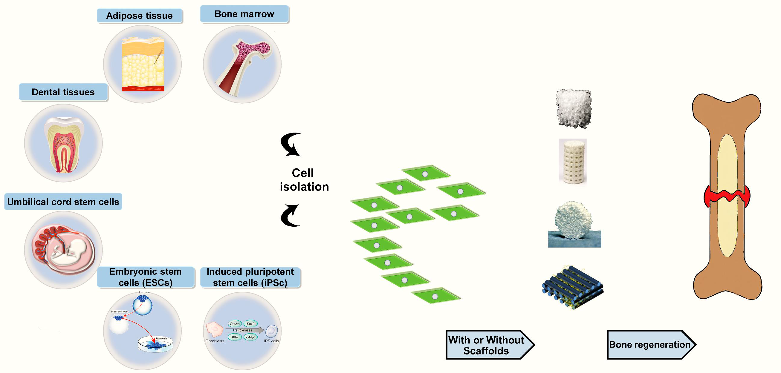
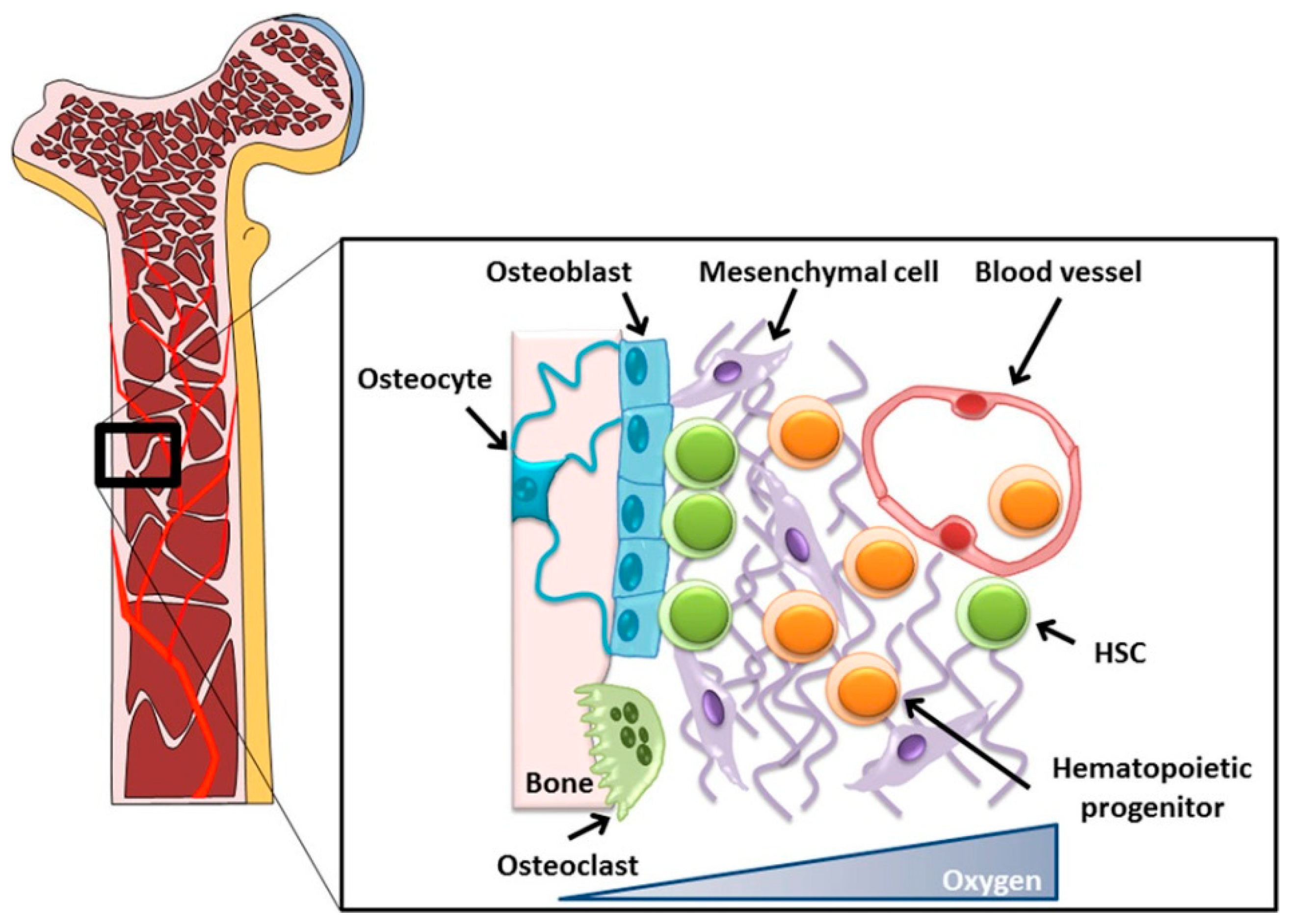
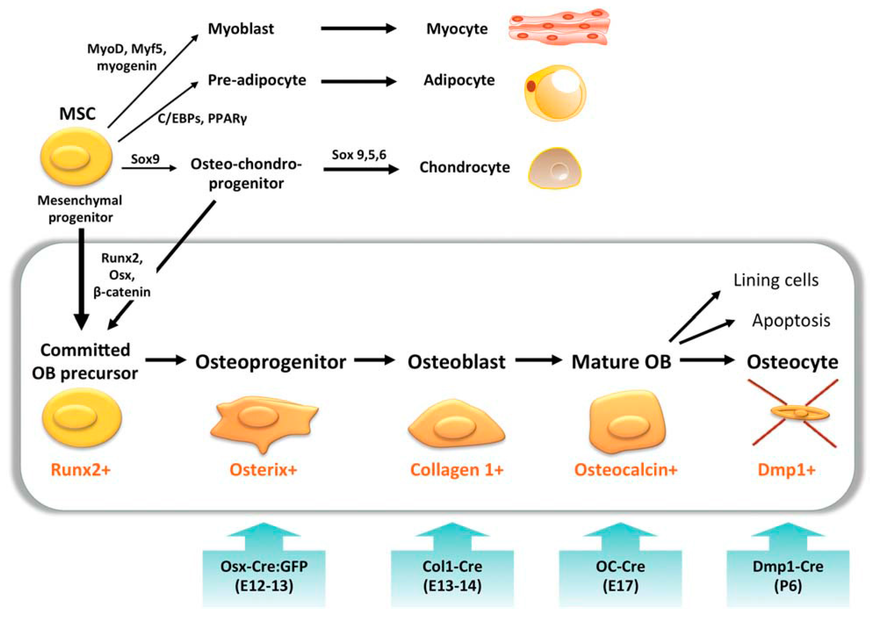
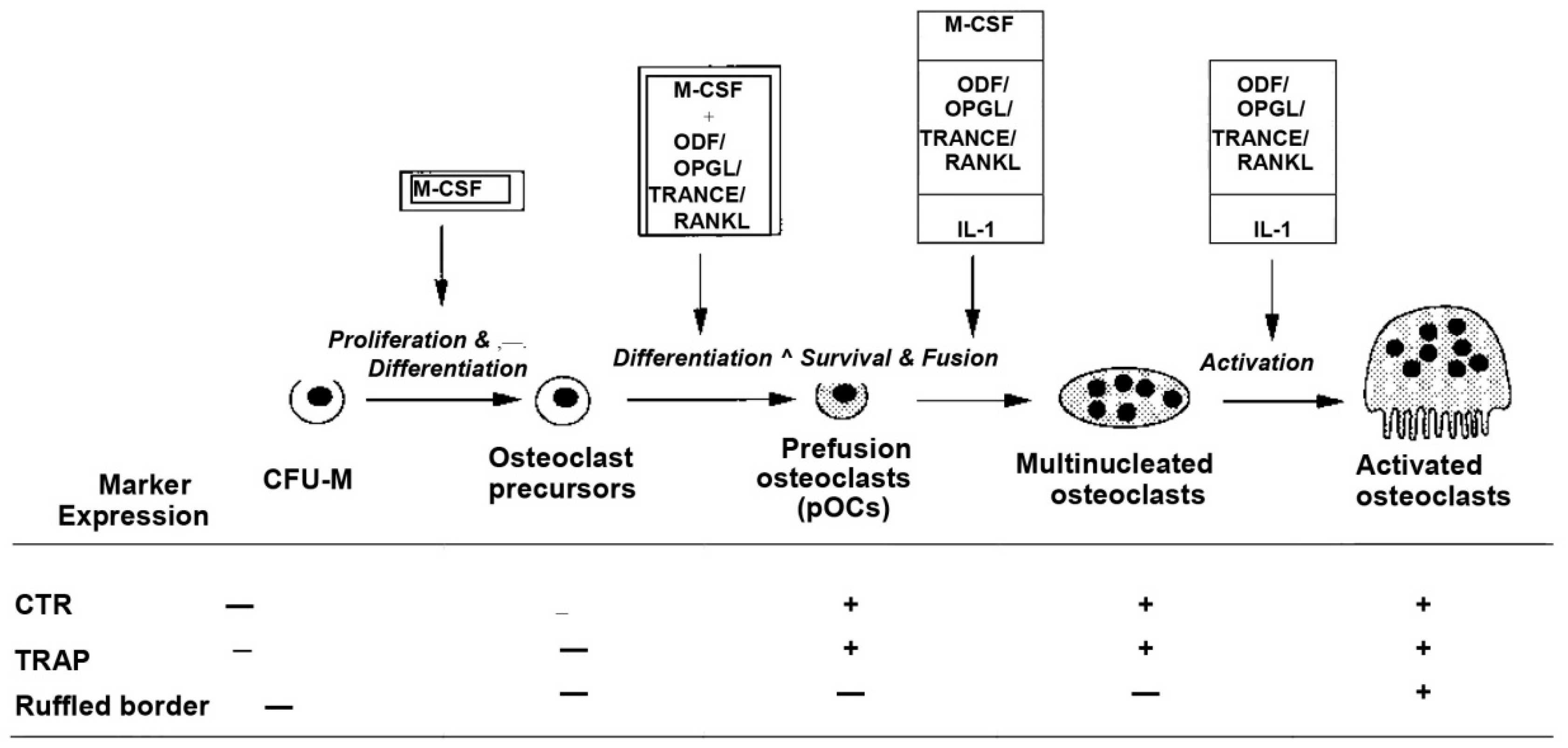
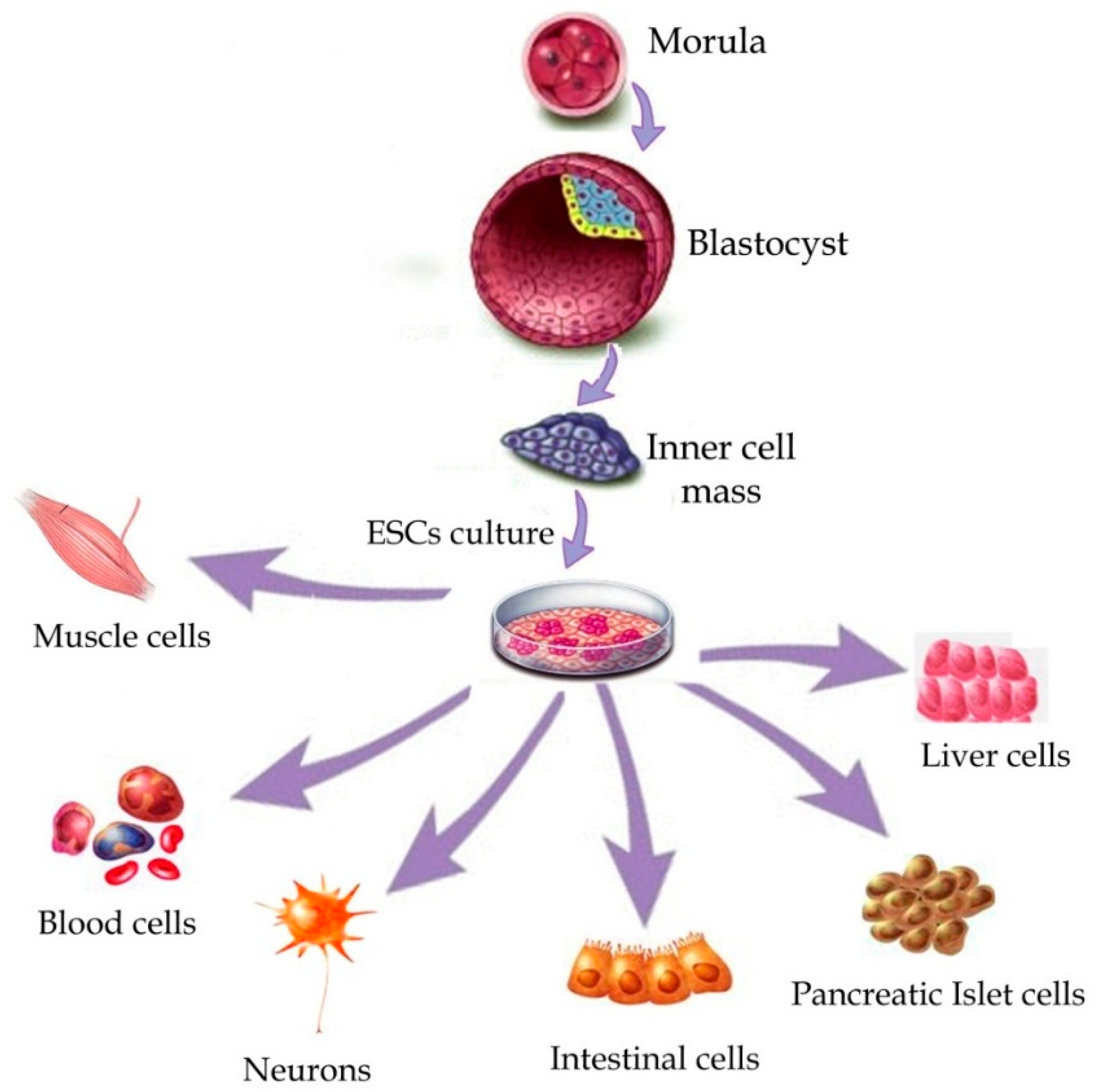
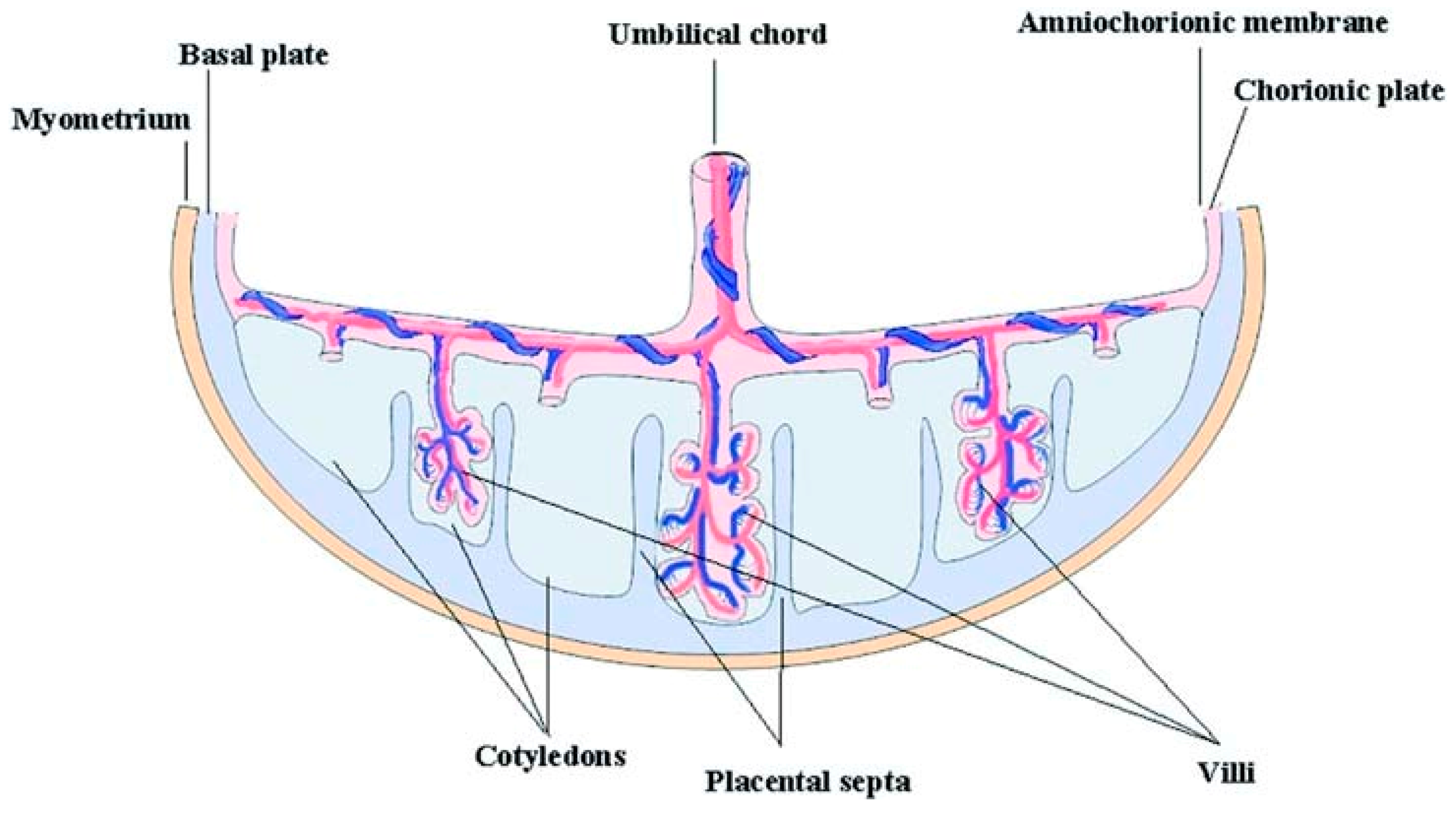
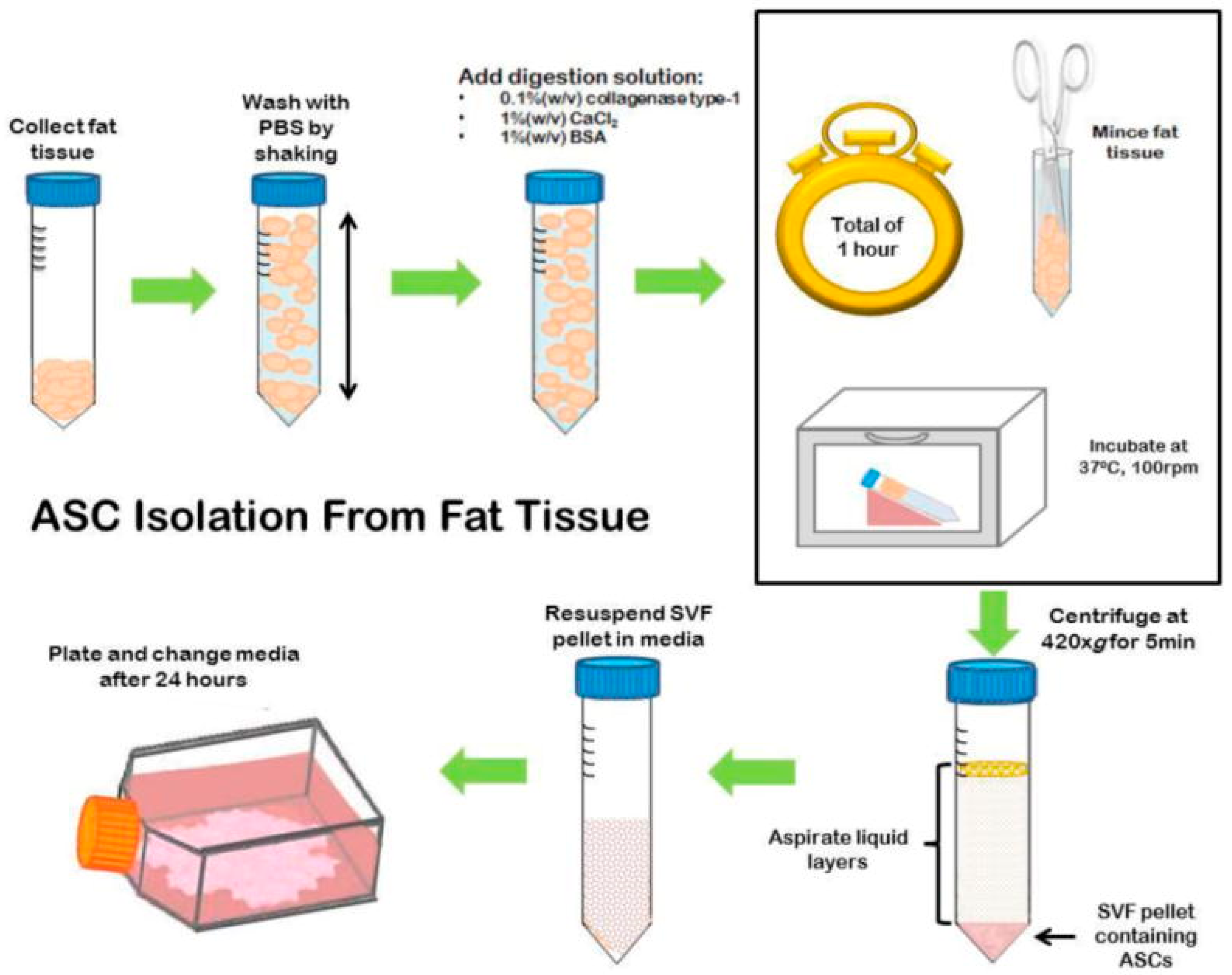
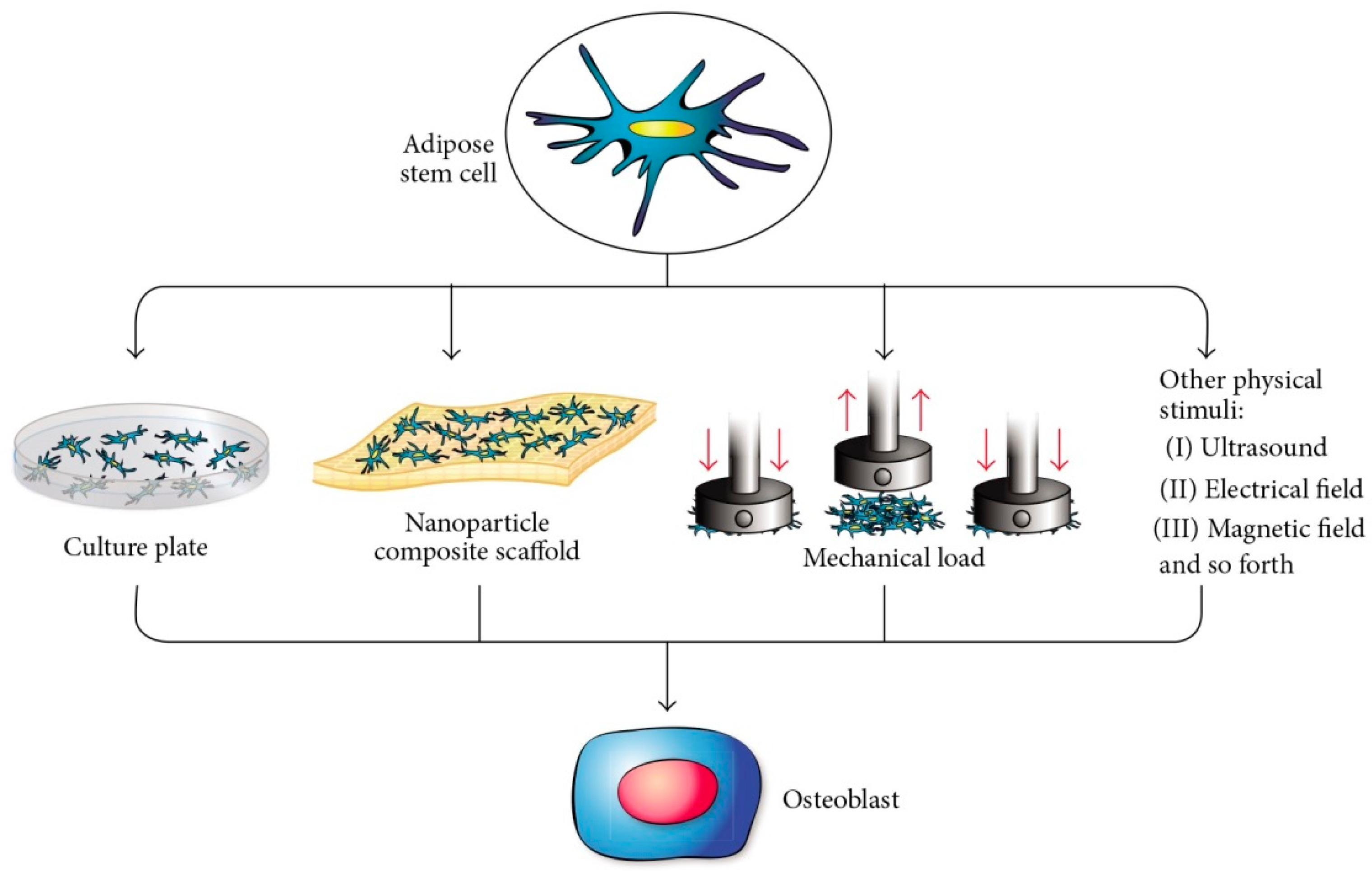
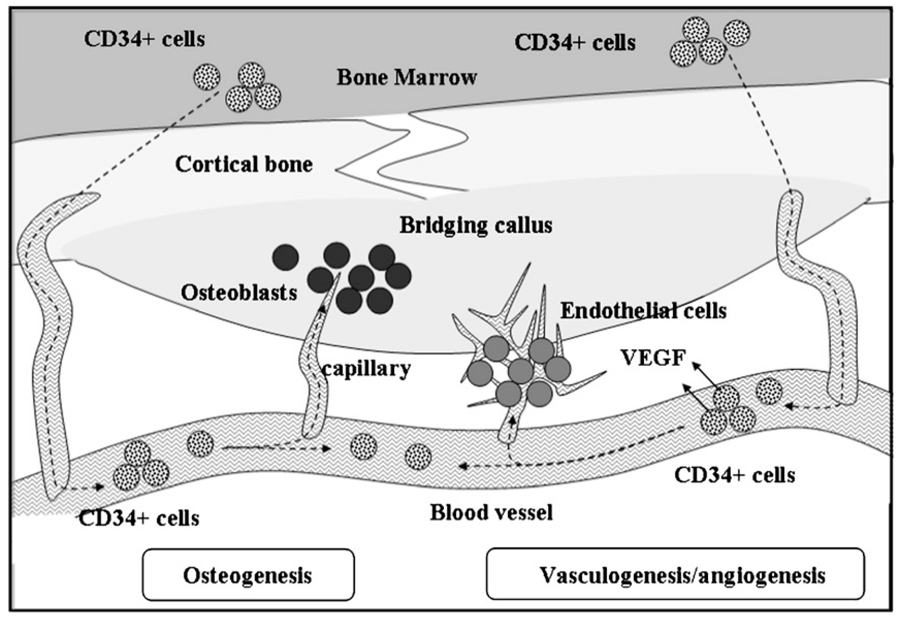
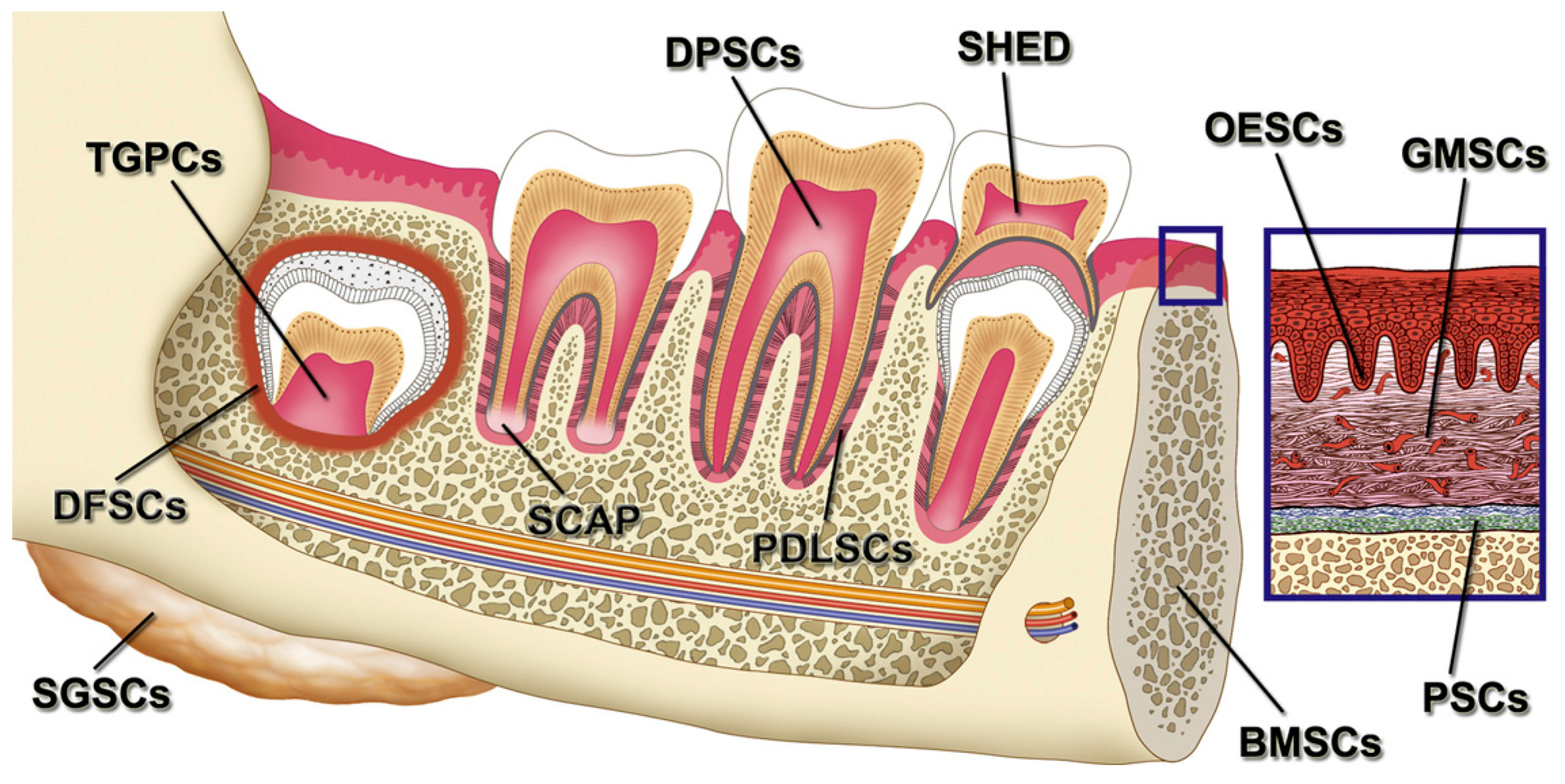
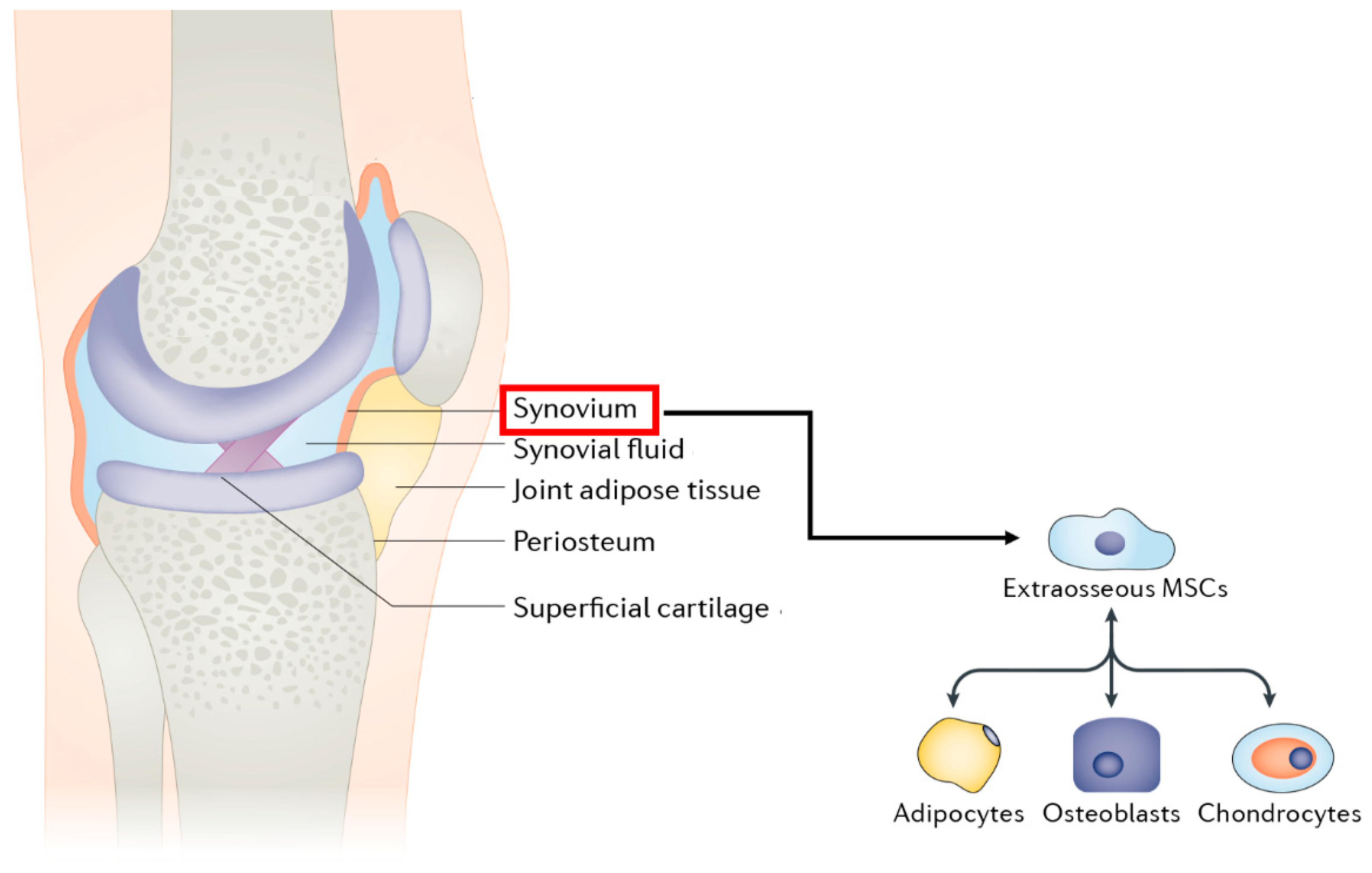
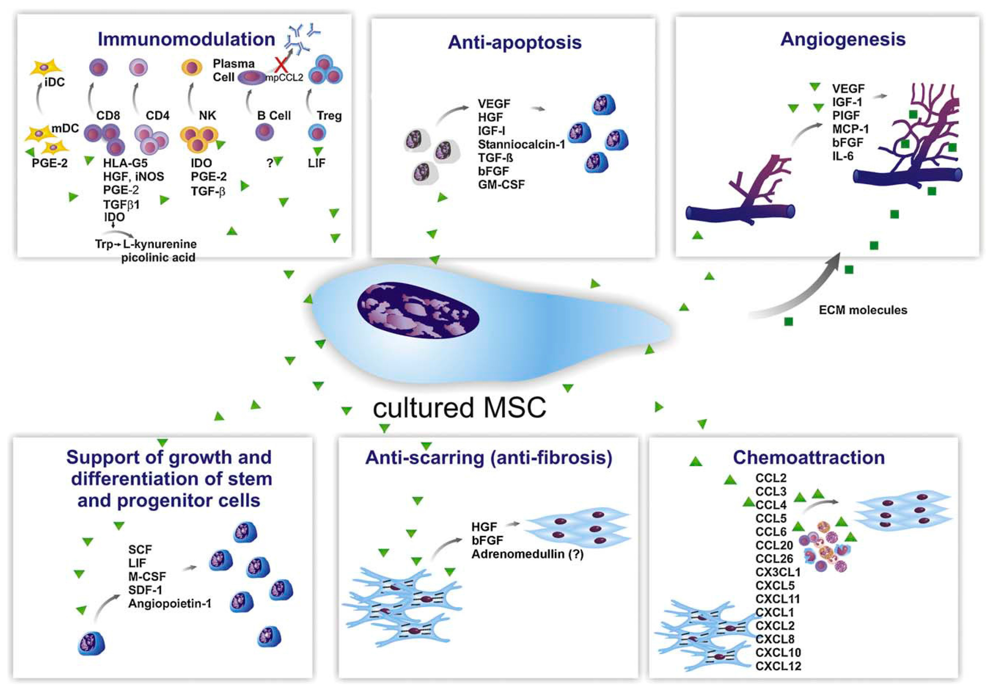
Figure 1. The cell niche within the bone tissue. Osteoblasts, osteocytes, osteoclasts, mesenchymal cells, and endothelial cells of blood vessels create the niches and contribute to the maintenance and differentiation of hematopoietic stem cells (HSCs). With permission from Reference [26].
Applsci 09 00174 g001
Figure 2. Schematic representation of the different steps of osteoblast (OB) differentiation from mesenchymal stem cells (MSC). With permission from Reference [35].
Applsci 09 00174 g002
Figure 3. The pathways and cytokines involved in the differentiation of osteoclast progenitors into functionally active osteoclast. With permission from Reference [58]. CTR: calcitonin receptors, TRAP: tartrate-resistant acid phosphatase.
Applsci 09 00174 g003
Figure 4. Schematic representation of embryonic stem cells (ESCs) differentiation. Human ESCs could differentiate into various cell types with origins of endoderm, mesoderm, and ectoderm. Reproduced with permission from Reference [72].
Applsci 09 00174 g004
Figure 5. Schematic illustration of the cross-section of human term placenta. All fetal stem cells are isolated from this part of the human fetus. Reproduced with permission from Reference [106].
Applsci 09 00174 g005
Figure 6. A representative schematic of ASCs isolation from fat tissue after digestion, isolation of stromal vascular fraction (SVF), and plating of SVF. Reproduced with permission from Reference [198].
Applsci 09 00174 g006
Figure 7. Schematic illustration of various stimulating factors affecting the differentiation ASCs towards osteoblasts. With permission from Reference [206].
Applsci 09 00174 g007
Figure 8. The systemic transplantation of human CD34+ cells/EPCs results in their recruitment to the fracture site. This causes a favorable environment for fracture healing by enhancing vasculogenesis/angiogenesis and osteogenesis, which leads to functional recovery from the fracture. With permission from Reference [262].
Applsci 09 00174 g008
Figure 9. Various stem cell sources within the oral and maxillofacial region. BMSCs: bone marrow-derived MSCs. DPSCs: dental pulp stem cells. SHED: stem cells from human exfoliated deciduous teeth. PDLSCs: periodontal ligament stem cells. DFSCs: dental follicle stem cells. TGPCs: tooth germ progenitor cells. SCAP: stem cells from the apical papilla. OESCs: oral epithelial progenitor/stem cells. GMSCs: gingiva-derived MSCs. PSCs: periosteum-derived stem cells. SGSCs: salivary gland-derived stem cells with permission from Reference [287].
Applsci 09 00174 g009
Figure 10. Representation of S-MSCs location in the knee joint and their potential in differentiation. Reproduced with some modifications from Reference [349].
Applsci 09 00174 g010
Figure 11. Schematic representation of therapeutic molecules secreted by MSCs to accelerate bone healing. With permission from Reference [374].
Applsci 09 00174 g011
Table 1. Overview of the somatic and stem cells used in BTE applications.
Cell TypeAdvantagesDisadvantages
Autologous osteoblasts
-
Lack of immune rejection
-
Low cost
-
No requirement for cell line development
-
Need for ex vivo expansion (a time-consuming procedure)
-
May be harmful
Embryonic stem cells
-
Ability to differentiate into cells of all three primary germ layers (ectoderm, mesoderm, and endoderm)
-
Immortality
-
Ethical issues
-
The risk of tumorigenicity
-
Difficult culturing
-
Maybe immunogenic
Fetal stem cells (AFSCs, AMSCs, Ch-MSCs)
-
High proliferation rate
-
Lack of ethical issues
-
Lack of tumorigenicity *
-
Immuno-privileged properties
-
Inability to differentiate into the cells of three germ layers
-
Lower potential regarding osteogenic differentiation than other cells
Adult stem cells (BM-MSCs, ASCs, WJMSCs, UCB-MSCs, EPCs, HUCPVCs, SMSCs, PDPCs, MD-SCs, Dental stem/progenitor cells)
-
Ease availability
-
Lack of ethical issues
-
Lack of tumorigenicity *
-
Immuno-privileged properties
-
The limited lifespan in vitro condition
-
Inability to differentiate into the cells of three germ layers
Induced pluripotent stem cells (iPSCs)
-
Ability to differentiate into cells of all three primary germ layers
-
Elimination of immunological rejection
-
Lack of ethical issues
-
The risk of tumorigenicity
-
The safety issues about the retroviral transfection system
* It has been reported that the proteins secreted by these cells may support tumor cells’ proliferation. Abbreviations: AFSCs: Amniotic fluid-derived stem cells, AMSCs: Amniotic membrane stems cells, Ch-MSCs: Chorion Mesenchymal Stem Cells, BM-MSCs: Bone marrow stem cells, ASCs: Adipose tissue-derived mesenchymal stem cells, WJMSCs: Umbilical cord Wharton’s jelly-derived mesenchymal stem cells, UCB-MSCs: Umbilical cord blood-derived mesenchymal stem cells, EPCs: Endothelial progenitor cells, HUCPVCs: Umbilical cord perivascular cells, PDPCs: Periosteum-derived progenitor cells, SMSCs: Synovium-derived mesenchymal stem cells, MD-SCs: Muscle-derived stem cells, iPSCs: Induced pluripotent stem cells.

