Increased Internal Porosity and Surface Area of Hydroxyapatite Accelerates Healing and Compensates for Low Bone Marrow Mesenchymal Stem Cell Concentrations in Critically-Sized Bone Defects

Abstract
:Featured Application
Abstract
1. Introduction
2. Materials and Methods
2.1. Implant Materials
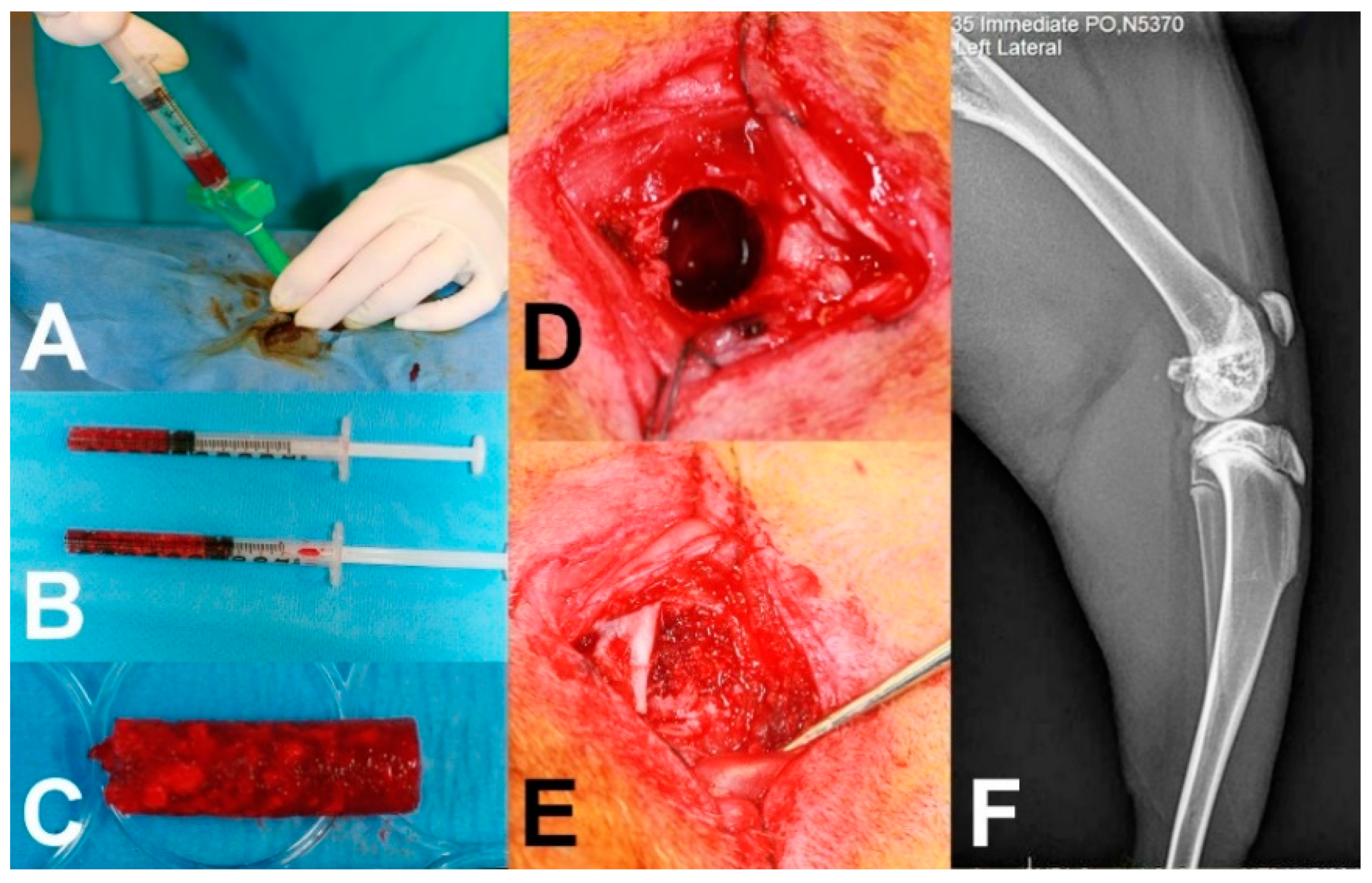
2.2. Surgical Protocol
2.3. Cellular Analysis of Bone Marrow Aspirate
2.4. MicroCT, Histological and Radiographic Analysis
2.5. Statistical Analysis
3. Results
3.1. Cellular Analysis of Bone Marrow Aspirate
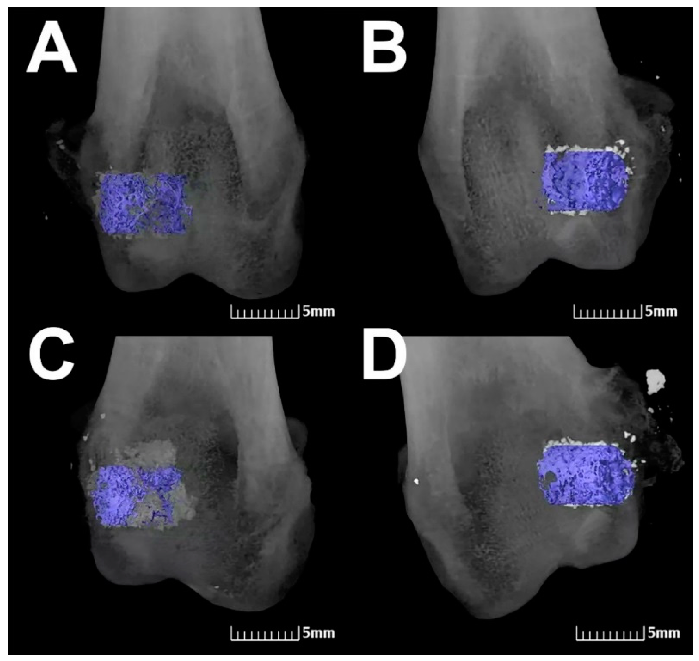
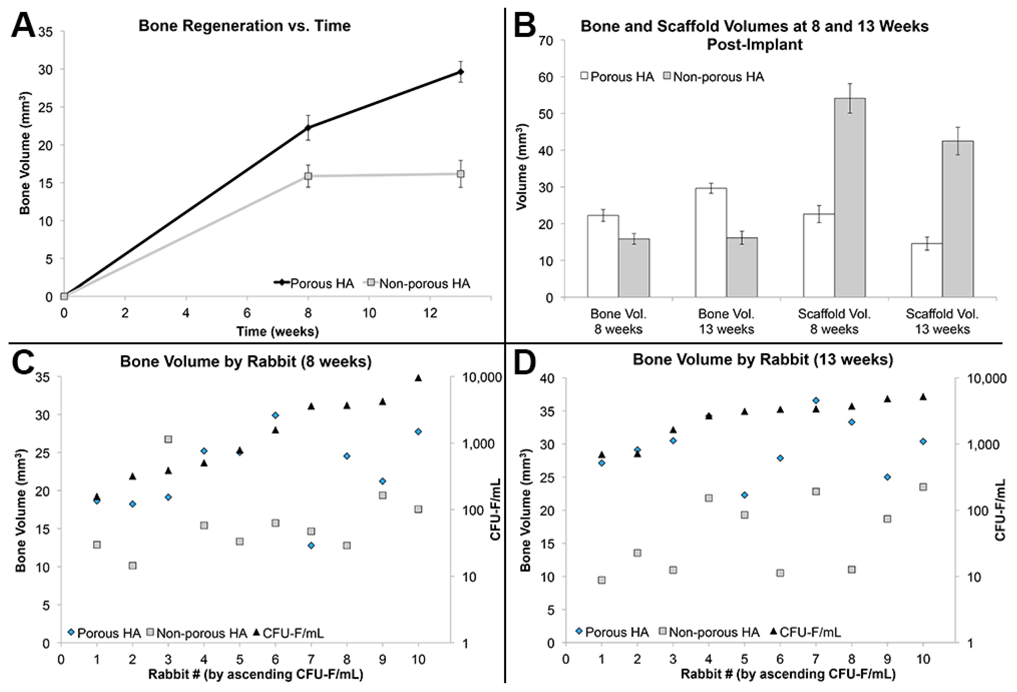
3.2. Bone Formation and Scaffold Degradation
3.3. Effect of MSC Concentration on Bone Formation
4. Discussion
5. Conclusions
Author Contributions
Funding
Acknowledgments
Conflicts of Interest
References
- Silber, J.S.; Anderson, D.G.; Daffner, S.D.; Brislin, B.T.; Leland, J.M.; Hilibrand, A.S.; Vaccaro, A.R.; Albert, T.J. Donor site morbidity after anterior iliac crest bone harvest for single-level anterior cervical discectomy and fusion. Spine 2003, 28, 134–139. [Google Scholar] [CrossRef] [PubMed]
- Heise, U.; Osborn, J.F.; Dawe, F. Hydroxyapatite ceramic as a bone substitute. Int. Orthop. 1990, 14, 329–338. [Google Scholar] [CrossRef] [PubMed]
- Oonishi, H. Orthopaedic applications of hydroxyapatite. Biomaterials 1991, 12, 171–178. [Google Scholar] [CrossRef]
- Appleford, M.R.; Oh, S.; Oh, N.; Ong, J.L. In vivo study on hydroxyapatite scaffolds with trabecular architecture for bone repair. J. Biomed. Mater. Res. Part A 2009, 89, 1019–1027. [Google Scholar] [CrossRef] [PubMed]
- Son, J.S.; Appleford, M.; Ong, J.L.; Wenke, J.C.; Kim, J.M.; Choi, S.H.; Oh, D.S. Porous hydroxyapatite scaffold with three-dimensional localized drug delivery system using biodegradable microspheres. J. Control. Release 2011, 153, 133–140. [Google Scholar] [CrossRef] [PubMed]
- Laurencin, C.T.; Ambrosio, A.M.A.; Borden, M.D.; Cooper, J.A., Jr. Tissue engineering: Orthopedic applications. Annu. Rev. Biomed. Eng. 1999, 1, 19–46. [Google Scholar] [CrossRef] [PubMed]
- Kuboki, Y.; Takita, H.; Kobayashi, D.; Tsuruga, E.; Inoue, M.; Murata, M.; Nagai, N.; Dohi, Y.; Ohgushi, H. BMP-induced osteogenesis on the surface of hydroxyapatite with geometrically feasible and nonfeasible structures: Topology of osteogenesis. J. Biomed. Mater. Res. 1998, 39, 190–199. [Google Scholar] [CrossRef]
- Chang, B.S.; Hong, K.S.; Youn, H.J.; Ryu, H.S.; Chung, S.S.; Park, K.W. Osteoconduction at porous hydroxyapatite with various pore configurations. Biomaterials 2000, 21, 1291–1298. [Google Scholar] [CrossRef]
- Karageorgiou, V.; Kaplan, D. Porosity of 3D biomaterial scaffolds and osteogenesis. Biomaterials 2005, 26, 5474–5491. [Google Scholar] [CrossRef] [PubMed]
- Woodard, J.R.; Hilldore, A.J.; Lan, S.K.; Park, C.J.; Morgan, A.W.; Eurell, J.A.C.; Clark, S.G.; Wheeler, M.B.; Jamison, R.D.; Johnson, A.J.W. The mechanical properties and osteoconductivity of hydroxyapatite bone scaffolds with multi-scale porosity. Biomaterials 2007, 28, 45–54. [Google Scholar] [CrossRef] [PubMed]
- Den Boer, F.C.; Wippermann, B.W.; Blokhuis, T.J.; Patka, P.; Bakker, F.C.; Henk, J.T.M. Healing of segmental bone defects with granular porous hydroxyapatite augmented with recombinant human osteogenic protein-1 or autologous bone marrow. J. Orthop. Res. 2003, 21, 521–528. [Google Scholar] [CrossRef]
- Gan, Y.; Dai, K.; Zhang, P.; Tang, T.; Zhu, Z.; Lu, J. The clinical use of enriched bone marrow stem cells combined with porous beta-tricalcium phosphate in posterior spinal fusion. Biomaterials 2008, 29, 3973–3982. [Google Scholar] [CrossRef] [PubMed]
- Kubo, T.; Doi, K.; Hayashi, K.; Morita, K.; Matsuura, A.; Teixeira, E.R.; Akagawa, Y. Comparative evaluation of bone regeneration using spherical and irregularly shaped granules of interconnected porous hydroxylapatite. A beagle dog study. J. Prosthodont. Res. 2011, 55, 104–109. [Google Scholar] [CrossRef] [PubMed]
- Murphy, M.B.; Blashki, D.; Buchanan, R.M.; Tasciotti, E. Engineering a better way to heal broken bones. Chem. Eng. Prog. 2010, 106, 37–43. [Google Scholar]
- Murphy, M.B.; Moncivais, K.; Caplan, A.I. Mesenchymal stem cells: Environmentally responsive therapeutics for regenerative medicine. Exp. Mol. Med. 2013, 45, e54. [Google Scholar] [CrossRef] [PubMed]
- Murphy, M.B.; Suzuki, R.K.; Sand, T.T.; Chaput, C.D.; Gregory, C.A. Short Term Culture of Human Mesenchymal Stem Cells with Commercial Osteoconductive Carriers Provides Unique Insights into Biocompatibility. J. Clin. Med. 2013, 2, 49–66. [Google Scholar] [CrossRef] [PubMed]
- Hernigou, P.; Poignard, A.; Beaujean, F.; Rouard, H. Percutaneous Autologous Bone-Marrow Grafting for Nonunions. J. Bone Jt. Surg. Incorp. 2005, 87, 1430–1437. [Google Scholar] [CrossRef]
- Hernigou, P.; Daltro, G.; Filippini, P.; Mukasa, M.M.; Manicom, O. Percutaneous implantation of autologous bone marrow osteoprogenitor cells as treatment of bone avascular necrosis related to sickle cell disease. Open Orthop. J. 2008, 2, 62–65. [Google Scholar] [CrossRef] [PubMed]
- Hernigou, P.; Lachaniette, C.H.F.; Delambre, J.; Zilber, S.; Duffiet, P.; Chevallier, N.; Rouard, H. Biologic augmentation of rotator cuff repair with mesenchymal stem cells during arthroscopy improves healing and prevents further tears: A case-controlled study. Int. Orthop. 2014, 38, 1811–1818. [Google Scholar] [CrossRef] [PubMed]
- Khademhosseini, A.; Langer, R.; Borenstein, J.; Vacanti, J.P. Microscale technologies for tissue engineering and biology. Proc. Natl. Acad. Sci. USA 2006, 103, 2480–2487. [Google Scholar] [CrossRef] [PubMed] [Green Version]
- Dawson, E.; Mapili, G.; Erickson, K.; Taqvi, S.; Roy, K. Biomaterials for stem cell differentiation. Adv. Drug Deliv. Rev. 2008, 60, 215–228. [Google Scholar] [CrossRef] [PubMed]
- Murphy, M.B.; Blashki, D.; Buchanan, R.M.; Fan, D.; De Rosa, E.; Shah, R.N.; Stupp, S.I.; Weiner, B.K.; Simmons, P.J.; Ferrari, M.; et al. Multi-Composite Bioactive Osteogenic Sponges Featuring Mesenchymal Stem Cells, Platelet-Rich Plasma, Nanoporous Silicon Enclosures, and Peptide Amphiphiles for Rapid Bone Regeneration. J. Funct. Biomater. 2011, 2, 39–66. [Google Scholar] [CrossRef] [PubMed] [Green Version]
- Guda, T.; Walker, J.A.; Pollot, B.E.; Appleford, M.R.; Oh, S.; Ong, J.L.; Wenke, J.C. In vivo performance of bilayer hydroxyapatite scaffolds for bone tissue regeneration in the rabbit radius. J. Mater. Sci. Mater. Med. 2011, 22, 647–656. [Google Scholar] [CrossRef] [PubMed]
- Bohner, M.; Baumgart, F. Theoretical model to determine the effects of geometrical factors on the resorption of calcium phosphate bone substitutes. Biomaterials 2004, 25, 3569–3582. [Google Scholar] [CrossRef] [PubMed]
- Kretlow, J.D.; Jin, Y.Q.; Liu, W.; Zhang, W.J.; Hong, T.H.; Zhou, G.; Baggett, L.S.; Mikos, A.G.; Cao, Y. Donor age and cell passage affects differentiation potential of murine bone marrow-derived stem cells. BMA Cell Biol. 2008, 9, 60. [Google Scholar] [CrossRef] [PubMed] [Green Version]
- Kretlow, J.D.; Spicer, P.P.; Jansen, J.A.; Vacanti, C.A.; Kasper, F.K.; Mikos, A.G. Uncultured Marrow Mononuclear Cells Delivered Rat Cranial Defects. Tissue Eng. Part A 2010, 16, 3555–3568. [Google Scholar] [CrossRef] [PubMed]
- Murphy, M.B.; Blashki, D.; Buchanan, R.M.; Yazdi, I.K.; Ferrari, M.; Simmons, P.J.; Tasciotti, E. Adult and umbilical cord blood-derived platelet-rich plasma for mesenchymal stem cell proliferation, chemotaxis, and cryo-preservation. Biomaterials 2012, 33, 5308–5316. [Google Scholar] [CrossRef] [PubMed]
- Weibrich, G.; Hansen, T.; Kleis, W.; Buch, R.; Hitzler, W.E. Effect of platelet concentration in platelet-rich plasma on peri-implant bone regeneration. Bone 2004, 34, 665–671. [Google Scholar] [CrossRef] [PubMed]
- Sarkar, M.R.; Augat, P.; Shefelbine, S.J.; Schorlemmer, S.; Huber-Lang, M.; Claes, L.; Kinzl, L.; Ignatius, A. Bone formation in a long bone defect model using a platelet-rich plasma-loaded collagen scaffold. Biomaterials 2006, 27, 1817–1823. [Google Scholar] [CrossRef] [PubMed]


| Data by Time Point: | 8-Week Groups (n = 10) | 13-Week Groups (n = 10) | ||||
| Average TNC/mL | 3.23 × 10⁷ ± 5.44 × 10⁶ | 3.18 × 10⁷ ± 3.41 × 10⁶ | ||||
| Average CFU-F/mL | 2486 ± 933 | 2935 ± 488 | ||||
| Porous | Non-porous | p-value | Porous | Non-porous | p-value | |
| Residual Scaffold Vol. (mm3) | 22.62 | 54.11 | <0.001 | 14.59 | 42.50 | <0.001 |
| Bone Vol. (BV) (mm3) | 22.25 | 15.87 | 0.009 | 29.63 | 16.16 | <0.001 |
| Bone Vol./Total Vol. | 0.23 | 0.16 | 0.009 | 0.31 | 0.17 | <0.001 |
| Connectivity Density (1/mm3) | 28.45 | 8.73 | <0.001 | 40.40 | 12.84 | <0.001 |
| Trabecular Thickness (mm) | 0.18 | 0.15 | 0.004 | 0.19 | 0.15 | <0.001 |
| Bone Surface Area (mm2) | 446.02 | 410.81 | 0.384 | 576.81 | 442.76 | 0.003 |
| Mean Bone Density (mgHA/cm3) | 730.84 | 615.17 | <0.001 | 771.76 | 644.94 | <0.001 |
| Data by Scaffold Type: | Porous HA (n = 10) | Non-porous HA (n = 10) | ||||
| 8 weeks | 13 weeks | p-value | 8 weeks | 13 weeks | p-value | |
| Residual Scaffold Vol. (mm3) | 22.62 | 14.59 | 0.014 | 54.11 | 42.50 | 0.049 |
| Bone Vol. (BV) (mm3) | 22.25 | 29.63 | 0.003 | 15.87 | 16.16 | 0.901 |
| Bone Vol./Total Vol. | 0.23 | 0.31 | 0.003 | 0.16 | 0.17 | 0.899 |
| Connectivity Density (1/mm3) | 28.45 | 40.40 | 0.010 | 8.73 | 12.84 | 0.078 |
| Trabecular Thickness (mm) | 0.18 | 0.19 | 0.207 | 0.15 | 0.15 | 0.884 |
| Bone Surface Area (mm2) | 446.02 | 576.81 | 0.003 | 410.81 | 442.76 | 0.447 |
| Mean Bone Density (mgHA/cm3) | 730.84 | 771.76 | 0.016 | 615.17 | 644.94 | 0.027 |
© 2018 by the authors. Licensee MDPI, Basel, Switzerland. This article is an open access article distributed under the terms and conditions of the Creative Commons Attribution (CC BY) license (http://creativecommons.org/licenses/by/4.0/).
Share and Cite
Dawson, E.R.; Suzuki, R.K.; Samano, M.A.; Murphy, M.B. Increased Internal Porosity and Surface Area of Hydroxyapatite Accelerates Healing and Compensates for Low Bone Marrow Mesenchymal Stem Cell Concentrations in Critically-Sized Bone Defects. Appl. Sci. 2018, 8, 1366. https://doi.org/10.3390/app8081366
Dawson ER, Suzuki RK, Samano MA, Murphy MB. Increased Internal Porosity and Surface Area of Hydroxyapatite Accelerates Healing and Compensates for Low Bone Marrow Mesenchymal Stem Cell Concentrations in Critically-Sized Bone Defects. Applied Sciences. 2018; 8(8):1366. https://doi.org/10.3390/app8081366
Chicago/Turabian StyleDawson, Eileen R., Richard K. Suzuki, Melissa A. Samano, and Matthew B. Murphy. 2018. "Increased Internal Porosity and Surface Area of Hydroxyapatite Accelerates Healing and Compensates for Low Bone Marrow Mesenchymal Stem Cell Concentrations in Critically-Sized Bone Defects" Applied Sciences 8, no. 8: 1366. https://doi.org/10.3390/app8081366