Plasmon Modulation Spectroscopy of Noble Metals to Reveal the Distribution of the Fermi Surface Electrons in the Conduction Band


Abstract
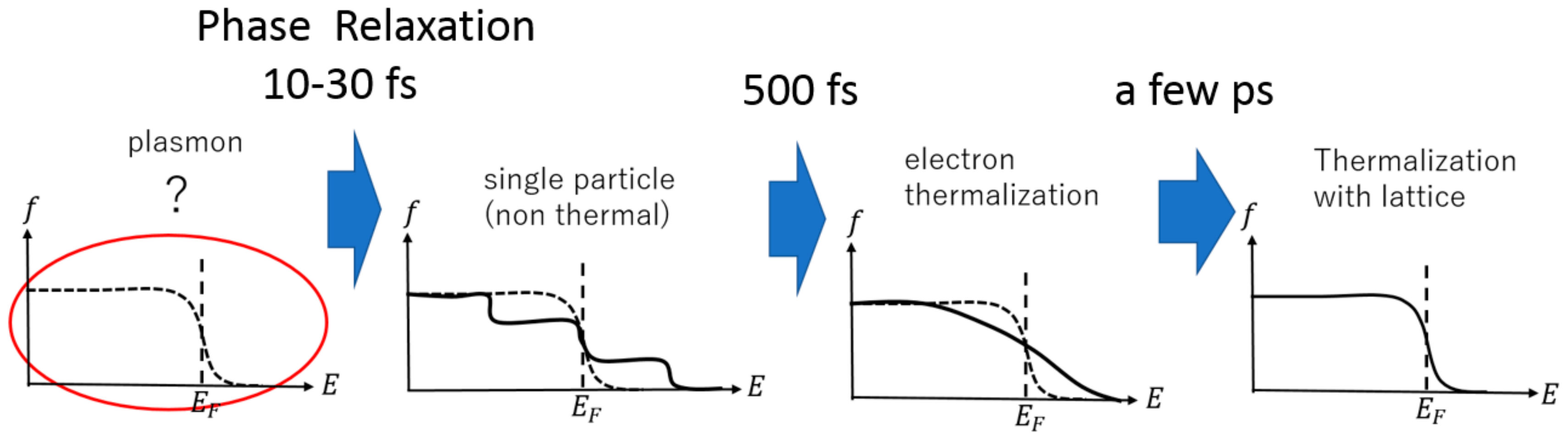
:1. Introduction
2. Experiment
2.1. Sample Preparation and Evaluation of Plasmon Resonance and Interband Transition
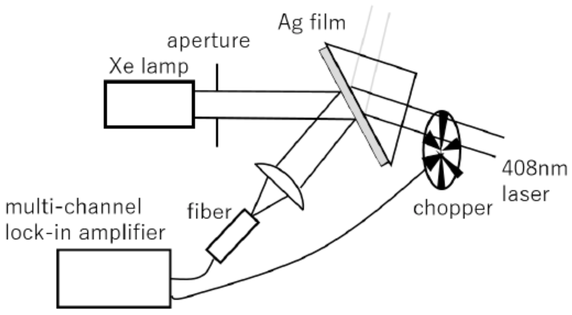
2.2. CW Experiment
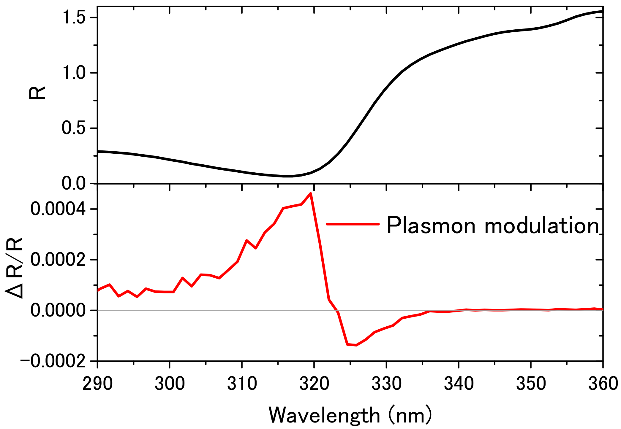
2.2.1. Plasmon Modulation (Reflectance Change in the Interband Transition Due to Surface Plasmon Excitation)
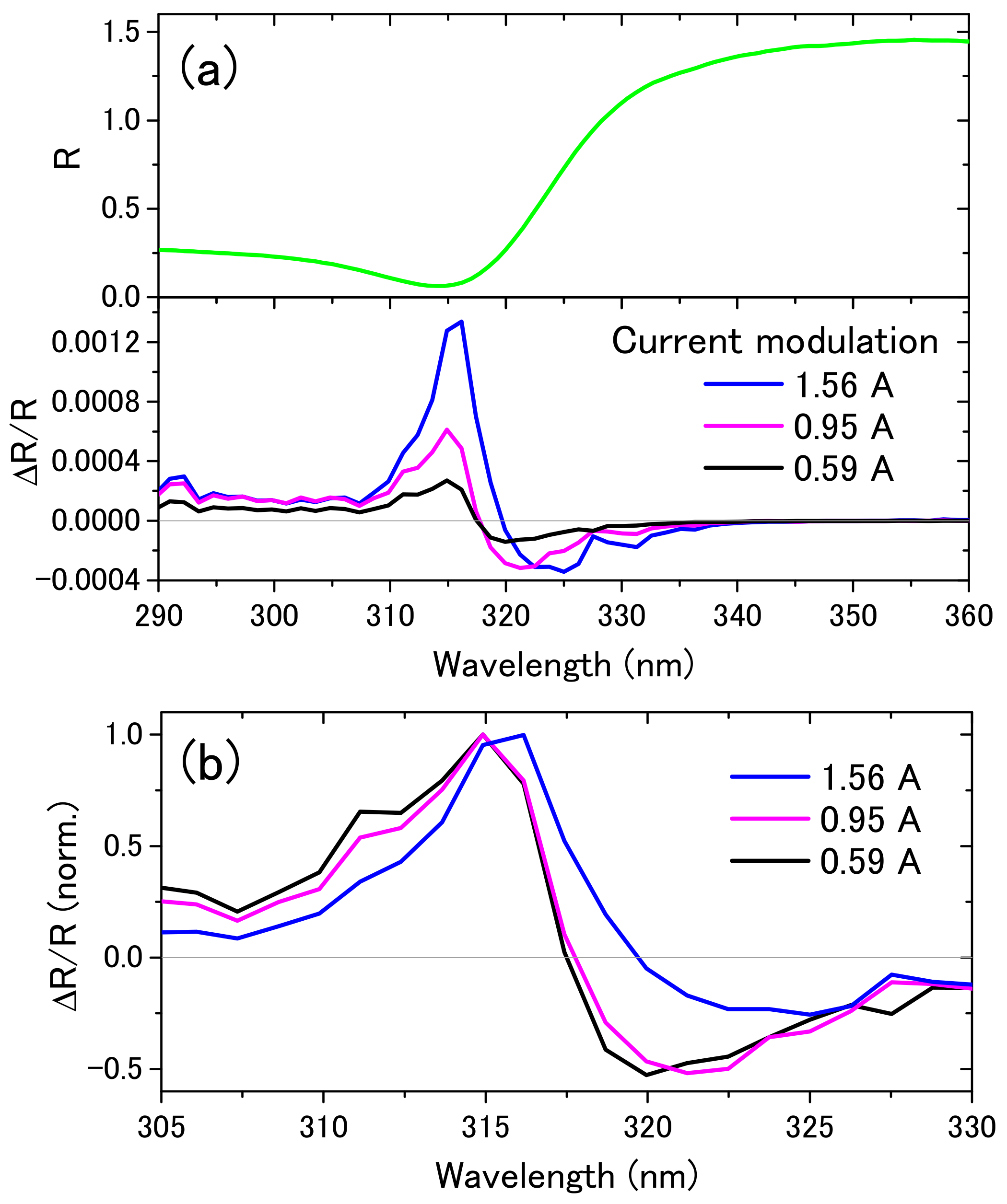
2.2.2. Current Modulation (Current Modulation Spectroscopy of Interband Transition)
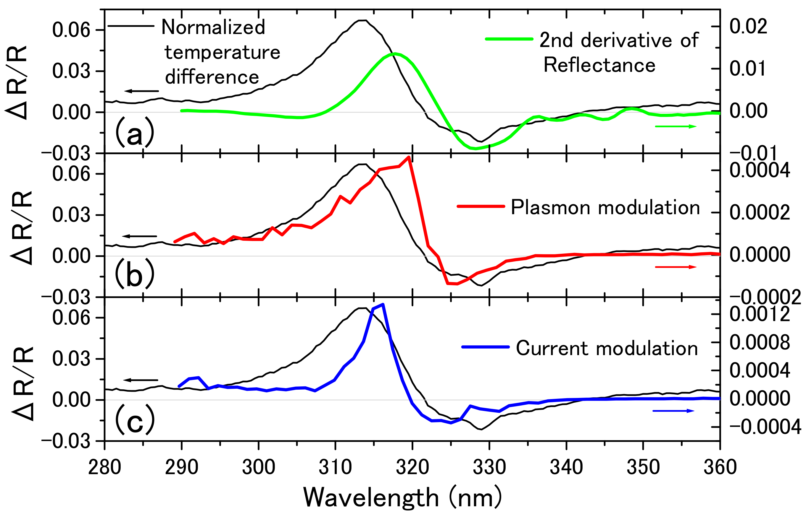
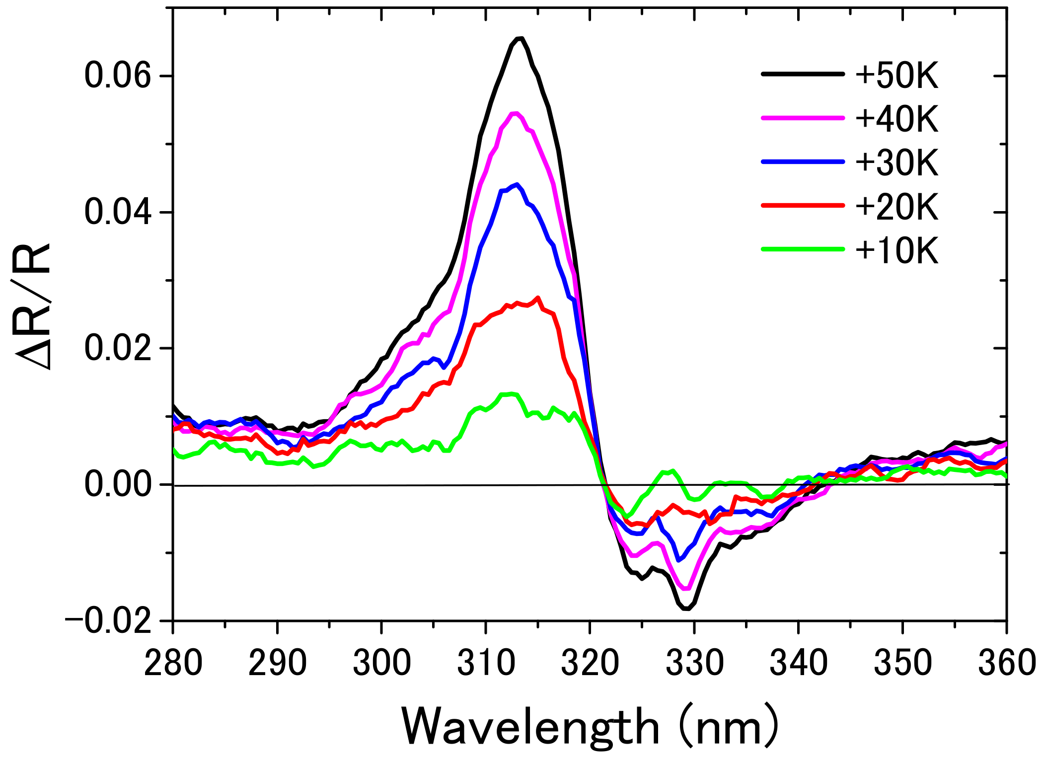
2.2.3. Temperature Difference Spectrum
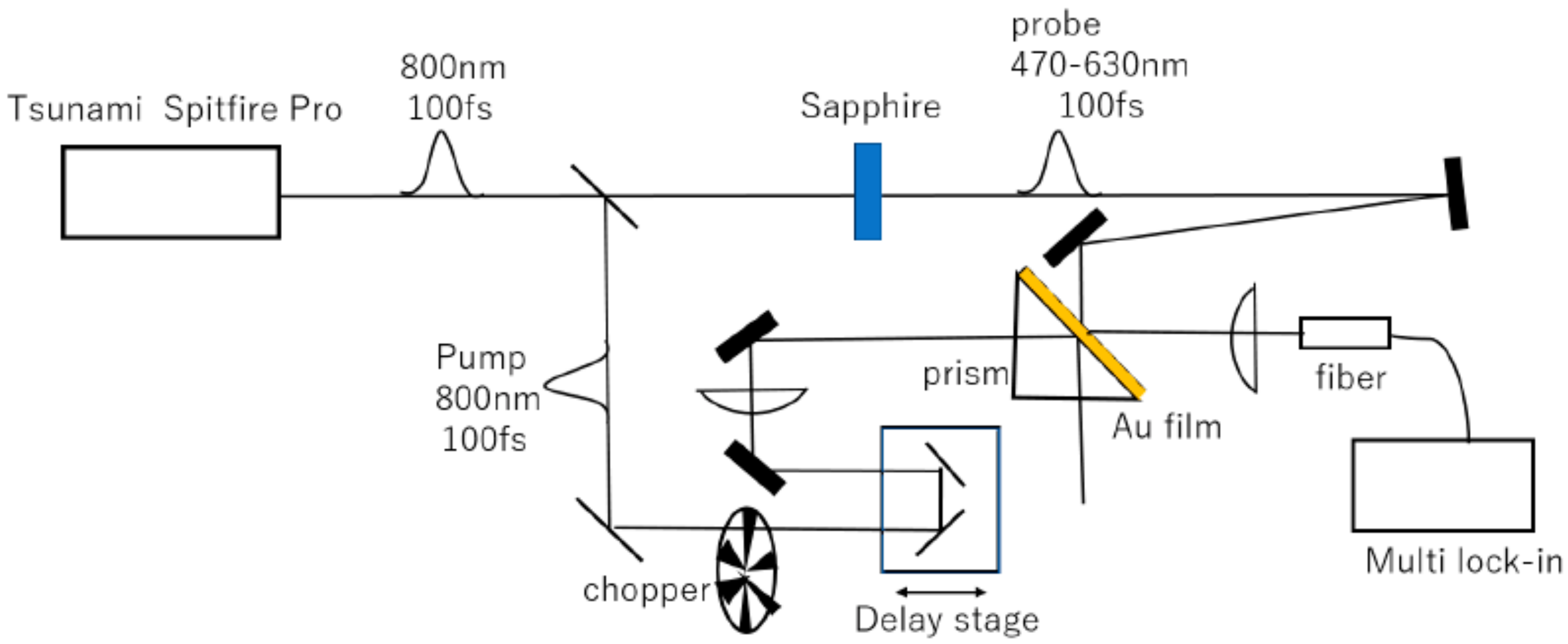
2.3. Time-Resolved Experiment (Femtosecond Pump-Probe Spectroscopy)
3. Results and Discussion
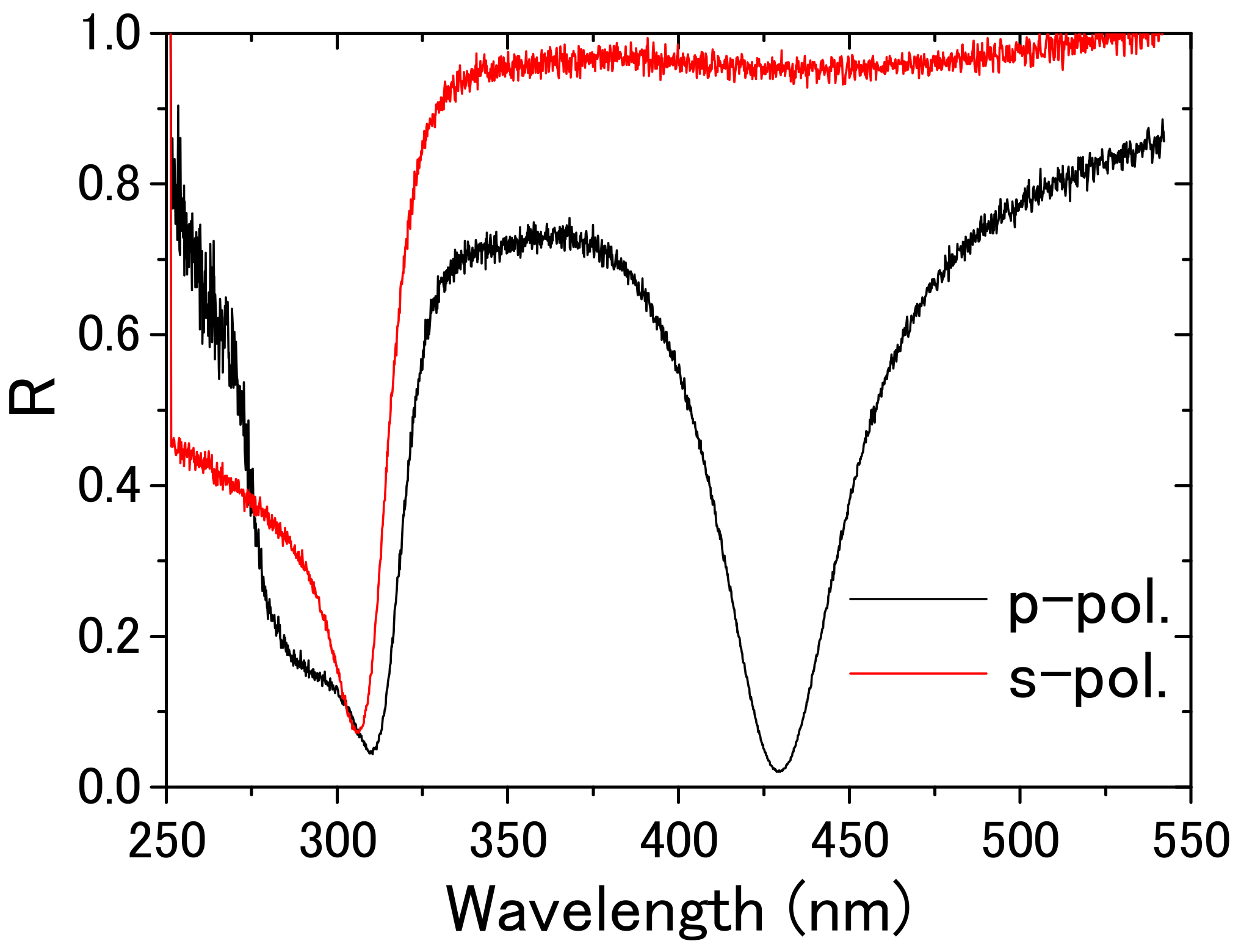
3.1. Observation of Surface Plasmon Resonance and Interband Transition Spectra
3.2. Plasmon Modulation Spectrum
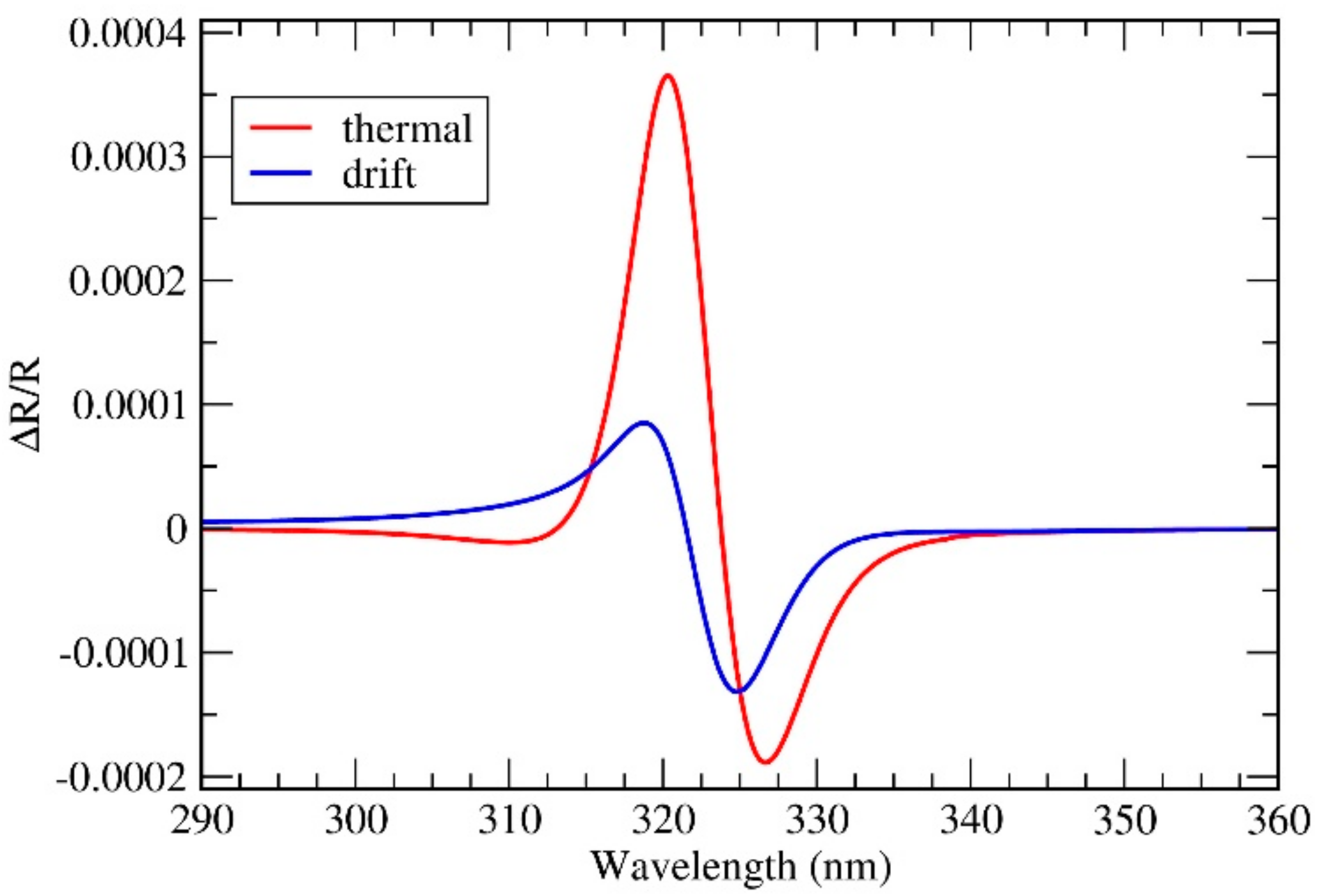
3.3. Current Modulation and Temperature Difference Spectra
3.4. CW Modulation Spectra
3.5. Calculation of Reflectivity Modulation
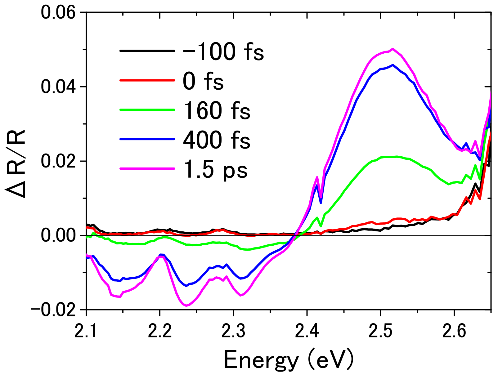
3.6. Femtosecond Time-Resolved Spectra
4. Conclusions and Prospects
Acknowledgments
Author Contributions
Conflicts of Interest
References
- Weber, W.H.; Ford, G.W. Optical electric-field enhancement at a metal surface arising from surface-plasmon excitation. Opt. Lett. 1981, 6, 122–124. [Google Scholar] [CrossRef] [PubMed]
- Willets, K.A.; van Duyne, R.P. Localized Surface Plasmon Resonance Spectroscopy and Sensing. Annu. Rev. Phys. Chem. 2007, 58, 267–297. [Google Scholar] [CrossRef] [PubMed]
- Gramotnev, D.K.; Bozhevolnyi, S.I. Plasmonics beyond the diffraction limit. Nat. Photonics 2010, 4, 83–91. [Google Scholar] [CrossRef]
- Luo, X.; Ishihara, T. Surface plasmon resonant interference nanolithography technique. Appl. Phys. Lett. 2004, 84, 4780–4872. [Google Scholar] [CrossRef]
- Kauranen, M.; Zayats, A.V. Nonlinear plasmonics. Nat. Photonics 2012, 6, 737–748. [Google Scholar] [CrossRef]
- Abe, S.; Kajikawa, K. Linear and Nonlinear Optical Properties of Gold Nanoparticles Immobilized on Metallic Surface. Phys. Rev. B 2006, 74. [Google Scholar] [CrossRef]
- Atwater, H.A.; Polman, A. Plasmonics for improved photovoltaic devices. Nat. Mater. 2010, 9, 205–213. [Google Scholar] [CrossRef] [PubMed]
- Tame, M.S.; McEnery, K.R.; Ozdemir, S.K.; Lee, J.; Maier, S.A.; Kim, M.S. Quantum plasmonics. Nat. Phys. 2013, 9, 329–340. [Google Scholar] [CrossRef]
- Kubo, A.; Pontius, N.; Petek, H. Femtosecond Microscopy of Surface Plasmon Polariton Wave Packet Evolution at the Silver/Vacuum Interface. Nano Lett. 2007, 7, 470–475. [Google Scholar] [CrossRef] [PubMed]
- Pitarke, J.M.; Silkin, V.M.; Chulkov, E.V.; Echenique, P.M. Theory of surface plasmons and surface-plasmon polaritons. Rep. Prog. Phys. 2007, 70, 1–87. [Google Scholar] [CrossRef]
- Sundararaman, R.; Narang, P.; Jermyn, A.S.; Goddard, W.A., III; Atwater, H.A. Theoretical predictions for hot-carrier generation from surface plasmon decay. Nat. Commun. 2014, 5. [Google Scholar] [CrossRef] [PubMed]
- Groeneveld, R.H.M.; Sprik, R.; Lagendijk, A. Effect of a nonthermal electron distribution on the electron-phonon energy relaxation process in noble metals. Phys. Rev. B 1992, 45, 5079–5082. [Google Scholar] [CrossRef]
- Groeneveld, R.H.M.; Sprik, R.; Lagendijk, A. Femtosecond spectroscopy of electron-electron and electron-phonon energy relaxation in Ag and Au. Phys. Rev. B 1995, 51, 11433–11445. [Google Scholar] [CrossRef]
- Sun, C.-K.; Vallee, F.; Acioli, L.H.; Ippen, E.P.; Fujimoto, J.G. Femtosecond-tunable measurement of electron thermalization in gold. Phys. Rev. B 1994, 50, 15337–15348. [Google Scholar] [CrossRef]
- Rethfeld, B.; Kaiser, A.; Vicanek, M.; Simon, G. Ultrafast dynamics of nonequilibrium electrons in metals under femtosecond laser irradiation. Phys. Rev. B 2002, 65. [Google Scholar] [CrossRef]
- Kretschmann, E.; Raether, H. Radiative decay of non radiative surface plasmons excited by light. Z. Naturforsch. A 1968, 23, 2135–2136. [Google Scholar] [CrossRef]
- Takagi, K.; Nair, S.V.; Watanabe, R.; Seto, K.; Kobayashi, T.; Tokunaga, E. Surface plasmon polariton resonance of gold, silver, and copper studied in the Kretschmann geometry: Dependence on wavelength, angle of incidence, and film thickness. J. Phys. Soc. Jpn. 2017, 86. [Google Scholar] [CrossRef]
- Shirakawa, M.; Nakata, K.; Suzuki, M.; Kobayashi, T.; Tokunaga, E. Nonlinear absorption spectroscopy of porphyrin J-aggregates in aqueous solution: Evidence for control of degree of association by light induced force. J. Phys. Soc. Jpn. 2017, 86. [Google Scholar] [CrossRef]
- Du, J.; Teramoto, T.; Nakata, K.; Tokunaga, E.; Kobayashi, T. Real-time vibrational dynamics in chlorophyll a studied with a few-cycle pulse laser. Biophys. J. 2011, 101, 995–1003. [Google Scholar] [CrossRef] [PubMed]
- Rosei, R.; Lynch, D.W. Thermomodulation Spectra of Al, Au, and Cu. Phys. Rev. B 1972, 5, 3883–3894. [Google Scholar] [CrossRef]
- Christensen, N.E. The band structure of silver and optical interband transitions. Phys. Status Solidi b 1972, 54. [Google Scholar] [CrossRef]
- Johnson, P.B.; Christy, R.W. Optical Constants of the Noble Metals. Phys. Rev. B 1972, 6. [Google Scholar] [CrossRef]
- Kobayashi, T.; Shirakawa, A.; Matsuzawa, H.; Nakanishi, H. Real-time vibrational mode-coupling associated with ultrafast geometrical relaxation in polydiacetylene induced by sub-5-fs pulses. Chem. Phys. Lett. 2000, 321, 385–393. [Google Scholar] [CrossRef]
- Kravets, V.G.; Jalil, R.; Kim, Y.-J.; Ansell, D.; Aznakayeva, D.E.; Thackray, B.; Britnell, L.; Belle, B.D.; Withers, F.; Radko, I.P.; et al. Graphene-protected copper and silver plasmonics. Sci. Rep. 2014, 4. [Google Scholar] [CrossRef] [PubMed]
- McPeak, K.M.; Jayanti, S.V.; Kress, S.J.P.; Meyer, S.; Iotti, S.; Rossinelli, A.; Norris, D.J. Plasmonic Films Can Easily Be Better: Rules and Recipes. ACS Photonics 2015, 2, 326–333. [Google Scholar] [CrossRef] [PubMed]


© 2017 by the authors. Licensee MDPI, Basel, Switzerland. This article is an open access article distributed under the terms and conditions of the Creative Commons Attribution (CC BY) license (http://creativecommons.org/licenses/by/4.0/).
Share and Cite
Takagi, K.; Nair, S.V.; Saito, J.; Seto, K.; Watanabe, R.; Kobayashi, T.; Tokunaga, E. Plasmon Modulation Spectroscopy of Noble Metals to Reveal the Distribution of the Fermi Surface Electrons in the Conduction Band. Appl. Sci. 2017, 7, 1315. https://doi.org/10.3390/app7121315
Takagi K, Nair SV, Saito J, Seto K, Watanabe R, Kobayashi T, Tokunaga E. Plasmon Modulation Spectroscopy of Noble Metals to Reveal the Distribution of the Fermi Surface Electrons in the Conduction Band. Applied Sciences. 2017; 7(12):1315. https://doi.org/10.3390/app7121315
Chicago/Turabian StyleTakagi, Kentaro, Selvakumar V. Nair, Jumpei Saito, Keisuke Seto, Ryosuke Watanabe, Takayoshi Kobayashi, and Eiji Tokunaga. 2017. "Plasmon Modulation Spectroscopy of Noble Metals to Reveal the Distribution of the Fermi Surface Electrons in the Conduction Band" Applied Sciences 7, no. 12: 1315. https://doi.org/10.3390/app7121315