Article

Effects of a Rehabilitation Exercise Program Using Electro Muscle Stimulation following Anterior Cruciate Ligament Reconstruction on the Circumference, Activity, and Function of the Quadriceps Muscle

1
Sports Medical Center, Konkuk University Medical Center, 120 Neungdong-ro, Gwangjin-gu, Seoul 05030, Republic of Korea
2
Department of Sports Medicine and Science, Graduate School, Konkuk University, 120 Neungdong-ro, Gwangjin-gu, Seoul 05029, Republic of Korea
3
Graduate School of Professional Therapy, Gachon University, 1342 Seongnamdaero, Sujeong-gu, Seongnam-si 13306, Gyeonggi-do, Republic of Korea
4
Taekwondo Research Institute, Kukkiwon, Seoul 06130, Republic of Korea
5
Department of Sports Convergence, Eulji University, 553 Sanseongdaero, Sujeong-gu, Seongnam-si 13135, Gyeonggi-do, Republic of Korea
6
Department of Senior Exercise Prescription, Dongseo University, 47, Jurye-ro, Sasang-gu, Busan 47011, Republic of Korea
*
Authors to whom correspondence should be addressed.
These authors contributed equally to this work.
Appl. Sci. 2023, 13(7), 4153; https://doi.org/10.3390/app13074153
Submission received: 2 March 2023 / Revised: 21 March 2023 / Accepted: 23 March 2023 / Published: 24 March 2023
(This article belongs to the Special Issue Biotechnology and Sports Engineering II)

Abstract

:
The current study compared the effects of exercise program and a rehabilitation exercise program (REP) with electro muscle stimulation (EMS) on participants who had anterior cruciate ligament (ACL) reconstruction. The control group (CONG, n = 12) and the electro muscle stimulation group (EMSG, n = 12) were equally assigned among the 24 patients who underwent ACL reconstruction. For 12 weeks, CONG executed the REP, and EMSG executed of added EMS into the REP. Every participant in the rehabilitative exercise program completed two 60 min sessions each week for a total of 12 weeks. Both a pre- and post-evaluation were completed before and after the workout session. IKDC (International Knee Documentation Committee) score, Lysholm score, thigh circumference, muscular activity, and isokinetic muscle function were the variables examined. The EMSG exposed a significant increase (p < 0.05) in Lysholm score, muscle circumference, muscle activity of vastus medialis (VM), rectus femoris (RM), and vastus lateralis (VL), and isokinetic function (peak torque, total work). However, CONG only showed a significant increase in IKDC score (p < 0.05). This study proved that the REP employing EMS following ACL reconstruction enhanced muscular activation during muscle contraction when compared to REP, and it showed that using EMS in rehabilitation exercise is an effective rehabilitation strategy.

1. Introduction

Rather than using a conservative approach, surgical reconstruction was used to treat anterior cruciate ligament (ACL) injuries because they cause rotation and anterior instabilities of the knee joint, which causes early deterioration and chronic weakness of the knee function [1]. However, after ACL reconstruction, edema and long-period non-movement may cause atrophy and weakness of the quadriceps and weakening of knee functions such as range of motion (ROM) [2,3], rehabilitation is necessary for recovery quickly after surgery. The danger of falling is particularly increased by the weakening of the quadriceps femoris muscle since it reduces one’s capacity to walk normally and their ability to maintain their postural stability [4]. The quadriceps femoris muscle weakening following ACL reconstruction is causing this issue [5]. Malone et al. [6] reported quadriceps muscle mass weakness by 20–30% during the about 3 months after ACL reconstruction and Lindström et al. [7] reported that quadriceps femoris muscle weakness after ACL reconstruction can persist for 1 year after 6 months of rehabilitation exercise program. According to these previous studies, regaining quadriceps muscle function—including strength, endurance, and power—is crucial for restoring knee function following ACL surgery and for successfully returning to everyday activities or athletics [8]. Due to knee pain, edema, swelling, and fear of re-injury, which might result in muscle atrophy and loss, functional motions may be limited who have undergone ACL reconstruction [9]. Therefore, previous studies related to rehabilitation after ACL reconstruction recommend that the rehabilitation exercise program be applied in the early stage after surgery [10].
However, the traditional rehabilitation exercise program for initial muscle function recovery after ACL reconstruction causes pain due to arthrogenic muscle inhibition and limitation of the joint ROM, and so, there are many difficulties in recovering muscle strength, endurance, and power [11]. In recent studies, rehabilitation exercise programs using electro muscle stimulation (EMS) were widely used to not only relieve pain but also to solve problems of muscle atrophy and decreased muscle function [12]. EMS is an effective way in which electrical pulses delivered to muscles through electrodes placed on the skin stimulate motor nerves to induce muscle contraction and generate functionally useful movements through electrical stimulation, which can generate large forces with minimal neuromuscular fatigue. Therefore, it is an effectively used tool for muscle recovery and rehabilitation exercise [13,14].
Various previous studies reported the recovery of the muscle function using EMS rehabilitation exercise program; however, the EMS stimulation frequency used for the recovery of muscle function are different [14,15,16]. Current parameters, which are important determinants, such current amplitude, pulse length, and frequency must be adjusted to produce the EMS effect. In addition, surface electrode properties such as size, type, and location should be considered [17]. Lieber et al. [15] reported that 50 Hz stimulation frequency was most effective in restoring muscle function using EMS; however, Mortitani et al. [16] confirmed that the stimulation frequency using EMS was more effective in restoring muscle function at a low intensity of 20 Hz than in the range of 50 Hz to 80 Hz. Previous studies reported that EMS using various stimulation frequencies is effective in enhancing or restoring muscle function; however, a program that can serve as a standard is still incomplete. In addition, there are insufficient studies to clearly confirm the effect of rehabilitation exercise program using EMS in the thigh muscle activity and function in patients with ACL reconstruction compared with a rehabilitation exercise program.
Therefore, this study examines the effects of adding EMS to rehabilitation exercises in patients who underwent ACL reconstruction on muscle circumference, activity, and function of the femoral quadriceps, as well as to establish a standard program for EMS effect.

2. Materials and Methods

2.1. Participants Characteristics

A total of 30 patients who were scheduled to receive ACL reconstruction using the same surgical procedure (own transplant) by an orthopedic surgeon participated in the research. The G*Power 3.1.9.2 software was used to determine the necessary number of participants for each population [18]. Based on the findings of the study by Thomé et al. [19], that confirmed the impact of isokinetic muscle function, we used an effect size of 0.35, a significance level of 0.05, and a power of 80%. Given that 10 participants per group seemed to be the minimum need, 15 subjects per group were ultimately chosen. The participants of this study included both acute and planned patients due to ACL injury, and all of them were treated by the same orthopedic knee specialist and rehabilitation center. The electro muscle stimulation group (EMSG, n = 15) and rehabilitative exercise program group (CONG, n = 15) were assigned to one of the 30 patients scheduled for ACL reconstruction at randomly assigned. Elite athletes, patients without a loss of basic muscular strength due to chronic injury, and patients who received sutures after an associated cartilage damage were excluded from the study. All evaluations were performed the same before and 12 weeks after ACL reconstruction. The pre-operative evaluation was performed on those who were able to walk and spontaneously contract quadriceps femoris muscle through exercise education before surgery after injury. After the 12-week rehabilitation exercise, the post-evaluation was conducted with those who regularly visited the center twice a week. Six patients who did not fall under this category were excluded from the participants, and a total of 24 participants were used for the analysis. Table 1 and Figure 1 shown participant characteristics and consolidated standards of reporting trials flow diagram.
The study’s goal, methodology, and potential adverse effects were explained to all participants, and their agreement was acquired. The analyses included all of the data as all individuals finished the trial. The Declaration of Helsinki and the ethical guidelines set by the competent commission on human experimentation were followed during every procedure. The Konkuk University Hospital’s Clinical Ethics Committee gave their clearance for this study to be carried out (KUH1060126).

2.2. Experimental Design

The 12-week rehabilitation exercise before and after, a 1-day post-testing session, and a 1-day pre-testing session comprised the experimental design. All individuals had their Lysholm score and IKDC score, thigh circumference, muscular activity, and isokinetic muscle function assessed on the days before and after testing (Figure 2).
For a total of 12 weeks, both groups engaged in 60 min rehabilitative exercise regimens twice a week at Konkuk University Hospital’s Sports Medicine Center. On the day following surgery until three weeks later, the ROM exercise was gradually increased within the range of pain management with a target of 130 degrees. The weight-bearing exercise was carried out immediately after surgery within the range that the participant could perform. From the 2nd to the 3rd day after surgery, weight shifting exercise was started with the knee extended in place, and the participants were allowed to walk normally at 2–3 weeks. For the muscle strength exercise, quadriceps femoris muscle setting exercise and hamstring muscle stretching exercise were performed in the early stage after surgery, and straight leg elevation exercise was performed in various directions from the time when complete knee extension was possible. In addition, after 1–2 weeks, starting with squats leaning against the wall in the 0–45° range, strength training was gradually performed under full weight bearing conditions. For open kinetic chain exercise, quadriceps femoris extension exercise using weights was performed, and from 6 weeks onward, the exercise was performed in the range of 60–90°, which is the angle at which the stress of the ACL is less applied. Participants focused on closed kinetic chain exercise from week 6. In addition, in order to recover proprioception in the early stage, weight shifting exercise started, and after 3 weeks, standing on one leg and centering exercise was performed. In a total of three sets, each exercise was performed 15 times. Between sets, there was a 30 s pause, and there was a 2 min rest in between exercises. In the same manner as CONG, EMSG carried out a rehabilitation exercise program for 40 min, and EMS was worn on the thighs to carry out electrical stimulation for 20 min. The entire EMS process is fully described in the EMS measuring technology.
All testing methods and sessions for the rehabilitation exercise program were carried out by the Konkuk University Hospital Sports Medicine Center in Korea. The rehabilitation exercise regimen that each participant underwent following an ACL reconstruction was shown in Table 2.

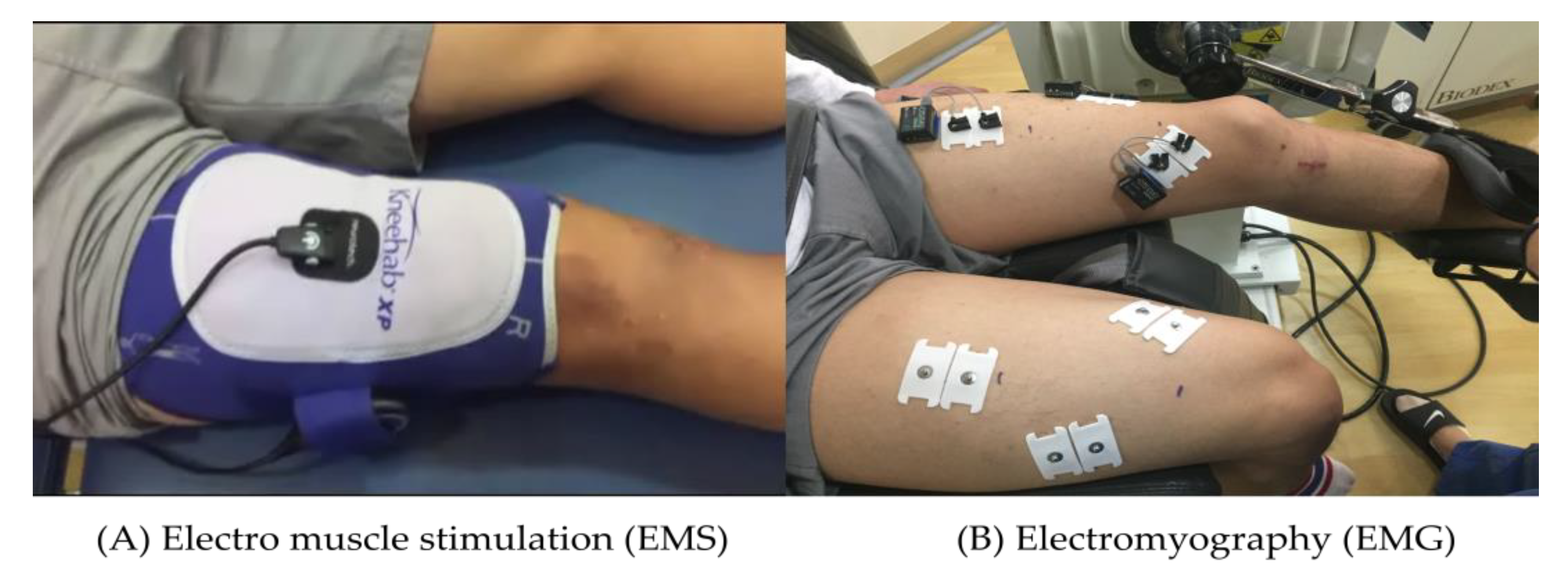
2.3. Electro Muscle Stimulation

EMS was performed using Kneehab (Biomedical Research Ltd., Galway, Ireland) equipment, and the frequency of electrical stimulation was set at 70 Hz. Stimulation-pulse duration (pulse wide) was 300 μs to 400 μs, and contraction for 10 s and relaxation for 50 s were repeated for 20 min (Figure 3A). The neuromuscular stimulation application group started on the second day after ACL reconstruction and was applied twice a week during a 12-week rehabilitation exercise [20].

2.4. Anthropometry

The height and weight were measured using Inbody’s BSM330 (Seoul, Korea) equipment. The participants were encouraged to remove any metal items from their bodies and to wear light clothing while having their measurements taken while standing straight.

2.5. Clinical Evaluation Parameters of the Subjective Knee Functional State

The subject’s subjective knee function status was assessed the Lysholm score and the IKDC score. ACL and meniscus injury patients’ symptoms and physical capabilities were assessed using the Lysholm score, which measures a variety of everyday job tasks, but excludes sports and leisure time. Higher scores on these eight-question assessments, which can provide a total score of 100, indicate less symptomatology and increased functioning.
The IKDC score assesses function throughout every day and motor tasks in individuals with knee ailments, including patellar-thigh pain or symptoms, ligament and semi-permanent cartilage loss, and other problems. Seven questions about knee problems, two about participating in sports, and two about bodily processes make up this question.

2.6. Thigh Circumference

A Balzer 80206F tape measure was used the circumference of the thighs (Hoechstmas, Sulzbach, Germany). The circumference was measured at two points, 5 cm and 15 cm up from the patella, after applying force, with all individuals standing with their legs as wide apart as their shoulders [21,22].

2.7. Muscle Activity

Surface myoelectricity tests were carried out to exert as much force on the femoral quadriceps as possible while seated on the floor in order to assess the level of muscle activation through isometric contraction of the femoral quadriceps, and the vastus medialis (VM), vastus lateralis (VL), and rectus femoris (RF) were also assessed (Figure 3B). A TeleMyo DTS (Noraxon Inc., Scottsdale, AZ, USA) device with a 22 mm diameter was used for wireless surface electromyography in order to analyze the EMG of the VM, VL, and RF. Each channel’s surface EMG analog data was transformed into a digital signal, which was then saved and subjected to analysis on a computer with software called MR-XP Clinical Edition 1.07 (USA) [23].

2.8. Isokinetic Muscular Function

The quadriceps muscle isokinetic muscular function, such as peak torque and total work, was assessed using an isokinetic dynamometer (Biodex Inc., Shirley, NY, USA). The quadriceps muscle peak torque (PT) was determined by completing four repetitions under a 60°/sec condition, and its total work (TW) was determined by completing 10 repetitions under a 180°/sec condition.

2.9. Statistical Analysis

The Windows SPSS program, version 25.0, was used to analyze the data (IBM Corp., Shirley, NY, USA). All data were given with their means and standard deviations (SD). The Shapiro–Wilk test was carried out and verified to ensure the homogeneity of all variables. The mean difference between the two groups as well as the dependent variables between the two tests were both analyzed concurrently using repeated two-way analysis of variance (ANOVA). The partial eta square (η2) value was used to show the effect size. The data with interactions and main effects were assessed using a paired t-test or an independent t-test. The 0.05 cutoff for significance was used.

3. Results

The participants of this study were a total of 24. There were nine males and three females in the CONG, 28.8 ± 10.3 years, height 171.1 ± 7.7 cm, and body weight 71.5 ± 13.6 kg. The EMSG were ten males and two females, 30.4 ± 13.0 years, height 173.3 ± 8.0 cm, and body weight 74.7 ± 12.9 kg. All participants were 29.6 ± 11.5 years, height 172.2 ± 7.8 cm and body weight 73.1 ± 13.0 kg.
Figure 4 shows the Lysholm and IKDC scores. The Lysholm score (p = 0.018, η2 = 0.228) and the IKDC score (p = 0.011, η2 = 0.258) both showed a major impact over time, despite the absence of a significant interaction. After ACL surgery, the post hoc analysis revealed that exercise rehabilitation significantly increased the Lysholm score (p = 0.049) in the EMSG and the IKDC score (p = 0.026) in the CONG in each group.
The findings of the thigh circumference are displayed in Figure 5. In the 5 cm circumference, the main effect of time was demonstrated (p = 0.049, η2 = 0.162), while at the 15 cm circumference, interactions were demonstrated (p = 0.010, η2 = 0.268). Post-analysis revealed that the circumference of the EMSG increased significantly at 5 cm (p = 0.001) and 15 cm (p = 0.013).
On the basis of Figure 6, there was an interaction in the muscle activity of VM during isokinetic contraction (p = 0.028, η2 = 0.202). Moreover, there was a significant main effect with time in the muscle activity of the VL (p = 0.004, η2 = 0.316), RF (p = 0.026, η2 = 0.207), and VM (p = 0.010, η2 = 0.268), respectively, during isometric contraction and isokinetic contraction. In the EMSG, post hoc analysis showed that the muscle activity was substantially greater in the VM during isokinetic contraction (p = 0.001) and in the VL (p = 0.015) and RF (p = 0.015) during isometric contraction, respectively.
In the quadriceps muscle peak torque, there was a significant interaction (p = 0.012, η2 = 0.253). The quadriceps femoris muscle’s peak torque and total work both showed a significant main impact that varied with time (p = 0.001, η2 = 0.405 and p = 0.008, η2 = 0.280, respectively). The PT (p = 0.002) and TW (p = 0.019) in EMSG both significantly increased, according to post hoc analysis (Figure 7).

4. Discussion

EMS was a training equipment known to improve neuromuscular functional activity by directly stimulating motor nerves and inducing muscle tetanus as a training method complementary to muscle strength development. It recently gained increasing popularity because it can be applied a topical or hole body [24,25]. It was recognized as an effective alternative approach to improving and maintaining muscle size and function, especially as diseases associated with muscle atrophy [26,27]. Hence, our study investigated if the complex use of EMS therapy and a standardized 12-week rehabilitation program for patients reduced quadriceps muscles due to ACL reconstruction would have any influence on the rapid recovery of muscular function.
Feil et al. [28] evaluated the complementary effect of adding electro muscle stimulation to a REP after ACL reconstruction using Kneehab. A substantial improvement in Lysholm was seen in the group using EMS compared to the group not using it in the clinical assessment findings of Lysholm and IKDC-2000. On the other hand, it was reported that there was an improvement in IKDC-2000, but there was no discernible variation in the main effect of time across the groups. In the our study, there was no interaction effect between groups in the Lysholm evaluation, but significant improvement in time was shown in EMSG, showing results consistent with previous studies. This is believed to have had a positive impact on the Lysholm score, which evaluates knee joint function in daily life, because of the rehabilitation of quadriceps femoris muscular strength and function [14,15]. On the other hand, the 12-week assessment revealed no significant differences across groups; however, the IKDC2000 examination revealed a considerable improvement in CONG. This could be because it takes three months following surgery to begin functional movements such as running and leaping, and there was no discernible evaluative disparity among the two groups.
Hasegawa et al. [29] examined the impact of early neuromuscular electrical stimulation after surgery and incorporated the EMS protocol at a 250 μs pulse length and 20 Hz frequency to the general rehabilitation program to prevent muscle degeneration after ACL reconstruction. The improvement in the thigh circumference after four weeks of treatment, five days a week, for 20 min, was noticeable. In addition, the vastus medialis muscle underwent neuromuscular electrical stimulation for 4 weeks, 5 days a week, for 15 min in patients with degenerative knee osteoarthritis, suggesting that it is effective based on the fact that the thigh circumference increased significantly and the quadriceps muscle strength increased [25]. Regarding these alterations, Martin et al. [30] suggested that long-term electrical stimulation of muscular neurons might turn type II fibers into type I fibers and result in type I fiber hypertrophy. The EMSG thigh circumference increased significantly in this study, as it did in the previous studies. Using a program in which multidirectional EMS was transmitted based on the vastus medialis and lateralis resulted in a notable increase in the circumference of the thigh at the 5 cm location centered on the patella compared to the general exercise group. It is believed that there is a connection between improved muscle strength as a result of a 12-week continuous exercise program and increased muscle activity of the VM and VL owing to neuromuscular adaptation [31,32].
Taradaj et al. [33] reported changes in femoral muscle strength reported that neuromuscular electrical stimulation was applied to soccer players that underwent anterior cruciate ligament surgery for four weeks, three days a week, three times a day, and 30 times a day. When applied as a program of 10 s muscle contraction, 50 s rest at a frequency of 50 Hz per minute, they reported significant improvements in quadriceps muscle strength and thigh circumference compared to controls. Similar outcomes were found in this investigation, although the quadriceps muscle’s isometric muscular strength function in the fully extended condition did not demonstrate a significant improvement in the VM muscle, but the VM muscle’s isokinetic muscle strength function did. This is connected to the fact that during open chain exercise (OKC), the vastus medialis muscle operates primarily on medial stability of the patella at the about 10–15° of knee joint [34]. Moreover, it was noted that during knee extension, the vastus medialis muscle (VM) had greater muscular activity than the vastus lateralis muscle (VL) [35]. According to Brownstein et al. [36] the vastus medialis (VM) showed a significant level of muscle activity when the knee was flexed between 60 and 90°, and a low level of activity when the knee was completely extended. Therefore, we found that more significant muscle activity occurred during more knee-extension movements in the full knee condition, and EMS exercise in particular is reported to increase muscle activity.
The present study revealed increased PT and TW in all groups after 12 weeks compared to pre-exercise, with the EMSG showing a significantly higher rate of increase in PT and TW than the CONG. According to Hammami et al. [37] a 6-week EMS exercise program enhanced maximal muscular strength much more than the normal exercise group. Werner et al. [38] discovered a slight but significant difference in the PT of lower limb muscular strength following the 10-week EMS training program. This was consistent with our study. Reviewing the results of such previous research and this experiment, it was concluded that an EMS exercise program of 6 weeks or longer has a positive effect on muscle function improvement. A previous study also reported that EMS was an efficient modality that elicited substantial improvements in isokinetic muscle function [39], suggesting that following ACL reconstruction, EMS paired with regular exercise will be an effective method for improving isokinetic muscle function within 4 weeks [40].
Toth et al. [41] reported that treatment of EMS for three weeks reduced atrophy of type II fibers and maintained preserved contractility of type I fibers to increase and, maintain contractile velocity and power output of muscle. As such, it is judged that the combined treatment of EMS and other dynamic exercises (e.g., plyometrics, sprints, vertical jumps) to improve muscle function in ACL patients can induce more muscle contractions. In addition, it is considered to be effective in that more fast-twitch fibers muscle are mobilized during muscle contraction, which helps to improve muscle hypertrophy and function. Therefore, it is determined that in order to increase voluntary muscular strength and anaerobic power output, future research should concentrate on the long-term effects of EMS dynamic contraction and the best way to combine EMS with dynamic training modalities.
Langeard et al. [42] reported that performing EMS training at least 4 weeks and two to four times a week, at a frequency 20 to 70 Hz is effective for muscle nerve improvement, and should be performed at a frequency above 50 Hz, and for at least 9 weeks, to induce effects on functional parameters such as walking. It was also said to be beneficial for improving muscle form changes and functions when employing appropriate frequencies and pulse widths, and typically calls for pulses of 100–400 μs, frequencies of 50–100 Hz, and contractions of 5–10 s [20,43]. Therefore, it is judged that the EMS program for 12 weeks of 10 sec contraction 50 sec relaxation at frequencies 70 Hz and 300–400 μs pulses in this study was positive for the improvement of related parameters.
There were some limitations of this study. The maximum current parameters, including current amplitude, pulse length, and pulse frequency, were applied in a similar manner to previous studies [44]. No participants showed hypersensitivity to electrical stimulation. The neurophysiological mechanisms of pain were not investigated in this study. We did not explore the link between the commencement of substantial muscle activity and changed sensory motor behavior or neuromuscular activity. Because each patient may have different goals or prefer some therapy modalities for rehabilitation over others, the underlying rehabilitation protocols followed for each patient may vary. Low sample sizes may also not accurately reflect the population as a whole. The EMS group may have produced better results because of the interactive nature of the program, which may possibly have affected motivational factors. Finally, this study needs further research to determine whether the effect of this intervention lasts for a long time. Follow-up, since simply measuring the perimeter is limited in determining the actual changed size of the muscle, future studies are expected to clarify the reason for the difference in thigh circumference by identifying the difference in actual muscle size with EMS through ultrasound or MRI.

5. Conclusions

Our research showed that improving muscle circumference, muscular activity, and muscle function may be accomplished more effectively with a rehabilitation program that incorporates EMS after ACL reconstruction. Nevertheless, there were no glaring disparities between the thigh circumference and the subjective knee function evaluation questionnaire. Further study is required to confirm the mechanism and effectiveness of a rehabilitation exercise program utilizing EMS on patients who underwent ACL reconstruction in terms of muscle hypertrophy and ligament strengthening.

Author Contributions

Conceptualization, S.-I.C., H.-W.M. and W.-S.J.; data curation, S.-I.C.; formal analysis, S.-I.C., J.-W.K., S.-S.N., H.-W.M. and W.-S.J.; investigation, S.-I.C., H.-W.M. and W.-S.J.; methodology, S.-I.C., J.-W.K., S.-S.N., H.-W.M. and W.-S.J.; writing—original draft, S.-I.C., H.-W.M. and W.-S.J.; writing—review and editing, S.-I.C., J.-W.K., S.-S.N., H.-W.M. and W.-S.J. All authors have read and agreed to the published version of the manuscript.

Funding

This research received no external funding.

Institutional Review Board Statement

The study was conducted according to the guidelines of the Declaration of Helsinki and was approved by the Institutional Review Board of Konkuk University Hospital (KUH1060126).

Informed Consent Statement

Informed consent was obtained from all participants involved in the study.

Data Availability Statement

The data presented in this study are available on request from the corresponding author.

Acknowledgments

We thank the staff of Konkuk University Hospital for their assistance in this study.

Conflicts of Interest

The authors declare no conflict of interest.

References

  1. Sandberg, R.; Balkfors, B.; Nilsson, B.; Westlin, N. Operative versus non-operative treatment of recent injuries to the ligaments of the knee. A prospective randomized study. J. Bone Joint Surg. Am. 1987, 69, 1120–1126. [Google Scholar] [CrossRef]
  2. Giove, T.P.; Miller, S.J., 3rd; Kent, B.E.; Sanford, T.L.; Garrick, J.G. Non-operative treatment of the torn anterior cruciate ligament. J. Bone Joint Surg. Am. 1983, 65, 184–192. [Google Scholar] [CrossRef] [PubMed]
  3. Tsepis, E.; Vagenas, G.; Giakas, G.; Georgoulis, A. Hamstring weakness as an indicator of poor knee function in ACL-deficient patients. Knee Surg. Sports Traumatol. Arthrosc. 2004, 12, 22–29. [Google Scholar] [CrossRef] [PubMed]
  4. Yang, D.J.; Park, S.K.; Uhm, Y.H.; Park, S.H.; Chun, D.W.; Kim, J.H. The correlation between muscle activity of the quadriceps and balance and gait in stroke patients. J. Phys. Ther. Sci. 2016, 28, 2289–2292. [Google Scholar] [CrossRef] [Green Version]
  5. Thomas, A.C.; Villwock, M.; Wojtys, E.M.; Palmieri-Smith, R.M. Lower extremity muscle strength after anterior cruciate ligament injury and reconstruction. J. Athl. Train. 2013, 48, 610–620. [Google Scholar] [CrossRef] [Green Version]
  6. Malone, T.; Blackburn, T.A.; Wallace, L.A. Knee rehabilitation. Phys. Ther. 1980, 60, 1602–1610. [Google Scholar] [CrossRef]
  7. Lindström, M.; Strandberg, S.; Wredmark, T.; Felländer-Tsai, L.; Henriksson, M. Functional and muscle morphometric effects of ACL reconstruction. A prospective CT study with 1 year follow-up. Scand. J. Med. Sci. Sports 2013, 23, 431–442. [Google Scholar] [CrossRef]
  8. Ithurburn, M.P.; Paterno, M.V.; Thomas, S.; Pennell, M.L.; Evans, K.D.; Magnussen, R.A.; Schmitt, L.C. Clinical measures associated with knee function over two years in young athletes after ACL reconstruction. Knee 2019, 26, 355–363. [Google Scholar] [CrossRef]
  9. Jenkins, S.M.; Guzman, A.; Gardner, B.B.; Bryant, S.A.; Del Sol, S.R.; McGahan, P.; Chen, J. Rehabilitation After Anterior Cruciate Ligament Injury: Review of Current Literature and Recommendations. Curr. Rev. Musculoskelet Med. 2022, 15, 170–179. [Google Scholar] [CrossRef] [PubMed]
  10. Hauger, A.V.; Reiman, M.P.; Bjordal, J.M.; Sheets, C.; Ledbetter, L.; Goode, A.P. Neuromuscular electrical stimulation is effective in strengthening the quadriceps muscle after anterior cruciate ligament surgery. Knee Surg. Sports Traumatol. Arthrosc. 2018, 26, 399–410. [Google Scholar] [CrossRef]
  11. Buckthorpe, M.; La Rosa, G.; Villa, F.D. Restoring knee extensor strength after anterior cruciate ligament reconstruction: A clinical commentary. Int. J. Sports Phys. Ther. 2019, 14, 159–172. [Google Scholar] [CrossRef] [Green Version]
  12. Bremner, C.B.; Holcomb, W.R.; Miller, M.G. Recommendations to Increase Neuromuscular Electrical Stimulation Training Intensity During Quadriceps Treatments for Orthopedic Knee Conditions. Clin. J. Sport Med. 2021, 31, 330–334. [Google Scholar] [CrossRef] [PubMed]
  13. Stevens, J.E.; Mizner, R.L.; Snyder-Mackler, L. Neuromuscular electrical stimulation for quadriceps muscle strengthening after bilateral total knee arthroplasty: A case series. J. Orthop. Sports Phys. Ther. 2004, 34, 21–29. [Google Scholar] [CrossRef] [PubMed] [Green Version]
  14. Glaviano, N.R.; Saliba, S. Can the Use of Neuromuscular Electrical Stimulation Be Improved to Optimize Quadriceps Strengthening? Sports Health 2016, 8, 79–85. [Google Scholar] [CrossRef]
  15. Lieber, R.L.; Silva, P.D.; Daniel, D.M. Equal effectiveness of electrical and volitional strength training for quadriceps femoris muscles after anterior cruciate ligament surgery. J. Orthop. Res. 1996, 14, 131–138. [Google Scholar] [CrossRef]
  16. Moritani, T.; Muro, M.; Kijima, A.; Gaffney, F.A.; Parsons, D. Electromechanical changes during electrically induced and maximal voluntary contractions: Surface and intramuscular EMG responses during sustained maximal voluntary contraction. Exp. Neurol. 1985, 88, 484–499. [Google Scholar] [CrossRef] [PubMed]
  17. Maffiuletti, N.A.; Vivodtzev, I.; Minetto, M.A.; Place, N. A new paradigm of neuromuscular electrical stimulation for the quadriceps femoris muscle. Eur. J. Appl. Physiol. 2014, 114, 1197–1205. [Google Scholar] [CrossRef] [PubMed]
  18. Faul, F.; Erdfelder, E.; Lang, A.G.; Buchner, A. G* Power 3: A flexible statistical power analysis program for the social, behavioral, and biomedical sciences. Behav. Res. Methods 2007, 39, 175–191. [Google Scholar] [CrossRef]
  19. Thomé, G.R.; Costa, R.A.; Marquezi, M.L.; Aparecido, J.M.; Durigan, J.L.Q.; Amorim, C.F.; Liebano, R.E. Effects of neuromuscular electrical stimulation on torque and performance in recreational distance runners: A randomized controlled trial. J. Bodyw. Mov. Ther. 2021, 28, 369–375. [Google Scholar] [CrossRef]
  20. Blazevich, A.J.; Collins, D.F.; Millet, G.Y.; Vaz, M.A.; Maffiuletti, N.A. Enhancing Adaptations to Neuromuscular Electrical Stimulation Training Interventions. Exerc. Sport Sci. Rev. 2021, 49, 244–252. [Google Scholar] [CrossRef]
  21. Soderberg, G.L.; Ballantyne, B.T.; Kestel, L.L. Reliability of lower extremity girth measurements after anterior cruciate ligament reconstruction. Physiother. Res. Int. 1996, 1, 7–16. [Google Scholar] [CrossRef] [PubMed]
  22. Wright, A.R.; Richardson, A.B.; Kikuchi, C.K.; Goldberg, D.B.; Marumoto, J.M.; Kan, D.M. Effectiveness of Accelerated Recovery Performance for Post-ACL Reconstruction Rehabilitation. Hawaii J. Health Soc. Welf. 2019, 78, 41–46. [Google Scholar] [PubMed]
  23. Tillin, N.A.; Folland, J.P. Maximal and explosive strength training elicit distinct neuromuscular adaptations, specific to the training stimulus. Eur. J. Appl. Physiol. 2014, 114, 365–374. [Google Scholar] [CrossRef] [PubMed]
  24. Matos, F.; Amaral, J.; Martinez, E.; Canário-Lemos, R.; Moreira, T.; Cavalcante, J.; Peixoto, R.; Pinheiro, B.N.; Junior, L.S.; Uchoa, P.; et al. Changes in Muscle Thickness after 8 Weeks of Strength Training, Electromyostimulation, and Both Combined in Healthy Young Adults. Int. J. Environ. Res. Public Health 2022, 19, 3184. [Google Scholar] [CrossRef]
  25. Lepley, L.K.; Wojtys, E.M.; Palmieri-Smith, R.M. Combination of eccentric exercise and neuromuscular electrical stimulation to improve quadriceps function post-ACL reconstruction. Knee 2015, 22, 270–277. [Google Scholar] [CrossRef] [Green Version]
  26. Xiong, J.; Zhang, Q.; Li, Y. Clinical Study of Neuromuscular Electrical Stimulation in the Prevention of Deep Venous Thrombosis of Lower Extremities after Anterior Cruciate Ligament Reconstruction. J. Healthc. Eng. 2022, 2022, 7857272. [Google Scholar] [CrossRef]
  27. Glattke, K.E.; Tummala, S.V.; Chhabra, A. Anterior Cruciate Ligament Reconstruction Recovery and Rehabilitation: A Systematic Review. J. Bone Jt. Surg. Am. 2022, 104, 739–754. [Google Scholar] [CrossRef]
  28. Feil, S.; Newell, J.; Minogue, C.; Paessler, H.H. The effectiveness of supplementing a standard rehabilitation program with superimposed neuromuscular electrical stimulation after anterior cruciate ligament reconstruction: A prospective, randomized, single-blind study. Am. J. Sports Med. 2011, 39, 1238–1247. [Google Scholar] [CrossRef]
  29. Hasegawa, S.; Kobayashi, M.; Arai, R.; Tamaki, A.; Nakamura, T.; Moritani, T. Effect of early implementation of electrical muscle stimulation to prevent muscle atrophy and weakness in patients after anterior cruciate ligament reconstruction. J. Electromyogr. Kinesiol. 2011, 21, 622–630. [Google Scholar] [CrossRef] [Green Version]
  30. Martin, L.; Cometti, G.; Pousson, M.; Morlon, B. Effect of electrical stimulation training on the contractile characteristics of the triceps surae muscle. Eur. J. Appl. Physiol. Occup. Physiol. 1993, 67, 457–461. [Google Scholar] [CrossRef]
  31. Oliveira, P.; Modesto, K.A.G.; Bottaro, M.; Babault, N.; Durigan, J.L.Q. Training Effects of Alternated and Pulsed Currents on the Quadriceps Muscles of Athletes. Int. J. Sports Med. 2018, 39, 535–540. [Google Scholar] [CrossRef] [PubMed]
  32. Mancinelli, R.; Toniolo, L.; Di Filippo, E.S.; Doria, C.; Marrone, M.; Maroni, C.R.; Verratti, V.; Bondi, D.; Maccatrozzo, L.; Pietrangelo, T.; et al. Neuromuscular Electrical Stimulation Induces Skeletal Muscle Fiber Remodeling and Specific Gene Expression Profile in Healthy Elderly. Front. Physiol. 2019, 10, 1459. [Google Scholar] [CrossRef] [PubMed] [Green Version]
  33. Taradaj, J.; Halski, T.; Kucharzewski, M.; Walewicz, K.; Smykla, A.; Ozon, M.; Slupska, L.; Dymarek, R.; Ptaszkowski, K.; Rajfur, J.; et al. The effect of neuromuscular electrical stimulation on quadriceps strength and knee function in professional soccer players: Return to sport after ACL reconstruction. BioMed Res. Int. 2013, 2013, 802534. [Google Scholar] [CrossRef]
  34. Deng, W.; Lin, M.; Yu, S.; Liang, H.; Zhang, Z.; Tang, C.; Liu, C. Effects of Hip Joint Angle on Quadriceps Recruitment Pattern During Knee Extension in Healthy Individuals: Analysis by Ultrasound-Based Shear-Wave Elastography. Front. Physiol. 2022, 13, 836435. [Google Scholar] [CrossRef] [PubMed]
  35. Laprade, J.; Culham, E.; Brouwer, B. Comparison of five isometric exercises in the recruitment of the vastus medialis oblique in persons with and without patellofemoral pain syndrome. J. Orthop. Sports Phys. Ther. 1998, 27, 197–204. [Google Scholar] [CrossRef] [PubMed] [Green Version]
  36. Brownstein, B.; Lamb, R.E.; Mangine, R.E. Quadriceps torque and integrated electromyography*. J. Orthop. Sports Phys. Ther. 1985, 6, 309–314. [Google Scholar] [CrossRef]
  37. Hammami, N.; Jdidi, H.; Khezami, M.A.; Ghidaoui, L.; Talbi, A.; Hannachi, C.; Farinha, P.M.; Behlouli, E.; Bouassida, A.; Dziri, C.; et al. Isokinetic strengthening and neuromuscular electrical stimulation protocol impact on physical performances, functional status and quality of life in knee osteoarthritis overweight/obese women. Knee 2022, 39, 106–115. [Google Scholar] [CrossRef] [PubMed]
  38. Werner, S.; Arvidsson, H.; Arvidsson, I.; Eriksson, E. Electrical stimulation of vastus medialis and stretching of lateral thigh muscles in patients with patello-femoral symptoms. Knee Surg. Sports Traumatol. Arthrosc. 1993, 1, 85–92. [Google Scholar] [CrossRef]
  39. Gondin, J.; Cozzone, P.J.; Bendahan, D. Is high-frequency neuromuscular electrical stimulation a suitable tool for muscle performance improvement in both healthy humans and athletes? Eur. J. Appl. Physiol. 2011, 111, 2473–2487. [Google Scholar] [CrossRef]
  40. Kim, K.M.; Croy, T.; Hertel, J.; Saliba, S. Effects of neuromuscular electrical stimulation after anterior cruciate ligament reconstruction on quadriceps strength, function, and patient-oriented outcomes: A systematic review. J. Orthop. Sports Phys. Ther. 2010, 40, 383–391. [Google Scholar] [CrossRef] [Green Version]
  41. Toth, M.J.; Tourville, T.W.; Voigt, T.B.; Choquette, R.H.; Anair, B.M.; Falcone, M.J.; Failla, M.J.; Stevens-Lapslaey, J.E.; Endres, N.K.; Slauterbeck, J.R.; et al. Utility of Neuromuscular Electrical Stimulation to Preserve Quadriceps Muscle Fiber Size and Contractility After Anterior Cruciate Ligament Injuries and Reconstruction: A Randomized, Sham-Controlled, Blinded Trial. Am. J. Sports Med. 2020, 48, 2429–2437. [Google Scholar] [CrossRef] [PubMed]
  42. Langeard, A.; Bigot, L.; Chastan, N.; Gauthier, A. Does neuromuscular electrical stimulation training of the lower limb have functional effects on the elderly?: A systematic review. Exp. Gerontol. 2017, 91, 88–98. [Google Scholar] [CrossRef] [PubMed]
  43. de Oliveira Melo, M.; Aragão, F.A.; Vaz, M.A. Neuromuscular electrical stimulation for muscle strengthening in elderly with knee osteoarthritis—A systematic review. Complement. Ther. Clin. Pract. 2013, 19, 27–31. [Google Scholar] [CrossRef] [PubMed]
  44. Nussbaum, E.L.; Houghton, P.; Anthony, J.; Rennie, S.; Shay, B.L.; Hoens, A.M. Neuromuscular Electrical Stimulation for Treatment of Muscle Impairment: Critical Review and Recommendations for Clinical Practice. Physiother. Can. 2017, 69, 1–76. [Google Scholar] [CrossRef] [Green Version]
Figure 1. The CONSORT Flow Diagram. control group; CONG, electro muscle stimulation group; EMSG.
Applsci 13 04153 g001
Figure 2. Experimental process. control group; CONG, electro muscle stimulation group; EMSG.
Applsci 13 04153 g002
Figure 3. Measurement method of Electro Muscle Stimulation (A) and Muscle Activity (B).
Applsci 13 04153 g003
Figure 4. The rehabilitative exercise programs’ effects on Lysholm score and IKDC score. (A) Compared in Lysholm score. (B) Compared in IKDC score. control group; CONG, electro muscle stimulation group; EMSG, International Knee Documentation Committee; IKDC. # p < 0.05: main effect or interaction; * p < 0.05: pre- and post-test.
Applsci 13 04153 g004
Figure 5. The rehabilitative exercise programs’ effects on circumference at 5 cm and 15 cm. (A) Compared in 5 cm circumference. (B) Compared in 15 cm circumference. control group; CONG electro muscle stimulation group; EMSG. # p < 0.05: main effect or interaction; * p < 0.05: pre- and post-test.
Applsci 13 04153 g005
Figure 6. The rehabilitative exercise programs’ effects on muscle activity of VM, VL, and RF during isometric and isokinetic contraction. (A) Compared in VM during isometric contraction. (B) Compared in VL during isometric contraction. (C) Compared in RF during isometric contraction. (D) Compared in VM during isokinetic contraction. (E) Compared in VL during isokinetic contraction. (F) Compared in RF during isokinetic contraction. control group; CONG, electro muscle stimulation group; EMSG, vastus medialis; VM, vastus lateralis; VL, rectus femoris; RF. # p < 0.05: main effect or interaction; * p < 0.05: pre- and post-test.
Applsci 13 04153 g006
Figure 7. The rehabilitative exercise programs’ effects on isokinetic muscle function. (A) Compared in quadriceps femoris muscle PT. (B) Compared in quadriceps femoris muscle TW. control group; CONG, electro muscle stimulation group; EMSG, peak torque; PT, total work; TW. # p < 0.05: main effect or interaction; * p < 0.05: pre- and post-test.
Applsci 13 04153 g007
Table 1. Participants’ characteristics. Data are means (±SD).
ParametersCONG (n = 12)EMSG (n = 12)p-Value
Gender (male/female)10/29/3-
Age (years)30.4 ± 13.028.8 ± 10.60.542
Height (cm)173.3 ± 8.0171.1 ± 7.70.611
Body weight (kg)66.6 ± 12.266.2 ± 18.30.917
Body mass index (kg·m−2)22.8 ± 3.222.2 ± 4.00.854
Tegner activity scale6.8 ± 1.66.4 ± 1.60.620
ACL reconstruction side (Rt/Lt)7/55/7-
Note. standard deviation; SD, anterior cruciate ligament; ACL, right; Rt, left; Lt.
Table 2. Rehabilitation exercise program.
ExerciseWeeks of Program
0–23–45–67–89–12
  • Range of motion
  0–90°o
  0–130° o
2.
Stretching (hamstring femoris and quadriceps muscle)
ooooo
3.
Muscle strength (quadriceps muscle setting exercise, straight leg raising, active leg extension)
oo
4.
Closed-kinetic chain exercise (gait retraining, squat, heel raise)
oooo
5.
Open-kinetic chain exercise
  Leg extension (90~60°) oo
  Leg curl (non-weight) oo
  Leg curl (weight) oo
6.
Proprioception training
  Weight shifting, cup walkingoo
  One leg balance ooo
7.
Perturbation
o
Disclaimer/Publisher’s Note: The statements, opinions and data contained in all publications are solely those of the individual author(s) and contributor(s) and not of MDPI and/or the editor(s). MDPI and/or the editor(s) disclaim responsibility for any injury to people or property resulting from any ideas, methods, instructions or products referred to in the content.

