Article

Evaluation of Dimensional Stability and Occlusal Wear of Additively and Subtractively Manufactured Resin-Based Crowns after Thermomechanical Aging

by
Mehmet Esad Güven
1,
Gülce Çakmak
2,
Mustafa Borga Dönmez
2,3,* and
Burak Yilmaz
2,4,5
1
Department of Prosthodontics, Faculty of Dentistry, Necmettin Erbakan University, 42090 Konya, Turkey
2
Department of Reconstructive Dentistry and Gerodontology, School of Dental Medicine, University of Bern, 3012 Bern, Switzerland
3
Department of Prosthodontics, Faculty of Dentistry, Istinye University, 34010 Istanbul, Turkey
4
Department of Restorative, Preventive, and Pediatric Dentistry, School of Dental Medicine, University of Bern, 3012 Bern, Switzerland
5
Division of Restorative and Prosthetic Dentistry, The Ohio State University, Columbus, OH 43210, USA
*
Author to whom correspondence should be addressed.
Appl. Sci. 2023, 13(21), 11849; https://doi.org/10.3390/app132111849
Submission received: 19 September 2023 / Revised: 23 October 2023 / Accepted: 24 October 2023 / Published: 30 October 2023
(This article belongs to the Special Issue CAD & CAM Dentistry)

Abstract

:
The knowledge on the surface deviations and wear of recently introduced additively or subtractively manufactured materials indicated for definitive prosthesis is limited. The aim of this present study was to evaluate the external surface and mesiodistal width deviation and the occlusal surface wear of one additively manufactured composite resin (MS) and three subtractively manufactured resins (nanographene-reinforced polymethylmethacrylate (GR), conventional polymethylmethacrylate (PMMA), and reinforced composite resin (BC)) after thermomechanical aging. Molar-shaped crowns were fabricated in the tested materials and digitized with an intraoral scanner (CEREC Primescan; Dentsply Sirona, Bensheim, Germany). Each crown was subjected to thermomechanical aging and rescanned with the same scanner. A three-dimensional analysis software (Geomagic Control X v.2022.1; 3D Systems, Rock Hill, SC, USA) was used to calculate the deviations on the external surface, mesiodistal width, and wear on the occlusal surfaces of the tested crowns. Data were analyzed using one-way ANOVA and Tukey’s tests (α = 0.05). MS had higher external surface deviations than PMMA and GR (p ≤ 0.038) and higher mesiodistal width deviations than PMMA and BC (p = 0.004). BC and GR had higher volume loss than PMMA (p ≤ 0.002). The additively manufactured composite resin was more prone to deviations, while reinforced composite resin had lower wear resistance than most of the tested materials.

1. Introduction

Computer-aided design and computer-aided manufacturing (CAD-CAM) technologies have diversified the materials that can be used to additively and subtractively fabricate definitive prostheses [1,2,3,4]. The incorporation of nanotechnologies into dentistry has also been a subject of interest, as well as a polymethyl methacrylate (PMMA) reinforced with nanographene-reinforced, which is indicated for definitive prostheses has been launched recently [5]. This new material (G-CAM; Graphenano DENTAL SL) comprises a crystalline form of carbon known as graphene, which forms a tightly packed lattice on a large surface area in the shape of a honeycomb [6]. This unique structure enhances the mechanical properties of PMMA [3,6,7]. Additive manufacturing has also become popular, given that this technology allows for the fabrication of products with complex geometries with less waste and cost than subtractive manufacturing [8,9,10]. Additively manufactured composite resins are among the latest materials indicated for definitive prostheses [4].
Wear is material loss from the surface, and it is a multifactorial process that can be either physiological or pathological when intraoral situations are considered [11]. An ideal restorative material should wear similar to that of natural dentition [12], and excessive wear might lead to the loss of both esthetics and function [13]. The wear behavior of a material depends on various properties, such as filler size, filler volume, filler hardness, elastic modulus, fracture toughness, and hardness [14].
Even though additively manufactured composite resins [4,15,16,17,18,19,20,21,22,23] and nanographene-reinforced PMMA [3,5,6,7,19,24,25,26,27] have been investigated previously, only one study has focused on the comparison between these materials as Çakmak et al. [19] investigated the fabrication trueness of these materials when fabricated as crowns. Given that restorative materials should maintain their integrity intraorally, a study based on how these new-generation resin-based CAD-CAM materials indicated for definitive prostheses behave after thermomechanical aging could elaborate the knowledge on their clinical applicability. However, to the authors’ knowledge, only two studies evaluated the wear of additively manufactured composite resins [21,22], and the wear of nanographene-reinforced PMMA has not been investigated. Therefore, the aim of this present study was to compare the external surface and mesiodistal width deviations (any change in the external surface or mesiodistal width determined using the greatest mesiodistal width of the occlusal third of the crowns due to leaching or water absorption during thermomechanical aging) and occlusal surface wear (material loss from the occlusal surface during thermomechanical aging) of one additively manufactured composite resin and three subtractively manufactured resins (one nanographene-reinforced PMMA, one PMMA, and one reinforced composite resin) after thermomechanical aging. The null hypotheses were that material type would not affect external surface deviations, mesiodistal width deviations, and occlusal surface wear after thermomechanical aging.

2. Materials and Methods

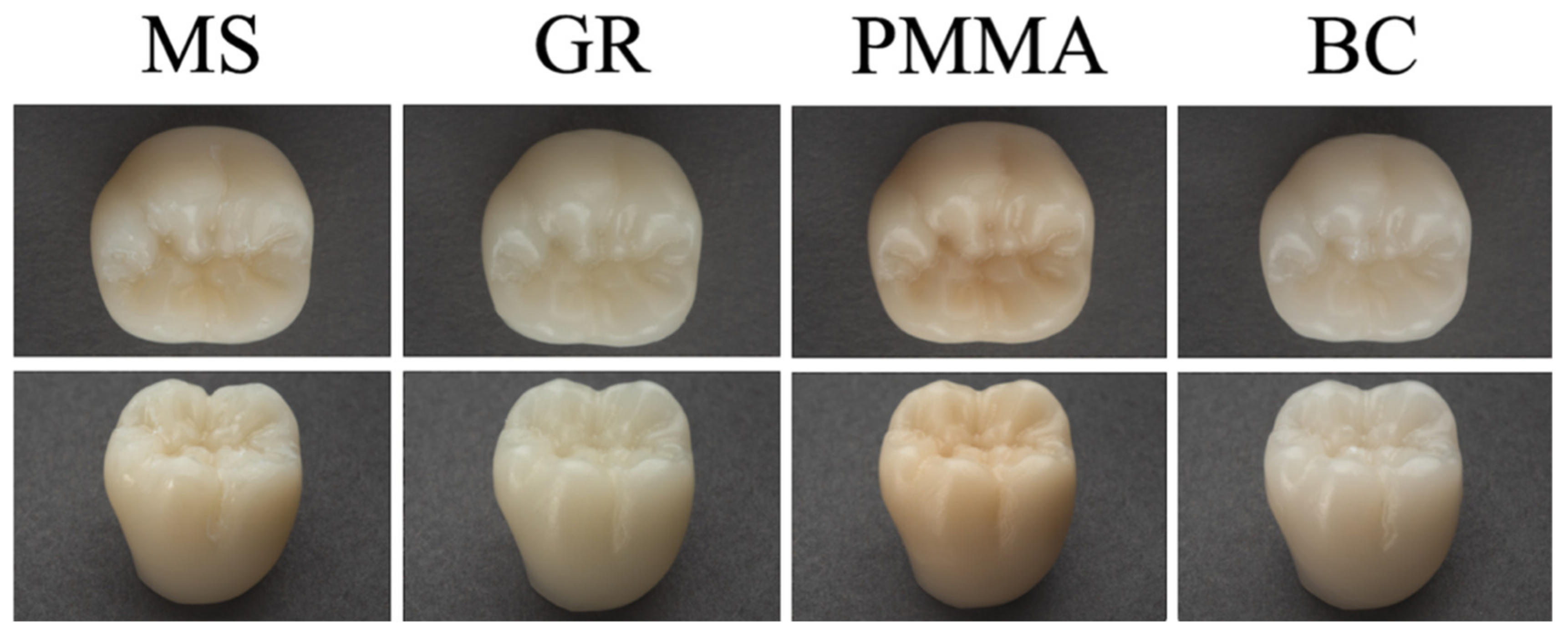
2.1. Specimen Preparation

A complete mandibular right first molar crown that had 30 µm cement gap [28], 3 mm thick axial walls, 1.5 mm thick margins, and 1 mm of minimum occlusal thickness [19] was designed in standard tessellation language (STL) format using dental design software (exocad DentalCAD v3.0; exocad GmbH, Darmstadt, Germany) and the STL of a prefabricated titanium abutment. A total of 40 crowns were fabricated in additively manufactured composite resin (Crowntec; Saremco Dental AG, Rebstein, Switzerland (MS)), graphene-reinforced PMMA (G-CAM; Graphenano DENTAL SL, Valencia, Spain (GR)), PMMA (breCAM.monoCOM; bredent GmbH, Senden, Germany (PMMA)), and reinforced composite resin (Brilliant Crios; Coltène AG, Altstätten, Switzerland (BC)) (Table 1) using this crown STL (n = 10). A priori power analysis (f = 0.90, 1 − β = 95%, α = 0.05) performed using the results of a previous study on the wear of additively and subtractively manufactured resins [21] deemed 7 specimens to be sufficient; however, the statistical power was increased by fabricating 10 specimens, which also allowed compensation in case of specimen loss during thermomechanical aging.
A digital light processing 3-dimensional printer (MAX UV; ASIGA, Sydney, Australia) was used to fabricate MS crowns with 50 µm layer thickness. A 5-axis milling unit (PrograMill PM7; Ivoclar AG, Schaan, Liechtenstein) was used to fabricate GR, PMMA, and BC crowns [19]. A cut-off wheel was used to remove the support structures, and any remnants on the external surface were smoothened under optical magnification loupes at ×3.5 magnification using a small round carbide bur at 10,000 rpm (Figure 1). A single operator (G.Ç.) performed all fabrication and adjustments.
The STL file of the abutment was used to fabricate 40 fiberglass-reinforced epoxy resin abutments (G10; McMaster-Carr, Atlanta, GA, USA) with the same 5-axis milling unit. The fit of the crowns to abutment dies was controlled with the same optical magnification loupes. Abutments were then cleaned for 10 min in an ultrasonic cleaner (Jelsonic; Jelenko, New Hyde Park, NY, USA), and autopolymerizing acrylic resin (PremEco; Merz Dental GmbH, Lütjenburg, Germany) was used to embed the crowns into the molds of the lower part of a mastication simulator. Abutment surfaces were etched with 35% phosphoric acid for 30 s (Etchant Gel S; Coltène AG, Altstätten, Switzerland), steam-cleaned, and air-dried. Intaglio surfaces of the crowns were also steam-cleaned and air-dried. Then, an adhesive (One Coat 7 Universal; Coltène AG, Altstätten, Switzerland) was applied with a microbrush to the abutment and crown surfaces, left undisturbed for 20 s, and gently air-dried for 5 s. A light-emitting diode curing unit (Bluephase; Ivoclar AG, Schaan, Liechtenstein), which has a power density of 950 mW/cm2, was used to light-polymerize the adhesive for 10 s. The crowns were then cemented onto their respective fiberglass-reinforced epoxy resin abutments using a self-adhesive dual-polymerizing resin cement (SoloCem; Coltène AG, Altstätten, Switzerland). A constant load of 2 N was applied during cementation with a brass holder [29]. Resin cement was light-polymerized for 3 s at each of two opposite sides to remove the excess cement with a scalpel (Surgical Scalpel Blade #11; Swann-Morton, Sheffield, UK) followed by additional light-polymerization for 20 s per surface. The crown-abutment complex was left for 10 min under the brass holder until the resin cement was completely set and then stored in distilled water (37 °C) for 24 h.

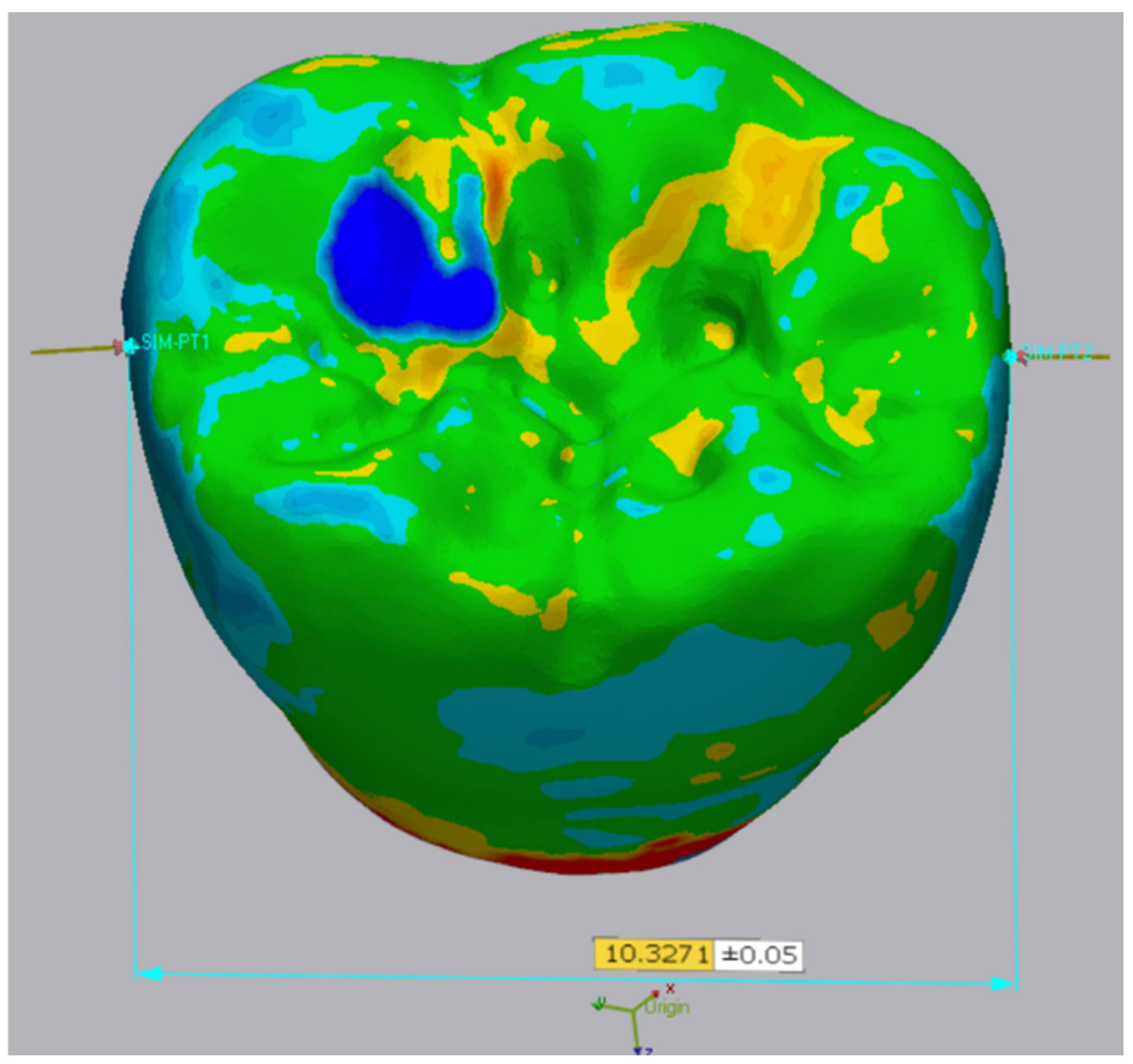
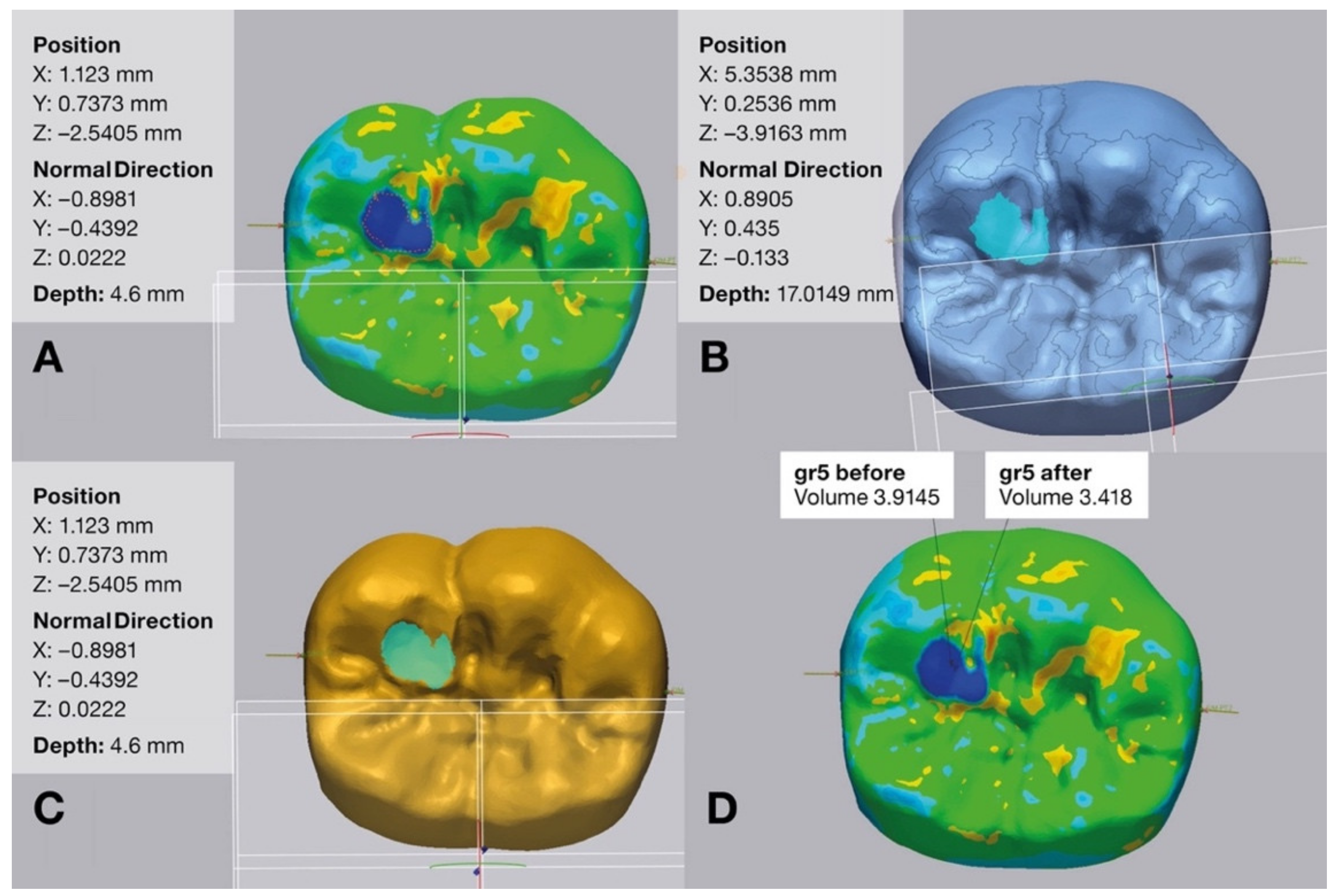
2.2. Thermomechanical Aging
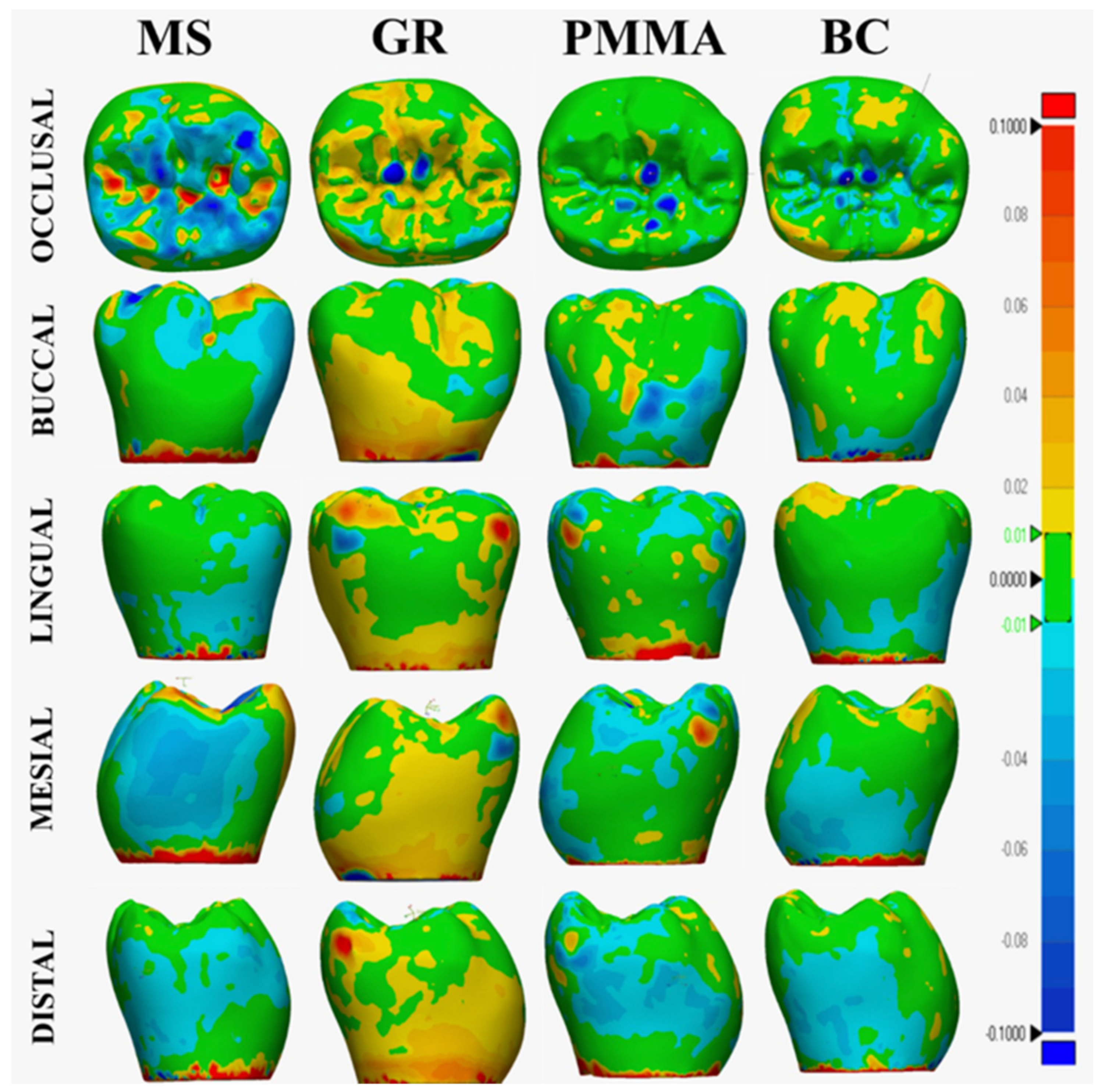
An experienced operator (M.E.G.) scanned the entire anatomy of the crowns in a temperature and humidity-controlled room under ambient light using the same intraoral scanner used to digitize the prefabricated abutment. The intraoral scanner was calibrated before starting the scans and recalibrated in every 5 crowns, and the operator took 5 min breaks after every 5 crowns to minimize fatigue-related deviations. The scans were checked for any error and saved as before thermomechanical aging test STL (BTMA-STL) files to further compare them with after thermomechanical aging STLs (ATMA-STLs) to evaluate external surface deviations and occlusal surface wear.
The crowns were randomly (Excel; Microsoft Corp, Seattle, WA, USA) mounted on a dual-axis computer-controlled mastication simulator (Dent Ar-Ge; Analitik Medical, İstanbul, Turkey), including a thermal-cycler. Crowns were aged at 1.7 Hz frequency for 1.2 million cycles under 50 N load with a lateral movement of 1 mm and a vertical and lateral sliding speed of 60 mm/s to simulate 5 years of functional loading [4,15,16,17]. The load was applied with 3 mm stainless steel sphere antagonists, which contacted the central fossae of the crowns. Force concentrations at asperities on the occlusal surface were avoided by placing a piece of tin foil between the crown and the steel sphere. Crowns were also simultaneously thermocycled for 6000 cycles between 5–55 °C with 30 s holding and 15 s transfer time in distilled water. After thermomechanical aging, crowns were visually inspected in a stereomicroscope (SMZ 1500; Nikon Corp., Tokyo, Japan) for the presence of any failures (Figure 2). Thereafter, crowns were scanned within 48 h with the same intraoral scanner to generate ATMA-STLs.
To evaluate the dimensional stability, which refers to the surface topography (external surface changes in RMS) and linear changes (mesiodistal width in microns) against liquid media storage and loading, and occlusal surface wear, which refers to the volume loss against liquid media storage and direct contact with the stainless sphere load application, of the crowns, an industrial-grade 3-dimensional (3D) quality control and dimensional inspection software program (Geomagic Control X v.2022.1; 3D Systems, Rock Hill, SC, USA) was used. For each specimen, BTMA-STLs were imported as the reference data. Then, ATMA-STLs of corresponding crown were imported and superimposed over BTMA-STL using initial alignment followed by best-fit alignment to minimize errors. Root mean square (RMS) method was used to calculate the 3D deviations at the external surfaces. The “3D Compare Tool” of the software was used to generate color maps for the quantitative evaluation of the deviations at the external surfaces. Overcontoured areas are indicated in red color, and undercontoured areas are indicated in blue color, with maximum/minimum deviation values set at 100 μm. The tolerance range was set at 10 μm and indicated in green color.
To measure the mesiodistal width deviations, BTMA-STL and ATMA-STL were superimposed as mentioned above, and one coordinate measuring machine (CMM) point was generated on the mesial and the distal contact surface on the superimposed data (Figure 3). These CMM points ensured the greatest mesiodistal width on the occlusal third, and the distance between these points was automatically calculated for both STLs. The absolute difference between these values was used for the statistical analyses. To calculate the volume loss, ATMA-STLs were superimposed over their respective BTMA-STLs, and the “measurement tool-volume inspection tool-enclosed volume” feature of the software was used to encapsulate the worn area. Thereafter, worn area was manually selected using the “paint brush tool” of the software on both BTMA-STL and ATMA-STL. Both STLs’ volumetric loss at the worn area was automatically calculated. Statistical analysis was performed using the absolute volume difference [21] (Figure 4).
Shapiro–Wilk test confirmed the normal distribution of data. Therefore, 1-way analysis of variance followed by Tukey honestly significant difference tests were used for further analyses. All analyses were performed with a statistical analysis software program (IBM SPSS Statistics v22.0; IBM Corp., Seattle, WA, USA) (α = 0.05).

3. Results

Table 2 summarizes descriptive statistics of tested parameters for each material. Significant differences were observed among the tested materials when external surface deviations (df = 3, F = 6.941, and p = 0.001), mesiodistal width deviations (df = 3, F = 15.7, and p < 0.001), and volume loss at wear area (df = 3, F = 9.616, and p < 0.001) were considered.
MS and BC had higher external surface deviations than PMMA (p = 0.001 for MS and p = 0.016 for BC). In addition, MS had higher deviations than GR (p = 0.038), and the difference in external surface deviations between other materials was nonsignificant (p ≥ 0.308). The color map of GR was predominantly yellow, which indicates a slightly overcontoured occlusal surface, a mesiobuccal groove that lies between the mesiobuccal and distobuccal cusps, and the area below the occlusal third of the crown. In addition, orange and red areas indicate overcontoured (moderate to high) lingual inclinations of the mesiolingual cusp. The color map of BC had yellow on the buccal inclination of the buccal cusp and on the lingual inclination of the mesiolingual cusp. PMMA also had yellow-colored areas at the buccal inclination of the distobuccal cusp. The overcontouring of varying magnitudes was evident at the margins, regardless of the material. MS had higher mesiodistal width deviations than PMMA and BC (p = 0.004). However, the deviations in mesiodistal width did not significantly differ when other material comparisons were made (p ≥ 0.218). MS had a distinct external surface color map with evident light blue and blue colors that covered a large area at the occlusal and interproximal surfaces (Figure 5).
Figure 6 shows the distribution of raw mesiodistal width deviation among test groups. BC had higher volume loss in the worn area than MS and PMMA (p ≤ 0.047), while GR had higher volume loss in the worn area than PMMA (p = 0.002). The differences in occlusal surface wear of remaining material comparisons were nonsignificant (p ≥ 0.104).

4. Discussion

The first and the second null hypotheses were rejected as tested resin-based CAD-CAM materials had significant differences when external surface deviations after thermomechanical aging were considered. In addition, MS had higher mesiodistal width deviations than other materials, except for GR.
MS had either significantly or nonsignificantly higher external surface and mesiodistal width deviations than tested subtractively manufactured resin-based materials. This could be related to the fact that subtractively manufactured disks or blocks are fabricated under standardized conditions [26], whereas additively manufactured resins require additional polymerization after fabrication [30]. Therefore, tested subtractively manufactured materials may have a higher degree of conversion and a more stable structure, which is supported by the color maps’ (Figure 5) qualitative interpretation, as MS’ color maps suggest degradation and leaching that is also supported by the mesiodistal width deviations and the distribution of raw mesiodistal width deviation data (Figure 6). This finding may lead to potential occlusal and interproximal contact issues becoming possibly lighter in the long term for MS crowns. When tested subtractively, manufactured resin-based materials were considered, BC had higher external surface deviations than PMMA, and GR had similar deviations to both BC and PMMA. The chemical compositions of subtractively manufactured materials may have led to these results, as BC has a more heterogeneous composition than PMMA. However, the manufacturer of GR did not disclose its composition, and this hypothesis needs to be supported by studies on the composition of these materials.
The color map findings suggest potential occlusal contact issues and irritation of the patient’s cheek for GR crowns and occlusal interferences for GR and BC crowns, particularly during laterotrusive movements of the mandible. Even though PMMA also had slightly overcontoured areas at the buccal inclination of the distobuccal cusp, they were relatively small compared with GR and BC. Therefore, it can be hypothesized that these overcontoured areas may interfere with the occlusion less than those of GR and BC crowns, and potential adjustments would take less time. The expansion at the margins may be related to potential stress concentration during thermomechanical aging or to the closeness of the expanded area to abutment–crown junction, which involves a preset cement gap. Nevertheless, how these dimensional changes affect the marginal integrity of the crowns should be clinically investigated. The mesiodistal width deviations were similar among tested subtractively manufactured materials, which resemble the interproximal surface color maps of those materials, considering that mesiodistal width deviations were measured from the occlusal third of the crowns. Distribution of colors also supports the raw mesiodistal width deviation data as GR mostly had positive deviations indicating tighter contacts over time and potential issues with flossing, whereas PMMA and BC had negative deviations indicating lighter contacts. Even though raw mesiodistal width deviation data do not disclose on which surface the deviation had occurred, color maps suggest interproximal issues at the distal surface of BC and at both surfaces of GR and PMMA. Previous studies have evaluated additively manufactured definitive composite resins [4,15,16,17,21,22,23] or nanographene-reinforced PMMA [24,25,26,27] after aging; however, the dimensional changes of these materials after aging have not been investigated. Therefore, the results of this present study could not be compared with previous studies.
The third null hypothesis was also rejected, as when volume loss at the worn area was considered, significant differences were observed among the tested materials, as BC had higher volume loss than the tested materials other than GR. In addition, GR had a higher volume loss than PMMA. Considering the fact that the tested materials have different chemical compositions, and even though GR’s composition is not disclosed, it differentiates from other materials with the presence of graphene, and their resistance to wear is also expected to be different from each other. There are studies on the hardness of most of the tested materials in this present study [5,14,23,24,25]; however, those studies either did not involve any aging or used thermocycling to age the specimens and were not based on direct comparisons among them. In addition, only the manufacturer of MS has disclosed their internal hardness data [31]. Therefore, this speculation should be supported by studies on the inherent and after-aging hardness of the tested materials. GR’s and BC’s volume losses being higher than PMMA may be related to their water sorption during thermomechanical aging, which might have increased the number of contact points with the indenter and caused a greater loss. However, it should be mentioned that this present study did not investigate the maximum wear depth; thus, higher volume loss of GR and BC does not necessarily mean a deeper worn area, as volume loss may have occurred on a wider surface considering the circular motion of the chewing simulator, and these differences may not lead to varying clinical effects. Despite the efforts to standardize the location of the contact point of the indenter during aging, worn areas were evident at different locations on the occlusal surfaces of the crowns (Figure 2), which supports this hypothesis.
In a recent study on the volume loss of MS and BC, it was concluded that BC had lower volume loss and wear depth than MS [21]. This difference with this present study may be associated with the design of test specimens, as cement-retained molar crowns were tested in this present study, whereas screw-retained premolar crowns were tested in the previous study [21]. In addition, a laser scanner was used in Diken Türksayar et al.’s [21] study, and the inherent inaccuracy of the scanner might have affected the results. Nevertheless, the intraoral scanner used to scan each crown in this study has precision similar to that of laboratory scanners [32], and it has also been used in previous studies on the fabrication trueness of additively and subtractively manufactured restorations [19]. The International Organization for Standardization standard 12836 referred to the metrology-grade 3D analysis software (Geomagic Control X v.2022.1; 3D Systems, Rock Hill, SC, USA) used in this present study for digital analyses [33], and previous studies on wear analysis have also used this software [13,21]. The best-fit algorithm was deliberately preferred to avoid any operator-related error during superimpositions, given that the worn area would have been selected manually. Considering these aspects, the authors think that the methodology to analyze the volume loss is justified.
None of the specimens was polished as this present study aimed to evaluate the inherent resistance of the tested materials to deviations and occlusal surface wear caused by thermomechanical aging, and it is difficult to standardize the polishing of complex geometries like crowns. However, polishing performed either at the chairside or the dental laboratory may lead to a more stable external surface and enhance the wear resistance of the tested materials due to improved surface roughness. In addition, polishing may reduce the amount of unpolymerized monomers on the external surface of tested resin-based materials, particularly those of MS, given that it was the only additively manufactured material that had to be postpolymerized. Another limitation was that the test design of this present study was based on two-body wear; however, a different design that involves three-body wear, such as brushing, may affect the results. Only one type of antagonist was used for thermomechanical aging, and the type of antagonist may affect the wear [14]. In addition, the wear of the antagonists has not been investigated in this present study. Even though all digital analyses were performed by a single experienced operator, some aspects of the volume loss evaluation were operator-dependent, which might have affected the results. Finally, other clinically relevant mechanical properties of the tested materials, such as their surface roughness and fracture strength, were not evaluated in this present study. Future in vivo studies are needed to corroborate the results of this present study and to substantiate the interpretations made based on these results.

5. Conclusions

Tested additively, manufactured composite resin crowns had either similar or higher external surface and mesiodistal width deviations than those made of subtractively manufactured materials. Subtractively manufactured reinforced composite resin crowns had higher volume loss than additively manufactured composite resin and polymethylmethacrylate crowns. However, the clinical effects of differences in deviation values and volume loss should be corroborated by clinical studies.

Author Contributions

Conceptualization, G.Ç., M.E.G. and B.Y.; methodology, M.E.G. and G.Ç.; software, M.E.G.; validation, G.Ç. and M.B.D.; formal analysis, M.E.G.; investigation, M.E.G. and G.Ç.; resources, M.B.D. and M.E.G.; data curation, M.E.G.; writing—original draft preparation, M.B.D. and G.Ç.; writing—review and editing, M.B.D. and B.Y.; visualization, M.E.G.; supervision, B.Y. and G.Ç. All authors have read and agreed to the published version of the manuscript.

Funding

This research received no external funding.

Institutional Review Board Statement

Not applicable.

Informed Consent Statement

Not applicable.

Data Availability Statement

Data sharing is not applicable to this paper.

Acknowledgments

The authors would like to thank med. dent. Ana Maria Rusa for their contributions during specimen preparation.

Conflicts of Interest

The authors declare no conflict of interest.

References

  1. Peng, C.C.; Chung, K.H.; Yau, H.T.; Ramos, V., Jr. Assessment of the internal fit and marginal integrity of interim crowns made by different manufacturing methods. J. Prosthet. Dent. 2020, 123, 514–522. [Google Scholar] [CrossRef]
  2. Guo, N.; Leu, M.C. Additive manufacturing: Technology, applications and research needs. Front. Mech. Eng. 2013, 8, 215–243. [Google Scholar] [CrossRef]
  3. Ciocan, L.T.; Ghitman, J.; Vasilescu, V.G.; Iovu, H. Mechanical properties of polymer-based blanks for machined dental restorations. Materials 2021, 14, 7293. [Google Scholar] [CrossRef] [PubMed]
  4. Corbani, K.; Hardan, L.; Eid, R.; Skienhe, H.; Alharbi, N.; Ozcan, M.; Salameh, Z. Fracture resistance of three-unit fixed dental prostheses fabricated with milled and 3D printed composite-based materials. J. Contemp. Dent. Pract. 2021, 22, 985–990. [Google Scholar]
  5. Agarwalla, S.V.; Malhotra, R.; Rosa, V. Translucency, hardness and strength parameters of PMMA resin containing graphene-like material for CAD/CAM restorations. J. Mech. Behav. Biomed. Mater. 2019, 100, 103388. [Google Scholar] [CrossRef]
  6. Di Carlo, S.; De Angelis, F.; Brauner, E.; Pranno, N.; Tassi, G.; Senatore, M.; Bossù, M. Flexural strength and elastic modulus evaluation of structures made by conventional PMMA and PMMA reinforced with graphene. Eur. Rev. Med. Pharmacol. Sci. 2020, 24, 5201–5208. [Google Scholar]
  7. Lee, J.-H.; Jo, J.-K.; Kim, D.-A.; Patel, K.D.; Kim, H.-W.; Lee, H.-H. Nano-graphene oxide incorporated into PMMA resin to prevent microbial adhesion. Dent. Mater. 2018, 34, e63–e72. [Google Scholar] [CrossRef] [PubMed]
  8. Shim, J.S.; Kim, J.E.; Jeong, S.H.; Choi, Y.J.; Ryu, J.J. Printing accuracy, mechanical properties, surface characteristics, and microbial adhesion of 3D-printed resins with various printing orientations. J. Prosthet. Dent. 2020, 124, 468–475. [Google Scholar] [CrossRef] [PubMed]
  9. Yu, B.Y.; Son, K.; Lee, K.B. Evaluation of intaglio surface trueness and margin quality of interim crowns in accordance with the build angle of stereolithography apparatus 3-dimensional printing. J. Prosthet. Dent. 2021, 126, 231–237. [Google Scholar] [CrossRef]
  10. Lerner, H.; Nagy, K.; Pranno, N.; Zarone, F.; Admakin, O.; Mangano, F. Trueness and precision of 3D-printed versus milled monolithic zirconia crowns: An in vitro study. J. Dent. 2021, 113, 103792. [Google Scholar] [CrossRef]
  11. D’Arcangelo, C.; Vanini, L.; Rondoni, G.D.; De Angelis, F. Wear properties of dental ceramics and porcelains compared with human enamel. J. Prosthet. Dent. 2016, 115, 350–355. [Google Scholar] [CrossRef]
  12. Stöckl, C.; Hampe, R.; Stawarczyk, B.; Haerst, M.; Roos, M. Macro- and microtopographical examination and quantification of CAD-CAM composite resin 2- and 3-body wear. J. Prosthet. Dent. 2018, 120, 537–545. [Google Scholar] [CrossRef]
  13. Diken Turksayar, A.A.; Hisarbeyli, D.; Seçkin Kelten, Ö.; Bulucu, N.B. Wear behavior of current computer-aided design and computer-aided manufacturing composites and reinforced high performance polymers: An in vitro study. J. Esthet. Restor. Dent. 2022, 34, 527–533. [Google Scholar] [CrossRef]
  14. Zaim, B.; Serin Kalay, T.; Purcek, G. Friction and wear behavior of chairside CAD-CAM materials against different types of antagonists: An in vitro study. J. Prosthet. Dent. 2022, 128, 803–813. [Google Scholar] [CrossRef] [PubMed]
  15. Corbani, K.; Hardan, L.; Skienhe, H.; Özcan, M.; Alharbi, N.; Salameh, Z. Effect of material thickness on the fracture resistance and failure pattern of 3D-printed composite crowns. Int. J. Comput. Dent. 2020, 23, 225–233. [Google Scholar]
  16. Zimmermann, M.; Ender, A.; Attin, T.; Mehl, A. Fracture load of three-unit full-contour fixed dental prostheses fabricated with subtractive and additive CAD/CAM technology. Clin. Oral Investig. 2020, 24, 1035–1042. [Google Scholar] [CrossRef]
  17. Zimmermann, M.; Ender, A.; Egli, G.; Özcan, M.; Mehl, A. Fracture load of CAD/CAM-fabricated and 3D-printed composite crowns as a function of material thickness. Clin. Oral Investig. 2019, 23, 2777–2784. [Google Scholar] [CrossRef] [PubMed]
  18. Atria, P.J.; Bordin, D.; Marti, F.; Nayak, V.V.; Conejo, J.; Benalcázar Jalkh, E.; Witek, L.; Sampaio, C.S. 3D-printed resins for provisional dental restorations: Comparison of mechanical and biological properties. J. Esthet. Restor. Dent. 2022, 34, 804–815. [Google Scholar] [CrossRef]
  19. Çakmak, G.; Rusa, A.M.; Donmez, M.B.; Akay, C.; Kahveci, Ç.; Schimmel, M.; Yilmaz, B. Trueness of crowns fabricated by using additively and subtractively manufactured resin-based CAD-CAM materials. J. Prosthet. Dent. 2022. [Google Scholar] [CrossRef] [PubMed]
  20. Grzebieluch, W.; Kowalewski, P.; Grygier, D.; Rutkowska-Gorczyca, M.; Kozakiewicz, M.; Jurczyszyn, K. Printable and machinable dental restorative composites for CAD/CAM application—Comparison of mechanical properties, fractographic, texture and fractal dimension analysis. Materials 2021, 14, 4919. [Google Scholar] [CrossRef]
  21. Diken Türksayar, A.A.; Demirel, M.; Donmez, M.B.; Olcay, E.O.; Eyüboğlu, T.F.; Özcan, M. Comparison of wear and fracture resistance of additively and subtractively manufactured screw-retained, implant-supported crowns. J. Prosthet. Dent. 2023. [Google Scholar] [CrossRef] [PubMed]
  22. Rosentritt, M.; Rauch, A.; Hahnel, S.; Schmidt, M. In-vitro performance of subtractively and additively manufactured resin-based molar crowns. J. Mech. Behav. Biomed. Mater. 2023, 141, 105806. [Google Scholar] [CrossRef]
  23. Al-Haj Husain, N.; Feilzer, A.J.; Kleverlaan, C.J.; Abou-Ayash, S.; Özcan, M. Effect of hydrothermal aging on the microhardness of high- and low-viscosity conventional and additively manufactured polymers. J. Prosthet. Dent. 2022, 128, 822.e821–822.e829. [Google Scholar] [CrossRef]
  24. Hernández, J.; Mora, K.; Boquete-Castro, A.; Kina, S. The effect of thermocycling on surface microhardness of PMMA doped with graphene: An experimental in vitro study. J. Clin. Dent. Res. 2020, 17, 152–161. [Google Scholar] [CrossRef]
  25. Çakmak, G.; Donmez, M.B.; Akay, C.; Abou-Ayash, S.; Schimmel, M.; Yilmaz, B. Effect of thermal cycling on the flexural strength and hardness of new-generation denture base materials. J. Prosthodont. 2023, 32, 81–86. [Google Scholar] [CrossRef] [PubMed]
  26. Çakmak, G.; Herren, K.V.; Donmez, M.B.; Kahveci, Ç.; Schimmel, M.; Yilmaz, B. Effect of coffee thermocycling on the surface roughness and stainability of nanographene-reinforced polymethyl methacrylate used for fixed definitive prostheses. J. Prosthet. Dent. 2023, 129, 507.e501–507.e506. [Google Scholar] [CrossRef]
  27. Ionescu, A.C.; Brambilla, E.; Pires, P.M.; López-Castellano, A.; Alambiaga-Caravaca, A.M.; Lenardi, C.; Sauro, S. Physical-chemical and microbiological performances of graphene-doped PMMA for CAD/CAM applications before and after accelerated aging protocols. Dent. Mater. 2022, 38, 1470–1481. [Google Scholar] [CrossRef]
  28. Alharbi, N.; Osman, R.B.; Wismeijer, D. Factors influencing the dimensional accuracy of 3D-printed full-coverage dental restorations using stereolithography technology. Int, J. Prosthodont. 2016, 29, 503–510. [Google Scholar] [CrossRef]
  29. Peutzfeldt, A.; Sahafi, A.; Flury, S. Bonding of restorative materials to dentin with various luting agents. Oper. Dent. 2011, 36, 266–273. [Google Scholar] [CrossRef]
  30. Revilla-León, M.; Özcan, M. Additive manufacturing technologies used for processing polymers: Current status and potential application in prosthetic dentistry. J. Prosthodont. 2019, 28, 146–158. [Google Scholar] [CrossRef]
  31. The Saremco Website. Available online: https://www.saremco.ch/wp-content/uploads/2021/11/TDS-CROWNTEC_EN_002.pdf (accessed on 6 September 2023).
  32. Nulty, A.B. A Comparison of full arch trueness and precision of nine intra-oral digital scanners and four lab digital scanners. Dent. J. 2021, 9, 75. [Google Scholar] [CrossRef] [PubMed]
  33. ISO 12836; Dentistry Digitizing Devices for CAD/CAM Systems for Indirect Dental Restorations: Test Methods for Assessing Accuracy. International Organization of Standardization: Geneva, Switzerland, 2015. Available online: https://www.iso.org/obp/ui/ (accessed on 17 June 2023).
Figure 1. Additively (MS) and subtractively (GR, PMMA, and BC) manufactured crowns from occlusal and buccal aspects. MS, Crowntec; GR, G-CAM; PMMA, breCAM.monoCOM; BC, Brilliant Crios.
Applsci 13 11849 g001
Figure 2. Occlusal wear of tested materials after thermomechanical aging. Blue arrows indicate area of wear, while bottom images show cross-sectional view of worn areas. MS, Crowntec; GR, G-CAM; PMMA, breCAM.monoCOM; BC, Brilliant Crios.
Applsci 13 11849 g002
Figure 3. Representative image of CMM points selected on mesial and distal proximal surfaces. CMM, Coordinate measuring machine.
Applsci 13 11849 g003
Figure 4. Volume loss measurement. (A) Encapsulation of worn area after superimposition of before and after thermomechanical aging data; (B) Manual selection of worn area before thermomechanical aging data; (C) Manual selection of worn area after thermomechanical aging data; (D) Automatic volumetric calculation of worn area on both data.
Applsci 13 11849 g004
Figure 5. Color maps generated after superimpositions. Overcontoured areas are indicated in red color, and undercontoured areas are indicated in blue color. Acceptable areas are indicated in green color. MS, Crowntec; GR, G-CAM; PMMA, breCAM.monoCOM; BC, Brilliant Crios.
Applsci 13 11849 g005
Figure 6. Distribution of raw mesiodistal width deviation (µm) data.
Applsci 13 11849 g006
Table 1. Detailed information on tested materials.
MaterialTypeComposition
Crowntec
(MS)
Additively manufactured composite resinEsterification products of 4,4′-isopropylidiphenol, ethoxylated and 2-methylprop-2enoic acid, silanized dental glass, pyrogenic silica, initiators. Total content of inorganic fillers (particle size 0.7 μm) is 30–50 wt%.
G-CAM
(GR)
Subtractively manufactured nanographene-reinforced polymethylmethacrylateNot disclosed
breCAM.monoCOM
(PMMA)
Subtractively manufactured polymethylmethacrylatePolymethylmethacrylate base with low filler content
Brilliant Crios
(BC)
Subtractively manufactured reinforced composite resin70.7 wt% barium glass (<1 µm) and amorphous silica (SiO2; <20 nm), Cross-linked methacrylates (Bis-GMA, Bis-EMA, TEGDMA)
Table 2. Descriptive statistics (mean ± standard deviation) of external surface deviations, mesiodistal width deviations, and volume loss at wear area for each material.
External Surface
Deviation
(µm)
Mesiodistal Width Deviation
(µm)
Volume Loss
(mm3)
MS149.2 ± 52.0 c23.3 ± 11.3 b0.7 ± 0.1 ab
GR105.0 ± 34.0 ab15.6 ± 10.3 ab0.9 ± 0.23 bc
PMMA82.9 ± 24.5 a9.0 ± 4.7 a0.5 ± 0.2 a
BC132.3 ± 31.1 bc8.7 ± 7.1 a1.0 ± 0.3 c
Significant differences in columns are indicated with different superscript lowercase letters (p < 0.05).
Disclaimer/Publisher’s Note: The statements, opinions and data contained in all publications are solely those of the individual author(s) and contributor(s) and not of MDPI and/or the editor(s). MDPI and/or the editor(s) disclaim responsibility for any injury to people or property resulting from any ideas, methods, instructions or products referred to in the content.

