
Has the Toxicity of Therapeutic Deep Eutectic Systems Been Assessed?
by
 , , , , and
Cristina B. García
,
Julia Concha
,
Laura Culleré
,
Laura Lomba
,
Estela Sangüesa
* and
Mª Pilar Ribate
Facultad de Ciencias de la Salud, Universidad San Jorge, Campus Universitario Villanueva de Gállego, Autov. A-23 km 299, Villanueva de Gállego, 50830 Zaragoza, Spain
*
Author to whom correspondence should be addressed.
Appl. Sci. 2023, 13(10), 5980; https://doi.org/10.3390/app13105980
Submission received: 31 March 2023
/
Revised: 5 May 2023
/
Accepted: 11 May 2023
/
Published: 12 May 2023
(This article belongs to the Special Issue Toxicity of Chemicals: Evaluation, Analysis and Impact)
Abstract
:Featured Application
As emerging therapeutic systems, the toxicity of THEDES at different levels should be assessed prior to using them clinically.
Abstract
Therapeutic Deep Eutectic Systems (THEDESs) are a mixture of components, including an active pharmaceutical ingredient, that have recently emerged because of their interesting properties for drug therapies. In general, they have been recognized to increase the solubility and permeability of some drugs, and consequently, their bioavailability. Moreover, they have also been used for novel formulations of pharmaceuticals. Despite the potential benefits of THEDESs, concerns about their safety and toxicity remain. In this review, we summarize previous studies that have investigated the toxicity of THEDESs. These studies evaluate the toxicity of THEDESs using various methods, including cell cultures, animal models, and human trials. The results of previous findings suggest that THEDESs are generally well-tolerated and have low toxicity. However, further research is needed to fully understand the long-term effects of these systems on human health and to identify any potential adverse effects.
Keywords:
eutectic; cytotoxicity; cell viability; gene expression; animal models; human therapeutics1. Introduction
The solubility of drugs in an aqueous medium is a major problem in the pharmaceutical industry. More than 50% of newly developed drug molecules suffer from low aqueous solubility [1,2,3]. The Biopharmaceutics Classification System (BCS) is a classification system that categorizes drugs based on their solubility and permeability properties, which are important determinants of drug absorption and bioavailability. The BCS is a classification system that categorizes drugs into four classes (I–IV) based on their solubility and permeability properties [4]. Class I drugs have high solubility and high permeability, while Class II drugs have low solubility and high permeability. Class III drugs have high solubility and low permeability, and Class IV drugs have low solubility and low permeability [5]. The poor solubility of drugs is a common problem, particularly for Class II and IV drugs [6].
There are several techniques available to improve the solubility of poorly soluble drugs, and this is important because drug solubility affects many pharmaceutical parameters such as pharmacokinetics or pharmacodynamics [7]. Some of the commonly used techniques to enhance drug solubility include physical modifications such as particle size reduction by micronization and nanosuspension, alteration of pH, manipulation of solid state, and complexation [8,9,10]. Other techniques for solubility enhancement include chemical modifications, such as salt formation, hydrotrophy, and precipitation inhibitors, as well as the use of surfactants, self-emulsifying drug delivery systems, microemulsions, and liquid solid methods [9,11].
During the last few years, a trend has emerged to improve drug solubilization techniques, and that is the use of Deep Eutectic Solvents (DESs) for increasing drug solubility [12,13]. These mixtures are formed by the hydrogen bond acceptor (HBA) and hydrogen bond donor (HBD), which, at a particular molar composition, become liquid at room temperature. Their liquefaction is reached only by the interaction between their components, not involving any chemical reaction. However, the stability of DESs is a crucial factor that must be considered when evaluating their potential uses, and it was found that the hydrogen bond plays an important role in its thermostability [14,15]. As it can be possible to choose the components, they are usually non-toxic, biodegradable, and biocompatible molecules [16]. The most used components are chemicals such as sugars, alcohols, urea, natural metabolites, organic acids, and choline chloride (ChCl), among others [17,18]. When one of these components is an active pharmaceutical ingredient (API), these systems are renamed THEDESs (Therapeutic Deep Eutectic Systems). The first THEDES was reported by Stott and co-workers in 1998 by mixing ibuprofen as an API and seven skin penetration enhancers to obtain eutectic systems [19]. In a THEDES, an API can be served both as an HBA and HBD. For example, aspirin can be used as the hydrogen bond donor to form DES with ChCl [20], while lidocaine and atropine can be used as the hydrogen bond acceptor to construct DES with carboxylic acid [21].
They offer a wide range of advantages when combined. In several pharmaceutical fields, DESs have been useful in order to increase the drug solubility, permeation, and absorption [22]. Compounds such as paracetamol, aspirin, and salicylic acid are very poorly soluble in water. However, by using a DES as a solvent, for example, mixtures of urea, ChCl could dissolve benzoic acid, danazol and itraconazole [23]. Duarte et al. prepared different THEDESs based on menthol complexed with different APIs (ibuprofen, phenylacetic acid, and benzoic acid). The solubility and permeability of the systems in an isotonic solution was evaluated and compared with the pure APIs [24]. The authors concluded that with the exception of the system containing phenylacetic acid, for the rest of the mixtures, the solubility of the APIs when they are in the THEDES can be improved up to 12-fold, and the permeability can be improved up to 3-fold in comparison with the pure APIs. Therefore, this work demonstrates the efficiency of a THEDES as a new formulation in order to improve the bioavailability of APIs by changing the physical state of the molecules from a solid dosage to a liquid system. However, only a few studies study the combination of two APIs as counterparts to obtain a THEDES. This is the case of the paper published by Yin et al., who constructed a new THEDES based on two APIs derived from natural products, osthole and paeonol. Osthole has a wide variety of pharmacological functions, such as antimicrobial, antioxidant, and anticancer functions, and paeonol is known as a great anti-inflammatory, antioxidant, and antibacterial agent [25].
DESs can have a variety of applications including human health [26]. Biomedical potential applications of hydrophobic THEDESs based in menthol and saturated fatty acids with different chain lengths are studied by different authors, and for example, Silva et al. found a huge versatility of THEDESs as an approach toward the development of many effective therapies [27]. THEDESs are used as an alternative to enhance the therapeutic action of certain antibiotics [28], and also to increase the antibacterial action [29]. Pereira et al. [30] showed that the production of new THEDESs involves combining terpenes with anticancer properties, such as safranal, menthol, and linalool, with nonsteroidal anti-inflammatory drugs, such as ibuprofen and ketoprofen. To evaluate the therapeutic potential of these THEDESs, it is necessary to carry out the evaluation of their physicochemical properties and bioactivity. Moreover, Pereira et al. observed that the combination of limonene with ibuprofen as a THEDES formulation presented anticancer activities, associated with increased ibuprofen solubility [31]. Different THEDESs of ibuprofen with several terpenes such as thymol and menthol were already reported. Menthol/ibuprofen (3:1) is able to increase Ibuprofen solubility, while THEDESs with thymol did not increase its solubility. Moreover, menthol and stearic acid were used to formulate a THEDES for wound healing [27]. Interestingly, it is rather promising to develop API-NADESs (Natural Deep Eutectic Solvents) formulations based on various components from Chinese herbal medicines to improve their targeted properties, such as solubility, bioavailability, stability, or fast drug delivery capacity to the target site [32].
THEDESs have been also used in the formulation of pharmaceuticals, including creams and suppositories, due to their ability to improve skin permeability or drug absorption, or even to provide controlled drug release or as an alternative strategy for drugs’ delivery [33,34,35,36,37]. In some other cases, the formulation could also improve the stability of APIs and even promote a controlled release to achieve the therapeutic effect. In the work from Mano et al. (2017) [38], the THEDES formed by ChCl-mandelic acid (1:2) and encapsulated in gelatin showed a fast-dissolving release profile in phosphate-buffered saline without cytotoxicity. The formulation also maintained the antibacterial effect of mandelic acid on both Gram-positive and -negative bacteria. Additionally, one THEDES based on a NADES (ChCl/ascorbic acid) and sunitinib malate as a drug was used to obtain an ion-gel system that could be used as a novel carrier for this anticancer drug. In addition, no cytotoxicity was found for the NADES mixture toward HN-5 cells [39]. The same NADES (ChCl/ascorbic acid) was also used to solubilize dexamethasone and a supercritical fluid sintering process was followed to obtain a controlled drug delivery system for this glucocorticoid with low water solubility. The authors highlighted the potential applications of this THEDES in biomaterials science beyond the pharmaceutical industry involving drugs [40].
To sum up, the potential advantages and main applications of THEDESs are collected in Table 1.
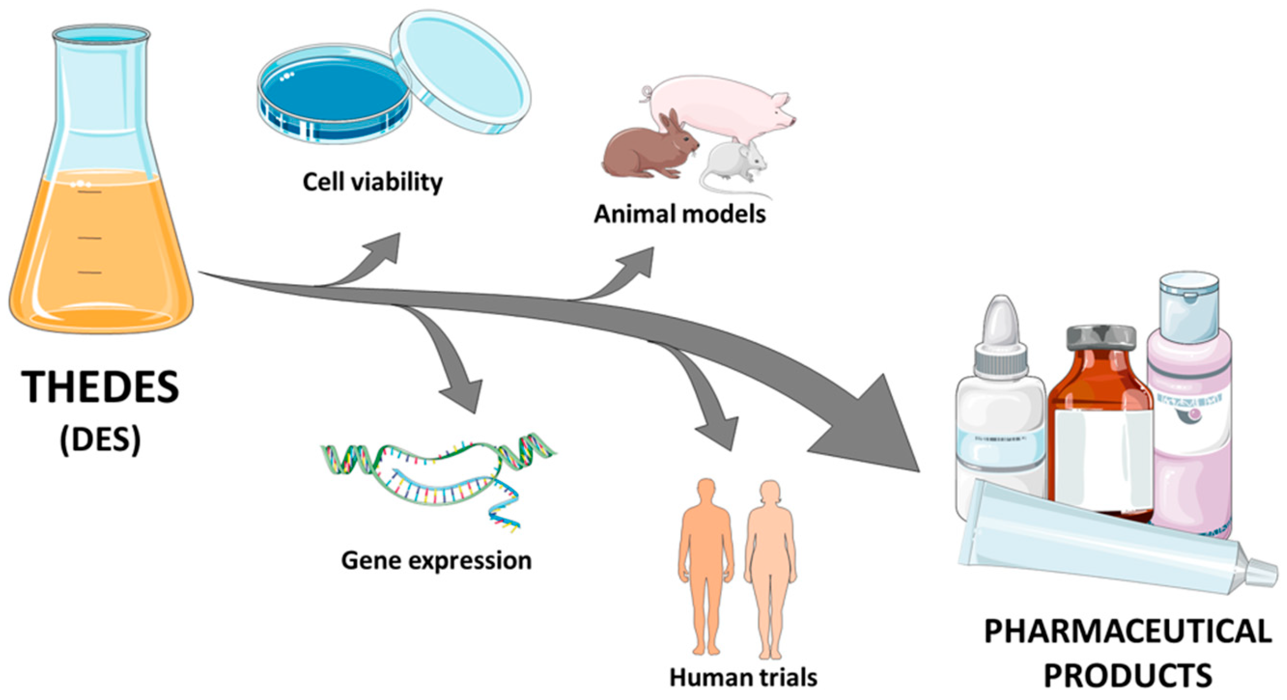
The main objective of this paper is to gather different studies regarding the toxicity of THEDESs and to check if there is enough supporting information to confirm their safety to be used for human therapies. Previously, Lomba et al. carried out an exhaustive review of similar information regarding DESs [41]. Therefore, the most attention was paid on the toxicity results of THEDESs including cell lines, animal models, and human trials, as shown in Figure 1.
2. Cell Lines
2.1. Cell Viability
The analysis of DESs’ cytotoxicity has been an objective since they were firstly described and potential applications were proposed [26]. In fact, each time that a new mixture is characterized, and the goodness of its properties is demonstrated, its toxicity should be also checked. Despite being a mixture of components that are not harmful, toxicological behavior of the final resulting DES can change the expected result, taking into account the components separately. For example, Hayyan et al. demonstrated a low cytotoxicity compared to ionic liquids, and they pointed that due to this fact, they could be useful for therapeutic applications. They examined the cytotoxicity of six different concentrations of several ammonium-based DESs toward seven cell lines (PC3, A375, HepG2, HT29, MCF-7, OKF6, and H413), most of them being cancer cell types, using MTT cell viability assay. When comparing the cytotoxicity results of DESs with their individual components, a synergistic effect was supposed to play a role, since the results could not be explained by the individual behavior of each one alone. Additionally, some components of the DES can show a higher toxicity, and even the effect in different cell lines can be variable [42]. One of the components that was pointed out as a potential factor that increases the cytotoxicity of DESs is oxalic acid, when fish (CCO) and human (MCF-7) cells are exposed to a eutectic mixture with ChCl. Nevertheless, the sensitivity of the cell lines was also different to this DES [43]. The HBA was considered as the main responsible factor of the cytotoxicity found by Macario et al. in their study involving several THEDESs toward two cell lines [44]. When compared to ionic liquids, NADESs have also been recognized with a low cytotoxicity profile, although some constituents, such as tartaric acid, could affect the metabolism of cells [45]. Similarly, one study about the cytotoxicity of NADESs showed that the cytotoxicity was lower than in the former version, DESs, at the same time that it highlighted the importance of several factors such as their composition (e.g., presence of organic acids) or physical properties (e.g., viscosity) influencing it [46].
Sometimes, the toxicity of DESs is searched to destroy bacteria. Hayyan et al. found that some phosphonium-based DESs were effective in the growth inhibition of both kinds of bacteria, Gram-positive (Bacillus subtilis and Staphylococcus aureus) and Gram-negative (Escherichia coli and Pseudomonas aeruginosa) [47]. Zhao et al. characterized different ChCl-based DESs and suggested that those DESs that are acid-based could be more harmful to bacteria [48]. If the attempt of confirming the antibacterial activity of DESs is successful, DESs can even enhance the antibacterial activity of drugs used for this purpose.
Occasionally, DESs are not used to solubilize any chemotherapy agent or to enhance their activity, but they show their own cytotoxic usefulness for destroying cancer cells. In such cases, selectivity is the main property that must be verified in order to avoid side-effects [42]. Moreover, DESs have been also used as reaction media for the preparation of an anticancer compound, such as in the case of a derivate of a quinazoline. The final product was also tested using MTT assay with three cell lines (MCF-7, A549, and MCF-10A) in order to find a higher cytotoxic effect on the cancer cell lines than in the non-malignant one (MCF-10A) [49]. Other studies concerning NADESs as potential anticancer agents evaluated their cytotoxicity toward six hepatic cell lines. As expected, it was found that there were different toxicity profiles depending on the composition of NADESs as well as the tested cell lines. The metabolic pathway of each component was specifically highlighted as a possible cause of the differences [50].
Finally, DESs proposed to be used as solvents of some APIs need to be analyzed to demonstrate that they are safe enough for their latter potential therapeutic use. For instance, in the case of lamotrigine (antiepileptic agent) that is effectively solubilized by DESs consisting of ChCl and urea/ethylene glycol or glycerol, the cytotoxicity of such DESs was tested. Using an MTT cell viability assay, a moderate toxicity was found for the A549 lung cell line when exposed to three different combinations of those THEDESs [51].
As DESs are being used as part of THEDESs, it is necessary for new cytotoxicity assessments to be developed to check the possible synergistic effect of the final product, similarly to DESs [31,40,52]. They have been compiled according to the applications of THEDESs.
There are some drugs that are better solubilized with DESs, such as coumarin. Different final THEDESs were obtained, and low toxicity was found for them toward a cancer cell line [53].
The use of anti-inflammatory drugs is widely spread among the general population. Many drugs with this application are not easily dissolved, and THEDESs have risen as a good alternative to solve the problem. In fact, DESs have also been used to solubilize ibuprofen and the absence of cytotoxicity in xylitol; ChCl:water toward HaCaT (immortalized human epidermal keratinocytes) and HepG2 (hepatocellular carcinoma) cell lines using a PrestoBlue cell viability assay was demonstrated [54]. Ibuprofen is one of the most extended drugs within THEDESs, and cytotoxicity assays were developed to test if such combinations are not cytotoxic [30,55]. Nevertheless, there is one study that was performed with dexamethasone that was successfully solubilized with ChCl and ascorbic acid. At some higher concentrations, cytotoxicity was found, while it was not shown at lower ones. Interestingly, a synergistic effect was found inside the components of THEDESs combining the antioxidant activity of ascorbic acid with the activity of dexamethasone [40].
Bacterial toxicity was an outcome explored with the use of several THEDESs, while cytotoxicity toward other cell lines was also explored. Olivares et al. analyzed the potential cytotoxic effect of THEDES including betaine/urea with imipenem. They found that at the same time that betaine/urea could serve as an effective stabilizer for imipenem, it also enhanced the antimicrobial activity toward some bacteria. Moreover, its cytotoxicity was low for human fibroblasts and even when using human skin explants with the absence of histopathological changes. In this case, as the cytotoxicity profile was tested with human fibroblasts and skin explants, the authors suggested that it could be used for topical applications [56]. Silva et al. combined menthol, a terpene which generally increases the permeation of other components, with three different acids, namely, stearic acid, myristic acid, and lauric acid. The assessment of cytotoxicity was performed toward keratinocytes cells because they were searching a specific application for wound healing. The antibacterial activity of the THEDESs was maintained, and relevant cytotoxic effects were not found in the analyzed cell line [27]. Pedro et al. found a reduced toxicity profile of ciprofloxacin within THEDES combinations (with proline/urea/malonic acid or citric acid/xylitol) when compared to the cytotoxicity of ciprofloxacin alone, while the therapeutic action against bacteria was maintained. They used immortalized human epidermal keratinocytes (HaCaT) as cell models [28,57]. In general, some THEDESs show good properties when the bacterial toxicity is the objective, while the low cytotoxicity of the other exposed cell lines is maintained. A similar situation was found when amoebae was the focus of the antimicrobial activity of the THEDESs [58].
As cancer remains as a challenging disease for effective therapies, some THEDESs have also checked for this therapeutic application. Pereira et al. explored the possibility of including limonene within a THEDES while trying to decrease its intrinsic toxicity for normal cells. Mixtures of limonene with capric acid or menthol did not seem to decrease its toxicity, whereas combinations of limonene with ibuprofen (1:4) did. Moreover, a synergistic effect of this latter THEDES was found that overcomes the simple mixture of its individual components, as previously shown with the DES. Even a different mechanism of action is followed by the THEDESs compared to the isolated ibuprofen and limonene, but their advantages for cancer therapy are preserved [31]. Notably, in a review, Oliveira et al. suggested to understand the cytotoxicity process of THEDESs when affecting different kinds of cell lines, malignant and non-malignant, in order to take advantage of this knowledge to design proper therapies for specific cancer types, such as colorectal cancer [59]. Later on, Pereira et al. evaluated the usefulness of different THEDES including terpenes and nonsteroidal anti-inflammatory drugs. They found that a specific mixture containing menthol/ibuprofen (3:1) was selectively cytotoxic toward cancer cells [30].
In diabetes mellitus, chlorpropamide and tolbutamide were solubilized with tromethamine and a proportion of water after trying other conformers. In the study developed by Sarraguça et al., these THEDESs showed a low toxicity profile in two cell lines involving murine fibroblasts and human cancer cells [60].
The treatment of tuberculosis is another field where THEDESs are being analyzed. Santos et al. combined citric acid with L-arginine or ethambutol, and the cytotoxicity was assessed using the Caco-2 cell line. Citric acid was found to be more toxic than ethambutol for this specific cell line. The acidity of the media was pointed out as a relevant factor that could increase the toxicity of THEDESs, as seen with previous studies with DESs [61].
In relation to the development of new drug delivery systems, Mano et al. found that the cell proliferation of fibroblast was not inhibited by the exposure to gelatin fibers with encapsulated THEDESs composed of ChCl and mandelic acid. At the same time, fibers impregnated with THEDES maintained the expected antibacterial activity [38]. In another study, a liquid L-arginine-based THEDES was encapsulated in a lipidic matrix. When checking its cytotoxicity toward a mouse fibroblast cell line, no toxicity was found [62]. In addition, Pedro et al. explored the cytotoxicity profile of a THEDES including ibuprofen in the form of hydrogel for transdermal administration. The main action of the ibuprofen was maintained while arginine/glycerol (1:4) was used to solubilize it. Human abdominal skin samples were used to check the enhancement of drug permeation. Additionally, murine macrophages were analyzed in relation to the cytotoxicity, and no toxicity was found [55].
A summary of the cytotoxicity studies carried out with THEDESs is gathered in Table 2.
2.2. Gene Expression
Although the above findings classify THEDESs as generally safe, little is known about their interaction at the cellular and molecular level, and how they can affect DNA structure and gene expression [63].
Considering the little evidence about genotoxicity, similar strategies to those carried out in the studies with NADESs could be followed. They must be focused on genes related to the mechanism of action of drugs whose solubility is intended to be increased.
The evidence to date for the genotoxicity of DESs/NADESs is more extensive, having been tested in different cell lines and methods. Between them, gene expression or DNA damage are measured. Grozdanova et al. (2020) [64] carried out a comet assay, a single-cell gel electrophoresis for measuring DNA stand breaks, to assess the genotoxicity of tested NADESs applied for the extraction of two medicinal plants as follows: ChCl: glycerol/Sideritis scardica, ChCl: glycerol/Plantago major, Citric acid-1,2—propanediol 1:4 (CAPD)/Sideritis scardica and CAPD/Plantago major. All tested NADESs were harmless for CCL1 cells, and the genotoxicity was concentration-dependent [64]. Ammonium-based DESs with glycerin, ethylene glycol, triethylene glycol, or urea were tested on MCF-7 cells to evaluate DNA integrity. The results obtained via DNA fragmentation analysis on agarose gel show that the cell deaths were not caused by DNA damage [42].
Trans-resveratrol is a natural polyphenol widely used in cosmetics and the pharmaceutical industry, both in its topical and oral forms (e.g., antioxidants and hypolipidemic supplements). When exposed to UV light, it is isomerized to trans-1,4,6-trihydroxyphenanthrene (THP), with genotoxic effects. Proline/glycerol NADES showed a photo-protective effect under UV exposure, inhibiting the isomerization of resveratrol and THP formation [65]. Moreover, another study evaluated resveratrol, improving its bioavailability via lipid conjugation. Endothelial permeability protection was evaluated by MMP-9 inhibition, which plays an important role in the inflammation and healing processes. Resveratrol-linoleic acid dissolved in NADESs, composed by 1,2-propanediol:ChCl:water, showed the highest MMP-9 inhibitory effect in THP-1 tested cells [66].
A study carried out by Szél et al. (2019) [67] tested the protective effect to hyperosmotic stress produced by sorbitol addition of glycerol and xylitol in keratinocyte HaCaT cells. The inflammatory cytokines’ expression was measured to evaluate stress cell levels. The results show that both polyols decreased IL-1α expression, while only glycerol affects IL-1β and NFAT5 expression, despite having similar chemical structures.
Lee et al. (2022) [68] tested a fermented NADES ginger extract on HTC-116/R cells (colorectal cancer model). The NADES ginger extract has a higher phenolic content and antioxidant activity than organic solvent extract. The NADESs were composed of a mixture of betaine/lactate/water. The fermented NADES extract were shown to inhibit the expression of NF-κB (via activity assay) and CXC chemokine receptor 4 (CXCR4) (using Real time PCR) genes, enhancing the therapeutic effects in oxipalatin-resistant CRC cells.
Likewise, similar studies, such as Wang et al. (2019) [69], were carried out in ionic liquids, demonstrating their influence on the expression of p53 and Bcl-2 family genes, and showing NADESs’ cytotoxic and apoptotic effect on hepatocellular carcinoma (HepG2) cells treated with f 1-dodecyl-3-methylimidazolium chloride.
To the best of our knowledge, the only study really considered as a THEDES formed by betaine-urea DESs containing imipenem was tested in monocyte cells, showing that this formulation increases therapeutic efficacy. The immunogenic response of the THP-1 cells was measured by the proinflammatory cytokines gene expressions (IL-1, IL-6, and TNF-α). This THEDES showed no immunogenic effect, suggesting its potential as a formulation [56].
3. Animal Models
Regarding the toxicity of DESs, to date, these compounds have been tested mainly in invertebrates and animal models (rodents).
Studies in invertebrate animals included Daphnia magna, Artemia salina, and Hydra sinensis with mixed results in the toxicity of the DES use. In reference to the Daphnia magna toxicity test, only two studies evaluated different DESs [70] or NADESs [71], and none of the tested combinations induced significant toxicity toward this model. Hayyan et al. tested different DESs in Artemia salina to determine the toxicity and the mortality. The results of the assays indicated that the toxicity of DESs was higher than their individual components, suggesting that the toxicity can be influenced by the composition of DESs by varying the salt/HBD combination and molar ratio [42,47]. The study of Hydra sinensis concluded that the survival times of Hydra sinensis were significantly shortened by exposure to a DES-containing aqueous solution, suggesting that the DES exhibited a toxic effect on the hydras. It was suspected that depending on the type of HBD of the eutectic mixtures, the charge will be delocalized in the HBA and modify the toxicity [72].
Regarding more complex animal models, Hayyan et al. also tested the toxic potential of ammonium-based DESs in vivo in a murine model. As in Artemia salina had shown previously, in mice tests, DESs were relatively more toxic in comparison to their individual components [38]. Consequently, Mbous et al. tested the toxicity of DESs and NADESs in mice, showing that NADESs presented higher toxicity than DESs, and induced liver failure due to their viscosity, which made the circulation of the mixture difficult [50]. In the same line, Benlebna et al. administered phenolic NADES extract from green coffee beans to Wistar rats, and its oral administration induced mortality in two rats, and short-term side effects such as excessive water consumption, weight loss, hepatomegaly, and plasma oxidative stress associated with hyperlipemia [73].
On the other hand, more recent articles observed less toxicity on the DESs studied. Nurunnabi et al. investigated the toxicity of oral CAGE (Choline bicarbonate and Geranic acid) administration in rats treated for 30 days. The results obtained indicate that the CAGE formulation was well tolerated, that no changes were found in the red blood cells and platelet count of treated rats compared with the controls [74]. Zhao et al. used metabolites of Coptidis rhizoma extract to artificially prepare the NADES. Then, the toxicity in the mice was evaluated, showing that after the oral administration of the NADES, the mice became irritable and presented dyskinesia, which means that the NADES itself showed an acute toxicity to the mice. However, the mice that were given 10% dilution of the NADES in water showed no toxicity, and none died after oral administration [75].
Different NADESs/DESs were used to improve the bioavailability and/or biocompatibility of different compounds [36,76,77,78,79,80]. Xiao et al. developed a novel hydrogel system incorporating an amino acid-based DES. The DESs were integrated into Carbomer® 940 hydrogels for the skin delivery of “sanwujiaowan” extract, a Chinese herbal medicine. Sanwujiaowan is composed of six ingredients, some of them with a certain degree of toxicity. By preparing the DES extract complex, it demonstrated good dissolution and skin permeability of the ingredients in the extracts. The authors used a collagen-induced-arthritis rat model, and the hydrogel with the DES extract complex exerted an enhanced therapeutic effect that reduced the inflammatory response with systemic toxicity of the extracts [32].
Due to their high stabilization and solubilization power, DESs offer the ability to tune the solubility, permeation, and absorption of APIs. For that, APIs with low solubility or permeability could be improved by including these solvents on their formulation in in vivo studies. The toxicity of THEDESs has been tested primarily on animals such as mice, rats, or pigs focused on the preclinical angle. Previous studies are shown according to their routes of administration and are summarized in Table 3.
3.1. Injection Routes: Transdermal/Subcutaneous/Intravenous Administration
Injection administrations require the drug to be delivered through the skin with a needle (subcutaneous, intravenous, or transdermal). Some drugs are delivered body-wide through a patch on the skin. These drugs are sometimes mixed with chemical compounds, as DESs, that enhances penetration through the skin into the bloodstream without any injection. According to transdermal skin delivery, it is difficult to assess the skin permeability of materials using only in vivo experiments. Consequently, numerous ex vivo models are frequently employed to assess drug skin permeation profiles and kinetic parameters [96].
Kim et al. used DESs to formulate a nanocomplex for the systemic administration of verteporfin, a chemotherapeutic drug. They investigated the efficacy of the nanocomplex administered intravenously in female mice bearing a 4T1 tumor in the mammary pad. The authors observed a better drug penetration into the tumor tissue, probably due to the DESs’ contribution to the fluidization of the tissue through the interaction between the DESs and membrane lipids. In addition, greater drug sensitivity and effective inhibition of orthotopic tumor growth was shown [81]. The same authors reported a deep eutectic-based formulation that slows the release of apomorphine after subcutaneous injection. In this case, CAGE1:2 and a mixture with the drug was prepared, which is called SEAPORT formulation. The skin from the dorsal side of Wistar rats was used to study apomorphine release from SEAPORT ex vivo and it was observed that CAGE 1:2 plays a critical role in its release. Moreover, in vivo pharmacokinetic of the apomorphine from subcutaneously injected SEAPORT was measured in the rats and pigs and showed a prolonged duration of a higher apomorphine concentration in plasma in comparison to the clinical comparator (reference drug formulation) [82].
Other studies also synthesized CAGE, as DESs, and included them in drug formulations for the application in ex vivo and in vivo models, mainly with insulin, because of its difficult transdermal delivery by conventional chemical permeation enhancers. Banerjee et al. reported that CAGE can deliver insulin across porcine skin and significantly reduce blood glucose levels in rats when insulin-CAGE is topically administered. The ex vivo penetration into the skin samples was measured using porcine skin; to measure the insulin-CAGE penetration, Fluorescein isothiocyanate-labeled insulin (FITIC-insulin) was added to CAGE and applied to the porcine skin. In vivo experiments were performed in male rats applying transdermal insulin in CAGE [83]. Tanner et al. synthesized variants of CAGE by controlling the ratio of a range of 1:4 to 2:1. In this case, an ex vivo pig’s skin model demonstrated that the 1:2 and 1:4 molar ratio variants were capable of transporting FITIC-insulin through to the stratum corneum, epidermis, and dermis (transdermal transport) [84]. Recently, Curreri et al. developed a new tool toward improving subcutaneous formulation using biocompatible deep eutectics. The DESs were synthesized using a salt metathesis reaction at the cation/anion ratios of 1:2 and were then formulated with solubilized 100 U/mL of regular insulin. Pharmacokinetics of subcutaneous insulin administration using deep eutectics (SPADES-insulin) was compared against the equivalent dose of the clinical gold standard of the fast-acting insulin analog (Humalog) in male Wistar non-fasting rats and improved the systemic absorption of subcutaneously injected insulin faster than Humalog. The toxicity was analyzed in healthy female Balb/c mice receiving a single subcutaneous injection of DES solutions and demonstrated that SPADES is a safe and non-toxic formulation [85]. In a new paper, DESs were introduced as novel chemical penetration enhancers for transdermal iontophoresis of insulin across rat skin. The results of the in vivo studies indicated a reduction in the blood glucose level in diabetic rats using ChCl/ethylene glycol and ChCl/glycerol in the iontophoresis delivery of insulin [86].
Al-Akayleh et al. investigated the skin permeability of risperidone, an antipsychotic drug, using eutectic systems. The prepared THEDES mixture of risperidone and capric acid was spread on the skin of male rodents and was found to be tolerable, less irritant, and safe for application to the skin compared with the control untreated skin. A histopathological study was performed that did not show changes in the state of the skin and morphological changes [87].
Rajan et al. synthesized DESs, which influenced the formation of folic acid (FA)-tagged g-beta-alanine-co-Poly ε-caprolactone (PCL) polymer (DES@FA-g-β-alanine-co-PCL polymeric system) to encapsulate the doxorubicin drug. The enhanced anticancer potential of doxorubicin-loaded polymeric micelle was studied in an in vivo breast cancer model. The advantage of this polymeric system is that it could increase the bioavailability of hydrophobic doxorubicin drugs. The intraperitoneal injection of doxorubicin-loaded polymeric micelle significantly reduced the body weight and tumor size when compared with dimethylbenzanthracene-induced mammary carcinoma-bearing rats and polymeric micelle-treated rats [76].
Recently, Li et al. reported a transdermal delivery strategy using a silica nanoparticle matrix (MSN) encapsulated with methotrexate and impregnated in DES hydrogels for the topical management of rheumatoid arthritis. This formulated DES-MSN hydrogel system was applied to the skin of rats to test the skin irritation and the in vivo toxicity of this system. They observed a sustained penetration and accumulation of MSNs at subcutaneous inflammation sites [88].
3.2. Oral Administration
Regarding in vivo pharmacokinetic studies, Liu et al. administered RA-XII dissolved in 5–20% v/v Betaine and Mandelic acid (NADES) solution in rats. The authors observed that the oral absorption of RA-XII dissolved in 20% NADES solution was significantly increased. They concluded that NADES solutions were effective approaches for improving solubility, permeability, and oral bioavailability of the anti-tumor candidate RA-XII [89]. Patel et al. prepared a novel eutectic of Diacerein with fumaric acid (FMA) as a coformer in various molar ratios. Diacerein and its active metabolite, called rhein, are anthraquinone derivatives used for treating osteoarthritis. Healthy Sprague Dawley rats were distributed in two groups and administered pure Diacerein and prepared eutectic suspension orally, respectively. Blood samples were collected, and pharmacokinetic parameters were analyzed after oral administration. The results of the pharmacokinetic evaluation suggested greater oral bioavailability of the prepared eutectic versus pure Diacerein [90]. These authors repeated the same study but changed the fumaric acid for 2,4- dihydroxybenzoic acid (DHA) as coformer, and again, the pharmacokinetic study in the rats proved higher oral bioavailability of the prepared eutectic [91]. Berberine is a quaternary benzylisoquinoline alkaloid with huge properties as an antihypertensive, antiarrhythmic, anti-inflammatory, and hypolipidemic, among other properties. Despite its therapeutic properties, berberine is poorly absorbed after oral administration and its plasma concentration in pharmacokinetic studies is extremely low. In this study, several NADESs were prepared and used to solubilize berberine. The NADESs selected were urea or organic acids (malic and lactic acid), and proline. The berberine solubilized in the selected eutectics mixtures were used to evaluate their pharmacokinetic properties in the female Balb/c mice and were compared with berberine. The plasma levels were determined up to 6 h after oral administration via gavage in the mice. The administration of NADESs with berberine yielded in a significant increase in the plasma level of berberine [36]. These authors were in concordance with another recent report that observed that the pharmacokinetic parameters of berberine were improved in the liver and indicated that NADES dilutions promoted the intestinal absorption of oral berberine by increasing solubility in mice. The authors suggested that NADESs may help to promote its use in oral drug delivery [75].
Another natural compound, the salvianolic acid B with anti-inflammatory and antioxidant properties, was studied, adding the model drug in ChCl/glycerol with the common molar ratio (ChCl/glycerol 1:2) as a drug vehicle for this poorly soluble compound. The results obtained demonstrated that the oral administration using gavage of ChCl/glycerol (1:2) was non-toxic with LD50 7733 mg/kg in mice. The results indicated that ChCl/glycerol (1:2) could accelerate the absorption and elimination of salvianolic acid B, maybe because the eutectic system could enhance the membrane penetration ability of drugs [92].
Barnejee et al. also studied the efficacy of insulin-CAGE administered through the oral route. For the oral efficacy in in vivo studies, enterically coated capsules were administered via oral gavage to adult nondiabetic male rats. In vivo, when 10 U/kg insulin-CAGE was administered, a 45% decrease in blood glucose was observed, and by delivering insulin-CAGE, the authors evade gastric degradation of insulin and enhance its intestinal permeability [93]. Other authors reported the fabrication of a biodegradable polymeric patch for the buccal delivery of insulin using CAGE, as DESs, to transport facilitator and chitosan as mucoadhesive matrix. They applied ex vivo and in vivo studies. The ex vivo permeation was performed using porcine buccal tissue to study the extent of drug transport. The results show that CAGE induced an increase in insulin transport and enhanced insulin penetration through buccal tissue. To test in vivo the performance of insulin delivered by the patches, the patches were placed in the pouches of the nondiabetic rats in the buccal cavity of the rats. The authors concluded that the CAGE/insulin patches placed in the rat buccal pouch lowered the blood glucose levels, and non-tissue damage was shown [94].
3.3. Nasal Administration
Nasal administration was proposed as an alternative administration route for insulin. Like the transdermal and oral routes, this route must be evaluated to determine if there is toxicity and to determine if it is a good route of administration. Li et al. developed a biocompatible DES as a carrier for improving the nasal delivery of insulin. Nasal toxicity was evaluated in vivo using rats, after intranasal epithelia administration of the solution. The authors concluded that the in vivo studies demonstrated that DES caused no evident toxicity to rat nasal epithelia and could improve the permeability of nasal epithelia, because DESs absorb water to facilitate the disassociation of the structure and the release of drugs upon contact with the nasal mucus [95].
Regarding delivery methods, the studies mentioned focused on oral, buccal mucosa, transdermal, intravenous injection, subcutaneous injection, and topical delivery with very good results and low toxicity. However, other potential routes require investigation, such as ocular, sublingual, or vaginal/anal drug delivery.
4. Human Trials
The properties of DESs have been widely demonstrated at different levels, yet there are only few studies carried out directly in humans, and none of them are considered as conventional THEDESs. The antimicrobial properties of DESs and their ability to deliver drugs transdermally have generated great interest in the dermatology industry, mainly in [33,55]. One of the first human trials focuses on the treatment of rosacea. The study of Ko et al. showed that DES with a composition of CAGE is a good candidate to improve the effectiveness of rosacea treatment due to the good results of in vitro studies with this ionic liquid/DES [97,98]. Its high ability to penetrate into the deep layers of the skin allows for the treatment of pathogens at this level [83]. The CGB-400 gel, with a CAGE1:2 composition, has managed to complete the “seven steps of translation”, reaching the final step of human studies for the treatment of rosacea. The high antibacterial activity of CAGE against Propionibacterium acnes, associated with the biological origin of rosacea, has also been demonstrated [98]. This clinical trial was conducted in 52 patients with rosacea, and a 34.6% reduction in the mean papules and pustules typical of this disorder was observed after only two weeks of therapy with CGB-400 gel. At 12 weeks, the reduction observed was much greater, specifically, 71.9%. This study and the process to obtain the final product open a door to other products based on the properties that have been demonstrated for CAGE. The use of CGB-400 for the reduction in facial redness and bumps and blemishes are still in phase one of clinical trials (https://www.cdek.liu.edu/api/119951/#trials, accessed on 24 March 2023) [99].
Human tissues were also used for ex vivo analysis of toxicity. For example, abdominal skin samples were obtained to carry out a permeation study of risperidone. Al-Akayleh et al. concluded that the amount of this drug permeated as part of a THEDES including capric acid was high [83]. Pedro et al. also performed a permeation assay of an alginate-based hydrogel containing ibuprofen using human abdominal skin as an ex vivo model. The authors showed that the permeation of ibuprofen was improved compared to that obtained with the hydrogel without the DES [51].
5. Conclusions
Despite being recognized as promising solvents with many potential biomedical applications, THEDESs have not been fully analyzed on their toxicity profile, although there are some studies about this topic. There is a lack of comprehensive toxicity studies available in the literature. DESs have received more attention about their toxic profile, but combinations with APIs are not always recognized as THEDESs, and further research is needed to fully assess the potential risks associated with their use. As soon as a new THEDES arrives, its toxicity should be assessed, especially if it is going to be used for therapeutics. It is suggested that toxicity should be checked systematically since these kinds of data are scarce. As the complexity of the models increased, a smaller number of studies were developed. Interestingly, some authors have found a synergistic effect when mixing original compounds of the THEDESs, and toxicity can change in both directions, so it is necessary to test any new combination.
Since each THEDES has its own composition with unique properties, it is difficult to establish a universal toxicity testing protocol that would be applicable to all THEDESs. However, the development of such model would aid in the safe and effective use of THEDESs in their various applications. As THEDESs are tailored to be used for specific therapeutic areas, the models used to carry out the toxicity assessment should vary. Notably, an effort must be made to maintain the individual bioactivity of the components of the THEDESs, whereas some additional advantages could be found. Regarding cell cultures and gene expression, cell lines and genes must be selected in relation to the intended application. This should be the first step before using more complex animal models or even human trials. In animal models, a variable toxicity was found, with injection being the most tested route of administration of the compounds.
This manuscript is focused on the toxicity of THEDESs specifically, but it is also essential to investigate their degradation products to ensure they are non-toxic as well. THEDESs can break down into various substances, some of which may have different toxic properties from the original compound. Therefore, the investigation of the toxicity of THEDESs’ degradation products is also crucial to ensure their safe use.
Author Contributions
Conceptualization, C.B.G. and L.L.; writing—original draft preparation, C.B.G., J.C., L.C., E.S. and M.P.R.; writing—review and editing, C.B.G., L.L. and E.S. All authors have read and agreed to the published version of the manuscript.
Funding
This work was partially funded by Departamento de Ciencia, Universidad y Sociedad del Conocimiento, from the Gobierno de Aragón (Spain) (Research Group B58_23D).
Institutional Review Board Statement
Not applicable.
Informed Consent Statement
Not applicable.
Data Availability Statement
Not applicable.
Conflicts of Interest
The authors declare no conflict of interest.
Abbreviations
| API | Active Pharmaceutical Ingredient |
| BCS | Biopharmaceutics Classification System |
| CAGE | Choline bicarbonate and Geranic acid |
| CAPD | Citric acid-1,2—propanediol 1:4 |
| ChCl | Choline Chloride |
| DES | Deep Eutectic Solvents |
| DHA | 2,4-Dihydroxybenzoic Acid |
| FA | Folic Acid |
| FMA | Fumaric Acid |
| FITIC-insulin | Fluorescein Isothiocyanate-Labeled Insulin |
| HBA | Hydrogen Bond Acceptor |
| HBD | Hydrogen Bond Donor |
| MSN | Silica Nanoparticle Matrix |
| NADES | Natural Deep Eutectic Solvents |
| PCL | Poly ε-Caprolactone |
| THEDES | Therapeutic Deep Eutectic Systems |
| THP | Trans-1,4,6-Trihydroxyphenanthrene |
References
- Paul, R.; Paul, S. Exploration on the drug solubility enhancement in aqueous medium with the help of endo -functionalized molecular tubes: A computational approach. Phys. Chem. Chem. Phys. 2021, 23, 18999–19010. [Google Scholar] [CrossRef] [PubMed]
- FDA. Formulating Drug Products for Optimized Absorption: Elucidating Amorphous Solid Dispersions. Available online: https://www.fda.gov/drugs/regulatory-science-action/formulating-drug-products-optimized-absorption-elucidating-amorphous-solid-dispersions (accessed on 5 May 2023).
- Kalepu, S.; Nekkanti, V. Insoluble drug delivery strategies: Review of recent advances and business prospects. Acta Pharm. Sin. B 2015, 5, 442–453. [Google Scholar] [CrossRef] [PubMed]
- Rajpoot, K.; Tekade, M.; Sreeharsha, N.; Sharma, M.C.; Tekade, R.K. Recent advancements in solubilization of hydrophobic drugs. In The Future of Pharmaceutical Product Development and Research; Elsevier: Amsterdam, The Netherlands, 2020; pp. 109–144. ISBN 9780128144558. [Google Scholar]
- Kumar, S.; Bhargava, D.; Thakkar, A.; Arora, S. Drug carrier systems for solubility enhancement of BCS class II drugs: A critical review. Crit. Rev. Ther. Drug Carrier Syst. 2013, 30, 217–256. [Google Scholar] [CrossRef] [PubMed]
- Sekharan, T.R.; Chandira, R.M.; Tamilvanan, S.; Rajesh, S.C.; Venkateswarlu, B.S. Deep eutectic solvents as an alternate to other harmful solvents. Biointerface Res. Appl. Chem. 2022, 12, 847–860. [Google Scholar] [CrossRef]
- Bhalani, D.V.; Nutan, B.; Kumar, A.; Singh Chandel, A.K. Bioavailability Enhancement Techniques for Poorly Aqueous Soluble Drugs and Therapeutics. Biomedicines 2022, 10, 2055. [Google Scholar] [CrossRef]
- Savjani, K.T.; Gajjar, A.K.; Savjani, J.K. Drug Solubility: Importance and Enhancement Techniques. ISRN Pharm. 2012, 2012, 10. [Google Scholar] [CrossRef]
- Sikarra, D.; Shukla, V.; Kharia, A.A.; Chatterjee, D.P. Techniques for Solubility Enhancement of Poorly Soluble Drugs: An Overview. JMPAS 2012, 1, 1–22. [Google Scholar]
- Kanikkannan, N. Technologies to Improve the Solubility, Dissolution and Bioavailability of Poorly Soluble Drugs. J. Anal. Pharm. Res. 2018, 7, 1. [Google Scholar] [CrossRef]
- Deshmukh, A.S.; Tiwari, K.J.; Mahajan, V.R. Solubility Enhancement Techniques for Poorly Water-Soluble Drugs. Int. J. Pharm. Sci. Nanotechnol. 2017, 10, 3701–3708. [Google Scholar] [CrossRef]
- Abdkarimi, F.; Haghtalab, A. Solubility measurement and thermodynamic modeling of sertraline hydrochloride and clopidogrel bisulfate in deep eutectic solvent of choline chloride and malonic acid. J. Mol. Liq. 2021, 344, 117940. [Google Scholar] [CrossRef]
- Chakraborty, S.; Chormale, J.H.; Bansal, A.K. Deep eutectic systems: An overview of fundamental aspects, current understanding and drug delivery applications. Int. J. Pharm. 2021, 610, 121203. [Google Scholar] [CrossRef] [PubMed]
- Chen, W.; Xue, Z.; Wang, J.; Jiang, J.; Zhao, X.; Mu, T. Investigation on the thermal stability of deep eutectic solvents. Acta Phys. Chim. Sin. 2018, 34, 904–911. [Google Scholar] [CrossRef]
- Chen, Y.; Mu, T. Revisiting greenness of ionic liquids and deep eutectic solvents. Green Chem. Eng. 2021, 2, 174–186. [Google Scholar] [CrossRef]
- Fourmentin, S.; Gomes, M.C.; Lichtfouse, E. Deep Eutectic Solvents for Medicine, Gas Solubilization and Extraction of Natural Substances; Fourmentin, S., Costa Gomes, M., Lichtfouse, E., Eds.; Environmental Chemistry for a Sustainable World; Springer International Publishing: Cham, Switzerland, 2021; Volume 56, ISBN 978-3-030-53068-6. [Google Scholar]
- Nystedt, H.L.; Grønlien, K.G.; Tønnesen, H.H. Interactions of natural deep eutectic solvents (NADES) with artificial and natural membranes. J. Mol. Liq. 2021, 328, 115452. [Google Scholar] [CrossRef]
- Gala, U.; Chuong, M.C.; Varanasi, R.; Chauhan, H. Characterization and Comparison of Lidocaine-Tetracaine and Lidocaine-Camphor Eutectic Mixtures Based on Their Crystallization and Hydrogen-Bonding Abilities. AAPS PharmSciTech 2015, 16, 528–536. [Google Scholar] [CrossRef]
- Stott, P.W.; Williams, A.C.; Barry, B.W. Transdermal delivery from eutectic systems: Enhanced permeation of a model drug, ibuprofen. J. Control. Release 1998, 50, 297–308. [Google Scholar] [CrossRef]
- Abbott, A.P.; Ahmed, E.I.; Prasad, K.; Qader, I.B.; Ryder, K.S. Liquid pharmaceuticals formulation by eutectic formation. Fluid Phase Equilib. 2017, 448, 2–8. [Google Scholar] [CrossRef]
- Farooq, M.Q.; Abbasi, N.M.; Smith, E.A.; Petrich, J.W.; Anderson, J.L. Characterizing the Solvation Characteristics of Deep Eutectic Solvents Composed of Active Pharmaceutical Ingredients as a Hydrogen Bond Donor and/or Acceptor. ACS Sustain. Chem. Eng. 2022, 10, 3066–3078. [Google Scholar] [CrossRef]
- Tuntarawongsa, S.; Phaechamud, T. Menthol, borneol, camphor and WS-3 eutectic mixture. In Proceedings of the Advanced Materials Research; Trans Tech Publications Ltd.: Wollerau, Switzerland, 2012; Volume 506, pp. 355–358. [Google Scholar]
- Morrison, H.G.; Sun, C.C.; Neervannan, S. Characterization of thermal behavior of deep eutectic solvents and their potential as drug solubilization vehicles. Int. J. Pharm. 2009, 378, 136–139. [Google Scholar] [CrossRef]
- Duarte, A.R.C.; Ferreira, A.S.D.; Barreiros, S.; Cabrita, E.; Reis, R.L.; Paiva, A. A comparison between pure active pharmaceutical ingredients and therapeutic deep eutectic solvents: Solubility and permeability studies. Eur. J. Pharm. Biopharm. 2017, 114, 296–304. [Google Scholar] [CrossRef]
- Yin, T.; Wu, J.; Yuan, J.; Wang, X. Therapeutic deep eutectic solvent based on osthole and paeonol: Preparation, characterization, and permeation behavior. J. Mol. Liq. 2022, 346, 117133. [Google Scholar] [CrossRef]
- Lomba, L.; García, C.B.; Ribate, M.P.; Giner, B.; Zuriaga, E. Applications of deep eutectic solvents related to health, synthesis, and extraction of natural based chemicals. Appl. Sci. 2021, 11, 10156. [Google Scholar] [CrossRef]
- Silva, J.M.; Pereira, C.V.; Mano, F.; Silva, E.; Castro, V.I.B.; Sá-Nogueira, I.; Reis, R.L.; Paiva, A.; Matias, A.A.; Duarte, A.R.C. Therapeutic Role of Deep Eutectic Solvents Based on Menthol and Saturated Fatty Acids on Wound Healing. ACS Appl. Bio Mater. 2019, 2, 4346–4355. [Google Scholar] [CrossRef]
- Pedro, S.N.; Gomes, A.T.P.C.; Oskoei, P.; Oliveira, H.; Almeida, A.; Freire, M.G.; Silvestre, A.J.D.; Freire, C.S.R. Boosting antibiotics performance by new formulations with deep eutectic solvents. Int. J. Pharm. 2022, 616, 121566. [Google Scholar] [CrossRef]
- Aroso, I.M.; Silva, J.C.; Mano, F.; Ferreira, A.S.D.; Dionísio, M.; Sá-Nogueira, I.; Barreiros, S.; Reis, R.L.; Paiva, A.; Duarte, A.R.C. Dissolution enhancement of active pharmaceutical ingredients by therapeutic deep eutectic systems. Eur. J. Pharm. Biopharm. 2016, 98, 57–66. [Google Scholar] [CrossRef] [PubMed]
- Pereira, J.; Miguel Castro, M.; Santos, F.; Rita Jesus, A.; Paiva, A.; Oliveira, F.; Duarte, A.R.C. Selective terpene based therapeutic deep eutectic systems against colorectal cancer. Eur. J. Pharm. Biopharm. 2022, 175, 13–26. [Google Scholar] [CrossRef] [PubMed]
- Pereira, C.V.; Silva, J.M.; Rodrigues, L.; Reis, R.L.; Paiva, A.; Duarte, A.R.C.; Matias, A. Unveil the Anticancer Potential of Limomene Based Therapeutic Deep Eutectic Solvents. Sci. Rep. 2019, 9, 14926. [Google Scholar] [CrossRef] [PubMed]
- Xiao, S.; Wang, L.; Han, W.; Gu, L.; Cui, X.; Wang, C. Novel Deep Eutectic Solvent–Hydrogel Systems for Synergistic Transdermal Delivery of Chinese Herb Medicine and Local Treatments for Rheumatoid Arthritis. Pharm. Res. 2022, 39, 2431–2446. [Google Scholar] [CrossRef]
- Wang, J.; Li, M.; Duan, L.; Lin, Y.; Cui, X.; Yang, Y.; Wang, C. Deep Eutectic Systems as Novel Vehicles for Assisting Drug Transdermal Delivery. Pharmaceutics 2022, 14, 2265. [Google Scholar] [CrossRef]
- Emami, S.; Shayanfar, A. Deep eutectic solvents for pharmaceutical formulation and drug delivery applications. Pharm. Dev. Technol. 2020, 25, 779–796. [Google Scholar] [CrossRef]
- Aroso, I.M.; Craveiro, R.; Rocha, Â.; Dionísio, M.; Barreiros, S.; Reis, R.L.; Paiva, A.; Duarte, A.R.C. Design of controlled release systems for THEDES−Therapeutic deep eutectic solvents, using supercritical fluid technology. Int. J. Pharm. 2015, 492, 73–79. [Google Scholar] [CrossRef] [PubMed]
- Sut, S.; Faggian, M.; Baldan, V.; Poloniato, G.; Castagliuolo, I.; Grabnar, I.; Perissutti, B.; Brun, P.; Maggi, F.; Voinovich, D.; et al. Natural Deep Eutectic Solvents (NADES) to enhance berberine absorption: An in vivo pharmacokinetic study. Molecules 2017, 22, 1921. [Google Scholar] [CrossRef] [PubMed]
- Durand, E.; Lecomte, J.; Upasani, R.; Chabi, B.; Bayrasy, C.; Baréa, B.; Jublanc, E.; Clarke, M.J.; Moore, D.J.; Crowther, J.; et al. Evaluation of the ROS Inhibiting Activity and Mitochondrial Targeting of Phenolic Compounds in Fibroblast Cells Model System and Enhancement of Efficiency by Natural Deep Eutectic Solvent (NADES) Formulation. Pharm. Res. 2017, 34, 1134–1146. [Google Scholar] [CrossRef] [PubMed]
- Mano, F.; Martins, M.; Sá-Nogueira, I.; Barreiros, S.; Borges, J.P.; Reis, R.L.; Duarte, A.R.C.; Paiva, A. Production of Electrospun Fast-Dissolving Drug Delivery Systems with Therapeutic Eutectic Systems Encapsulated in Gelatin. AAPS PharmSciTech 2017, 18, 2579–2585. [Google Scholar] [CrossRef]
- Mokhtarpour, M.; Shekaari, H.; Shayanfar, A. Design and characterization of ascorbic acid based therapeutic deep eutectic solvent as a new ion-gel for delivery of sunitinib malate. J. Drug Deliv. Sci. Technol. 2020, 56, 101512. [Google Scholar] [CrossRef]
- Silva, J.M.; Reis, R.L.; Paiva, A.; Duarte, A.R.C. Design of Functional Therapeutic Deep Eutectic Solvents Based on Choline Chloride and Ascorbic Acid. ACS Sustain. Chem. Eng. 2018, 6, 10355–10363. [Google Scholar] [CrossRef]
- Lomba, L.; Ribate, M.P.; Zaragoza, E.; Concha, J.; Garralaga, M.P.; Errazquin, D.; García, C.B.; Giner, B. Deep eutectic solvents: Are they safe? Appl. Sci. 2021, 11, 10061. [Google Scholar] [CrossRef]
- Hayyan, M.; Looi, C.Y.; Hayyan, A.; Wong, W.F.; Hashim, M.A. In Vitro and in Vivo toxicity profiling of ammonium-based deep eutectic solvents. PLoS ONE 2015, 10, e0117934. [Google Scholar] [CrossRef]
- Radošević, K.; Cvjetko Bubalo, M.; Gaurina Srček, V.; Grgas, D.; Landeka Dragičević, T.; Redovniković, R.I. Evaluation of toxicity and biodegradability of choline chloride based deep eutectic solvents. Ecotoxicol. Environ. Saf. 2015, 112, 46–53. [Google Scholar] [CrossRef]
- Macário, I.P.E.; Oliveira, H.; Menezes, A.C.; Ventura, S.P.M.; Pereira, J.L.; Gonçalves, A.M.M.; Coutinho, J.A.P.; Gonçalves, F.J.M. Cytotoxicity profiling of deep eutectic solvents to human skin cells. Sci. Rep. 2019, 9, 3932. [Google Scholar] [CrossRef]
- Paiva, A.; Craveiro, R.; Aroso, I.; Martins, M.; Reis, R.L.; Duarte, A.R.C. Natural deep eutectic solvents-Solvents for the 21st century. ACS Sustain. Chem. Eng. 2014, 2, 1063–1071. [Google Scholar] [CrossRef]
- Hayyan, M.; Mbous, Y.P.; Looi, C.Y.; Wong, W.F.; Hayyan, A.; Salleh, Z.; Mohd-Ali, O. Natural deep eutectic solvents: Cytotoxic profile. Springerplus 2016, 5, 1–12. [Google Scholar] [CrossRef] [PubMed]
- Hayyan, M.; Hashim, M.A.; Al-Saadi, M.A.; Hayyan, A.; AlNashef, I.M.; Mirghani, M.E.S. Assessment of cytotoxicity and toxicity for phosphonium-based deep eutectic solvents. Chemosphere 2013, 93, 455–459. [Google Scholar] [CrossRef] [PubMed]
- Zhao, B.Y.; Xu, P.; Yang, F.X.; Wu, H.; Zong, M.H.; Lou, W.Y. Biocompatible Deep Eutectic Solvents Based on Choline Chloride: Characterization and Application to the Extraction of Rutin from Sophora japonica. ACS Sustain. Chem. Eng. 2015, 3, 2746–2755. [Google Scholar] [CrossRef]
- Arafa, W.A.A. Deep eutectic solvent for an expeditious sono-synthesis of novel series of bis-quinazolin-4-one derivatives as potential anti-cancer agents. R. Soc. Open Sci. 2019, 6, 182046. [Google Scholar] [CrossRef]
- Mbous, Y.P.; Hayyan, M.; Wong, W.F.; Looi, C.Y.; Hashim, M.A. Unraveling the cytotoxicity and metabolic pathways of binary natural deep eutectic solvent systems. Sci. Rep. 2017, 7, 41257. [Google Scholar] [CrossRef]
- Shekaari, H.; Zafarani-Moattar, M.T.; Mokhtarpour, M.; Faraji, S. Exploring cytotoxicity of some choline-based deep eutectic solvents and their effect on the solubility of lamotrigine in aqueous media. J. Mol. Liq. 2019, 283, 834–842. [Google Scholar] [CrossRef]
- Van Osch, D.J.G.P.; Zubeir, L.F.; Van Den Bruinhorst, A.; Rocha, M.A.A.; Kroon, M.C. Hydrophobic deep eutectic solvents as water-immiscible extractants. Green Chem. 2015, 17, 4518–4521. [Google Scholar] [CrossRef]
- Khorsandi, M.; Shekaari, H.; Mokhtarpour, M.; Hamishehkar, H. Cytotoxicity of some choline-based deep eutectic solvents and their effect on solubility of coumarin drug. Eur. J. Pharm. Sci. 2021, 167, 106022. [Google Scholar] [CrossRef]
- Lomba, L.; Garralaga, M.P.; Werner, Á.; Giner, B.; Baptista, P.M.; Sánchez-Romero, N. Ibuprofen solubility and cytotoxic study of deep eutectic solvents formed by xylitol, choline chloride and water. J. Drug Deliv. Sci. Technol. 2023, 82, 104327. [Google Scholar] [CrossRef]
- Pedro, S.N.; Mendes, M.S.M.; Neves, B.M.; Almeida, I.F.; Costa, P.; Correia-Sá, I.; Vilela, C.; Freire, M.G.; Silvestre, A.J.D.; Freire, C.S.R. Deep Eutectic Solvent Formulations and Alginate-Based Hydrogels as a New Partnership for the Transdermal Administration of Anti-Inflammatory Drugs. Pharmaceutics 2022, 14, 827. [Google Scholar] [CrossRef] [PubMed]
- Olivares, B.; Martínez, F.A.; Ezquer, M.; Morales, B.J.; Fuentes, I.; Calvo, M.; Campodónico, P.R. Betaine-urea deep eutectic solvent improves imipenem antibiotic activity. J. Mol. Liq. 2022, 350, 118551. [Google Scholar] [CrossRef]
- Wu, J.; Yin, T. Insight into the physicochemical properties and bioactivities of therapeutic deep eutectic solvents based on matrine and fatty acids. J. Mol. Liq. 2022, 360, 119560. [Google Scholar] [CrossRef]
- Siddiqui, R.; Makhlouf, Z.; Akbar, N.; Khamis, M.; Ibrahim, T.; Khan, A.S.; Khan, N.A. Antiamoebic properties of salicylic acid-based deep eutectic solvents for the development of contact lens disinfecting solutions against Acanthamoeba. Mol. Biochem. Parasitol. 2022, 250, 111493. [Google Scholar] [CrossRef] [PubMed]
- Oliveira, F.; Santos, F.; Duarte, A.R.C. Therapeutic deep eutectic systems towards the treatment of tuberculosis and colorectal cancer: Opportunities and challenges. Molecules 2021, 26, 7022. [Google Scholar] [CrossRef] [PubMed]
- Sarraguça, M.C.; Ribeiro, P.R.S.; Nunes, C.; Seabra, C.L. Solids Turn into Liquids—Liquid Eutectic Systems of Pharmaceutics to Improve Drug Solubility. Pharmaceuticals 2022, 15, 279. [Google Scholar] [CrossRef]
- Santos, F.; Leitão, M.I.P.S.; Duarte, A.R.C. Properties of therapeutic deep eutectic solvents of L-arginine and ethambutol for tuberculosis treatment. Molecules 2019, 24, 55. [Google Scholar] [CrossRef]
- Roda, A.; Santos, F.; Matias, A.A.; Paiva, A.; Duarte, A.R.C. Design and processing of drug delivery formulations of therapeutic deep eutectic systems for tuberculosis. J. Supercrit. Fluids 2020, 161, 104826. [Google Scholar] [CrossRef]
- De Oliveira, F.S.N.; Duarte, A.R.C. A Look on Target-Specificity of Eutectic Systems Based on Natural Bioactive Compounds, 1st ed.; Elsevier Ltd.: Amsterdam, The Netherlands, 2021; Volume 97, ISBN 9780128216910. [Google Scholar]
- Grozdanova, T.; Trusheva, B.; Alipieva, K.; Popova, M.; Dimitrova, L.; Najdenski, H.; Zaharieva, M.M.; Ilieva, Y.; Vasileva, B.; Miloshev, G.; et al. Extracts of medicinal plants with natural deep eutectic solvents: Enhanced antimicrobial activity and low genotoxicity. BMC Chem. 2020, 14, 1–9. [Google Scholar] [CrossRef]
- Mattioli, R.; Di Risola, D.; Federico, R.; Ciogli, A.; Gasparrini, F.; Villani, C.; Fontana, M.; Maggiore, A.; D’erme, M.; Mosca, L.; et al. Effect of Natural Deep Eutectic Solvents on trans-Resveratrol Photo-Chemical Induced Isomerization and 2,4,6-Trihydroxyphenanthrene Electro-Cyclic Formation. Molecules 2022, 27, 2348. [Google Scholar] [CrossRef]
- Shamseddin, A.; Crauste, C.; Durand, E.; Villeneuve, P.; Dubois, G.; Pavlickova, T.; Durand, T.; Vercauteren, J.; Veas, F. Resveratrol-Linoleate protects from exacerbated endothelial permeability via a drastic inhibition of the MMP-9 activity. Biosci. Rep. 2018, 38, BSR20171712. [Google Scholar] [CrossRef] [PubMed]
- Szél, E.; Danis, J.; Sőrés, E.; Tóth, D.; Korponyai, C.; Degovics, D.; Prorok, J.; Acsai, K.; Dikstein, S.; Kemény, L.; et al. Protective effects of glycerol and xylitol in keratinocytes exposed to hyperosmotic stress. Clin. Cosmet. Investig. Dermatol. 2019, 12, 323–331. [Google Scholar] [CrossRef] [PubMed]
- Lee, K.C.; Wu, K.L.; Chang, S.F.; Chang, H.I.; Chen, C.N.; Chen, Y.Y. Fermented Ginger Extract in Natural Deep Eutectic Solvent Enhances Cytotoxicity by Inhibiting NF-κB Mediated CXC Chemokine Receptor 4 Expression in Oxaliplatin-Resistant Human Colorectal Cancer Cells. Antioxidants 2022, 11, 2057. [Google Scholar] [CrossRef] [PubMed]
- Wang, P.; Wan, R.; Huo, W.; Dong, H.; Chang, Z.; Xia, X. Cytotoxicity, genotoxicity, oxidative stress, and apoptosis in HepG2 cells induced by the imidazole ionic liquid 1-dodecyl-3-methylimidazolium chloride. Environ. Toxicol. 2020, 35, 665–672. [Google Scholar] [CrossRef] [PubMed]
- Lapeña, D.; Errazquin, D.; Lomba, L.; Lafuente, C.; Giner, B. Ecotoxicity and biodegradability of pure and aqueous mixtures of deep eutectic solvents: Glyceline, ethaline, and reline. Environ. Sci. Pollut. Res. 2021, 28, 8812–8821. [Google Scholar] [CrossRef] [PubMed]
- Vieira Sanches, M.; Freitas, R.; Oliva, M.; Mero, A.; De Marchi, L.; Cuccaro, A.; Fumagalli, G.; Mezzetta, A.; Colombo Dugoni, G.; Ferro, M.; et al. Are natural deep eutectic solvents always a sustainable option? A bioassay-based study. Environ. Sci. Pollut. Res. 2022, 30, 17268–17279. [Google Scholar] [CrossRef]
- Wen, Q.; Chen, J.X.; Tang, Y.L.; Wang, J.; Yang, Z. Assessing the toxicity and biodegradability of deep eutectic solvents. Chemosphere 2015, 132, 63–69. [Google Scholar] [CrossRef]
- Benlebna, M.; Ruesgas-Ramón, M.; Bonafos, B.; Fouret, G.; Casas, F.; Coudray, C.; Durand, E.; Cruz Figueroa-Espinoza, M.; Feillet-Coudray, C. Toxicity of Natural Deep Eutectic Solvent Betaine: Glycerol in Rats. J. Agric. Food Chem. 2018, 66, 6205–6212. [Google Scholar] [CrossRef]
- Nurunnabi, M.; Ibsen, K.N.; Tanner, E.E.L.; Mitragotri, S. Oral ionic liquid for the treatment of diet-induced obesity. Proc. Natl. Acad. Sci. USA 2019, 116, 25042–25047. [Google Scholar] [CrossRef]
- Zhao, J.; Zhou, T.; Lu, J.Z.; Ye, D.; Mu, S.; Tian, X.H.; Zhang, W.D.; Ma, B.L. Intra-Herb Interactions: Primary Metabolites in Coptidis Rhizoma Extract Improved the Pharmacokinetics of Oral Berberine Hydrochloride in Mice. Front. Pharmacol. 2021, 12, 1426. [Google Scholar] [CrossRef]
- Pradeepkumar, P.; Rajendran, N.K.; Alarfaj, A.A.; Munusamy, M.A.; Rajan, M. Deep Eutectic Solvent-Mediated FA-g-β-Alanine-co-PCL Drug Carrier for Sustainable and Site-Specific Drug Delivery. ACS Appl. Bio Mater. 2018, 1, 2094–2109. [Google Scholar] [CrossRef] [PubMed]
- Tong, X.; Yang, J.; Zhao, Y.; Wan, H.; He, Y.; Zhang, L.; Wan, H.; Li, C. Greener extraction process and enhanced in vivo bioavailability of bioactive components from Carthamus tinctorius L. by natural deep eutectic solvents. Food Chem. 2021, 348, 129090. [Google Scholar] [CrossRef] [PubMed]
- Gao, H.; Zhou, H.M.; Yue, S.J.; Feng, L.M.; Guo, D.Y.; Li, J.J.; Zhao, Q.; Huang, L.; Tang, Y.P. Oral Bioavailability-Enhancing and Anti-obesity Effects of Hydroxysafflor Yellow A in Natural Deep Eutectic Solvent. ACS Omega 2022, 7, 19225–19234. [Google Scholar] [CrossRef] [PubMed]
- Da Silva, D.T.; Smaniotto, F.A.; Costa, I.F.; Baranzelli, J.; Muller, A.; Somacal, S.; Monteiro, C.S.A.; Vizzotto, M.; Rodrigues, E.; Barcia, M.T.; et al. Natural deep eutectic solvent (NADES): A strategy to improve the bioavailability of blueberry phenolic compounds in a ready-to-use extract. Food Chem. 2021, 364, 130370. [Google Scholar] [CrossRef] [PubMed]
- Semenov, A.L.; Tyndyk, M.L.; Von, J.D.; Ermakova, E.D.; Dorofeeva, A.A.; Tumanyan, I.A.; Radetskaya, E.A.; Yurova, M.N.; Zherebker, A.; Gorbunov, A.Y.; et al. Effects of Isoflavone-Rich NADES Extract of Pueraria lobata Roots and Astaxanthin-Rich Phaffia rhodozyma Extract on Prostate Carcinogenesis in Rats. Plants 2023, 12, 564. [Google Scholar] [CrossRef]
- Kim, J.; Shi, Y.; Kwon, C.J.; Gao, Y.; Mitragotri, S. A Deep Eutectic Solvent-Based Approach to Intravenous Formulation. Adv. Healthc. Mater. 2021, 10, 2100585. [Google Scholar] [CrossRef]
- Kim, J.; Gao, Y.; Zhao, Z.; Rodrigues, D.; Tanner, E.E.L.; Ibsen, K.; Sasmal, P.K.; Jaladi, R.; Alikunju, S.; Mitragotri, S. A deep eutectic-based, self-emulsifying subcutaneous depot system for apomorphine therapy in Parkinson’s disease. Proc. Natl. Acad. Sci. USA 2022, 119, e2110450119. [Google Scholar] [CrossRef]
- Banerjee, A.; Ibsen, K.; Iwao, Y.; Zakrewsky, M.; Mitragotri, S. Transdermal Protein Delivery Using Choline and Geranate (CAGE) Deep Eutectic Solvent. Adv. Healthc. Mater. 2017, 6, 1601411. [Google Scholar] [CrossRef]
- Tanner, E.E.L.; Ibsen, K.N.; Mitragotri, S. Transdermal insulin delivery using choline-based ionic liquids (CAGE). J. Control. Release 2018, 286, 137–144. [Google Scholar] [CrossRef]
- Curreri, A.M.; Kim, J.; Dunne, M.; Angsantikul, P.; Goetz, M.; Gao, Y.; Mitragotri, S. Deep Eutectic Solvents for Subcutaneous Delivery of Protein Therapeutics. Adv. Sci. 2023, 10, 2205389. [Google Scholar] [CrossRef]
- Khamoushian, S.; Madrakian, T.; Afkhami, A.; Ghoorchian, A.; Ghavami, S.; Tari, K.; Samarghandi, M.R. Transdermal Delivery of Insulin Using Combination of Iontophoresis and Deep Eutectic Solvents as Chemical Penetration Enhancers: In Vitro and in Vivo Evaluations. J. Pharm. Sci. 2023, in press. [Google Scholar] [CrossRef]
- Al-Akayleh, F.; Adwan, S.; Khanfer, M.; Idkaidek, N.; Al-Remawi, M. A Novel Eutectic-Based Transdermal Delivery System for Risperidone. AAPS PharmSciTech 2021, 22, 1–11. [Google Scholar] [CrossRef] [PubMed]
- Li, M.; Cui, H.; Cao, Y.; Lin, Y.; Yang, Y.; Gao, M.; Zhang, W.; Wang, C. Deep eutectic solvents—Hydrogels for the topical management of rheumatoid arthritis. J. Control. Release 2023, 354, 664–679. [Google Scholar] [CrossRef] [PubMed]
- Liu, M.; Lai, Z.; Zhu, L.; Ding, X.; Tong, X.; Wang, Z.; Bi, Q.; Tan, N. Novel amorphous solid dispersion based on natural deep eutectic solvent for enhancing delivery of anti-tumor RA-XII by oral administration in rats. Eur. J. Pharm. Sci. 2021, 166, 105931. [Google Scholar] [CrossRef] [PubMed]
- Patel, R.D.; Raval, M.K.; Sheth, N.R. Formation of Diacerein − fumaric acid eutectic as a multi-component system for the functionality enhancement. J. Drug Deliv. Sci. Technol. 2020, 58, 101562. [Google Scholar] [CrossRef]
- Patel, R.D.; Raval, M.K.; Pethani, T.M.; Sheth, N.R. Influence of eutectic mixture as a multi-component system in the improvement of physicomechanical and pharmacokinetic properties of diacerein. Adv. Powder Technol. 2020, 31, 1441–1456. [Google Scholar] [CrossRef]
- Chen, J.; Wang, Q.; Liu, M.; Zhang, L. The effect of deep eutectic solvent on the pharmacokinetics of salvianolic acid B in rats and its acute toxicity test. J. Chromatogr. B 2017, 1063, 60–66. [Google Scholar] [CrossRef]
- Banerjee, A.; Ibsen, K.; Brown, T.; Chen, R.; Agatemor, C.; Mitragotri, S. Ionic liquids for oral insulin delivery. Proc. Natl. Acad. Sci. USA 2018, 115, 7296–7301. [Google Scholar] [CrossRef]
- Vaidya, A.; Mitragotri, S. Ionic liquid-mediated delivery of insulin to buccal mucosa. J. Control. Release 2020, 327, 26–34. [Google Scholar] [CrossRef]
- Li, Y.; Wu, X.; Zhu, Q.; Chen, Z.; Lu, Y.; Qi, J.; Wu, W. Improving the hypoglycemic effect of insulin via the nasal administration of deep eutectic solvents. Int. J. Pharm. 2019, 569, 118584. [Google Scholar] [CrossRef]
- Godin, B.; Touitou, E. Transdermal skin delivery: Predictions for humans from in vivo, ex vivo and animal models. Adv. Drug Deliv. Rev. 2007, 59, 1152–1161. [Google Scholar] [CrossRef] [PubMed]
- Ko, J.; Mandal, A.; Dhawan, S.; Shevachman, M.; Mitragotri, S.; Joshi, N. Clinical translation of choline and geranic acid deep eutectic solvent. Bioeng. Transl. Med. 2021, 6, e10191. [Google Scholar] [CrossRef] [PubMed]
- Zakrewsky, M.; Banerjee, A.; Apte, S.; Kern, T.L.; Jones, M.R.; Sesto, R.E.D.; Koppisch, A.T.; Fox, D.T.; Mitragotri, S. Choline and Geranate Deep Eutectic Solvent as a Broad-Spectrum Antiseptic Agent for Preventive and Therapeutic Applications. Adv. Healthc. Mater. 2016, 5, 1282–1289. [Google Scholar] [CrossRef] [PubMed]
- API|cgb-400. Available online: https://www.cdek.liu.edu/api/119951/#trials (accessed on 24 March 2023).
Figure 1.
Focus of the review about toxicity studies developed with THEDESs.

Table 1.
Potential advantages and applications of THEDESs.
| Potential Advantages | Applications |
|---|---|
| Higher API solubility | Enhancement of therapeutic action of API |
| Higher API permeability | Faster drug delivery |
| Improved API absorption | Controlled drug release |
| Improved API bioavailability |
Table 2.
Cytotoxicity studies of THEDESs using different cell lines ordered by their therapeutic areas and dates from oldest to newest.
Table 2.
Cytotoxicity studies of THEDESs using different cell lines ordered by their therapeutic areas and dates from oldest to newest.
| Therapeutic Area | Components | Cell Lines | Cytotoxicity Assay | Reference |
|---|---|---|---|---|
| Anticoagulation | ChCl: urea/ethylene glycol/glycerol + coumarin | Melanoma skin cell line | MTT cell viability assay | [53] |
| Anti-inflammation | ChCl: ascorbic acid + dexamethasone | Immortalized mouse lung fibroblasts cell line (L929) | MTS cell viability assay | [40] |
| Anti-inflammation | Arginine: glycerol + ibuprofen | Murine raw 264.7 macrophages | Resazurin cell viability assay | [55] |
| Antimicrobial | ChCl + mandelic acid | Immortalized mouse lung fibroblasts cell line (L929) | MTS cell viability assay | [38] |
| Antimicrobial | Menthol + stearic acid/myristic acid/lauric acid | Immortalized human epidermal keratinocytes (HaCaT) | MTS cell viability assay | [27] |
| Antimicrobial | Betaine: urea + imipenem | Human primary dermal fibroblasts | CellTiter-Blue® Cell Viability Assay (resazurin) | [56] |
| Antimicrobial | Proline: urea: malonic acid/citric acid: xylitol + ciprofloxacin | Immortalized human epidermal keratinocytes cell line (HaCaT) | MTT cell viability assay | [28] |
| Antimicrobial | Trioctylphosphine/trihexylamine/trioctylamine + malonic acid/salicylic acid | Henrietta Lacks cervical cancer cell line (HeLa) | LDH cytotoxicity assay | [58] |
| Cancer | Menthol/capric acid/ibuprofen + limonene | Human colon adenocarcinoma cell lines (Caco-2 and HT29) | MTS cell viability assay (and antiproliferative assay) | [31] |
| Cancer | Safranal/menthol/linalool + ibuprofen/ketoprofen/flurbiprofen | Human colon adenocarcinoma cell lines (Caco-2 and HT29) | MTS cell viability assay (and antiproliferative assay) | [30] |
| Diabetes mellitus | Tromethamide: water + clorpropamide/tolbutamide | Immortalized mouse lung fibroblasts cell line (L929) and human colon adenocarcinoma cell line (Caco-2) | MTT cell viability assay | [60] |
| Tuberculosis | Citric acid + L-arginine/ethambutol | Human colon adenocarcinoma cell line (Caco-2) | MTS cell viability assay | [61] |
| Tuberculosis | Citric acid + L-arginine | Immortalized mouse lung fibroblasts cell line (L929) | MTS cell viability assay | [62] |
Table 3.
Animal model studies with API dissolved in DESs.
| Route of Administration | Components | Animal Model | Reference |
|---|---|---|---|
| Injection: intravenous (in vivo) | Choline bicarbonate: oleic acid (CODES) + verteporfin | Balb/c female mice | [81] |
| Injection: subcutaneous (in vivo) + skin from dorsal side (ex vivo) | CAGE + Apomorphine | Wistar rats and pigs | [82] |
| Injection: transdermal (in vivo) + porcine skin (ex vivo) | CAGE + insulin | Male Wistar rats and Porcine skin | [83] |
| Injection: transdermal skin (ex vivo) | CAGE + insulin | Porcine skin | [84] |
| Injection: subcutaneous (in vivo) | Choline glycolate, acetylcholine glycolate, choline lactate, acetyl-choline lactate, choline propionate, acetylcholine propionate, choline hexenoate, acetylcholine hexenoate, CAGE, and acetylcholine geranate (aCAGE) + insulin. | Balb/c female mice | [85] |
| Injection: transdermal iontophoresis (in vivo) | ChCl/urea, ChCl/glycerol, and ChCl/ethylene glycol + insuline | Diabetic rats | [86] |
| Injection: skin topical (in vivo) | Capric acid: Risperidone | Male rodents | [87] |
| Injection: intraperitoneal (in vivo) | 3-(4-(4- (bis(2chloroethyl) amino)phenyl)butanoyloxy)-N,N,N-trimethyl propane-1-aminium chloride (CABAL), 1,4-butanediol + doxorubicin | Adult female Sprague Dawley rats | [76] |
| Injection: topical and subcutaneous (in vivo) | Arginine (Arg)-citric acid (CA) + Methotrexate | Male Wistar rats | [88] |
| Oral (in vivo) | Betaine, Mandelic acid (Bet-Man NADES) + RA-XII | Healthy male Sprague Dawley rats | [89] |
| Oral (in vivo) | Fumaric acid + Diacerein 2,4-dihydroxybenzoic acid + Diacerein | Healthy Sprague Dawley rats | [90,91] |
| Oral (in vivo) | Mixtures of sugars (glucose, xylitol, sorbitol), amino acids (glutamic acid, proline), organic acids (citric, malic, oxalic, and tartaric acid), and other nitrogen-containing compounds (urea, ChCl, acetylcarnitine and carnitine) + Berberine | Balb/c female mice | [36] |
| Oral (in vivo) | Malic acid + Berberine | Grade II ICR mice (male and female) | [75] |
| Oral (in vivo) | ChCl/glycerol + salvianolic acid B | Mice (male and female) and Sprague Dawley male rats | [92] |
| Oral (in vivo) | CAGE + insulin | Adult nondiabetic male Wistar rats | [93] |
| Oral (in vivo) + porcine buccal tissue (ex vivo) | CAGE + chitosan + insulin | Nondiabetic adult male Wistar rats and Yorkshire pigs | [94] |
| Nasal (in vivo) | ChCl and malic acid (CM-DES) + insulin | Male Sprague Dawley rats | [95] |
Disclaimer/Publisher’s Note: The statements, opinions and data contained in all publications are solely those of the individual author(s) and contributor(s) and not of MDPI and/or the editor(s). MDPI and/or the editor(s) disclaim responsibility for any injury to people or property resulting from any ideas, methods, instructions or products referred to in the content. |
© 2023 by the authors. Licensee MDPI, Basel, Switzerland. This article is an open access article distributed under the terms and conditions of the Creative Commons Attribution (CC BY) license (https://creativecommons.org/licenses/by/4.0/).
Share and Cite
MDPI and ACS Style
García, C.B.; Concha, J.; Culleré, L.; Lomba, L.; Sangüesa, E.; Ribate, M.P. Has the Toxicity of Therapeutic Deep Eutectic Systems Been Assessed? Appl. Sci. 2023, 13, 5980. https://doi.org/10.3390/app13105980
AMA Style
García CB, Concha J, Culleré L, Lomba L, Sangüesa E, Ribate MP. Has the Toxicity of Therapeutic Deep Eutectic Systems Been Assessed? Applied Sciences. 2023; 13(10):5980. https://doi.org/10.3390/app13105980
Chicago/Turabian StyleGarcía, Cristina B., Julia Concha, Laura Culleré, Laura Lomba, Estela Sangüesa, and Mª Pilar Ribate. 2023. "Has the Toxicity of Therapeutic Deep Eutectic Systems Been Assessed?" Applied Sciences 13, no. 10: 5980. https://doi.org/10.3390/app13105980
Note that from the first issue of 2016, this journal uses article numbers instead of page numbers. See further details here.