Article

Evaluation of Microleakage of a New Bioactive Material for Restoration of Posterior Teeth: An In Vitro Radioactive Model

by
Pedro Neves
1,*,
Salomé Pires
2,3,4,5,
Carlos Miguel Marto
1,2,3,4,5,6,
Inês Amaro
1,3,4,5,
Ana Coelho
1,3,4,5,
José Sousa
1,
Manuel Marques Ferreira
3,4,5,7,
Maria Filomena Botelho
2,3,4,5,
Eunice Carrilho
1,3,4,5,
Ana Margarida Abrantes
2,3,4,5 and
Anabela Baptista Paula
1,3,4,5,*
1
Institute of Integrated Clinical Practice, Faculty of Medicine, University of Coimbra, 3000-354 Coimbra, Portugal
2
Institute of Biophysics, Faculty of Medicine, University of Coimbra, 3000-354 Coimbra, Portugal
3
Institute for Clinical and Biomedical Research (iCBR), Area of Environment Genetics and Oncobiology (CIMAGO), Faculty of Medicine, University of Coimbra, 3000-354 Coimbra, Portugal
4
Clinical Academic Center of Coimbra (CACC), 3000-354 Coimbra, Portugal
5
Center for Innovative Biomedicine and Biotecnhology (CIBB), 3000-354 Coimbra, Portugal
6
Institute of Experimental Pathology, Faculty of Medicine, University of Coimbra, 3000-354 Coimbra, Portugal
7
Institute of Endodontics, Faculty of Medicine, University of Coimbra, 3000-354 Coimbra, Portugal
*
Authors to whom correspondence should be addressed.
Appl. Sci. 2022, 12(22), 11827; https://doi.org/10.3390/app122211827
Submission received: 26 October 2022 / Revised: 16 November 2022 / Accepted: 17 November 2022 / Published: 21 November 2022
(This article belongs to the Section Applied Dentistry and Oral Sciences)

Abstract

:
Hybrid bioactive composite resins combine the benefits of glass ionomer cements with composite resins. Its self-adhesiveness is achieved through functional polyacrylic acids and carboxylic groups, hybridizing the smear layer and establishing ionic interactions between calcium and dentin. These materials are defined as having good aesthetics, moisture tolerance, durability, simplicity in technique and handling and are able to maintain a low microfiltration rate while releasing calcium, phosphate and fluorine. The aim of the present study was to evaluate microleakage in restorations using Surefill One™ bioactive resin. The null hypothesis is that this type of resin does not obtain a lower microleakage rate when compared to other materials. An in vitro study was carried out using thirty-six premolars and molars extracted for orthodontic reasons. Identical preparations were thus performed in all of them (Class V with 4 mm mesio-distal, 3 mm occluso-gingival and 3 mm in depth) and divided into different experimental groups: one positive control, one negative control and two tests with bioactive composite resin and conventional composite resin (Surefill One™ and Spectra™ ST HV, respectively). Through quantitative techniques using nuclear medicine, it was possible to evaluate microleakage through the use of a radioactive isotope, technetium. Radioactivity emitted by the specimens was detected by a gamma camera. The different groups were compared using the Kruskal–Wallis test and the Games–Howell test for multiple comparisons. The results of the experimental study point to statistically significant differences between the test groups (p = 0.002) with increased microleakage in the bioactive composite resin group. Based on the present microleakage study, it was possible to conclude that the bioactive composite resin (Surefill One™) does not reduce the microleakage rate when compared to a conventional nanohybrid composite resin (Spectra™ ST HV). However, Surefill One™ can be used in temporary restorations, primary teeth and in cases of tissue remineralization, thus avoiding more invasive procedures.

1. Introduction

Composite resins have been used clinically for approximately half a century, and the evolution of this type of material was largely motivated by the gaps they presented in clinical practice. The first two decades were dedicated to the development of composite resins with better mechanical and polishing properties [1]. Then the focus became the production of resins with less polymerization shrinkage as a way to reduce post-operative sensitivity, cusp deflection and the formation of gaps and increase the durability of these materials [1,2]. Composite resins reached a degree of development that allowed them to have excellent aesthetic characteristics [2,3], optics, wear resistance, radiopacity, biocompatibility, adhesion to dental tissue, handling and polishing [3].
New trends in dentistry are focused on non-invasive and minimally invasive procedures [4], and thus adhesive restorations continue to dominate and evolve in the market. These restorations allow for a more conservative approach to caries removal and cavity preparation [1,3,4,5].
In References [2,5], microleakage is defined as the undetectable clinical passage of bacteria, fluids, molecules and ions between the cavity wall and the restorative material [3,6]. It can be measured with help of nuclear medicine, through the decay rate (half-life) of radioactive isotopes [3]. It is considered one of the most important factors in the longevity of restorations, so that marginal sealing should be one of the main goals of the clinician [3,5]. The performance of composite resins depends mainly on the adhesive technique and humidity control, using absolute isolation [4,7]. Some studies have revealed limitations in its use, especially in posterior teeth [8,9].
Glass ionomer cements (GIC) are the most used self-adhesive material in direct restorations, with two components, an acid (aqueous solution of polyacrylic acid) and a base (fluoroaluminosilicate powder) that mix in an acid–base reaction [10,11,12]. Its chemical adhesion to the tooth structure is achieved through the carboxylic groups (−COOH) present in its acid component, which establish chemical bonds with the calcium of hydroxyapatite [1,13]. This adhesion is responsible for promoting a good marginal seal and reduced microleakage, but it is not considered strong enough to retain the material in the cavity [1,10,14]. They can reject isolation protocol, as they are less sensitive to moisture and acid conditioning is also redundant [3,10,14]. Studies report few cases of postoperative sensitivity and low cytotoxicity to the pulp–dentin complex [15,16,17]. In addition to their self-adhesiveness, GICs also show bioactivity with the release of fluorine, aluminum and calcium ions from the fluoro–alumino–silicates (FAS) contained in their composition, inducing dental tissue remineralization and a cariostatic effect [10,13]. Their acid–base mixing reaction allows these materials to have a “bulk-fill” application, that is, in a single layer [13]. However, they present worse mechanical, aesthetic and polishing properties and adhesion strength when compared to resins [3,4,7,10].
In literature [10,11,13], conventional GICs are not indicated for definitive restorations due to their strong tendency to abrasion, fracture and detachment [18]. RM-GICs have better adhesion and flexural characteristics [19] but still show low abrasion resistance and should be applied according to the manufacturer’s instructions on the temporary restorations of permanent teeth or the definitive restorations of primary teeth [20].
The ideal would be to combine the characteristics of both dental materials described above, thus obtaining a single material with good aesthetics, good mechanical properties, moisture tolerance, self-adhesive, durable, simple technical application and handling and with low microfiltration. This way, bioactive resins came to light, such as Surefill One™ (Dentsply-Sirona, Konstanz, Germany), a hybrid self-adhesive (enamel and dentin) hybrid composite “bulk-fill”, with dual polymerization and ionic fluoride release. The reduction in clinical steps and chair time is due to the fact that, regardless of the cavity, material stratification (“bulk-fill”) or specific adhesive protocol are not required [1,4,7,10]. This “smart material” aims to simplify the technique, making it less sensitive, less subject to errors and faster. Surefill One™ differs in its chemical composition from GICs [10,13,21] and may represent an alternative with some advantages [20].
These materials have been presented with an enormous clinical relevance in pediatric dentistry, where collaboration and action time to solve problems are both reduced [1,4,7,10,20]. Surefill One™ (Dentsply-Sirona, Konstanz, Germany) is supplied in a pre-dosed liquid/powder mixing capsule with a minimum quantity of 0.3 g [7,10].
This new bioactive resin provides clinicians with the use of a material capable of rivaling the performance of composite resins with the biological and handling qualities of glass ionomer cements [2,7,10], overcoming the two major disadvantages of resins, cytotoxicity and microleakage. Several “in vitro” and “in vivo” studies show that this type of antibacterial and remineralizing material has lower cytotoxicity, with good biocompatibility with the dentin–pulp complex, which is desirable in deeper cavities [2,15,22,23]. However, no studies related to microleakage were found.
The aim of the present study is to evaluate microleakage in dental restorations using the bioactive resin Surefill One™ (Dentsply-Sirona, Konstanz, Germany). The null hypothesis is that this type of resin does not obtain a lower microleakage rate when compared to other conventional composite resins. It is intended to contribute, in this way, to a better solidification of scientific knowledge regarding this subject.

2. Materials and Methods

Ethics Committee of the Faculty of Medicine of the University of Coimbra approved this study under the number CE-086/2019.
To objectively evaluate microleakage, nuclear medicine was utilized through the use of radioactive isotopes. Technetium-99m (99mTc) is an artificial element, obtained by the radioactive decay of molybdenum, which, in turn, is a radioactive metallic element belonging to the transition metals. Technetium occupies position 43 on the periodic table, being the radioactive element with the lowest atomic number. It has a half-life of 6.04 h, and its decay occurs by the isometric transition and emission and 140.5 keV of gamma radiation [24].
Thirty-six intact human molars or premolars (extracted without caries) were manually curetted and stored in 0.9% normal saline at 5 °C, no more than 4 months after extraction [25]. Subsequently, class V cavities were prepared on the buccal surface of each tooth. A clear resin mold was made to design the cavities on each tooth surface. Each cavity had the following dimensions: 4 mm mesiodistal, 3 mm occluso-gingival and 3 mm deep. An internal line angle of 90 degrees was maintained to create occlusal and gingival margin walls of approximately 3 mm [26]. All margins finished in enamel.
Carbide FG C21-314-010-4.4 burs (Coltène, 9450, Altstätten, Switzerland, LOT C19102) were used in the cavity preparation and were replaced every five preparations. The prepared specimens were randomly divided into each group. Twenty-six specimens were used for the study groups and ten specimens for the control groups, five for positive control group and five for negative control group. One investigator was calibrated by doing three preliminary preparations and restorations, under the scrutiny of three evaluators. After this calibration, the same investigator performed all samples tested.
The experimental design defined was then the following:
Test Groups—groups 1 and 2
Group 1: Surefill One™ (Table 1) (Dentsply-Sirona, 78467, Konstanz, Germany, LOT 2106000297/2106000948) was used to restore class V of 13 specimens. The procedure was performed according to the instructions recommended by the manufacturer. The capsule was activated and immediately placed in a capsule mixer (4200–4600 osc/min) for 10 s. Then, using the Capsule Extruder 2 (Dentsply-Sirona, Konstanz, Germany) the material was dispensed into the deepest part of the cavity in a unique way, without ever removing the material application tip. It was applied in excess and spread towards the margins during the working time (1:30 min). The surface layer was light cured using Bluephase® Style (Ivoclar Vivadent, 5VDC, Liechtenstein, Austria) for 20 s (1100 mW/cm2). The light device was calibrated with a radiometer (Bluephase® Meter II, Ivoclar Vivadent, Liechtenstein, Austria). Restorations were polished with the Enhance® and Enhance®PoGo® disc rubber system (Dentsply-Sirona, Konstanz, Germany, LOT 00026514/ 00017898). The first system prepared composites surface for their final polish and the second created the final polish with a diamond-impregnated system.
Group 2: Spectra™ ST HV nanohybrid composite (Table 1) (Dentsply-Sirona, Konstanz, Germany, LOT 1136) was used to restore class V cavities from 13 specimens. The enamel was selectively etched for 30 s with 37% orthophosphoric acid gel (3M ESPE, N301289, St. Paul, MN, USA, LOT 643417). Then it was washed with an air/water jet for 20 s, followed by light drying with an air jet. This was followed by the application of a universal adhesive system, Prime & Bond Active™ (Dentsply-Sirona, Konstanz Germany, LOT1908001303) applied according to the manufacturer’s instructions. The solvent was evaporated with a gentle stream of air before being light cured for 20 s (1100 mW/cm2) with the Bluephase® Style (Ivoclar Vivadent, 5VDC, Liechtenstein, Austria). The light device was calibrated with a radiometer (Bluephase® Meter II, Ivoclar Vivadent, Liechtenstein, Austria). The nanohybrid composite resin was placed in 2 mm increments and light cured for 20 s (1100 mW/cm2) using Bluephase® Style (Ivoclar Vivadent, 5VDC, Liechtenstein, Austria) [27]. The restorations were polished using the same rubber system as the previous group.
Negative Control Group
Group 3: Spectra™ ST HV nanohybrid composite (Dentsply-Sirona, Konstanz, Germany, LOT 1136) was used to restore the class V cavities of 5 specimens. The enamel was etched in the same way as described for group 2. This was followed by the application of a universal adhesive system, Prime & Bond Active™ (Dentsply-Sirona, Konstanz, Germany, LOT1908001303) used according to the protocol described for group 2. The nanohybrid composite resin was placed in 2 mm increments and light cured for 20 s (1100 mW/cm2) using Bluephase® Style (Ivoclar Vivadent, 5VDC, Liechtenstein, Austria). The light device was calibrated with a radiometer (Bluephase® Meter II, Ivoclar Vivadent, Liechtenstein, Austria). The restorations were polished using the same rubber system mentioned above.
Positive Control Group
Group 4: Class V cavities of five specimens have not undergone any intervention, so they have not been restored.
As previously mentioned, only one operator performed all cavity preparation and restorative procedures. The specimens in groups 1, 2 and 4 were all coated with two layers of varnish (Cliché, 1100-063, Lisbon, Portugal, LOT IJ257) up to a 2 mm margin around the restorations. Group 3 specimens were coated with the same varnish over their entire surface. Specimens from all groups were immersed in a solution of sodium pertechnetate (99mTc) for 3 h. Then the varnish was completely removed.
Radioactivity emitted by the specimens was detected by a gamma camera (GE Millennium MG, Milwaukee, WI, USA) controlled by an acquisition computer (GenieAcq, GE, Milwaukee, WI, USA). For each specimen, a static image was acquired for two minutes using a 512 × 512 matrix and 1.33 zoom. Regions of interest (ROIs) in each image were drawn over each specimen to obtain total counts (XelerisTM, GE, Milwaukee, WI, USA). The total counts obtained from each image were used to quantify the microleakage of the restorations [26].
The sample was characterized through the presentation of the mean with standard deviation. The different groups were compared using the Kruskal–Wallis test and the Games–Howell test for multiple comparisons. The option for non-parametric tests was based on the lack of adjustment of the sample quantitative distributions to normal distributions, and this assumption was evaluated by the Shapiro–Wilk test. The analysis was performed using SPSS, version 27, and analyzed at a significance level of 5%.

3. Results

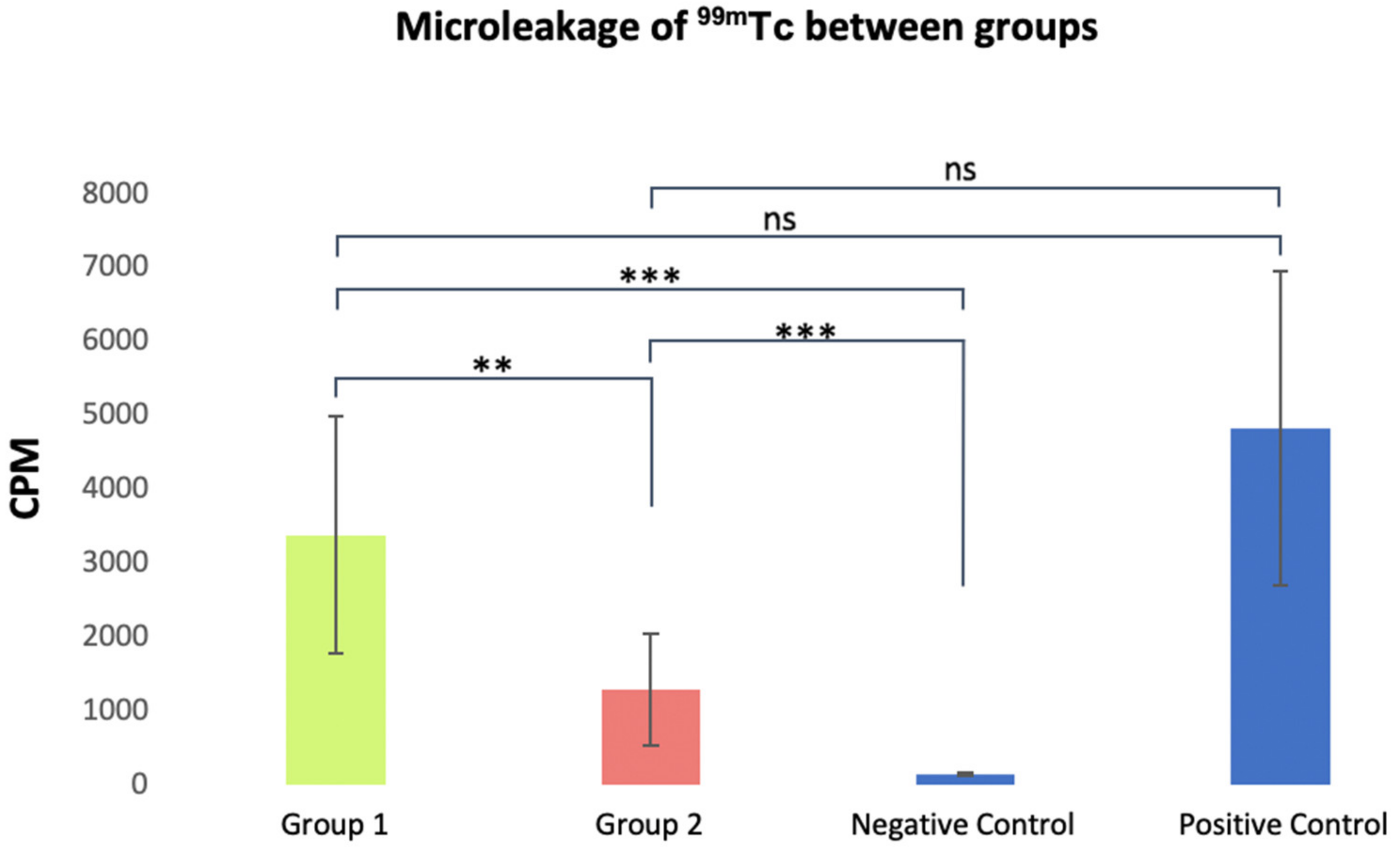
After processing the images (Figure 1) and statistical analysis, it was possible to recognize, according to Table 2, average values (±the standard deviation) for the total counts. The averages and standard deviations of the counts per minute (CPM) of analyzed samples are given in Figure 2.
According to Figure 1, it was possible to detect that the positive control group presented the highest values, followed by group 1 (Surefill One™), group 2 (Spectra™ ST HV composite) and finally the negative control. When comparing the positive control with groups 1 and 2, there were no statistically significant differences between the positive control and group 1 (p = 0.553) and between the positive control and group 2 (p = 0.064).
Regarding the comparison between the negative control and groups 1 and 2, it was found that the negative control group has a statistically significant lower total count value compared to group 1 (p = 0.000) and group 2 (p = 0.000).
Finally, when comparing the values of the total counts of groups 1 and 2, it was found that group 1 presents statistically higher infiltration values than group 2, with statistical significance (p = 0.002).
In this methodology, the greater infiltration of radioisotopes correlates, directly, to the highest values of total counts. Therefore, the higher these values are, the higher the isotope microleakage rate associated with the respective restoration/material.
The key findings of this study concern the two experimental groups, where the Surefill One™ group (3371 ± 1607) had a higher microleakage rate and statistically significant differences in total counts (p < 0.01) when compared to the Spectra™ group. ST HV nanohybrid composite (1286 ± 751).
Regarding the positive control group (4817 ± 2131), groups 1 and 2 did not show a statistically significant decrease in total counts (p > 0.05). However, when compared with the negative control group (140 ± 13), groups 1 and 2 obtained values with statistically significant differences (p < 0.001).

4. Discussion

Microleakage at the tooth-restoration interface is considered a major factor in the longevity of dental restorations [28]. The consequences of microleakage can be diverse, such as marginal pigmentation, the early rupture of restorations, secondary caries at the tooth –restoration interface, postoperative sensitivity and the inflammatory pathology of the pulp organ [8,29]. The integrity of the marginal seal is therefore essential to increasing the longevity of restorations [3,30]. The integrity and internal adaptation of the restoration to dental walls is compromised when microleakage occurs, normally resulting from the polymerization contraction of the restorative material. It is, therefore, considered, among other factors, as the most common cause of failure to direct restorations in posterior teeth [3,5,8].
Microleakage can be studied through several methods, such as the use of dyes, glucose penetration, bacterial or toxin infiltration, protein microleakage, electrochemical microleakage or scanning electron microscopy [31,32,33,34].
The most common methods in the evaluation of microleakage, in recent studies, are infiltration with a dye and bacterial infiltration. However, penetration with the methylene blue dye has some limitations, such as the need to destroy the samples, the subjective evaluation of the results by a specialized operator and the semi-quantitative nature of the microleakage measurement [35]. The methodology that uses bacterial infiltration, although more clinically and biologically relevant when compared to penetration with dye, can only be applied to materials that have antimicrobial activity for the specific type of bacteria used and usually presents a qualitative rather than quantitative assessment [32].
The present study was designed to evaluate the ability of marginal sealing and the consequent microleakage of different restorative strategies/materials, using an uncommon radioactive isotopes technique. Spectra™ ST HV nanohybrid composite resin (Dentsply-Sirona, Konstanz, Germany) and the bioactive resin Surefill One™ (Dentsply-Sirona, Konstanz, Germany) were used in dental restorations with standardized cavities, using a solution of sodium pertechnetate (99mTc).
The use of radioactive isotopes is a method with a quantitative and non-destructive nature, allowing the measurement of microleakage of the same specimens at different intervals, for long periods and without the need to destroy the samples, in contrast to the two methods mentioned above [36]. It also allows the quantitative measurement of microleakage, detected quickly, even when present in very small concentrations, representing an advantage over the penetration method with a dye [37,38]. The adopted method showed lower mean values for the negative control group and higher mean values for the positive control group, with a statistically significant difference (p = 0.027), thus validating the selected methodology.
Polymerization shrinkage is a very complex phenomenon, reliant on the conditions of the cavity margins due to the difference in composition of dental substrates; the amount of material in the polymerization reaction; and the formulation and setting reaction mechanism of the selected material [3,39]. The reduction of polymerization shrinkage has been one of the focuses in the development of new materials [8,40]. However, different restorative materials have different formulations and, consequently, different polymerization shrinkages, which makes their “side by side” comparison complex [3,40].
The shrinkage process essentially occurs due to the conversion of monomers into a polymer network, replacing van der Waals bonds by stronger covalent bonds. This process creates contraction in the restorative material and the maladaptation of the margin, leading to an internal tensional stress on the surrounding tooth structure and consequent microleakage [9,41,42]. The magnitude of these stresses depends on several factors, such as the configuration of the cavities but also the effect of light-curing mode of the material [3,40]. In this study, all cavities had the same dimensions, as they had the same C factor (index between free and adhered faces).
The null hypothesis was rejected, because the study resin, Surefill One™, did not obtain a lower microleakage rate when compared to other conventional composite resins. Even more, the higher values of microleakage observed in group 1 (Surefill One™) when compared to group 2 (Spectra™ ST HV composite) suggest that the new bioactive resin may present greater polymerization shrinkage compared to a conventional nanohybrid composite resin. Dual polymerization was used for Surefill One™ (recommended by the manufacturer) which, by itself, can induce a higher polymerization shrinkage stress in cavities with a high C factor and a greater challenge for adhesion to the dentin of the cavity floor, when compared to self-curing techniques [21,43,44]. This way, composite resins achieve better control over polymerization shrinkage, reducing microgaps and associated stress, thus managing to produce restorations with less microleakage and consequently greater longevity.
However, when restoring cavities of this type, other parameters must be taken into consideration, such as the mechanical properties of the materials and the strength of adhesion to dentin [11,45]. The adhesion of Surefill One™ depends mainly on high molecular weight polyacrylic acids, capable of facilitating the hybridization of the smear layer and ionic interactions between the dentin calcium and the carboxylic groups of the MOPOS (Modified Polyacid System), also reported in RM-GICs [11,14]. Despite containing water in its formulation, this self-adhesive hybrid bulk-fill resin requires some moisture to activate the functional acids [14,46]. This way dentin must not be completely dehydrated. However, it is much more difficult to control the moisture level in a deep, narrow cavity than on a flat surface, making it difficult to obtain the ideal moistened dentin [15,47]. The above-mentioned factors can help to justify the higher values of microleakage with statistically significant differences obtained between the test groups (p = 0.002).
Test groups 1 and 2, when compared with the positive control group, did not show values with a statistically significant difference (p > 0.05), unlike when analyzed with the negative control group where there is a statistically significant difference (p < 0.001). From this analysis, we can infer that both restorative materials under study are far from what could be considered an ideal restorative material, where microleakage values would potentially be close to null. Additionally, recent research showed that Biomimetic Hydroxyapatite [48] and Casein phosphopeptide-amorphous calcium phosphate [49] compounds showed the deposition of hydroxyapatite on polymeric composite, thus preventing caries on the margins of the composite frameworks. Additionally, the influence of these compounds should be tested in future microleakage investigations.
The standardization of in vitro conditions is an essential step to control possible bias factors, optimize the statistical analysis and allow the reproducibility of the study [31,50]. As recommended by several authors [51,52,53,54], the restorative procedures were performed by the same operator, reducing the associated “human” error.
However, this study has some limitations: not being an “in vivo” protocol, the restorative process ends up not being influenced by several factors present in the oral cavity, such as contaminants (saliva, blood, etc.), humidity, quality of isolation protocol and access and location of the lesion. In addition to these factors, the cavities obtained after the removal of the lesions would never be standardized and the restorations would be subject to cyclic fatigue. No ageing process was carried out, contrary to some studies, in order to mimic the previously mentioned factors [3,55,56]. In addition, increasing the sample could increase the robustness of the study, as well as performing microleakage studies with dyes. Therefore, we must regard the limitations inherent to the present “in vitro” study when interpreting the results.
The laboratory technique proved to be simple and fast and met the objective of a quantitative method to the evaluation of microleakage. The performance of Surefill One™ and the intrinsic results of the present study must be substantiated with a new study, where the same samples would be used, but this time using dye penetration, thus comparing the two methods of microleakage evaluation. Subsequently, carrying out a new “in vitro” study with same samples using a thermocycling process to evaluate the possible modification to results after aging the samples would be valuable. Additionally, long-term clinical studies would be beneficial to consolidating the results and the topic of study.

5. Conclusions

Based on the present microleakage study, it was possible to conclude that the new bioactive composite resin (Surefill One™) does not reduce the microleakage rate when compared to conventional nanohybrid composite resin (Spectra™ ST HV). However, Surefill One™ can be used in temporary restorations, primary teeth and in cases of tissue remineralization, avoiding more invasive procedures. This new “bulk-fill” bioactive composite resin represents an evolution over the old generation of bioactive materials and an asset in areas such as pediatric dentistry.

Author Contributions

Conceptualization, S.P., C.M.M., M.F.B., A.M.A. and A.B.P.; Data curation, P.N. and M.M.F.; Formal analysis, A.C., J.S. and M.M.F.; Funding acquisition, M.F.B. and E.C.; Investigation, P.N., S.P., A.M.A. and A.B.P.; Methodology, P.N., S.P. and A.M.A.; Project administration, C.M.M., M.M.F., M.F.B. and A.B.P.; Resources, C.M.M., I.A. and A.C.; Software, I.A., A.C. and J.S.; Supervision, C.M.M., M.F.B., E.C. and A.B.P.; Validation, S.P., E.C. and A.M.A.; Visualization, S.P., J.S., E.C. and A.B.P.; Writing—original draft, P.N.; Writing—review and editing, A.M.A. All authors have read and agreed to the published version of the manuscript.

Funding

This research received no external funding.

Institutional Review Board Statement

The study was conducted in accordance with the Declaration of Helsinki and approved by the Ethics Committee of the Faculty of Medicine of the University of Coimbra and was approved under the number CE-086/2019.

Informed Consent Statement

Informed consent was obtained from all subjects involved in the study.

Data Availability Statement

The data presented in this study are available on request from the corresponding author.

Acknowledgments

All authors are grateful to Dentsply Sirona, Portugal, for the donation of the Surefill One™, the Spectra™ ST HV nanohybrid composite and other materials used in this study.

Conflicts of Interest

The authors declare no conflict of interest.

References

  1. Maas, M.S.; Alania, Y.; Natale, L.C.; Rodrigues, M.C.; Watts, D.C.; Braga, R.R. Trends in Restorative Composites Research: What Is in the Future? Braz. Oral Res. 2017, 31, 23–36. [Google Scholar] [CrossRef] [Green Version]
  2. Zhang, K.; Zhang, N.; Weir, M.D.; Reynolds, M.A.; Bai, Y.; Xu, H.H.K. Bioactive Dental Composites and Bonding Agents Having Remineralizing and Antibacterial Characteristics. Dent. Clin. N. Am. 2017, 61, 669–687. [Google Scholar] [CrossRef] [PubMed]
  3. Carrilho, E.; Abrantes, M.; Paula, A.; Casalta-Lopes, J.; Botelho, M.; Ferreira, M. Microleakage Study of a Restorative Material via Radioisotope Methods. Rev. Port. Estomatol. Med. Dentária Cir. Maxilofac. 2014, 55, 129–134. [Google Scholar] [CrossRef] [Green Version]
  4. Pawar, M.; Saleem Agwan, M.A.; Ghani, B.; Khatri, M.; Bopache, P.; Aziz, M.S. Evaluation of Class II Restoration Microleakage with Various Restorative Materials: A Comparative In vitro Study. J. Pharm. Bioallied Sci. 2021, 13 (Suppl. 2), S1210–S1214. [Google Scholar] [CrossRef] [PubMed]
  5. Delgado, A.H.S.; Jamal, H.; Young, A.; Ashley, P. Scoping Review of Trials Evaluating Adhesive Strategies in Pediatric Dentistry: Where Do Simplified Strategies Lie? BMC Oral Health 2021, 21, 33. [Google Scholar] [CrossRef]
  6. Frankenberger, R.; Dudek, M.-C.; Winter, J.; Braun, A.; Krämer, N.; von Stein-Lausnitz, M.; Roggendorf, M.J. Amalgam Alternatives Critically Evaluated: Effect of Long-Term Thermomechanical Loading on Marginal Quality, Wear, and Fracture Behavior. J. Adhes. Dent. 2020, 22, 107–116. [Google Scholar] [CrossRef]
  7. Ayna, B.; Celenk, S.; Atas, O.; Tümen, E.C.; Uysal, E.; Toptanci, I.R. Microleakage of Glass Ionomer Based Restorative Materials in Primary Teeth: An In Vitro Study. Niger. J. Clin. Pract. 2018, 21, 1034–1037. [Google Scholar] [CrossRef]
  8. Pfefferkorn, F. Scientific Manual Surefil OneTM Self-Adhesive Composite Hybrid; Dentsply Sirona: Charlotte, NC, USA, 2019. [Google Scholar]
  9. Radhika, M.; Sajjan, G.S.; Kumaraswamy, B.; Mittal, N. Effect of Different Placement Techniques on Marginal Microleakage of Deep Class-II Cavities Restored with Two Composite Resin Formulations. J. Conserv. Dent. 2010, 13, 9. [Google Scholar] [CrossRef] [Green Version]
  10. Arora, R.; Kapur, R.; Sibal, N.; Juneja, S. Evaluation of Microleakage in Class II Cavities using Packable Composite Restorations with and without use of Liners. Int. J. Clin. Pediatr. Dent. 2012, 5, 178–184. [Google Scholar] [CrossRef]
  11. François, P.; Remadi, A.; le Goff, S.; Abdel-Gawad, S.; Attal, J.P.; Dursun, E. Flexural Properties and Dentin Adhesion in Recently Developed Self-Adhesive Bulk-Fill Materials. J. Oral Sci. 2021, 63, 139–144. [Google Scholar] [CrossRef] [PubMed]
  12. Mickenautsch, S. High-Viscosity Glass-Ionomer Cements for Direct Posterior Tooth Restorations in Permanent Teeth: The Evidence in Brief. J. Dent. 2016, 55, 121–123. [Google Scholar] [CrossRef] [PubMed]
  13. Iaculli, F.; Salucci, A.; di Giorgio, G.; Luzzi, V.; Ierardo, G.; Polimeni, A.; Bossù, M. Bond Strength of Self-Adhesive Flowable Composites and Glass Ionomer Cements to Primary Teeth: A Systematic Review and Meta-Analysis of In Vitro Studies. Materials 2021, 14, 6694. [Google Scholar] [CrossRef] [PubMed]
  14. Francois, P.; Fouquet, V.; Attal, J.P.; Dursun, E. Commercially Available Fluoride-Releasing Restorative Materials: A Review and a Proposal for Classification. Materials 2020, 13, 2313. [Google Scholar] [CrossRef] [PubMed]
  15. Yao, C.; Ahmed, M.H.; Okazaki, Y.; van Landuyt, K.L.; Huang, C.; van Meerbeek, B. Bonding Efficacy of a New Self-Adhesive Restorative onto Flat Dentin vs Class-I Cavity-Bottom Dentin. J. Adhes. Dent. 2020, 22, 65–77. [Google Scholar] [CrossRef] [PubMed]
  16. Vilkinis, V.; Hörsted-Bindslev, P.; Baelum, V. Two-Year Evaluation of Class II Resin-Modified Glass Ionomer Cement/Composite Open Sandwich and Composite Restorations. Clin. Oral Investig. 2000, 4, 133–139. [Google Scholar] [CrossRef]
  17. Banomyong, D.; Palamara, J.E.A.; Messer, H.H.; Burrow, M.F. Fluid Flow after Resin-Composite Restoration in Extracted Carious Teeth. Eur. J. Oral Sci. 2009, 117, 334–342. [Google Scholar] [CrossRef]
  18. Sidhu, S.K. Clinical Evaluations of Resin-Modified Glass-Ionomer Restorations. Dent. Mater. 2010, 26, 7–12. [Google Scholar] [CrossRef]
  19. Sidhu, S.; Nicholson, J. A Review of Glass-Ionomer Cements for Clinical Dentistry. J. Funct. Biomater. 2016, 7, 16. [Google Scholar] [CrossRef]
  20. Xie, D.; Brantley, W.A.; Culbertson, B.M.; Wang, G. Mechanical Properties and Microstructures of Glass-Ionomer Cements. Dent. Mater. 2000, 16, 129–138. [Google Scholar] [CrossRef]
  21. Yao, C.; Ahmed, M.H.; Zhang, F.; Mercelis, B.; van Landuyt, K.L.; Huang, C.; van Meerbeek, B. Structural/Chemical Characterization and Bond Strength of a New Self-Adhesive Bulk-Fill Restorative. J. Adhes. Dent. 2020, 22, 85–97. [Google Scholar] [CrossRef]
  22. Klee, J.E.; Renn, C.; Elsner, O. Development of Novel Polymer Technology for a New Class of Restorative Dental Materials. J. Adhes. Dent. 2020, 22, 35–45. [Google Scholar] [CrossRef]
  23. Li, F.; Weir, M.D.; Fouad, A.F.; Xu, H.H.K. Time-Kill Behaviour against Eight Bacterial Species and Cytotoxicity of Antibacterial Monomers. J. Dent. 2013, 41, 881–891. [Google Scholar] [CrossRef] [Green Version]
  24. Imazato, S.; Tarumi, H.; Ebi, N.; Ebisu, S. Cytotoxic Effects of Composite Restorations Employing Self-Etching Primers or Experimental Antibacterial Primers. J. Dent. 2000, 28, 61–67. [Google Scholar] [CrossRef]
  25. Mazzi, U.; Schibli, R.; Pietzsch, H.J.; Künstler, J.U.; Spies, H. Technetium in Medicine. In Technetium-99m Pharmaceuticals: Preparation and Quality Control in Nuclear Medicine; Springer: Berlin/Heidelberg, Germany, 2007; pp. 7–58. [Google Scholar] [CrossRef]
  26. Pagella, P.; Stadlinger, B.; Mitsiadis, T.A. Isolation of Dental Pulp and Periodontal Cells from Human Teeth for Single-Cell RNA Sequencing. STAR Protoc. 2021, 2, 100953. [Google Scholar] [CrossRef]
  27. Wannous, M.; Abboud, S.A. Curing Depth and Degree of Conversion of Different Nano-Hybrid Composites. J. Stomatol. 2021, 74, 147–152. [Google Scholar] [CrossRef]
  28. Hameed, H.; Babu, B.P.; Sagir, V.M.M.; Chiriyath, K.J.; Mathias, J.; Shaji, A.P. Microleakage in Resin Composite Restoration Following Antimicrobial Pre-Treatments with 2% Chlorhexidine and Clearfil Protect Bond. J. Int. Oral Health 2015, 7, 71–76. [Google Scholar]
  29. BALLAL, N.V. Microleakage of Composite Resin Restorations. Aust. Dent. J. 2008, 53, 369. [Google Scholar] [CrossRef]
  30. Majety, K.K.; Pujar, M. In Vitro Evaluation of Microleakage of Class II Packable Composite Resin Restorations Using Flowable Composite and Resin Modified Glass Ionomers as Intermediate Layers. J. Conserv. Dent. 2011, 14, 414–417. [Google Scholar] [CrossRef] [PubMed] [Green Version]
  31. Pereira, I.R.; Carvalho, C.; Paulo, S.; Martinho, J.P.; Coelho, A.S.; Paula, A.B.; Marto, C.M.; Carrilho, E.; Botelho, M.F.; Abrantes, A.M.; et al. Apical Sealing Ability of Two Calcium Silicate-Based Sealers Using a Radioactive Isotope Method: An In Vitro Apexification Model. Materials 2021, 14, 6456. [Google Scholar] [CrossRef] [PubMed]
  32. Jafari, F.; Rahimi, S.; Shahi, S.; Jafari, S. Endodontic Microleakage Studies: Correlation among Different Methods, Clinical Relevance, and Potential Laboratory Errors. Minerva Stomatol. 2017, 66, 169–177. [Google Scholar] [CrossRef] [PubMed]
  33. Gogna, R.; Jagadis, S.; Shashikal, K. A Comparative in Vitro Study of Microleakage by a Radioactive Isotope and Compressive Strength of Three Nanofilled Composite Resin Restorations. J. Conserv. Dent. 2011, 14, 128. [Google Scholar] [CrossRef] [Green Version]
  34. Rengo, C.; Goracci, C.; Ametrano, G.; Chieffi, N.; Spagnuolo, G.; Rengo, S.; Ferrari, M. Marginal Leakage of Class V Composite Restorations Assessed Using Microcomputed Tomography and Scanning Electron Microscope. Oper. Dent. 2015, 40, 440–448. [Google Scholar] [CrossRef]
  35. Camps, J.; Pashley, D. Reliability of the Dye Penetration Studies. J. Endod. 2003, 29, 592–594. [Google Scholar] [CrossRef]
  36. Afonso, T.; Lusitana, M.; de Andrade, S.; Pega, M.; Michelotto, A.L.D.C.; Margarida, A.; Abrantes, C.; Oliveiros, B.; Virgínia, E.; Carrilho, P.; et al. Effect of Calcium Hydroxide as Intracanal Medication on the Apical Sealing Ability of Mineral Trioxide Aggregate (MTA): An in Vitro Apexification Model Efeito Da Medicação Intra-Canal de Hidróxido de Cálcio Na Capacidade Seladora Do Mineral Trióxido Agregado (MTA): Um Modelo de Apicificação in Vitro. J. Health Sci. Inst. 2012, 30, 318–340. [Google Scholar]
  37. Ferreira, M.M. Quantitative Scintigraphic Analysis of the Apical Seal in Thermafil/Topseal and RealSeal 1/Realseal Filled Root Canals. World J. Stomatol. 2013, 2, 30. [Google Scholar] [CrossRef]
  38. Prasad, B.K.; Sudhakaran, S. Radioactive Isotope Evaluation of Coronal Leakage after Endodontic Treatment in Teeth Restored with Three Different Intracoronal Restorative Materials: An in Vitro Study. World J. Dent. 2011, 2, 35–38. [Google Scholar] [CrossRef]
  39. Schneider, L.F.J.; Cavalcante, L.M.; Silikas, N. Shrinkage Stresses Generated during Resin-Composite Applications: A Review. J. Dent. Biomech. 2010, 2010, 131630. [Google Scholar] [CrossRef]
  40. Kleverlaan, C.J.; Feilzer, A.J. Polymerization Shrinkage and Contraction Stress of Dental Resin Composites. Dent. Mater. 2005, 21, 1150–1157. [Google Scholar] [CrossRef]
  41. Alvarez-Gayosso, C.; Barceló-Santana, F.; Guerrero-Ibarra, J.; Sáez-Espínola, G.; Canseco-Martínez, M.A. Calculation of Contraction Rates Due to Shrinkage in Light-Cured Composites. Dent. Mater. 2004, 20, 228–235. [Google Scholar] [CrossRef]
  42. Lopes, L.G.; Franco, E.B.; Pereira, J.C.; Mondelli, R.F.L. Effect of Light-Curing Units and Activation Mode on Polymerization Shrinkage and Shrinkage Stress of Composite Resins. J. Appl. Oral Sci. 2008, 16, 35–42. [Google Scholar] [CrossRef] [Green Version]
  43. Giachetti, L.; Bertini, F.; Bambi, C.; Russo, D.S. A Rational Use of Dental Materials in Posterior Direct Resin Restorations in Order to Control Polymerization Shrinkage Stress. Minerva Stomatol. 2007, 56, 129–138. [Google Scholar]
  44. Wattanawongpitak, N.; Yoshikawa, T.; Burrow, M.F.; Tagami, J. The effect of thermal stress on bonding durability of resin composite adaptation to the cavity wall. Dent. Mater. J. 2007, 26, 445–450. [Google Scholar] [CrossRef]
  45. Papadopoulos, C.; Dionysopoulos, D.; Tolidis, K.; Kouros, P.; Koliniotou-Koumpia, E.; Tsitrou, E.A. Structural Integrity Evaluation of Large MOD Restorations Fabricated With a Bulk-Fill and a CAD/CAM Resin Composite Material. Oper. Dent. 2019, 44, 312–321. [Google Scholar] [CrossRef] [PubMed]
  46. Maletin, A.; Markovic, D.; Neskovic, I.; Ramic, B.; Veljovic, T.; Ristic, I. Application of a Novel Modification of the Microbond Test for Evaluation of Adhesive Bond Strength Between Fiber Posts and Dual-Cure Dental Resin Cement. Med. Sci. Monit. 2019, 25, 3397–3405. [Google Scholar] [CrossRef]
  47. de Munck, J.; van Landuyt, K.; Coutinho, E.; Poitevin, A.; Peumans, M.; Lambrechts, P.; van Meerbeek, B. Micro-Tensile Bond Strength of Adhesives Bonded to Class-I Cavity-Bottom Dentin after Thermo-Cycling. Dent. Mater. 2005, 21, 999–1007. [Google Scholar] [CrossRef]
  48. Butera, A.; Pascadopoli, M.; Gallo, S.; Lelli, M.; Tarterini, F.; Giglia, F.; Scribante, A. SEM/EDS Evaluation of the Mineral Deposition on a Polymeric Composite Resin of a Toothpaste Containing Biomimetic Zn-Carbonate Hydroxyapatite (MicroRepair®) in Oral Environment: A Randomized Clinical Trial. Polymers 2021, 13, 2740. [Google Scholar] [CrossRef]
  49. Khanduri, N.; Kurup, D.; Mitra, M. Quantitative Evaluation of Remineralizing Potential of Three Agents on Artificially Demineralized Human Enamel Using Scanning Electron Microscopy Imaging and Energy-Dispersive Analytical X-Ray Element Analysis: An in Vitro Study. Dent. Res. J. 2020, 17, 366–372. [Google Scholar] [CrossRef]
  50. Ribeiras, I.; Vasconcelos, I.; Ramos, M.; Lopes, M.; Ginjeira, A. Estudo Comparativo Da Adaptação Marginal de 2 Cimentos Endodônticos. Rev. Port. Estomatol. Med. Dentária Cir. Maxilofac. 2015, 56, 173–181. [Google Scholar] [CrossRef] [Green Version]
  51. Vicente, A.; Ortiz, A.J.; Bravo, L.A. Microleakage beneath Brackets Bonded with Flowable Materials: Effect of Thermocycling. Eur. J. Orthod. 2009, 31, 390–396. [Google Scholar] [CrossRef] [PubMed] [Green Version]
  52. Roggendorf, M.J.; Krämer, N.; Appelt, A.; Naumann, M.; Frankenberger, R. Marginal Quality of Flowable 4-Mm Base vs. Conventionally Layered Resin Composite. J. Dent. 2011, 39, 643–647. [Google Scholar] [CrossRef] [PubMed]
  53. Burke, F.J.T.; Crisp, R.J.; James, A.; MacKenzie, L.; Pal, A.; Sands, P.; Thompson, O.; Palin, W.M. Two Year Clinical Evaluation of a Low-Shrink Resin Composite Material in UK General Dental Practices. Dent. Mater. 2011, 27, 622–630. [Google Scholar] [CrossRef] [PubMed]
  54. Ernst, C.P.; Brandenbusch, M.; Meyer, G.; Canbek, K.; Gottschalk, F.; Willershausen, B. Two-Year Clinical Performance of a Nanofiller vs a Fine-Particle Hybrid Resin Composite. Clin. Oral Investig. 2006, 10, 119–125. [Google Scholar] [CrossRef]
  55. Amaral, F.L.B.; Colucci, V.; Palma-Dibb, R.G.; Corona, S.A.M. Assessment of In Vitro Methods Used to Promote Adhesive Interface Degradation: A Critical Review. J. Esthet. Restor. Dent. 2007, 19, 340–353. [Google Scholar] [CrossRef] [PubMed]
  56. Yuasa, T.; Iijima, M.; Ito, S.; Muguruma, T.; Saito, T.; Mizoguchi, I. Effects of Long-Term Storage and Thermocycling on Bond Strength of Two Self-Etching Primer Adhesive Systems. Eur. J. Orthod. 2010, 32, 285–290. [Google Scholar] [CrossRef] [PubMed]
Figure 1. Visual representation of radioactivity emitted by the specimens detected by a gamma camera of one tooth per group (CPM—Counts Per Minute).
Applsci 12 11827 g001
Figure 2. Visual representation of mean values and standard deviation of total counts in each group and significance level between groups: ** p < 0.01; *** p < 0.001; ns—meaningless. Group 1: Surefill One™; Group 2: Spectra™ ST HV composite; CPM: counts per minute.
Applsci 12 11827 g002
Table 1. Summary of the main chemical, physical and mechanical characterizations of the materials tested.
CharacterizationChemicalPhysical and Mechanical
Surefill One™Composition:
Aluminum phosphorus crystals, magnesium, electrolyte, sodium fluoride and sodium silicate
Water
Highly dispersed silicon dioxide
Acrylic acid
Polycarboxylic acid
Ytterbium fluoride
Bifunctional acrylate
Self-adjusting launcher
4-tert-butyl-N,N-dimethylaniline
Iron oxide pigments
Barium sulfate pigment
manganese pigment
Camphorquinone (photoinitiator)
Stabilizer
Self-Adhesive composite hybrid
Dual curing
Releases fluor
Recharge fluor
Indicated for Class I, II and V, and for the construction of cores.
Application in large increments, and dual configuration to fill any depth
Spectra™ ST HVComposition:
Methacrylate modified polysiloxane (organically modified ceramic)
Dimethacrylate resins
Fluorescent pigment UV stabilizer
Stabilizer
Camphorquinone
Ethyl-4(dimethylamino) benzoate
Bis-(4-methyl-phenyl)-iodonium hexafluorophosphate
Barium-aluminum-borosilicate glass
Ytterbium fluoride
Iron oxide pigments and titanium oxide pigments according to shade
Nano-ceramic
Light-cured
Radiopaque
Universal composite with novel SphereTEC® filler technology (granulated spherical fillers in combination with an optimized resin matrix)
Indicated: Direct anterior and posterior restorations (including occlusal surfaces); Core build-ups; Splinting; Indirect restorations including inlays, onlays and veneers
Table 2. Statistical analysis of laboratory results. Values in CPM (counts per minute).
GroupAverageStandard DeviationMinimum ValuesMaximum Values
Group 1
(n = 13)
3371.1±154812706717
Group 2
(n = 13)
1285.9±724.12932489
Negative
Control
(n = 5)
139.6±12.3120156
Positive
Control
(n = 5)
4817.2±1906.423698090
Publisher’s Note: MDPI stays neutral with regard to jurisdictional claims in published maps and institutional affiliations.