Article

Thymoquinone, but Not Metformin, Protects against Gentamicin-Induced Nephrotoxicity and Renal Dysfunction in Rats

by
Mansour Alsharidah
1,*,
Abdel-Moneim Hafez Abdel-Moneim
1,2,
Ashwag Saleh Alsharidah
1,
Mugahid A. Mobark
3,4,
Arshad Husain Rahmani
5,
Ahmed Shata
6,7,
Ahmed A. H. Abdellatif
8,9,
Mahmoud Zaki El-Readi
10,11,
Khalid M. Mohany
12 and
Osamah Al Rugaie
13
1
Department of Physiology, College of Medicine, Qassim University, Buraydah 51452, Saudi Arabia
2
Department of Physiology, Faculty of Medicine, Mansoura University, Mansoura 35516, Egypt
3
Department of Pharmacy Practice, College of Pharmacy, Qassim University, Buraydah 51452, Saudi Arabia
4
Department of Pathology, Faculty of Medicine, University of Kordofan, Kordofan 13314, Sudan
5
Department of Medical Laboratories, College of Applied Medical Science, Qassim University, Buraydah 51452, Saudi Arabia
6
Department of Clinical Pharmacology, Faculty of Medicine, Mansoura University, Mansoura 35516, Egypt
7
Department of Clinical Pharmacy, Faculty of Pharmacy, Delta University for Science and Technology, Gamasa 11152, Egypt
8
Department of Pharmaceutics, College of Pharmacy, Qassim University, Buraydah 51452, Saudi Arabia
9
Department of Pharmaceutics and Industrial Pharmacy, Faculty of Pharmacy, Al-Azhar University, Assiut 71524, Egypt
10
Department of Biochemistry, Faculty of Medicine, Umm Al-Qura University, Abdia, Makkah 21955, Saudi Arabia
11
Department of Biochemistry, Faculty of Pharmacy, Al-Azhar University, Assiut 71524, Egypt
12
Department of Medical Biochemistry and Molecular Biology, Faculty of Medicine, Assiut University, Assiut 71515, Egypt
13
Department of Basic Medical Sciences, College of Medicine and Medical Sciences, Qassim University, Unaizah 51911, Saudi Arabia
*
Author to whom correspondence should be addressed.
Appl. Sci. 2021, 11(9), 3981; https://doi.org/10.3390/app11093981
Submission received: 12 March 2021 / Revised: 21 April 2021 / Accepted: 22 April 2021 / Published: 27 April 2021

Abstract

:
Background: Gentamicin (GM) is an antibiotic that is widely used to treat many Gram-negative bacteria, such as those involved in urinary tract infections. However, being nephrotoxic, GM dose adjustment and reno-protective elements must be concurrently administered with GM to minimize kidney damage. Oxidative stress plays a pivotal role in the pathogenesis of GM-induced nephrotoxicity. Thymoquinone (TQ) is a promising therapeutic substance, that is being extensively studied in many diseases, such as diabetes mellitus, cancer, hypertension, and others. The powerful antioxidant properties of TQ may greatly help in minimizing GM nephrotoxicity. Metformin (MF) is a well-known, clinically approved oral hypoglycaemic drug that has many other actions, including antioxidant properties. The aim of this work was to evaluate the possible antioxidant and reno-protective effects of TQ and metformin in GM-induced nephrotoxicity in the same model (rats) at the same time. In addition, we aimed to further understand the effects underlying GM-induced nephrotoxicity. Methods: Twenty male rats were randomly divided into four equal groups: the first group (control) received distilled water; the second group received GM only; the third group received concurrent oral TQ and GM; and the fourth group received concurrent oral MF and GM. After 4 weeks, renal function and histopathology, as well as levels of the oxidative markers glutathione peroxidase-1 (GLPX1), superoxide dismutase (SOD), and malondialdehyde (MDA) in the kidney tissues, were assessed. Results: Compared with the control group, and as expected, the GM-injected rats showed significant biochemical and histological changes denoting renal damage. Compared with GM-injected rats, the concurrent administration of TQ with GM significantly reduced the levels of serum creatinine, serum urea, and tissue MDA and significantly increased the levels of GLPX1 and SOD. Concurrent metformin administration with GM significantly increased the levels of both GLPX1 and SOD and significantly decreased the levels of tissue MDA but had no significant effect on serum creatinine and urea levels. Compared with GM-injected rats, the addition of either TQ or MF resulted in a reduction in endothelial proliferation and mesangial hypercellularity. Conclusions: Both TQ and MF effectively alleviated the oxidative stress in GM-induced nephrotoxicity in rats, with TQ but not MF producing a complete reno-protective effect. Further studies for evaluation of different reno-protective mechanisms of TQ should be conducted.

1. Background

Antibiotics, especially aminoglycosides, have been notably linked to the development of nephrotoxicity that may end in acute kidney injury (AKI). Gentamicin (GM), a major aminoglycoside family member with low bacterial resistance rates (when compared with the older aminoglycoside members, such as streptomycin [1]). Moreover, the prevalence of aminoglycoside resistance remained relatively low when compared with that for cephalosporins or fluoroquinolones [2]. Although, GM is commonly used in the treatment of life threatening conditions caused mainly by Gram-negative bacteria, its clinical uses are greatly limited because of its nephrotoxicity [3].
Although GM nephrotoxicity has been attributed mainly to tubular cytotoxicity that ranges from loss of brush border of epithelial cells to tubular necrosis [4], the exact main site of tubular damage is still under investigation. Very early, in 1980, Moir’ et al., proposed a central mechanism for the gentamicin-induced AKI that involved the accumulation of the drug in the lysosomes of the cells of the proximal tubule with subsequent inhibition of specific enzymes activities. In addition, lysosomal accumulation of the drug led to altered properties of the lysosomal membrane permeability [5]. More recently, gentamicin-induced AKI had been attributed mainly to apoptosis of the proximal tubule and collecting cells [6]. Thus, the precise mechanism of GM-induced nephrotoxicity is still not fully clarified, although the principle pathophysiology involves the generation of reactive oxygen species (ROS), apoptosis, increased endothelin-1 levels, and increased cellular infiltration [7]. Karadeniz et al. showed that increased ROS generation resulting from GM-induced nephrotoxicity results in the inactivation of many enzymes involved in oxidative stress [8]. It had been reported that GM promotes AKI through the generation of ROS, such as superoxide anions [9], hydroxyl radicals, hydrogen peroxide [10], and reactive nitrogen species (RNS) in the kidney [11]. Excess generation of ROS is responsible for the pathogenesis of acute tubular necrosis (ATN) and a reduced glomerular filtration rate (GFR), which are hallmarks of drug-induced nephrotoxicity [12]. Therefore, studies aiming to minimise GM-induced nephrotoxicity should be based on the concurrent use of reno-protective agents, including ROS scavengers and antioxidant agents. At the histopathological level, the previous studies revealed that gentamicin-induced nephrotoxicity involved both glomerular and tubular changes in the form of tubular necrosis, apoptosis of glomerular mesangial cells [13], tubular degeneration associated with swelling, cytosis, and tubular irregularities [14].
TQ, the main volatile product of Nigella sativa, and MF (a well-known oral hypoglycaemic drug) both have known antioxidant effects, and as such could potentially help alleviate kidney damage in patients undergoing treatment with GM. In 2011, Sankaranarayanan and Pari reported that TQ has antioxidant effects and possesses a notable capability to prevent beta-cell damage in diabetic rats [15]. In addition, TQ has been extensively investigated as an anti-inflammatory [16], gastroprotective [17], and tumour anti-proliferative therapeutic agent [18]. MF, on the other hand, has been reported to have novel antioxidant and anti-inflammatory effects in type 2 diabetic patients [19].
Based on the findings that TQ and MF have antioxidant properties, the present study aimed to evaluate the efficacy of both TQ and metformin as possible reno-protective agents in GM-induced nephropathy in the same model (rats) at the same time. In addition, we aimed to further understand the effects underlying GM-induced nephrotoxicity and whether or not the antioxidant effect alone could protect against this nephrotoxicity.

2. Material and Methods

2.1. Animals

Twenty male Sprague-Dawley rats with average age between 5 and 6 months and weighing between 250 and 300 g were included in this study. The rats were obtained from the Animal House of College of Pharmacy, Qassim University, Saudi Arabia. The rats were housed in well-aerated plastic cages (5 rats/cage) under standard housing conditions (temperature of 24–25 °C, 50–80% humidity, with a 12 h light/dark cycle). The rats were allowed free access to standard rat food and water throughout the experiments. This study was approved by the Subcommittee of Health Research Ethics, Deanship of Scientific Research, Qassim University (Approval No: 18-04-07) in accordance with National Research Council (US) Guide for the Care and Use of Laboratory Animals [20].

2.2. Experimental Groups

The rats included in this study were randomly divided into four equal groups (n = 5 rats in each group). One group of rats was given 1 mL of distilled water per 100 g body weight per day by oral gavage for 4 weeks and served as the control group (CT). The other three groups were given a single daily intraperitoneal (IP) dose of GM (100 mg/kg body weight for 4 weeks [21]; gentamicin 80 mg/2 mL, Memphis Pharmaceutical and Chemical Industries, Egypt) The third group of rats, in addition to GM, was given concurrent 2-isopropyl-5-methylbenzoquinone (TQ; Sigma-Aldrich, St. Louis, MO, USA). TQ was dissolved in dimethyl sulfoxide (DMSO), followed by normal saline (the final concentration of DMSO was less than 0.5%). The prepared solution was given once daily by gastric gavage at a dose of 50 mg/kg body weight for 4 weeks [22]. The fourth group of rats, in addition to GM, was given concurrent MF (Sigma-Aldrich, St. Louis, MO, USA) at a dose of 60 mg/kg body weight [23] orally by oral gavage for 4 weeks, in addition to GM. At the end of the experiment, the rats were anaesthetized with ether, scarified, and blood samples were collected via the medial canthus of the eye and from heart puncture and transferred into sterilized tubes for serum separation for evaluation of renal function. After scarifying the animals, the kidneys were carefully dissected and stored in 10% neutral buffered formalin. Parts of one kidney sample were prepared for the histopathological examination. Other parts of the same used kidney samples were carefully weighed and homogenized in ice-cold 100 mM phosphate buffer (pH 7.4) using a Potter-Elvehjem homogeniser fitted with a Teflon Plunger. The homogenates were centrifuged, and the resulting supernatants were divided into aliquots and stored at –80 °C until use for later oxidative stress enzymes analysis.

2.3. Renal Function

Serum creatinine was measured using a creatinine kit (Crescent diagnostics; cat. no. CS604, KSA), using a kinetic method without deproteinization—the Jaffe reaction [24]. In the Jaffe reaction, creatinine reacts with alkaline precipitate to produce a reddish-orange colour, the intensity of which at 490 nm is directly proportional to creatinine concentration.
Serum urea was measured using a urea kit (Crescent diagnostics; cat. no. CS612, KSA), with an enzymatic, colorimetric endpoint—the Berthelot method [25]. Urease catalyses the conversion of urea to ammonia. In a modified Berthelot reaction, the ammonia ions react with a mixture of salicylate, hypochlorite, and nitroprusside to yield a blue-green dye (Indophenol). The intensity of this dye is proportional to the concentration of urea in the sample.

2.4. Oxidative Status Analysis

The homogenized kidney tissue was centrifuged for 20 min at 1000× g. The supernatants collected and the samples stored in aliquot at −80 °C till assessment. The collected supernatants were used for the evaluation of three oxidative enzymes in this study (GLPX1, SOD, and MDA) using ELISAs (Cloud-Clone Corp; USA) according to the manufacturer’s instructions as follows: For GLPX1, the kit was a sandwich enzyme immunoassay for in vitro quantitative measurement in tissue homogenates. At the end of the procedure, the colour change was measured spectrophotometrically at a wavelength of 450 ± 10 nm. The concentration of SOD in the samples was then determined by comparing the optical density (OD) of the samples with the standard curve. The same double-antibody sandwich was also applied for assessment of SOD spectrophotometrically at the same wavelength (450 ± 10 nm).
For MDA, the assay employed the competitive inhibition enzyme immunoassay technique. A monoclonal antibody specific to MDA was pre-coated onto a microplate. A competitive inhibition reaction was launched between biotin-labelled MDA and unlabelled MDA (standards or samples) with the pre-coated antibody specific to MDA. After addition of the substrate solution, the intensity of colour developed was reverse proportional to the concentration of MDA in the sample.

3. Histopathological Evaluation of the Kidneys

The formalin-fixed tissues were processed using an automated tissue processor machine (Leica TP1020) and embedded in paraffin blocks. Serial 5 µm sections were prepared using a microtome (Leica RM2245), stained with haematoxylin and eosin (H&E) and examined using a light microscope (Olympus BX41), digital image camera (5 MP Binocular Microscope Electronic Eyepiece USB Video CMOS Camera for Image Capture), and ToupView image analyser. All specimens were examined for glomerular and tubular changes (glomerular endothelial and mesangial proliferation Bowman’s space, vacuolar degeneration, tubular dilatation, and hyaline droplets).

Statistical Analysis

Statistical analysis was performed using Statistical Package for the Social Sciences (SPSS) (Version 19; Chicago, IL, USA). The mean (M) ± standard deviation (SD) was used to describe the studied variables, and one-way ANOVA was used for statistical comparisons. A p < 0.05 was considered statistically significant.

4. Results

To confirm that our rat model accurately represented GM-induced nephrotoxicity, we first assessed renal function and markers of oxidative stress in rats that had been treated with GM daily for four weeks. As expected, rats treated with GM showed both significant biochemical and histological changes when compared with the control group.

4.1. Biochemical Changes in the Different Groups

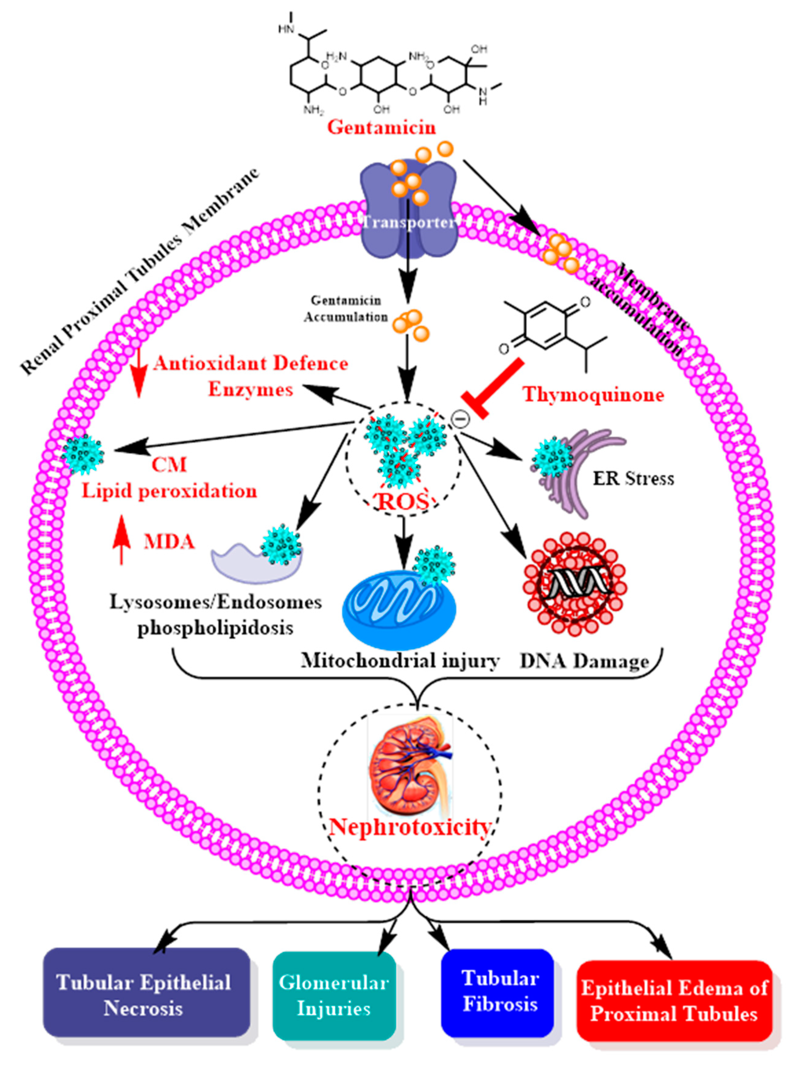
Nephrotoxic Effects and Oxidative Stress of GM in Treated Rats

The IP injection of GM at a single daily dose of 100 mg/kg body weight for four successive weeks resulted in a significant reduction in renal function, as indicated by the significantly increased serum creatinine in the GM-treated rats (M ± SD = 2.25 ± 0.46 mg/dl) compared with rats of the control group (M ± SD = 0.65 ± 0.05 mg/dl, p < 0.001) and the significantly increased levels of serum urea (M ± SD 148.31 ± 21.16 mg/dl) compared with those in the control group (M ± SD= 73 ± 1.3 mg/dl, p < 0.001; Table 1).
Additionally, injections of GM caused a significant reduction in oxidative buffering capacity in the treated rats, as indicated by a significant reduction in GLPX1 and SOD (M ± SD = 76.79 ± 14.22 and 1.25 ± 0.54, respectively) compared with that in the control group (M ± SD = 106.51 ± 5.07 and 2.76 ± 0.17, respectively, p < 0.05) and the significantly increased levels of tissue MDA (M ± SD= 356.32 ± 12.5 ng/mL) compared with those in the control group (M ± SD = 325.93 ± 8.83 ng/mL, p < 0.002; Table 1). These findings document the nephrotoxic effects of GM on the treated rats.

4.2. Renoprotective Effects of TQ on GM-Treated Rats

Next, we asked how treatment with TQ affected the biochemical changes induced by GM treatment. The concurrent oral administration of TQ in GM-treated rats at a dose of 50 mg/kg body weight daily for four successive weeks markedly alleviated the GM-induced nephrotoxicity, as indicated by the significantly reduced serum creatinine level (M ± SD= 0.70 ± 0.14 mg/dl) compared with that in the GM-treated rats (M ± SD= 2.25 ± 0.46 mg/dl, p < 0.001) and the significantly reduced level of serum urea (M ± SD= 74.37 ± 4.14 mg/dl) compared with that in the GM-treated rats (M ± SD = 148.31 ± 21.16 mg/dl, p < 0.00; Table 1).
Additionally, the administration of TQ improved the oxidative buffering capacity in the rats treated with GM, as indicated by the significant increase in GLPX1 and SOD (M ± SD = 111.88 ± 8.97 and 2.61 ± 0.17) compared with that in the GM-treated rats (M ± SD = 76.79 ± 14.22 and 1.25 ± 0.54, respectively, p < 0.05) and the significantly reduced levels of tissue MDA in TQ-treated rats (M ± SD 319.41 ± 8.21 ng/mL) compared with GM-treated rats (M ± SD = 356.32 ± 12.5 ng/mL, p < 0.00; Table 1). These findings suggest that TQ succeeded in reversing the biochemical changes (renal function and oxidative status) induced by GM injections.

4.3. Renoprotective Effects of MF on GM-Treated Rats

Next, we asked how treatment with MF affected the biochemical changes induced by GM treatment. The concurrent administration of MF to the GM-treated rats at a dose of 60 mg/kg body weight daily for four successive weeks produced no significant differences in serum creatinine and urea (M ± SD= 1.58 ± 0.55 and 126.86 ± 25.16 mg/dl) levels compared with those in the GM-treated rats (M ± SD = 2.25 ± 0.466 and 148.31 ± 21.16 mg/dl for creatinine and urea, respectively; Table 1).
Additionally, the administration of MF improved the oxidative buffering capacity in the rats treated with GM, as indicated by the significant increase in GLPX1 and SOD (M ± SD = 104.80 ± 5.01 and 2.61 ± 0.13) compared with that in the GM-treated rats (M ± SD = 76.79 ± 14.22 and 1.25 ± 0.54, respectively, p < 0.05) and the significantly reduced levels of tissue MDA (M ± SD 327.82 ± 15.04 ng/mL) compared with those in the GM-treated rats (M ± SD = 356.32 ± 12.5 ng/mL, p < 0.12; Table 1).
The comparison of the TQ-treated rats with the metformin-treated rats revealed no significant differences regarding the oxidative buffering capacity, but the TQ-treated rats showed better renal function (M ± SD=0.70 ± 0.14 and 74.37 ± 4.14 mg/dl) than the metformin-treated rats (M ± SD =1.58 ± 0.55 and 126.86 ± 25.16 mg/dl with p < 0.5 for serum creatinine and urea; Table 1). These findings suggest that MF could reverse the biochemical changes induced by GM injections at the level of oxidative stress but did not reverse the changes in renal function with the drug dose and experimental conditions used for this study.
Next, we asked how treatment with GM, TQ, or MF affected the renal histopathology of the treated rats.

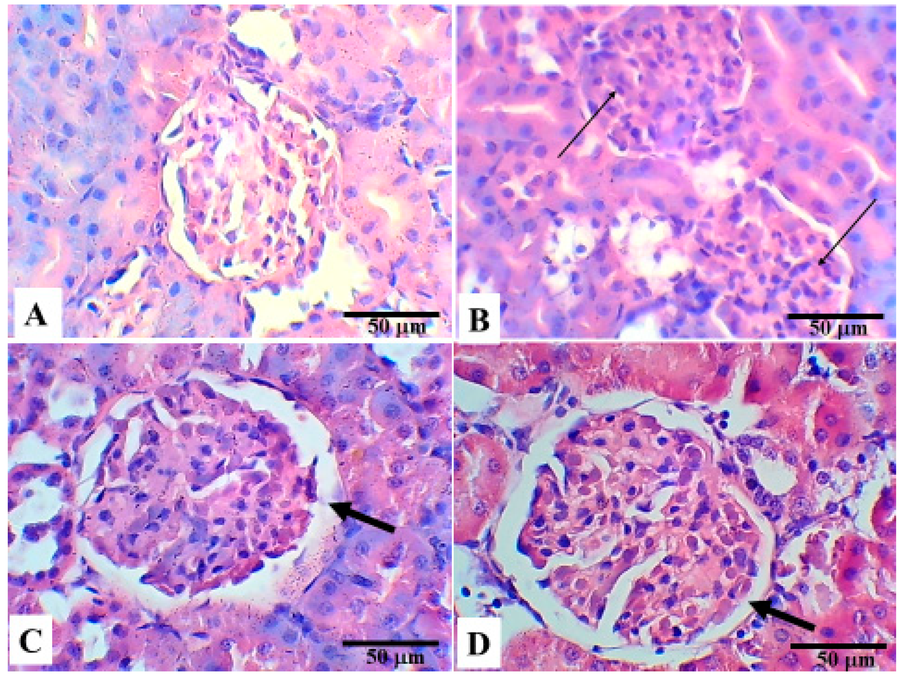
5. Histopathological Changes in the Different Groups

Figure 1 shows the glomerular changes in the four groups. Compared with the control group, GM induced mesangial cell hypercellularity and glomerular endothelial cell proliferation. When compared with the GM-treated group, Bowman’s space was noticeably preserved in the TQ-treated and MF-treated groups as a result of the reduction in endothelial proliferation and mesangial hypercellularity. These finding suggest that both TQ and MF persevered glomerular changes induced by GM treatment.
Figure 2 shows the observed tubular changes in the four groups. When compared with the control group, GM induced vacuolar degeneration of the tubular epithelium with evidence of tubular necrosis and tubular irregularities. However, the hyaline droplets seen in the GM-treated group were visibly reduced in the TQ-treated and MF-treated groups. In addition, the tubular regularity was preserved, the tubular vacuolar degeneration was reduced, and the ameliorative effects were increased in the TQ-treated group. These finding suggest that TQ has more protective effects especially at the tubular level than those of MF.

6. Discussion

In this study, we showed that both TQ and metformin efficiently alleviated oxidative stress in GM-induced nephrotoxicity in rats. However, only TQ produced reno-protective effects. We also confirmed the effects of GM on kidney function. The accumulation of GM, either intracellularly or in the cell membrane, lead to an increase in the production of ROS that interact with cellular macromolecules (lipids, protein, DNA). ROS increase denaturation of proteins, DNA damage, and production of MDA as a marker of lipid peroxidation (LPO) of the cell membrane’s lipids. These events lead to cellular injury and necrosis cascades that are commonly known as gentamicin-mediated nephrotoxicity (Figure 3) [26]. In addition, increases in the production of ROS by GM lead to depletion of antioxidant enzymes activity, such as superoxide dismutase (SOD), glutathione peroxidase (GPx), glutathione reductase (GR), and catalase (CAT) Figure 1 [27]. The most common markers indicating renal dysfunction and GM-nephrotoxicity are the increase of serum levels of creatinine and blood urea nitrogen [28].
In the present study, the daily injection of GM for four weeks resulted in nephrotoxicity as manifested by increased serum urea and creatinine concentrations. In addition, GM injection appeared to generate excess ROS, which resulted in a significant reduction in GLPX-1 and SOD levels, together with a significant increase in the MDA level. Histopathologically, GM affected both the glomerular and tubular structure of the kidney. The glomerular changes manifested as mesangial cell hypercellularity and glomerular endothelial cell proliferation with a resultant reduction in Bowman’s space. As reported in another study, GM simultaneously induced the proliferation and apoptosis of mesangial cells in vitro and glomerular mesangial cells in vivo [13]. This study also revealed that, in the renal tubules, GM induced vacuolar degeneration of the tubular epithelium with evidence of tubular necrosis and tubular irregularities. In a study performed by Alarifi S. et al. in 2012, tubular alterations caused by GM treatment appeared early in the form of necrosis, degeneration, and vacuolisation, and over time the degeneration progressed to severe necrosis. The degenerated tubules showed swelling, cytolysis, and tubular irregularity [14]. Thus, our results (glomerular and tubular changes) add support to the main pathophysiologic theme in GM-induced nephrotoxicity and are consistent with many previously published data [7,29,30,31]. Balakumar and his co-workers, attributed GM-induced nephrotoxicity to ROS generation and increased endothelin-1 level, in addition to cellular infiltration and apoptosis [7]. Thus, it seems that any animal trial aiming to reduce GM-induced nephrotoxicity should involve a substance with antioxidant activity and ROS scavenger properties [26].
In the current work, we tested the ability of well-known substances (TQ and MF) with antioxidant properties to reduce GM-induced nephrotoxicity. This is the first time that the two drugs (QT and MF) have been compared side-by-side for prevention of nephropathy in the same model (rats) at the same time. MF is widely used in diabetes, and many novel studies recommend TQ in treatment of diabetes [15,22] and prevention of diabetic complications, especially diabetic nephropathy. The concurrent administration of TQ greatly alleviated the GM-induced nephrotoxicity, possibly by protecting the kidney from lipid peroxidation, oxidative stress, and ROS production as well as by increasing the antioxidant enzymes activity (Figure 3), as indicated by a significant reduction in serum urea and creatinine levels and restoration of the disturbed oxidative stress status to normal levels. These findings are consistent with those of previous studies [21,29] reporting that TQ has a protective antioxidant effect and anti-inflammatory effect against GM-induced nephrotoxicity. Samarghandian and his colleagues performed a study to examine the cytokines profile (anti-inflammatory mechanism), renal function, and oxidative status in GM-induced acute renal failure. Findings of the study revealed that GM caused various changes, including renal failure [21].
This study also revealed that both TQ and MF ameliorated the effects of GM on the kidney. The glomerulus in the TQ and MF groups showed preservation of Bowman’s space when compared with the GM-group. In the renal tubules, the ameliorative effect of TQ was more pronounced than that of MF, as the hyaline droplets seen in GM-treated rats were visibly reduced; furthermore, the tubular regularity was preserved and the tubular vacuolar degeneration was reduced. Our findings support the principle that TQ is an agent that ameliorates or prevents GM-induced nephrotoxicity [32], and they are in agreement with a study showing that that TQ ameliorates acute renal failure in GM-treated male adult rats [21]. Another study revealed the ameliorative effect of MF on a mitochondrial pathway and showed that in rats treated with GM and MF, most of the proximal tubules contained completely viable cells; furthermore, necrosis was observed in less than 10% of cells, and fewer structural alterations occurred in renal tissues [33]. In contrast, the results of the current work show that the concurrent administration of MF improves the oxidative stress status but does not reduce the increased urea and creatinine levels. These findings increase the debate about the exact mechanism of GM-induced nephrotoxicity, as MF has been reported to have antioxidative and anti-inflammatory effects on pancreatic beta cells [19]. Thus, it appears that both antioxidative and anti-inflammatory effects are necessary to protect against GM-induced nephrotoxicity but are not the only required protective effects. TQ as well as metformin play a significant role in alleviating oxidative stress in GM-induced AKI, with only TQ providing reno-protection. Moreover, TQ shows a reno-protective role through modulating various activities, including oxidative stress and maintenance of architecture of kidney tissues. Thus, it appears that mechanisms other than oxidative stress may be involved in GM-induced nephrotoxicity, which opens the door for more research.

7. Conclusions

Our findings show that both TQ and metformin efficiently alleviate oxidative stress in rats with GM-induced nephrotoxicity, with TQ but not metformin displaying reno-protective effects. Additionally, it appears that mechanisms other than ROS generation are responsible for GM-induced nephrotoxicity. Further investigations with large sample size and different doses of tested drugs are needed to delineate the exact mechanism of GM-induced nephrotoxicity and other possible protective mechanisms of TQ and MF, especially in diabetic rats.

Author Contributions

Conceptualization, M.A.; Formal analysis, M.A. and M.A.M.; Methodology, M.A., A.-M.H.A.-M., A.S.A., M.A.M., A.H.R., A.S., A.A.H.A. and O.A.R.; Supervision, A.S.A., M.Z.E.-R. and O.A.R.; Validation, A.S.; Writing—original draft, M.A. and M.A.M.; Writing—review & editing, M.A., A.-M.H.A.-M., A.S.A., A.A.H.A., M.Z.E.-R., K.M.M. and O.A.R. All authors have read and agreed to the published version of the manuscript.

Funding

This research received no external funding.

Institutional Review Board Statement

The study was conducted according to the guidelines of the Declaration of Helsinki and was approved by the Ethical Committee (18-04-07) of Qassim University.

Informed Consent Statement

Not applicable.

Data Availability Statement

The data used to support the findings of this study are included within the article.

Acknowledgments

Researchers would like to thank the Deanship of Scientific Research, Qassim University, for funding the publication of this research.

Conflicts of Interest

The authors declare no conflict of interest.

Ethics Approval

The animals were cared for in accordance with the Guide for the Care and Use of Laboratory Animals published by the US National Institute of Health.

References

  1. Gad, G.F.; Mohamed, H.A.; Ashour, H.M. Aminoglycoside Resistance Rates, Phenotypes, and Mechanisms of Gram-Negative Bacteria from Infected Patients in Upper Egypt. PLoS ONE 2011, 6, e17224. [Google Scholar] [CrossRef] [Green Version]
  2. Harbarth, S.; Rohner, P.; Safran, E.; Garbino, J.; Auckenthaler, R.; Pittet, D. Resistance to amikacin and gentamicin among Gram-negative bloodstream isolates in a university hospital between 1989 and 1994. Clin. Microbiol. Infect. 1998, 4, 199–204. [Google Scholar] [CrossRef] [PubMed]
  3. Al Suleimani, Y.M.; Abdelrahman, A.M.; Karaca, T.; Manoj, P.; Ashique, M.; Nemmar, A.; Ali, B.H. The effect of the dipeptidyl peptidase-4 inhibitor sitagliptin on gentamicin nephrotoxicity in mice. Biomed. Pharmacother. 2018, 97, 1102–1108. [Google Scholar] [CrossRef] [PubMed]
  4. Quiros, Y.; Vicente-Vicente, L.; Morales, A.I.; López-Novoa, J.M.; López-Hernández, F.J. An Integrative Overview on the Mechanisms Underlying the Renal Tubular Cytotoxicity of Gentamicin. Toxicol. Sci. 2011, 119, 245–256. [Google Scholar] [CrossRef] [Green Version]
  5. Morin, J.P.; Viotte, G.; Vandewalle, A.; Van Hoof, F.; Tulkens, P.; Fillastre, J.P. Gentamicin-induced nephrotoxicity: A cell biology approach. Kidney Int. 1980, 18, 583–590. [Google Scholar] [CrossRef] [Green Version]
  6. Huang, H.; Jin, W.W.; Huang, M.; Ji, H.; Capen, D.E.; Xia, Y.; Yuan, J.; Păunescu, T.G.; Lu, H.A.J. Gentamicin-Induced Acute Kidney Injury in an Animal Model Involves Programmed Necrosis of the Collecting Duct. J. Am. Soc. Nephrol. 2020, 31, 2097–2115. [Google Scholar] [CrossRef]
  7. Balakumar, P.; Rohilla, A.; Thangathirupathi, A. Gentamicin-induced nephrotoxicity: Do we have a promising therapeutic approach to blunt it? Pharmacol. Res. 2010, 62, 179–186. [Google Scholar] [CrossRef]
  8. Karadeniz, A.; Yildirim, A.; Simsek, N.; Kalkan, Y.; Celebi, F. Spirulina platensisprotects against gentamicin-induced nephrotoxicity in rats. Phytother. Res. 2008, 22, 1506–1510. [Google Scholar] [CrossRef]
  9. Nitha, B.; Janardhanan, K. Aqueous-ethanolic extract of morel mushroom mycelium Morchella esculenta, protects cisplatin and gentamicin induced nephrotoxicity in mice. Food Chem. Toxicol. 2008, 46, 3193–3199. [Google Scholar] [CrossRef]
  10. Yaman, I.; Balikci, E. Protective effects of nigella sativa against gentamicin-induced nephrotoxicity in rats. Exp. Toxicol. Pathol. 2010, 62, 183–190. [Google Scholar] [CrossRef]
  11. Balakumar, P.; Chakkarwar, V.A.; Kumar, V.; Jain, A.; Reddy, J.; Singh, M. Experimental models for nephropathy. J. Renin Angiotensin Aldosterone Syst. 2008, 9, 189–195. [Google Scholar] [CrossRef] [Green Version]
  12. Lopez-Novoa, J.M.; Quiros, Y.; Vicente, L.; Morales, A.I.; Lopez-Hernandez, F.J. New insights into the mechanism of aminoglycoside nephrotoxicity: An integrative point of view. Kidney Int. 2011, 79, 33–45. [Google Scholar] [CrossRef] [PubMed] [Green Version]
  13. Martínez-Salgado, C.; Eleno, N.; Morales, A.I.; Pérez-Barriocanal, F.; Arévalo, M.; Lopez-Novoa, J.M. Gentamicin treatment induces simultaneous mesangial proliferation and apoptosis in rats. Kidney Int. 2004, 65, 2161–2171. [Google Scholar] [CrossRef] [PubMed] [Green Version]
  14. Alarifi, S.; Al-Doaiss, A.; Alkahtani, S.; Al-Farraj, S.A.; Al-Eissa, M.S.; Al-Dahmash, B.; Al-Yahya, H.; Mubarak, M. Blood chemical changes and renal histological alterations induced by gentamicin in rats. Saudi J. Biol. Sci. 2012, 19, 103–110. [Google Scholar] [CrossRef] [Green Version]
  15. Sankaranarayanan, C.; Pari, L. Thymoquinone ameliorates chemical induced oxidative stress and β-cell damage in experimental hyperglycemic rats. Chem. Biol. Interact. 2011, 190, 148–154. [Google Scholar] [CrossRef]
  16. Al-Ghamdi, M.S. The anti-inflammatory, analgesic and antipyretic activity of Nigella sativa. J. Ethnopharmacol. 2001, 76, 45–48. [Google Scholar] [CrossRef]
  17. Kanter, M.; Demir, H.; Karakaya, C.; Ozbek, H. Gastroprotective activity ofNigella sativaL oil and its constituent, thymoquinone against acute alcohol-induced gastric mucosal injury in rats. World J. Gastroenterol. 2005, 11, 6662–6666. [Google Scholar] [CrossRef] [PubMed]
  18. Dutta, S.; Padhye, S.; Priyadarsini, K.I.; Newton, C. Antioxidant and antiproliferative activity of curcumin semicarbazone. Bioorganic Med. Chem. Lett. 2005, 15, 2738–2744. [Google Scholar] [CrossRef]
  19. Chakraborty, A.; Chowdhury, S.; Bhattacharyya, M. Effect of metformin on oxidative stress, nitrosative stress and inflammatory biomarkers in type 2 diabetes patients. Diabetes Res. Clin. Pract. 2011, 93, 56–62. [Google Scholar] [CrossRef]
  20. Od, N.; Oer, O. Guide Laboratory Animals for The Care and Use of Eighth Edition Committee for the Update of the Guide for the Care and Use of Laboratory Animals Institute for Laboratory Animal Research Division on Earth and Life Studies; 2011; ISBN 9780309154000. Available online: http://www.nap.edu (accessed on 3 January 2019).
  21. Samarghandian, S.; Azimi-Nezhad, M.; Mehrad-Majd, H.; Mirhafez, S.R. Thymoquinone Ameliorates Acute Renal Failure in Gentamicin-Treated Adult Male Rats. Pharmacology 2015, 96, 112–117. [Google Scholar] [CrossRef]
  22. Fararh, K.M.; Shimizu, Y.; Shiina, T.; Nikami, H.; Ghanem, M.M.; Takewaki, T. Thymoquinone reduces hepatic glucose production in diabetic hamsters. Res. Vet. Sci. 2005, 79, 219–223. [Google Scholar] [CrossRef] [PubMed]
  23. Sena, C.M.; Matafome, P.; Louro, T.; Nunes, E.; Fernandes, R.; Seiça, R.M. Metformin restores endothelial function in aorta of diabetic rats. Br. J. Pharmacol. 2011, 163, 424–437. [Google Scholar] [CrossRef] [PubMed] [Green Version]
  24. Fabiny, D.L.; Ertingshausen, G. Automated Reaction-Rate Method for Determination of Serum Creatinine with the CentrifiChem. Clin. Chem. 1971, 17, 696–700. [Google Scholar] [CrossRef]
  25. Tabacco, A.; Meiattini, F.; Moda, E.; Tarli, P. Simplified enzymic/colorimetric serum urea nitrogen determination. Clin. Chem. 1979, 25, 336–337. [Google Scholar] [CrossRef] [PubMed]
  26. Mestry, S.N.; Gawali, N.B.; Pai, S.A.; Gursahani, M.S.; Dhodi, J.B.; Munshi, R.; Juvekar, A.R. Punica granatum improves renal function in gentamicin-induced nephropathy in rats via attenuation of oxidative stress. J. Ayurveda Integr. Med. 2020, 11, 16–23. [Google Scholar] [CrossRef]
  27. Farombi, E.O.; Ekor, M. Curcumin attenuates gentamicin-induced renal oxidative damage in rats. Food Chem. Toxicol. 2006, 44, 1443–1448. [Google Scholar] [CrossRef] [PubMed]
  28. Al-Shabanah, O.A.; Aleisa, A.M.; Al-Yahya, A.A.; Al-Rejaie, S.S.; Bakheet, S.A.; Fatani, A.G.; Sayed-Ahmed, M.M. Increased urinary losses of carnitine and decreased intramitochondrial coenzyme A in gentamicin-induced acute renal failure in rats. Nephrol. Dial. Transplant. 2010, 25, 69–76. [Google Scholar] [CrossRef] [Green Version]
  29. Mahmoud, A.M.; Ahmed, O.M.; Galaly, S.R. Thymoquinone and curcumin attenuate gentamicin-induced renal oxidative stress, inflammation and apoptosis in rats. EXCLI J. 2014, 13, 98–110. [Google Scholar]
  30. Kalayarasan, S.; Prabhu, P.N.; Sriram, N.; Manikandan, R.; Arumugam, M.; Sudhandiran, G. Diallyl sulfide enhances antioxidants and inhibits inflammation through the activation of Nrf2 against gentamicin-induced nephrotoxicity in Wistar rats. Eur. J. Pharmacol. 2009, 606, 162–171. [Google Scholar] [CrossRef]
  31. Polat, A.; Parlakpinar, H.; Tasdemir, S.; Colak, C.; Vardi, N.; Ucar, M.; Emre, M.H.; Acet, A. Protective role of aminoguanidine on gentamicin-induced acute renal failure in rats. Acta Histochem. 2006, 108, 365–371. [Google Scholar] [CrossRef]
  32. Ali, B.H.; Al Za’Abi, M.; Blunden, G.; Nemmar, A. Experimental Gentamicin Nephrotoxicity and Agents that Modify it: A Mini-Review of Recent Research. Basic Clin. Pharmacol. Toxicol. 2011, 109, 225–232. [Google Scholar] [CrossRef] [PubMed]
  33. Morales, A.I.; Detaille, D.; Prieto, M.; Puente, A.; Briones, E.; Arévalo, M.; Leverve, X.; López-Novoa, J.M.; El-Mir, M.-Y. Metformin prevents experimental gentamicin-induced nephropathy by a mitochondria-dependent pathway. Kidney Int. 2010, 77, 861–869. [Google Scholar] [CrossRef] [PubMed]
Figure 1. The glomerular changes in the four groups. When compared with the control group, the GM induced mesangial and glomerular endothelial cell proliferation (thin arrows). Additionally, note that the Bowman’s space was preserved in the TQ-treated and MF-treated groups (thick arrows). (A): Control group; (B): GM-treated group; (C): TQ-treated group; (D): MF-treated group. H&E staining.
Applsci 11 03981 g001
Figure 2. Tubular changes in in the four groups. When compared with the control group, GM induced vacuolar degeneration (thin arrow) and tubular irregularities (dotted arrow). The hyaline droplets seen in the GM-treated group (thick arrow) were reduced in the TQ-treated and MF-treated groups, and the tubular regularity preservation was more pronounced in the TQ-treated group. (A): Control group; (B): GM-treated group; (C): TQ-treated group; (D): MF-treated group. H&E staining.
Applsci 11 03981 g002
Figure 3. Gentamicin induction of nephrotoxicity by ROS pathway. TQ protects the proximal tubules by acting as a scavenger of ROS and preventing nephrotoxicity.
Applsci 11 03981 g003
Table 1. Comparison of renal function and oxidative stress status between control (CL), gentamicin (GM), thymoquinone (TQ), and metformin (MF)-treated rats. Data expressed as Mean ± SD.
CL
(n = 5)
GM
(n = 5)
TQ
(n = 5)
MF
(n = 5)
p
Renal function
Creatinine (mg/dL)0.65 ± 0.052.25 ± 0.460.70 ± 0.141.58 ± 0.55CL vs. GM < 0.001 *
GM vs. TQ < 0.001 *
GM vs. MF = 0.073
TQ vs. MF = 0.010 *
Urea (mg/dL)73 ± 1.3148.31 ± 21.1674.37 ± 4.14126.86 ± 25.16CL vs. GM < 0.001 *
GM vs. TQ < 0.001 *
GM vs. MF = 0.183
TQ vs. MF = 0.002 *
Oxidative Stress status
Glutathione peroxidase-1 (GLPX1) (pg/mL)106.51 ± 5.0776.79 ± 14.22111.88 ± 8.97104.80 ± 5.01CL vs. GM = 0.002 *
GM vs. TQ = 0.002 *
GM vs. MF = 0.003 *
TQ vs. MF = 0.162
Superoxide dismutase (SOD) (ng/mL)2.76 ± 0.171.25 ± 0.542.61 ± 0.172.61 ± 0.13CL vs. GM < 0.001 *
GM vs. TQ = 0.001 *
GM vs. MF = 0.001 *
TQ vs. MF = 0.969
Malondialdehyde (MDA) (ng/mL)325.93 ± 8.83356.32 ± 12.5319.41 ± 8.21327.82 ±15.04 CL vs. GM = 0.002 *
GM vs. TQ = 0.001 *
GM vs. MF = 0.012 *
TQ vs. MF = 0.305
* means the p-value is significant (<0.05).
Publisher’s Note: MDPI stays neutral with regard to jurisdictional claims in published maps and institutional affiliations.

