


Abstract
This systematic review aimed to assess the literature on the benefit of adjuvant nonsteroidal anti-inflammatory drugs (NSAIDs) during conventional periodontal therapy in terms of probing pocket depth (PD). A literature search according to PRISMA guidelines in Medline (PubMed), Embase (Ovid), and Cochrane library identified seven studies to be included in this review. In terms of the main outcome, PD, three studies found a larger reduction after NSAID administration compared to non-NSAID control patients. In two studies, no difference in PD reduction between NSAID and placebo was found. Overall, the NSAID patients showed no significant difference with an estimated 0.11 mm larger reduction in PD than the control [95% CI: −0.22 mm, 0.44 mm]. The secondary outcomes, bleeding on probing (BOP) and clinical attachment gain, also showed comparable results in all studies between patients receiving NSAID and those that did not. The estimated additional PD reduction of 0.11 mm in the NSAID group is very small and not statistically significant. It is unlikely that PD can be improved by adjuvant NSAID treatment after root surface debridement, yet the evidence to date is limited and warrants further investigation.
1. Introduction
Periodontitis is one of the major biological causes for adult tooth loss [1]. It represents an inflammatory disease triggered by biofilms and influenced by several environmental and systemic factors (e.g., stress, smoking, genetic susceptibility). An imbalance between bacterial attack and host response may cause an excessive inflammatory host reaction to plaque (PL) and lead to irreversible periodontal tissue damage [2,3]. Typical local signs of periodontal inflammation are edema and redness of the gingiva, bleeding on probing (BOP), pocket depth (PD) formation, and loss of clinical attachment (CAL). Finally, several studies have shown a relationship with diabetes [4,5], cardiovascular diseases [6,7], obesity [8,9], and pre-term delivery [10,11].
The major therapeutic goal is to positively influence the causative dysbiosis, mainly by debriding root surfaces and a modulation of risk factors, if existing. To mitigate excessive inflammatory host response, the use of adjuvant pharmacological substances has been suggested, such as non-steroidal anti-inflammatory drugs (NSAIDs), which block proinflammatory pathways [12] and prevent tissue degradation.
NSAIDs have an analgesic, antipyretic, and antiphlogistic effect [13]. They block the enzyme cyclooxygenase (COX) and thus the synthesis of prostaglandins, which are involved in inflammatory processes and the development of pain and fever. Two types of cyclooxygenase are implied in different ways: COX-1 is constitutively expressed and is responsible for prostaglandin synthesis in the healthy organism, while COX-2 is mainly expressed in inflamed tissues. It is involved in physiological processes, such as cell proliferation and wound healing, and can be triggered very quickly in connection with inflammatory events. As a result, the intake of NSAIDs leads to pain relief and reduces inflammation [14].
However, taking NSAID can also lead to undesirable side effects, especially with long-term use. The prostaglandin synthesis takes place physiologically in many organs; the blocking of COX induces side effects in these areas, such as the stomach (prostaglandin E2—necessary for regulation of gastric acid), kidney (increases the glomerular filtration rate), encephalon, or platelets (thromboxane A2). Since the majority of side effects are caused by blocking COX-1, selective COX-2 inhibitors allow management of anti-inflammatory therapy with reduced side effects [15,16].
Already in the 1970s and 1980s, studies were undertaken that revealed that prostaglandins play a role in inflammatory processes and are relevant for the progression of periodontal disease [17,18]. Several studies have been conducted in animals and humans to demonstrate the positive effect of NSAIDs on reducing periodontal inflammation [19,20]. The group of non-steroidal anti-inflammatory drugs, especially COX-2 inhibitors, seems to be the most promising “host-modulating agents” to date [21].
Therefore, the purpose of this study was to systematically assess the effect of NSAIDs on periodontal inflammation when used as an adjunct to scaling and root planning (SRP). We hypothesized that the intake of NSAID, in combination with conventional periodontal therapy, leads to a reduced PD compared to placebo-controlled conventional therapy.
2. Materials and Methods
The review considered the PRISMA checklist [22] and the focused question applied the criteria of the PICO method [23]. The focused question of this systematic review was: “In patients with periodontitis, how does anti-inflammatory medication (NSAID) in conjunction with scaling and root planing affect PD as compared to scaling and root planing without medication?”
2.1. Literature Search Strategy and Study Selection
A systematic literature search was conducted and studies dating from 1958 to October 2018 were included. Studies were identified by searching the electronic databases Cochrane Library, Embase, Pubmed, and Medline. The reference lists of all included articles in order to identify possibly missed but cited papers were cross-checked. The search was limited to human subjects, clinical trials, and English and German language. The following MeSH terms were applied:
Population: periodontitis OR paradontitis OR parodontitis OR periodontal disease OR periodont OR parodont OR paradont OR pericemetitides OR pericementitis OR periodontitides OR periodontoses OR periodontosis;
Intervention: root planing OR dental scaling OR subgingival OR supragingival;
Comparison: with substances and synonyms.
Two reviewers (N.M., S.G.) independently screened titles and abstracts for inclusion. The same reviewers selected the full manuscript of those studies meeting the inclusion criteria. Any disagreement was resolved by discussion with a third reviewer (PRS (Appendix A).
2.2. Inclusion and Exclusion Criteria Screening and Selection
Abstracts were considered if the following inclusion criteria were fulfilled:
Population: patients with periodontitis.
Intervention: scaling and root planing with anti-inflammatory medication (NSAID).
Comparison: scaling and root planing without anti-inflammatory medication or placebo.
Outcome: recording and evaluation of PD.
Study design: Randomized controlled clinical trial with a minimum follow-up time of 10 days after scaling and root planing with or without anti-inflammatory medication.
Studies were excluded for the following reasons: animal studies, in vitro studies, case reports, commentaries, non-randomized controlled clinical trial study designs, and surgical periodontal therapy (Appendix B).
2.3. Outcome Measures
The primary outcome measure was PD reduction between baseline and follow-up, to assess the benefit of adjuvant use of NSAID vs. conventional periodontal therapy. In addition to this primary outcome, secondary parameter outcomes, such as BOP, attachment loss gain, and plaque index, were described.
2.4. Data Extraction
The following data for each study were extracted: number of subjects, type of NSAID medication (test), dose and duration of administered drug, study period, PD, CAL, BOP, and intervention. PD reduction was calculated as the difference between PD at baseline and at follow-up, respectively. Its standard deviation was conservatively estimated as the square root of the sum of the squared standard deviations at baseline and follow up. CAL gain and PI change were calculated analogously.
2.5. Data Analysis and Synthesis
Random effects meta-analyses were performed for the endpoints PD reduction and CAL gain to estimate the pooled effect of the NSAID treatment against placebo (mean differences) across the seven studies. The inverse variance method was used for pooling and the restricted maximum-likelihood estimator was used to assess between-study heterogeneity, expressed as I2 index and tested with the Q statistic. The models were diagnosed using funnel, radial, and qq-plots and met modelling assumptions well. All analyses and plots were computed with the statistical software R [24], including the package metaphor [25]. The statistical significance level was set to α = 0.05.
2.6. Assessment of Quality of Studies
The studies of interest were evaluated for quality with the Oxford quality scoring system by two authors (N.M. and S.G.) [26] (Appendix C).
3. Results
3.1. Study Selection
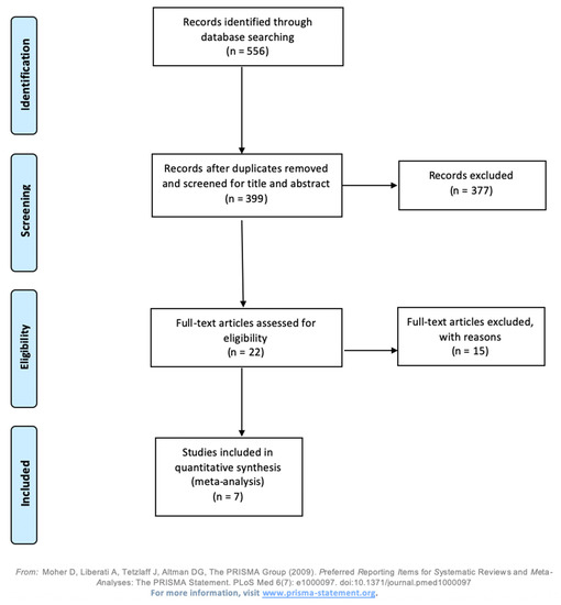
Initially, 556 studies were identified by an electronic data search. After reviewing the titles and abstracts, 22 potentially relevant studies were subjected to a full text evaluation. Of these, 15 studies were excluded based on their study design, research question, or missing numerical data for all the variables to be assessed in this study (PD, BOP, clinical attachment level, and plaque index), hence only seven studies with a detailed listing of such data could be included in the quantitative comparison (Figure 1).
Figure 1.
Prisma flow chart of the study selection process for this systematic review.
3.2. Description of Characteristics
The methodological characteristics of the selected studies are presented in Table 1. The study characteristics relevant to the specific research question are described as follows:
Table 1.
Characteristics of the included publications comparing nonsteroidal anti-inflammatory drugs (NSAID)f to placebo.
Population: Five of the studies included were performed in Turkey [27,28,29,30,31], one in Brazil [32], and one in the United States of America with mostly Caucasian followed by black subjects [33]. The study by Azoube et al. 2008 investigated patients with aggressive periodontitis [32]. All remaining studies included patients with chronic periodontitis [27,28,29,30,31,33]. All participants included in the study were systemically healthy, did not have hypersensitivity to NSAIDs, and were not pregnant or lactating. Four studies [27,28,30,32] excluded smokers from the study, whereas three studies [29,31,33] included smokers. Kurtis et al. not only included smokers but also divided smokers and non-smokers between the test and control group [29].
Overall, a total of 276 subjects were assessed in the meta-analysis. In detail, 135 subjects were assigned in the control group and received a placebo during scaling and root planing and 141 participants in the test group received an NSAID.
Intervention/Comparison: All studies except for Buduneli et al. gave oral hygiene instruction before or after the treatment. All participants in the studies received scaling and root planing with either an NSAID (test group) or a placebo (control group) [27,28,29,30,31,32,33]. The dose and duration of NSAID application varied between the studies depending on the length of the follow-up period. Patients were instructed to take either one or two tablets a day for 7 [32] or 10 [28,29,30,31] days. Aras et al. prescribed a six-week NSAID intake of one tablet a day and Yen et al. a six-month NSAID intake of one tablet a day. Study periods ranged from 4 weeks to 12 months and were categorized according to an early follow-up (7 days to 4 weeks) or a late follow-up (6 to 12 weeks) (Table 2).
Table 2.
NSAID dose and duration of follow-up in studies included in the quantitative assessment.
Outcome: All studies recorded PD, PI, and some method of gingiva inflammation recording (BOP, PBI, GBI) at baseline, different follow-up intervals, and at the final examination. Kurtis et al. took measurements at the beginning and 10 days after the intervention as a final recording of the study, so that it represented the shortest follow-up time [29]. Yen et al. distinguished PD recordings between moderate and deep pockets. Six out of seven studies documented CAL [28,29,30,31,32,33]. Some studies examined gingival crevicular fluid (GCF) [27,29] and prostaglandin E2 (PGE2) [28,32]. Buduneli et al. studied interleukin-1-beta (IL-1b) levels whereas Özgören et al. investigated levels of MMP-8 and TNF-α in subjects with chronic periodontitis.
Study design: All studies included a control group, which used a placebo. Five controlled clinical trials were double blinded [28,30,31,32,33].
3.3. Primary Outcome of the Intervention on Probing Pocket Depth (PD) Reduction
All seven studies assessed the PD at baseline and at the endpoint. The frequency of follow-up assessments varied from one to four follow-up appointments. Re-evaluation took place after 10 days, 4 weeks, or 12 weeks. Studies with the same follow-up period are compared in Table 3.
Table 3.
Probing pocket depth in comparison to non-NSAID and NSAID groups at three different follow-up timepoints.
Buduneli et al. [28] compared 20 patients that received NSAIDs with 20 non-NSAID patients in the control group. The reduction in PD was 1.3 mm in the control group and 1.6 mm in the NSAID group after 10 days, whereas the reduction of PD was much less after 30 days. Kurtis el. al [29] divided the control and NSAID groups further into smokers and non-smokers, with 15 patients in each non-NSAID group and 14 patients in each NSAID group. Only minimal changes of ±0.01 mm were observed, where both the smokers and non-smokers in the non-NSAID group showed an increase in PD of 0.01 mm while the non-smokers in the NSAID group achieved a reduction of 0.01 mm in PD. At four weeks of follow-up, two studies demonstrated only moderate or no differences in the probing pocket depth between the non-NSAID and NSAID groups. Azoubel et al. [32] showed an average reduction of 2.48 mm (SD 0.99 mm) in the control group compared to 2.17 mm (SD 0.44) in the NSAID group, while Özgören et al. [30] found the exact same average reduction of 0.72 mm (SD non-NSAID: 0.77; SD NSAID: 0.64) in both groups. Vardar et al. [31] analyzed 3 groups of 10 patients each, either receiving placebo (control group), Nimesulide, or Naproxen (test group). At the 12-week follow-up, the control group had a PD decrease of 4.1 mm (SD 1.39 mm), while PD decreased by 3.25 mm (SD 1.09 mm) in the Nimesulide group and by 4.45 mm (SD 1.33) in the Naproxen group. Yen et al. [33] observed differences in the reduction of the PD in cases with deep pockets, where the NSAID group showed a reduction of 3.27 mm (SD 11.46 mm) compared to 1.89 mm (10.97 mm) in the control group at the 12-week follow-up. The reduction of the probing depth in patients with moderate pockets was comparable between the NSAID group (1.14 mm reduction, SD 6.76 mm) and the non-NSAID control group (0.98 mm reduction, SD 6.17 mm).
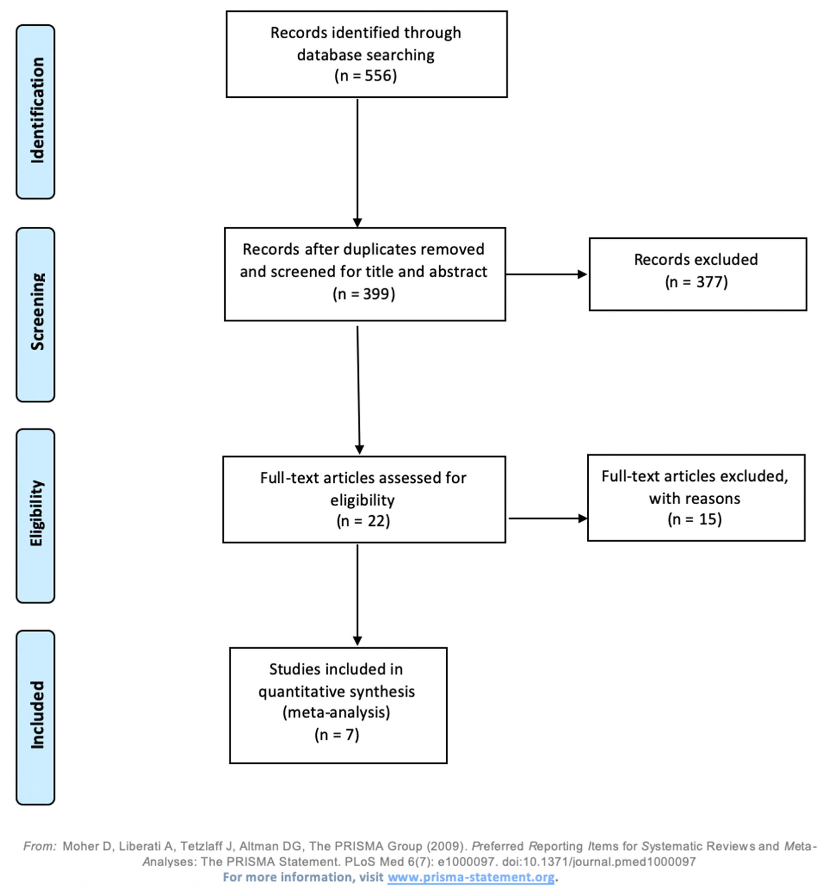
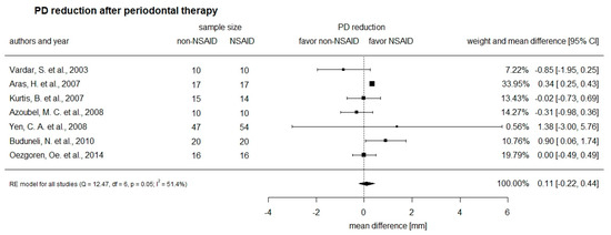
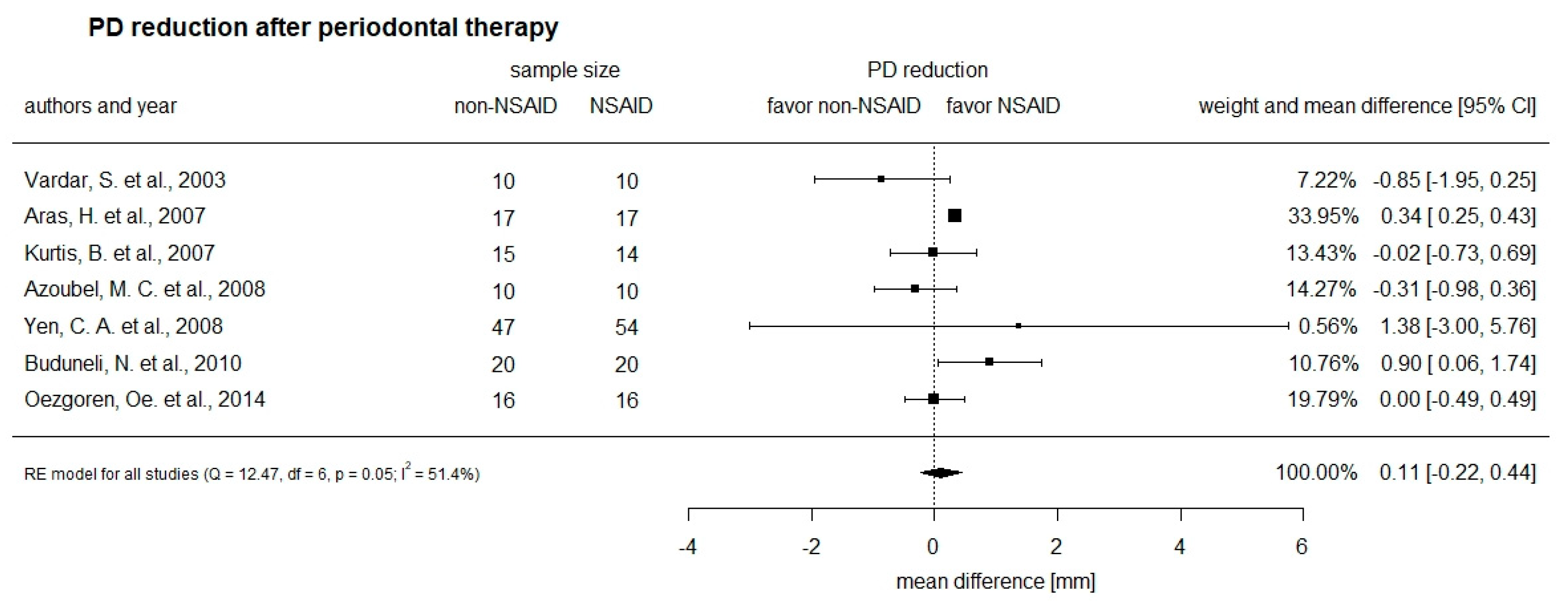
Numerical data from seven studies were quantitatively evaluated to compare PD reduction between the NSAID intervention group and the control group. The data are presented graphically in Figure 2 to show the differences between the studies and the study groups.
Figure 2.
Forest plot comparing probing pocket depth (PD) reduction between non-NSAID and NSAID groups.
The study by Kurtis et al. [29] collected data on smokers, while all other studies investigated only non-smokers. Three of the seven studies evaluated showed a larger PD reduction in the NSAID-treated group compared to the non-NSAID control group. Two studies showed a similar PD reduction for both the control and the NSAID groups. Two studies found a larger PD reduction in the control group with patients that had not received NSAID compared to those that did. The overall calculated difference in PD reduction was 0.11 mm [95% confidence interval −0.22 mm; 0.44 mm] greater in the NSAID group than the non-NSAID group. Kurtis et al. [29] was the study with the shortest follow-up time, showing similar results compared to the other studies and proving to be both neither significant nor clinically relevant.
3.4. Secondary Outcome of the Intervention on Bleeding on Probing (BOP)
Five studies presented quantitative data on BOP at three different follow-up timepoints (Table 4). Budunelli et al. [28] re-evaluated BOP using the papilla bleeding index (PBI) in 20 non-NSAID and 20 NSAID patients and found a comparable reduction of the index in both groups (non-NSAID: 1.6 SD 0.99; NSAID: 1.5 SD 1.13). Kurtis et al. [34] categorized the patients according to their smoking status and found a reduction in BOP reflected by the gingival index (GI) of 1.5 (SD 0.41) in non-NSAID smokers and 1.6 (SD 0.16) in non-NSAID non-smokers, while the reduction in the NSAID group was smaller, with 1.32 (SD 0.40) in smokers and 1.28 (0.69) in non-smokers. Two studies assessed bleeding on probing after four weeks of follow-up, yet both studies are difficult to compare as Azoubel et al. [32] reported the relative BOP as a percentage, while Özgören et al. used the GI to assess this parameter. Of the two studies with a patient re-evaluation after 12 weeks, only the study by Vardar et al. [31] reported results on BOP, which was reduced by 1.85 (SD 1.28) in the non-NSAID group and by 2.1 (SD 1.00) and 1.85 (SD 1.13) in the Nimesulide and Naproxen groups, respectively, based on the PBI. Overall, all studies report no significant difference in BOP among the NSAID and the non-NAID group.
Table 4.
Bleeding on probing (BOP) in comparison to non-NSAID and NSAID groups at three different follow-up timepoints.
3.5. Secondary Outcome of the Intervention on Clinical Attachment Gain (CAL)
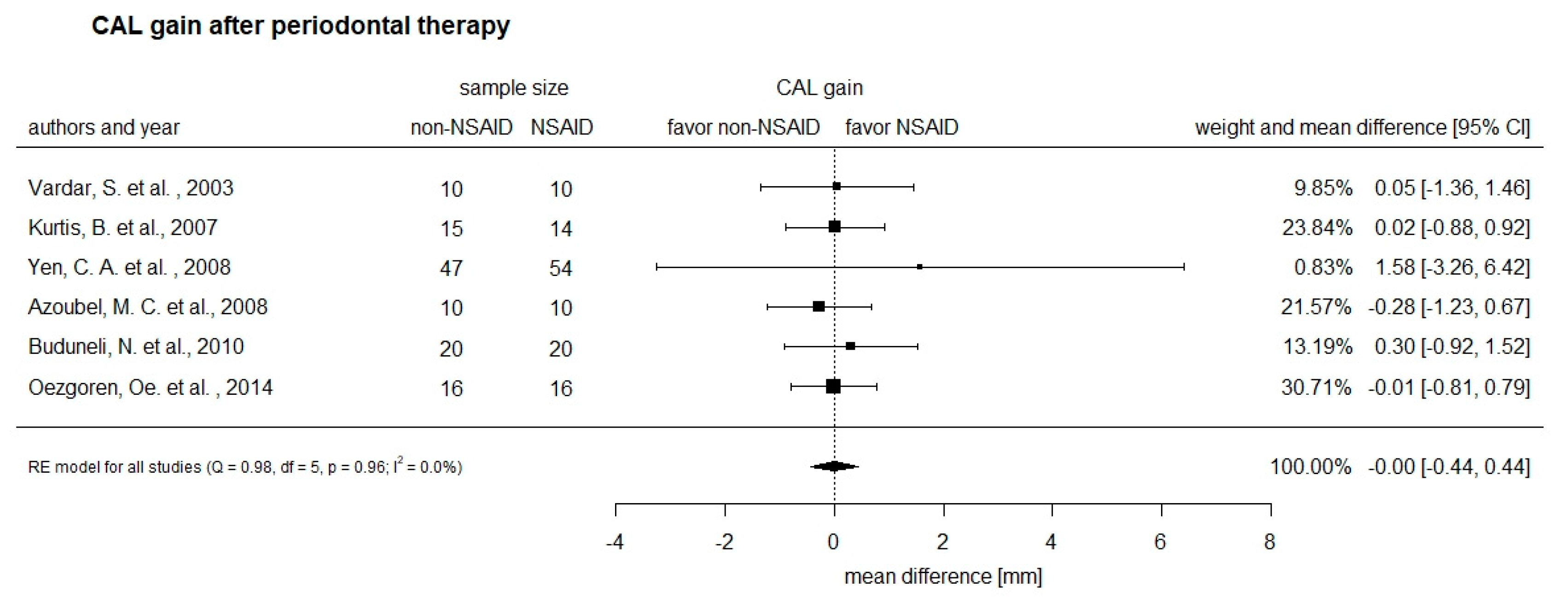
In total, 4 of the 12 included studies did not assess the CAL gain, and 2 studies that did assess this parameter did not present numerical data, leaving 6 studies for a quantitative comparison of this parameter. The frequency of follow-up assessments varied from one to four follow-up appointments. Studies with the same follow-up period of 10 days, 4 weeks, or 12 weeks are compared in Table 5.
Table 5.
Clinical attachment level in comparison of non-NSAID and NSAID groups at three different follow-up timepoints.
Two studies evaluated the patients 10 days after the initiation of NSAID treatment. Buduneli et al. [28] reported a gain in CAL by 0.5 mm (SD 1.98 mm) in the non-NSAID group compared to 1.2 mm (SD 2.13 mm) in the NSAID group after 10 days and greater after 30 days. Kurtis et al. [29] observed a gain of 0.29 mm (SD 0.51) in the CAL in the smoking non-NSAID group, while the non-smoker control group showed a clinical attachment gain of 0.01 mm (SD 1.16). In contrast, the non-smokers of the NSAID group showed no difference in CAL gain compared to the smokers in this group with 0.01 mm (SD 1.5).
At four weeks of follow-up, the observed CAL was comparable between the non-NSAID group and the NSAID group in the two studies with a re-evaluation at this timepoint. Azoubel et al. [32] reported a CAL gain of 2.23 mm (SD 1.36 mm) in the control group compared to 1.95 mm (SD 0.71) in the NSAID group, while Özgören et al. [30] observed only minimal changes in this parameter four weeks after initiation of the treatment, with a gain in CAL of 0.009 mm (SD 1.02 mm) and on average no change in the NSAID group (SD 1.27). Two studies evaluated the CAL after a follow-up period of 12 weeks. Vardar et al. [31] reported a gain in CAL for all three groups (placebo/non-NSAID group, Nimesulide, or Naproxen) of 1.3, 1.35, and 1.15 mm, respectively. Yen et al. [33] found the highest gain in CAL in the NSAID group with deep PD at 3.04 mm (SD 11.83 mm). The control group with deep PD showed an average gain of 1.46 mm (SD 12.82). The gain of the CAL in patients with moderate pockets was comparable between the NSAID-group (−1.06 mm, SD 7.42 mm) and the non-NSAID control group (−0.83 mm, SD 7.42 mm).
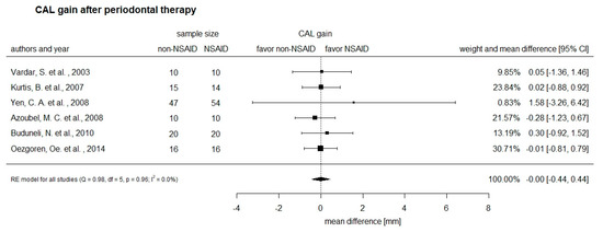
Numerical data from six studies could be quantitatively evaluated for the CAL between the NSAID and control groups. The data are plotted in Figure 3 to demonstrate the observed gain in CAL in these six studies. Three of the six evaluated studies showed close to no difference in the average clinical attachment gain between the non-NSAID and NSAIFD groups. In one study, the clinical attachment gain was slightly larger in the control group compared to the NSAID group (mean difference: −0.28, confidence interval −1.23, 0.67), while in another study the opposite was observed with a slightly larger gain in CAL in the NSAID group compared to the control (mean difference: 0.30, confidence interval −0.92, 1.52). Only one study reported a larger CAL gain in the NSAID group than in the control group (mean difference: 1.58, confidence interval −3.26, 6.42).
Figure 3.
Forest plot comparing clinical attachment (CAL) gain between non-NSAID and NSAID groups.
3.6. Secondary Outcome of the Intervention on Plaque Index (PI)
The results of the seven studies are listed in Table 6. Unfortunately, the results of these studies are not directly comparable, because the PI was assessed at different follow-up timepoints and, importantly, different indices were used to assess the index. Looking at the studies individually, only one of the studies (Aras et al.) showed twice the reduction in the plaque index in the NSAID group than that determined for the non-NSAID groups [27]. In the other six studies, the indices were comparable between patients receiving NSAID and those who did not.
Table 6.
Plaque index in comparison of non-NSAID and NSAID groups.
4. Discussion
Periodontitis may result in a dysbiosis of the biofilm and host, eventually causing inflammation and destruction of periodontal tissues [34,35]. The conventional treatment of periodontitis entails root surface debridement to allow for a recovery of the symbiosis between the host and the oral microbiome [36,37]. This therapy may be supported by pharmacological treatment of the inflammation using NSAID. The aim of the present study was to assess the evidence for the benefits of such an adjuvant pharmacological therapy in the literature in terms of PD and the secondary parameters CAL level, PI, and BOP.
Twelve studies were initially identified and further assessed in terms of their suitability and quality. Seven studies met the inclusion criteria and provided numerical data on at least the main outcome parameter, PD.
The results of this systematic review showed that in two of the seven included studies, there was a significant reduction of PD with adjunctive NSAID treatment than with just conventional scaling and root planing. In two studies, the opposite was the case, where PD showed a larger reduction in the control group than in the NSAID group, while for two studies, the changes in PD were comparable. However, taking the data from these seven studies together, there were no significant differences in terms of the PD reduction between patients who received NSAID and those that did not. Overall, the reduction in PD was only 0.11 mm larger in the NSAID group compared to the controls. The largest average reduction was observed in the study by Yen et al. [33], with a 1.38 mm greater PD reduction in the NSAID group compared to the control group. Nonetheless, the PD reduction also showed the widest range in this study, with an estimated 95% confidence interval of −3.00 to 5.76 mm, indicating that there were large differences between the patients. The studies assessed the probing pocket depth at three different follow-up timepoints: 10 days, 4 weeks, and 12 weeks. This extended time span could have an impact on the PD results, as one might expect a larger reduction after a longer time-period and hence a longer intake of NSAIDs. Nonetheless, there were no obvious differences or trends between these different follow-up periods. In fact, the study by Buduneli et al. [28] with a follow-up period of only 10 days found a larger reduction of the PD in the NSAID group, while the study by Vadar et al. [31], assessing patients after a 12-week follow-up period, found a larger reduction in the control group. A systematic review published by Donos et al. [38] investigated the adjunctive use of host modulators in non-surgical periodontal therapy. Among drugs, such as bisphosphonate, sub-antimicrobial doxycycline, and probiotics, NSAIDs applied locally and systemically were evaluated. Due to the heterogeneity of the NSAIDs being used and dosage no metanalysis was directed.
The secondary parameters were not assessed in all seven studies, with BOP reported in five studies and CAL gain in six studies. NSAID intake did not appear to have an impact on BOP, as the differences at the follow-up timepoints compared to baseline were comparable between the NSAID and control groups in the five studies. The same was true for the CAL gain, which showed no difference between patients that received NSAID adjuvant therapy and those that did not.
The quality of the initially identified 12 studies was assessed using the Oxford quality scoring system. Three of the seven studies reviewed, Yen et al. [33], Azoubel et al. [32], and Bunduneli et al. [28], received the highest scores, not addressing randomization, blinding, and drop-out rate. Two of these studies, those authored by Yen et al. [33] and Bunduneli et al. [28], observed an impact of adjuvant NSAID use on PD reduction and CAL gain, while the study by Azoubel et al. [32] saw a larger improvement of these parameters in the control group. The other four studies scored comparatively low, with scores ranging from zero to three. Nonetheless, the Oxford quality scoring system is only one of such systems and does not consider factors, such as the clear formulation of a research question and the risk of bias.
The present systematic review has several limitations that may explain the results and should be addressed. First of all, seven studies with relatively few patients could be identified that met all the inclusion criteria, and therefore the numerical data for a statistical comparison was limited. The low number of suitable studies also meant that in order to quantitatively assess differences between NSAID and non-NSAID patients, the results of all seven studies had to be pooled for this comparison independent of the timepoint at which the assessment took place. It is therefore difficult to directly compare the results, as there were only two studies available for each timepoint and parameter. Furthermore, the type, dose, and duration of administration of the NSAID may have an impact on the outcome, yet the patients in the seven studies took different NSAIDs (Etoricoxib, Meloxicam, Flurbiprofen, Tenoxicam, Naproxen, Nimesulide, Celexocib) at different dosages (7.5–325 mg). It therefore remains unclear whether the impact of NSAID administration may have been more pronounced if the same duration of the follow-up timepoint and the same NSAID with the same dosage would have been compared.
5. Conclusions
The present study evaluated the evidence of NSAID adjuvant to scaling and root planing in the treatment of periodontitis. Only a few studies could be identified in the literature addressing this topic, of which a maximum of seven studies could be used to quantitatively assess the main and secondary outcomes. Of these, only three studies showed an improvement in the main outcome parameter, probing pocket depth, in patients receiving NSAIDs compared to those that did not. Overall, the data presented indicate that NSAID has no quantifiable beneficial effect as an adjunct to scaling and root planing. Future studies with a larger number of patients, standardized follow-up periods, as well as NSAID types and dosages are required to assess the suitability of NSAID as an adjuvant therapy in root surface debridement.
Author Contributions
Writing—original draft preparation, N.M.; writing—review and editing, S.G.; data analysis, D.B.W.; supervision, P.S.; correction, T.A. All authors have read and agreed to the published version of the manuscript.
Funding
This research received no external funding.
Conflicts of Interest
The authors declare no conflict of interest. The funders had no role in the design of the study; in the collection, analyses, or interpretation of data; in the writing of the manuscript, or in the decision to publish the results.
Appendix A
Table A1.
Pubmed Search Strategy.
Appendix B
Table A2.
Excluded studies.
Appendix C
Table A3.
Quality assessment via the Oxford quality scoring system.
References
- Liu, R.; Li, N.; Liu, N.; Zhou, X.; Dong, Z.M.; Wen, X.J.; Liu, L.C. Effects of systemic ornidazole, systemic and local compound ornidazole and pefloxacin mesylate on experimental periodontitis in rats. Med. Sci. Monit. 2012, 18, BR95–BR102. [Google Scholar] [CrossRef] [Scilit]
- Hajishengallis, G. Immunomicrobial pathogenesis of periodontitis: Keystones, pathobionts, and host response. Trends Immunol. 2014, 35, 3–11. [Google Scholar] [CrossRef] [Scilit]
- Lamont, R.J.; Koo, H.; Hajishengallis, G. The oral microbiota: Dynamic communities and host interactions. Nat. Rev. Microbiol. 2018, 16, 745–759. [Google Scholar] [CrossRef] [Scilit]
- Lamster, I.B.; Lalla, E.; Borgnakke, W.S.; Taylor, G.W. The relationship between oral health and diabetes mellitus. J. Am. Dent. Assoc. 2008, 139, 19S–24S. [Google Scholar] [CrossRef] [Scilit]
- Lalla, E.; Papapanou, P.N. Diabetes mellitus and periodontitis: A tale of two common interrelated diseases. Nat. Rev. Endocrinol. 2011, 7, 738–748. [Google Scholar] [CrossRef] [Scilit]
- Gotsman, I.; Lotan, C.; Soskolne, W.A.; Rassovsky, S.; Pugatsch, T.; Lapidus, L.; Novikov, Y.; Masrawa, S.; Stabholz, A. Periodontal destruction is associated with coronary artery disease and periodontal infection with acute coronary syndrome. J. Periodontol. 2007, 78, 849–858. [Google Scholar] [CrossRef] [Scilit]
- Libby, P.; Ridker, P.M.; Maseri, A. Inflammation and atherosclerosis. Circulation 2002, 105, 1135–1143. [Google Scholar] [CrossRef] [Scilit]
- Pink, C.; Kocher, T.; Meisel, P.; Dörr, M.; Markus, M.R.; Jablonowski, L.; Grotevendt, A.; Nauck, M.; Holtfreter, B. Longitudinal effects of systemic inflammation markers on periodontitis. J. Clin. Periodontol. 2015, 42, 988–997. [Google Scholar] [CrossRef] [Scilit]
- Suvan, J.E.; Petrie, A.; Nibali, L.; Darbar, U.; Rakmanee, T.; Donos, N.; D’Aiuto, F. Association between overweight/obesity and increased risk of periodontitis. J. Clin. Periodontol. 2015, 42, 733–739. [Google Scholar] [CrossRef] [Scilit]
- Corbella, S.; Taschieri, S.; Del Fabbro, M.; Francetti, L.; Weinstein, R.; Ferrazzi, E. Adverse pregnancy outcomes and periodontitis: A systematic review and meta-analysis exploring potential association. Quintessence Int. 2016, 47, 193–204. [Google Scholar] [CrossRef] [Scilit]
- Michalowicz, B.S.; Hodges, J.S.; DiAngelis, A.J.; Lupo, V.R.; Novak, M.J.; Ferguson, J.E.; Buchanan, W.; Bofill, J.; Papapanou, P.N.; Mitchell, D.A.; et al. Treatment of periodontal disease and the risk of preterm birth. N. Engl. J. Med. 2006, 355, 1885–1894. [Google Scholar] [CrossRef] [Scilit] [PubMed]
- Van Dyke, T.E. The management of inflammation in periodontal disease. J. Periodontol. 2008, 79, 1601–1608. [Google Scholar] [CrossRef] [Scilit] [PubMed]
- Tegner, W.S. The Treatment of the Rheumatic Diseases in the United States and the Continent of Europe. Ann. Rheum. Dis. 1939, 1, 249–303. [Google Scholar] [CrossRef] [Scilit] [PubMed]
- Atkinson, T.J.; Fudin, J. Nonsteroidal Antiinflammatory Drugs for Acute and Chronic Pain. Phys. Med. Rehabil. Clin. N. Am. 2020, 31, 219–231. [Google Scholar] [CrossRef] [Scilit]
- Cascorbi, I. Drug interactions—Principles, examples and clinical consequences. Dtsch. Arztebl. Int. 2012, 109, 546–555. [Google Scholar] [CrossRef] [Scilit]
- Mutschler/Geisslinger/Menzel/Ruth/Schmidtko. Pharmakologie Kompakt Allgemeine und Klinische Pharmakologie, Toxikologie; Wiss. Verl: Stuttgart, Germany, 2016. [Google Scholar]
- Offenbacher, S.; Odle, B.M.; Gray, R.C.; Van Dyke, T.E. Crevicular fluid prostaglandin E levels as a measure of the periodontal disease status of adult and juvenile periodontitis patients. J. Periodontal Res. 1984, 19, 1–13. [Google Scholar] [CrossRef] [Scilit]
- Offenbacher, S.; Odle, B.M.; Van Dyke, T.E. The use of crevicular fluid prostaglandin E2 levels as a predictor of periodontal attachment loss. J. Periodontal Res. 1986, 21, 101–112. [Google Scholar] [CrossRef] [Scilit]
- Feldman, R.S.; Szeto, B.; Chauncey, H.H.; Goldhaber, P. Non-steroidal anti-inflammatory drugs in the reduction of human alveolar bone loss. J. Clin. Periodontol. 1983, 10, 131–136. [Google Scholar] [CrossRef] [Scilit]
- Vogel, R.I.; Copper, S.A.; Schneider, L.G.; Goteiner, D. The effects of topical steroidal and systemic nonsteroidal anti-inflammatory drugs on experimental gingivitis in man. J. Periodontol. 1984, 55, 247–251. [Google Scholar] [CrossRef] [Scilit]
- Alani, A.; Seymour, R. Systemic medication and the inflammatory cascade. Periodontology 2000 2014, 64, 198–210. [Google Scholar] [CrossRef] [Scilit]
- Moher, D.; Shamseer, L.; Clarke, M.; Ghersi, D.; Liberati, A.; Petticrew, M.; Shekelle, P.; Stewart, L.A.; Group, P.-P. Preferred reporting items for systematic review and meta-analysis protocols (PRISMA-P) 2015 statement. Syst. Rev. 2015, 4, 1. [Google Scholar] [CrossRef]
- Miller, S.A.; Forrest, J.L. Enhancing your practice through evidence-based decision making: PICO, learning how to ask good questions. J. Evid. Based Dent. Pract. 2001, 1, 136–141. [Google Scholar] [CrossRef] [Scilit]
- R Core Team. R: A Language and Environment for Statistical Computing; R Foundation for Statistical Computing: Vienna, Austria, 2018; Available online: https://www.R-project.org/ (accessed on 16 July 2019).
- Viechtbauer, W. Conducting meta-analyses in R with the metafor package. J. Stat. Softw. 2010, 36, 1–48. [Google Scholar] [CrossRef] [Scilit]
- Jadad, A.R.; Moore, R.A.; Carroll, D.; Jenkinson, C.; Reynolds, D.J.; Gavaghan, D.J.; McQuay, H.J. Assessing the quality of reports of randomized clinical trials: Is blinding necessary? Control. Clin. Trials 1996, 17, 1–12. [Google Scholar] [CrossRef] [Scilit]
- Aras, H.; Caglayan, F.; Guncu, G.N.; Berberoglu, A.; Kilinc, K. Effect of systemically administered naproxen sodium on clinical parameters and myeloperoxidase and elastase-like activity levels in gingival crevicular fluid. J. Periodontol. 2007, 78, 868–873. [Google Scholar] [CrossRef] [Scilit]
- Buduneli, N.; Buduneli, E.; Cetin, E.O.; Kirilmaz, L.; Kutukculer, N. Clinical findings and gingival crevicular fluid prostaglandin E2 and interleukin-1-beta levels following initial periodontal treatment and short-term meloxicam administration. Expert Opin. Pharm. 2010, 11, 1805–1812. [Google Scholar] [CrossRef] [Scilit]
- Kurtis, B.; Tuter, G.; Serdar, M.; Pinar, S.; Demirel, I.; Toyman, U. GCF MMP-8 levels in smokers and non-smokers with chronic periodontitis following scaling and root planing accompanied by systemic use of flurbiprofen. J. Periodontol. 2007, 78, 1954–1961. [Google Scholar] [CrossRef] [Scilit]
- Ozgoren, O.; Develioglu, H.; Guncu, G.; Akman, A.; Berker, E. The adjunctive effect of tenoxicam during non-surgical periodontal treatment on clinical parameters and gingival crevicular fluid levels of MMP-8 and TNF-alpha in patients with chronic periodontitis - randomized, double-blind clinical trial. Adv. Clin. Exp. Med. 2014, 23, 559–565. [Google Scholar] [CrossRef] [Scilit]
- Vardar, S.; Baylas, H.; Huseyinov, A. Effects of selective cyclooxygenase-2 inhibition on gingival tissue levels of prostaglandin E2 and prostaglandin F2alpha and clinical parameters of chronic periodontitis. J. Periodontol. 2003, 74, 57–63. [Google Scholar] [CrossRef] [Scilit]
- Azoubel, M.C.; Sarmento, V.A.; Cangussu, V.; Azoubel, E.; Bittencourt, S.; Cunha, F.Q.; Ribeiro, R.A.; Brito, G.A. Adjunctive benefits of systemic etoricoxib in non-surgical treatment of aggressive periodontitis: Short-term evaluation. J. Periodontol. 2008, 79, 1719–1725. [Google Scholar] [CrossRef] [Scilit]
- Yen, C.A.; Damoulis, P.D.; Stark, P.C.; Hibberd, P.L.; Singh, M.; Papas, A.S. The effect of a selective cyclooxygenase-2 inhibitor (celecoxib) on chronic periodontitis. J. Periodontol. 2008, 79, 104–113. [Google Scholar] [CrossRef] [Scilit]
- Marsh, P.D.; Moter, A.; Devine, D.A. Dental plaque biofilms: Communities, conflict and control. Periodontology 2000 2011, 55, 16–35. [Google Scholar] [CrossRef] [Scilit]
- Hajishengallis, G.; Liang, S.; Payne, M.A.; Hashim, A.; Jotwani, R.; Eskan, M.A.; McIntosh, M.L.; Alsam, A.; Kirkwood, K.L.; Lambris, J.D.; et al. Low-abundance biofilm species orchestrates inflammatory periodontal disease through the commensal microbiota and complement. Cell Host Microbe 2011, 10, 497–506. [Google Scholar] [CrossRef] [Scilit]
- Waerhaug, J. Healing of the dento-epithelial junction following subgingival plaque control: II: As observed on extracted teeth. J. Periodontol. 1978, 49, 119–134. [Google Scholar] [CrossRef] [Scilit]
- Waerhaug, J. Healing of the dento-epithelial junction following subgingival plaque control: I: As observed in human biopsy material. J. Periodontol. 1978, 49, 1–8. [Google Scholar] [CrossRef] [Scilit]
- Donos, N.; Calciolari, E.; Brusselaers, N.; Goldoni, M.; Bostanci, N.; Belibasakis, G.N. The adjunctive use of host modulators in non-surgical periodontal therapy. A systematic review of randomized, placebo-controlled clinical studies. J. Clin. Periodontol. 2019. [Google Scholar] [CrossRef] [Scilit]

Publisher’s Note: MDPI stays neutral with regard to jurisdictional claims in published maps and institutional affiliations. |
© 2020 by the authors. Licensee MDPI, Basel, Switzerland. This article is an open access article distributed under the terms and conditions of the Creative Commons Attribution (CC BY) license (http://creativecommons.org/licenses/by/4.0/).