Article

PEG-b-PLGA Nanoparticles Loaded with Geraniin from Phyllanthus Watsonii Extract as a Phytochemical Delivery Model

by
Kogilavanee Devasvaran
1,
Sreegayathri Jairaman
1,
Nur Azirah Yahaya
1,
Indu Bala S. Jaganath
2,
Yit Lung Khung
3,
Vuanghao Lim
1 and
Siti Hawa Ngalim
1,*
1
Advanced Medical and Dental Institute, Universiti Sains Malaysia (USM), Bertam 13200, Kepala Batas Penang, Malaysia
2
Malaysian Agricultural Research and Development Institute (MARDI), Seri Kembangan 43400, Selangor, Malaysia
3
Department of Biological Science and Technology, China Medical University, Taichung 40402, Taiwan
*
Author to whom correspondence should be addressed.
Appl. Sci. 2020, 10(14), 4891; https://doi.org/10.3390/app10144891
Submission received: 9 June 2020 / Revised: 11 July 2020 / Accepted: 13 July 2020 / Published: 16 July 2020
(This article belongs to the Section Food Science and Technology)

Abstract

:

Featured Application

A simplified delivery model of phytochemicals using PLGA-PEG scaffolds for future cell study.

Abstract

The study outlined a standardized double emulsion method for simple poly(ethylene glycol)-block-poly(lactic-co-glycolic acid) (PEG-b-PLGA) nanoparticle (NP) synthesis. The PEG-b-PLGA NP was also used for entrapment of geraniin as a simple model system for phytochemical delivery. PEG-b-PLGA NPs were prepared using the double emulsion method. The yields and particle sizes of PEG-b-PLGA NPs obtained with and without encapsulation of geraniin were 57.6% and 134.20 ± 1.45 nm and 66.7% and 102.70 ± 12.36 nm, respectively. High-performance liquid chromatography of geraniin that was extracted from Phyllanthus watsonii was detected at 64 min. Geraniin burst release began at 40 min and fully released at 3 h. PEG-b-PLGA NP was non-cytotoxic, while cytotoxicity of geraniin was dose dependant towards normal human epithelial colon cells, CCD 841 CoN cells.

Graphical Abstract

1. Introduction

The shift from micro- to nano-sized particles is one of the recent advances in biomedical applications. Nanoparticles (NPs) offer numerous advantages in the pharmaceutical and biomedical industries. For example, the physical and chemical characteristics of NPs can mask poorly water-soluble drugs, alter the pharmacokinetics of a drug, reduce the immunogenicity of a drug by increasing its half-life, produce a target-specific drug that reduces side effects, and increase the bioavailability to shorten metabolism of the drug [1].
Polymers with amphiphilic properties are ideal for drug delivery. Poly(lactic-co-glycolic acid) (PLGA) is a biodegradable polymer that has been approved by the US Food and Drug Administration (FDA) for use in drug delivery systems. PLGA is known for its biocompatibility, bioavailability, enhancement of drug content specificity, and immunosuppressive potential [2]. Poly(ethylene glycol) (PEG) is known for its stealth characteristic in drug delivery and is classified as Generally Regarded as Safe by the FDA. PEGylation of PLGA polymers has been shown to prolong NP bioavailability by acting as a shield to the NP surface, thereby preventing NP aggregation and opsonization by immune cells [3]. PEG-b-PLGA, which have termini that are hydrophobic and hydrophilic, respectively, are safe for consumption due to their excellent biodegradability, biocompatibility, and non-toxicity [4]. Chemical bonds present in the PEG-b-PLGA polymer are carboxylic acid, ether, ester, and methyl and carbonyl groups. Thus, a combination of polar and non-polar bonds makes the polymer amphiphilic [5]. In this study, PEG-b-PLGA polymers were prepared using a modified double emulsion method.
Encapsulation of phytochemicals derived from tropical plants into PEG-b-PLGA NPs may be a potential simple model for a phytochemical delivery system. Other approaches to deliver phytochemicals [6,7] include micelles (closed-loop, single layer amphiphiles) and liposomes (closed-loop, lipid bilayer that can come in unilamellar, multilamellar or multi vesicles features) and phytosomes (closed-loop primarily of liposomes that are grafted to phytochemicals). Meanwhile, microemulsion and solid lipid NPs employ various shell types but with an optimized lipid, surfactant, and phytochemical formulation, in liquid core and solid core, respectively. There are also polymer NPs that form the shell or core of the NP. Moreover, there are inorganic NPs that are made of metals, ceramics, or carbons and turn into various shapes, porosity, and configurations to load drugs and phytochemicals.
Tropical countries such a Malaysia have abundant supplies of local plants with medicinal value, such as Phyllanthus spp. (locally known as dukung anak), which is reported to have anti-diabetic, anti-viral, and anti-cancer properties [8]. Phyllanthus watsonii (P. watsonii) has been shown to have potent anti-cancer activity when compared to other species of Phyllanthus. The main ellagitannin present in P. watsonii is geraniin, which has been shown to contribute to the growth arrest of certain cancers, including colon cancer [8]. Geraniin (molecular formula: C41H28O27) is a bioactive compound and a potent antioxidant [9], and it has anti-cancer, anti-hypertensive, anti-microbial, anti-viral, liver processing-friendly, anti-hyperglycemic, and anti-diabetic properties [10,11,12,13,14,15,16,17]. In addition, geraniin decreases carbohydrate metabolism by inhibiting activities of α-glucosidase, α-amylase, and aldol reductase and production of advanced glycation end-products [16]. The interest of this research is to generate a simple PEG-b-PLGA NPs shell. Therefore, encapsulating phytochemicals such as geraniin derived from P. watsonii into PEG-b-PLGA NPs can be a simple research model to study the use of PEG-b-PLGA NPs as a carrier for phytochemicals too. We initially hypothesized that geraniin encapsulated in the PEG-b-PLGA NPs could be used as a general nutraceutical irrespective of the dosage.

2. Materials and Methods

All subsequent analysis and results presented here were acquired from nanoparticles (NPs) stored after freeze-drying.

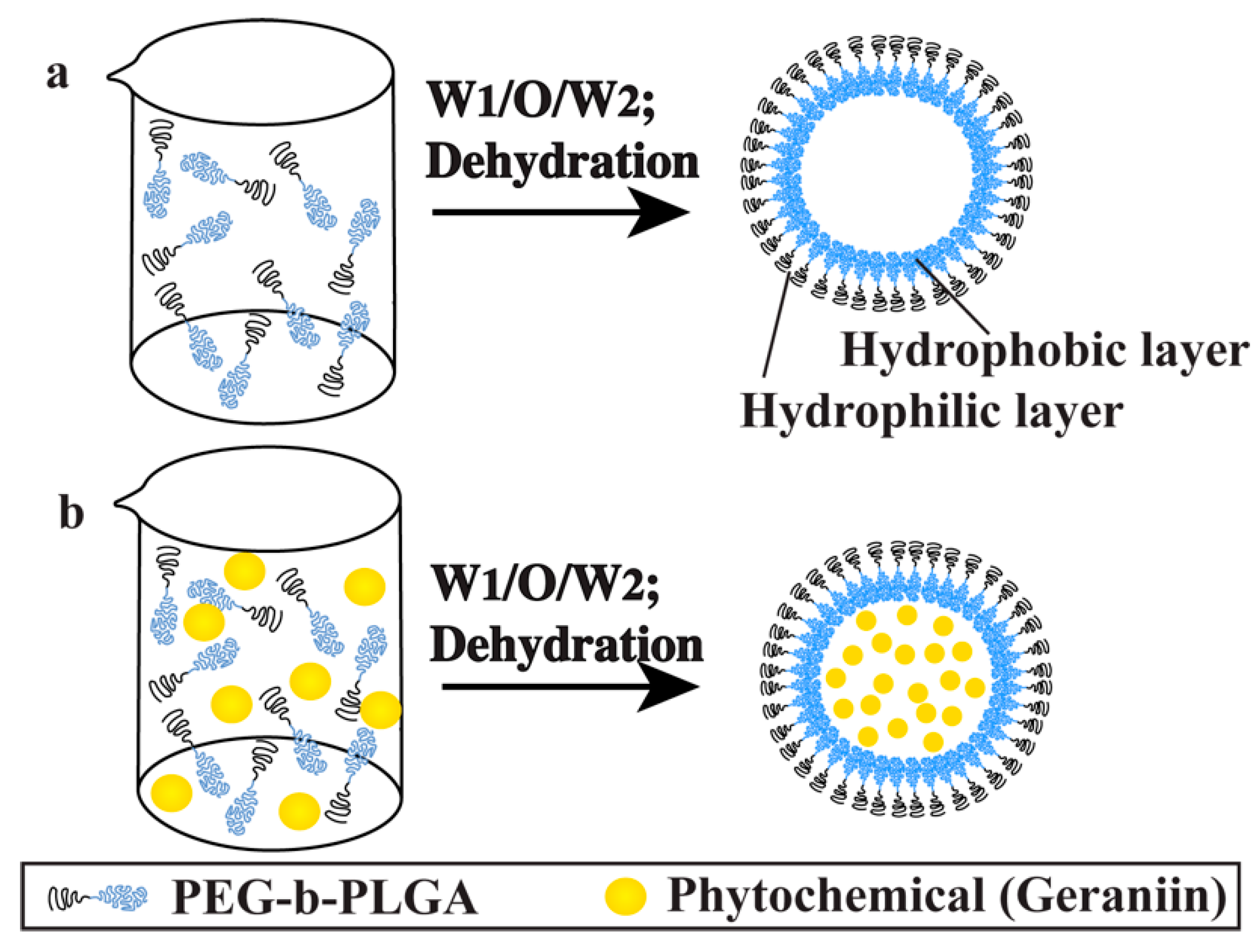
2.1. Synthesis of poly(ethylene glycol)-block-poly(lactic-co-glycolic acid) (PEG-b-PLGA) Nanoparticles (NPs)

Double emulsion, or the water/oil/water (W/O/W) technique (Figure 1), was used to synthesize the NPs. In brief, the PEG-b-PLGA polymer has hydrophilic and hydrophobic parts at the PEG and PLGA, respectively. For this reason, there is a need to create the emulsion of the PEG-b-PLGA, with the PEG and PLGA regions constituting the outer and inner shell of the NP, respectively. A more polar solvent (near water polarity) like acetonitrile acts as a continuous phase for PEG and a disperse phase for PLGA. Therefore, first, 30 mg of PEG (average Mn 2000)-b-PLGA (average Mn 11,500) polymer (Sigma Aldrich, St. Louis, MO, USA) were dissolved in 3 mL of acetonitrile (Fisher Chemical, Fair Lawn, NJ, USA), and 600 µL of a Pluronic F-127 solution (Molecular Probes, Eugene, OR, USA) was added and dissolved by sonicating (150VT Ultrasonic Homogenizer, BioLogics Inc., Manassas, VA, USA) for 20 s in an ice bath to obtain the first emulsion (W1). Next, 800 µL of Pluronic solution was added and sonicated for 30 s at 60% amplitude. Here, Pluronic was used as a surfactant or stabilizing agent for the PEG-b-PLGA emulsion in acetonitrile. Meanwhile, non-polar solvent (near oil polarity) like hexane (Fisher Scientific, Fair Lawn, NJ, USA) acts as a disperse phase for PEG. Then, 90 mg of sodium dodecyl sulfate (Fisher Scientific, Fair Lawn, NJ, USA) were added to 60 mL of hexane, and the resultant double emulsion was poured into the 60 mL of hexane and evaporated by stirring for 1 h at 950 rpm (O). Sodium dodecyl sulfate was used as a surfactant or stabilizing agent for the PEG-b-PLGA emulsion in hexane. The nanoemulsion was then centrifuged at 6000 rpm to separate the NPs from the microparticles. After centrifugation, the pellet was washed and centrifuged with distilled water three times (W2). Finally, the pellet (PEG-b-PLGA NPs) was suspended in distilled water [18,19], freeze-dried (Eyela FDU-1200 Freeze Dryer, Eyela, Bohemia, NY, USA) for 24 h, and stored at 4 °C before use in further experiments.

2.2. Encapsulation of Geraniin into PEG-b-PLGA Nanoparticles (NPs)

Geraniin isolate from Phyllanthus watsonii was used as a phytochemical example in NP encapsulation; it was a gift from Dr. Indu Bala S. Jaganath of Malaysian Agricultural Research and Development Institute. The ratio of geraniin to PEG-b-PLGA used was 1:10. For encapsulation, 3 mg of geraniin were dissolved in 3 mL of deionized water, and 600 µL of Pluronic solution were added. Concurrently, 30 mg of PEG-b-PLGA polymer was dissolved in 3 mL of acetonitrile. Both the phytochemical and polymer solutions were mixed and sonicated on ice for 20 s to obtain the first emulsion. Consequent steps of encapsulation of geraniin via the double emulsion technique followed the procedure described in Section 2.1.

2.3. Characterization of the Nanoparticles (NPs)

2.3.1. Particle Size and Zeta Potential

NP size and zeta potential were determined using the Zetasizer Nano ZS90 (Malvern Instruments, Malvern, UK), which uses dynamic light scattering (DLS) to measure particle size at a 90-degree scattering angle and static light scattering to measure zeta potential. In brief, the NPs (encapsulated with or without geraniin) were dissolved in deionized water at a 1:3 ratio and filtered using a polytetrafluoroethylene (PTFE) membrane syringe filter of diameter 13 mm and pore size 0.25 um (Millex HV; Milipore, Milford, CT, USA). The pH was measured, and 1 mL of the solution was added to the folded capillary zeta cell to be read. The mean value of three readings was reported [21].

2.3.2. Transmission Electron Microscopy (TEM)

TEM (Libra 120, Carl Zeiss, Oberkochen, Germany) was performed to identify the shape of the NPs. Briefly, 1 mg of PEG-b-PLGA NPs (encapsulated with or without geraniin) was suspended in 2 mL of deionized water and sonicated for 30 s. This solution was filtered using a 0.25 um PTFE syringe filter, and a drop of the filtrate was placed on a copper grid. Air drying and blotting of the grid with a filter paper were conducted to remove excess water before viewing [21].

2.3.3. Field Emission Scanning Electron Microscopy (FESEM)

FESEM (Verios 460L, FEI, Hillsboro, OR, USA) was performed to visualize the morphology of the NPs. Preparation of the NPs (PEG-b-PLGA with or without geraniin) was conducted, as described in Section 2.3.2. A drop of filtered solution was placed on a glass slide and left to dry in a vacuum desiccator overnight. The NPs were then coated with platinum, and the results were observed under the microscope.

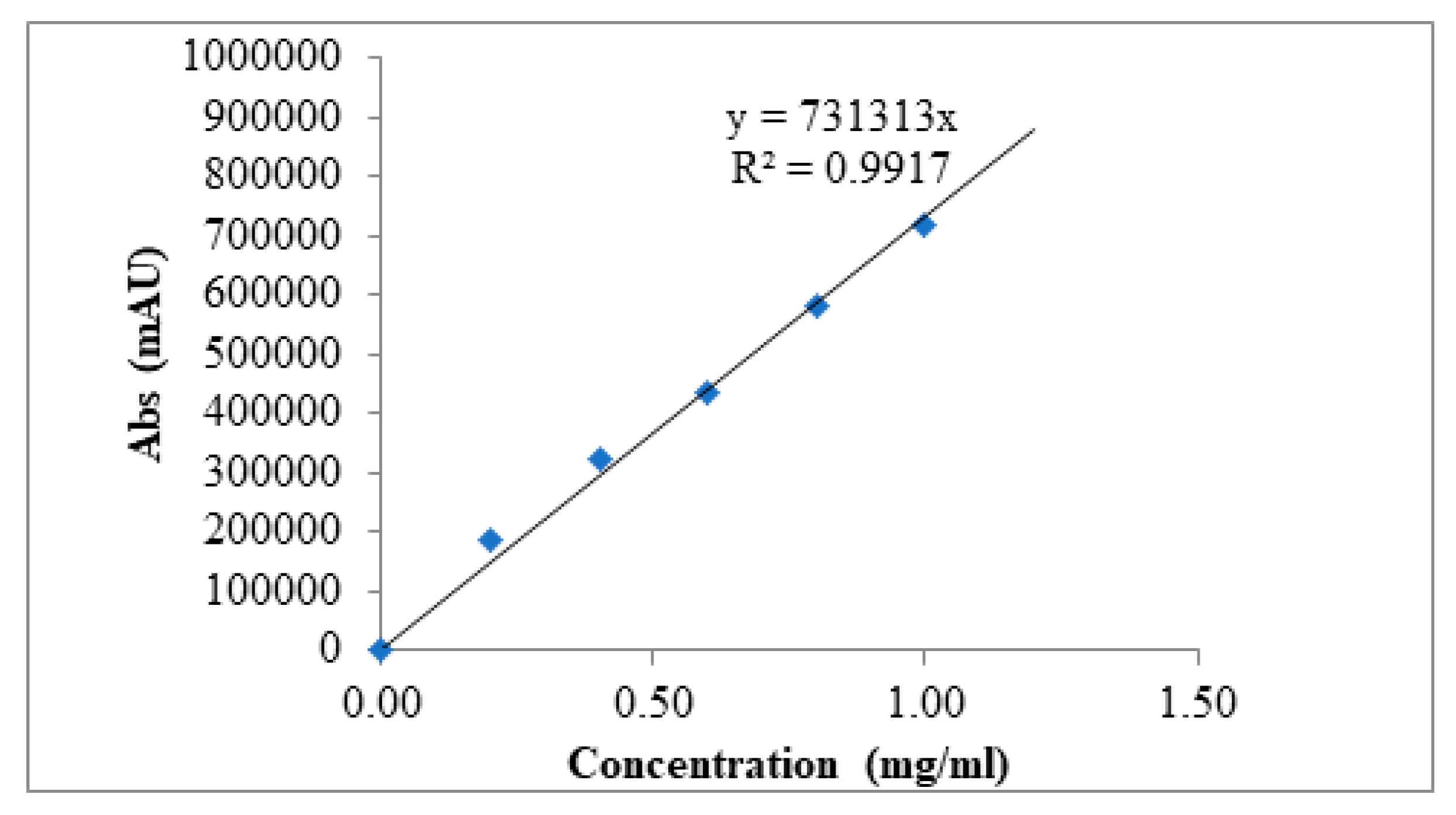
2.4. High-Performance Liquid Chromatography (HPLC)

Four concentrations of standard solutions (0.8, 0.6, 0.4, and 0.2 mg/mL) were prepared from a stock solution containing 1 mg/mL of geraniin in the mobile phase to determine the amount of geraniin encapsulated in PEG-b-PLGA NPs. The solution was filtered, and reverse-phase HPLC was conducted to measure the amount of geraniin encapsulated in the NPs, of which protocol can be found in Feng et al. [22]. The HPLC core components included a binary pump, an autosampler injector compartment, and a photodiode array detector. This is the applicable protocol of the physio-chemical technique for the determination of encapsulation content for geraniin when used with the percentage yield formula in McCall and Sirianni [18]. The reverse-phase used a C-18, 250 mm × 4.6 mm i.d. 5 µm particle size Thermo Hypersil GOLD column (Thermo Scientific, Hemel Hempstead, UK). The mobile phase consisted of sterile deionized water (Solvent A) and acetonitrile (Solvent B), and the solvent gradient flow rate was set to 1 mL/min. The total volume of the sample injected was 20 µL, and the data showed a peak at 274 nm. For the analysis, 5 mg/mL of the polymeric NPs containing geraniin were prepared at the mobile phase. The standard solution and sample solutions were injected into the chromatograph, and the concentration of geraniin in the polymeric NPs was measured [23]. A standard curve was plotted by running HPLC using six different concentrations of geraniin. The encapsulated content was evaluated by comparing the area under the peak of the geraniin HPLC data to the standard curve.

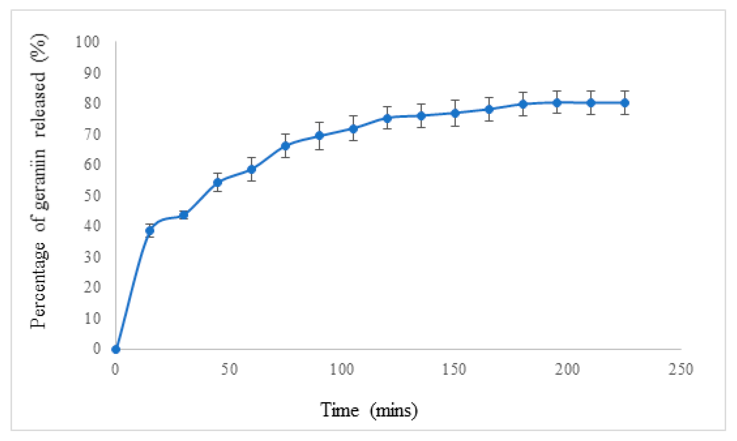
2.5. In Vitro Drug Release of poly(ethylene glycol)-block-poly(lactic-co-glycolic acid) (PEG-b-PLGA) Nanoparticles (NPs) Encapsulated with Geraniin

A stock solution of 1 mg/mL of geraniin was prepared in phosphate-buffered saline (PBS) (Invitrogen, Waltham, MA, USA). Six concentrations of working solutions (0.4, 0.2, 0.1, 0.05, 0.025, and 0.0125 mg/mL) were obtained by dilution from the stock solution. Next, 5 mg/mL of NPs were loaded with geraniin and solubilized with PBS buffer in a dialysis bag. The dialysis bag was immersed in a falcon tube with 10 mL of PBS as the external medium. At every 15 min interval, 1 mL of the external medium was withdrawn, which was replaced with 1 mL of fresh PBS. The absorbance of the sample was recorded using a double beam spectrophotometer (LAMDA 25, Perkin Elmer, Waltham, MA, USA) [24].

2.6. Cell Culture of CCD 841 CoN

Normal human colon epithelial cells (CCD 841 CoN, No. CRL-1790™) were purchased from the American Type Culture Collection (Manassas, VA, USA). They were cultured in Eagle’s minimal essential medium (EMEM) supplemented with 5% fetal bovine serum (v/v) and 1% penicillin/streptomycin (v/v). Unless otherwise stated, cells were always incubated in a humidified 5% CO2 and 95% air (v/v) incubator at 37 °C. Cells were passaged upon reaching approximately 80% confluence and were dissociated with 0.25% of Trypsin-EDTA in PBS (v/v). Low passage CCD 841 CoN cells (<10 passages) were used in the subsequent experiments.

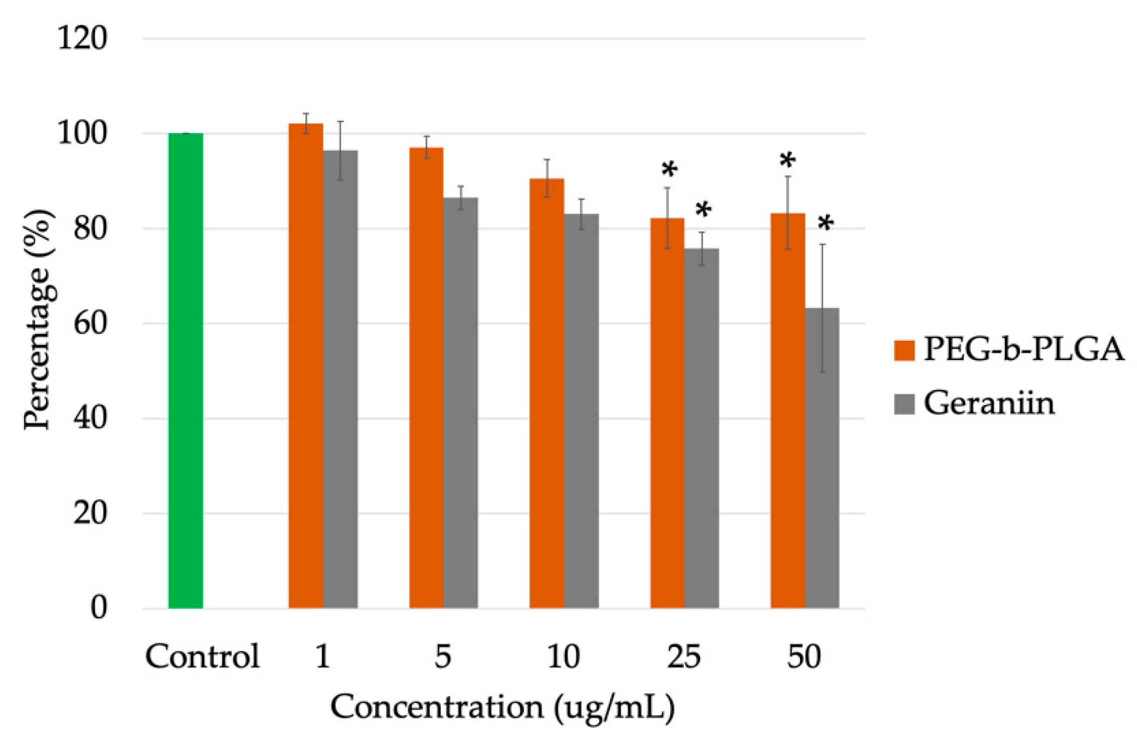
2.7. Cell Viability Assay

CCD 841 CoN cells were seeded at 4 × 10⁴ cells/well in a 24-well plate and incubated for 3 h. Desired concentrations of geraniin (1, 5, 10, 25, and 50 µg/mL) were then created in the respective wells. The negative control used in this experiment was untreated cells, and the blank was growth medium only. The duration of incubation for all samples, control, and blank was 6 h. After 6 h, 40 µL of thiazolyl blue tetrazolium bromide (also known as MTT dye) was added to the wells and incubated for 45 min. The reaction was stopped with the addition of 100 µL of isopropanol, and the absorbance was measured at 570 nm using an ELISA microplate reader (FLUOStar Omega, BMG Labtech, Ortenberg, Germany) [25]. Cell viability was expressed as the percentage of formazan absorbance [26].

2.8. Statistical Analysis

Analysis of variance was used for multiple group comparisons followed by post-hoc tests. Dunnett’s test was performed to compare the groups with the control. p < 0.05 was considered to be statistically significant. All statistical analysis was performed using the IBM SPSS (Statistics Package for Social Science, version 22) (Chicago, IL, USA).

3. Results

3.1. Synthesis of poly(ethylene glycol)-block-poly(lactic-co-glycolic acid) (PEG-b-PLGA) Nanoparticles (NPs) Encapsulated with and without Geraniin

The estimated percentage yield of PEG-b-PLGA NPs was ~66.7%, and that of PEG-b-PLGA NPs encapsulated with geraniin was 57.6%. The difference could be due to the increased size of the geraniin encapsulated NPs, which may have led to the decreased percentage yield.

3.2. Characterization of the poly(ethylene glycol)-block-poly(lactic-co-glycolic acid) (PEG-b-PLGA) Nanoparticles (NPs)

3.2.1. Particle Size Analysis

PEG-b-PLGA NPs and PEG-b-PLGA NPs encapsulated with geraniin were of optimal size for drug delivery, with values of 102.7 ± 12.36 nm and 134.2 ± 1.45 nm, respectively. The analysis was done on freeze-dried samples. No abnormality in sizes was detected. The DLS data provides an analysis of NP sizes of all NPs loaded at the time. Therefore, DLS data is ideal at representing an overall NP size (Supplementary data; Figures S1 and S2). Polydispersity index (PDI) values were 0.30 ± 0.02 and 0.24 ± 0.01, respectively. According to De Jong and Borm (2008), the NP size range of 100 to 200 nm is the most suitable for oral drug delivery. The PDI indicates the size distribution of the particles, and a PDI value > 0.5 indicates a broad size distribution that may contain large particles or aggregates that could be gradually sedimenting, which is not suitable for DLS analysis. Thus, the data showed that both blank PEG-b-PLGA NPs and PEG-b-PLGA NPs encapsulated with geraniin were within the optimal size range for drug delivery. The size of NPs and the PDI value play crucial roles in drug delivery applications because physical properties enable the drug carrier to cross various biological barriers within the body.

3.2.2. Zeta Potential Analysis

Zeta potential indicates the surface charge of particles in a solution, and it measures the repulsion or attraction of charges between particles. Thus, smaller particles with higher zeta potential confer good particle stability and resist particle aggregation [27]. According to previous reports, NPs with zeta potential values > +25 mV or < –25 mV have high degrees of particle stability [28]. In the current study, blank PEG-b-PLGA NPs had a zeta potential value of −39.1 ± 1.05 mV, and PEG-b-PLGA NPs encapsulated with geraniin had a zeta potential of -40.8 ± 0.90 mV. Thus, both values were < −25 mV, which indicated the negative surface charge and stability of the NPs. NP surface charge represents another design feature that can be tailored to prolong circulation lifetimes of the NPs and selectively enhance the accumulation of NPs at specific sites of interest. NPs with neutral and negative surface charges have been shown to resist the adsorption of serum proteins, resulting in longer circulation half-lives [29].

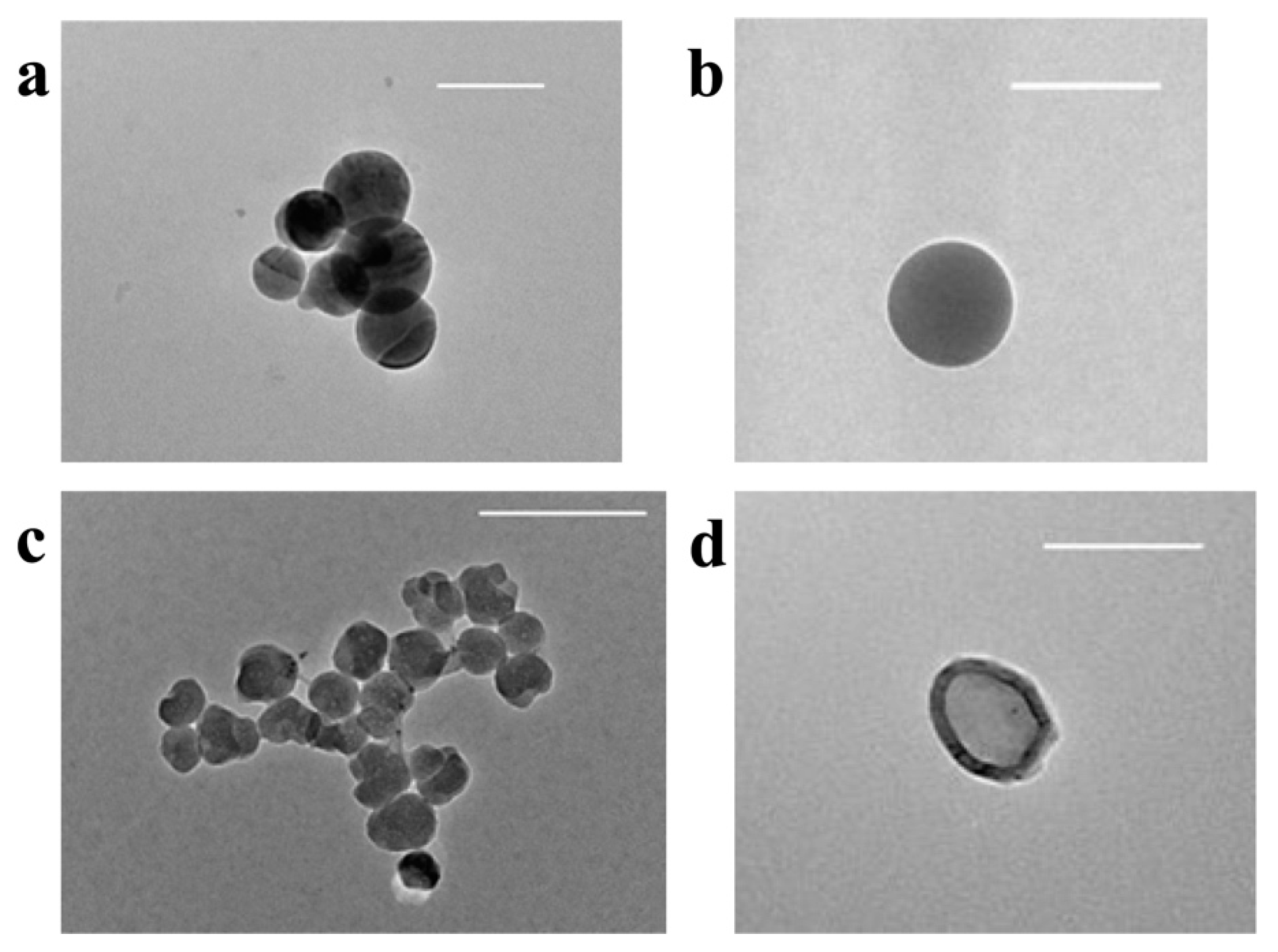
3.2.3. Transmission Electron Microscopy (TEM)

Figure 2a,b show that the NPs were spherical in shape and independent from each other. Potentially, the preparation method and NP interaction with stabilizers influenced the shape of the NPs. Figure 2c,d shows that the outer layer of the geraniin-encapsulated PEG-b-PLGA NPs was distinctly darker than the core area. This observation suggests the possibility that two physicochemically different constituents were present within the NP at the shell and core, such as PEG-b-PLGA and geraniin. Geraniin’s hydrophilic nature enables it to be contained in the hydrophobic part of the chain of PLGA, which is linked to PEG (Figure 1). The analysis was done on freeze-dried samples. As shown in Figure 2, considering the NP sizes with respective scale bars, no abnormality in sizes are observed.

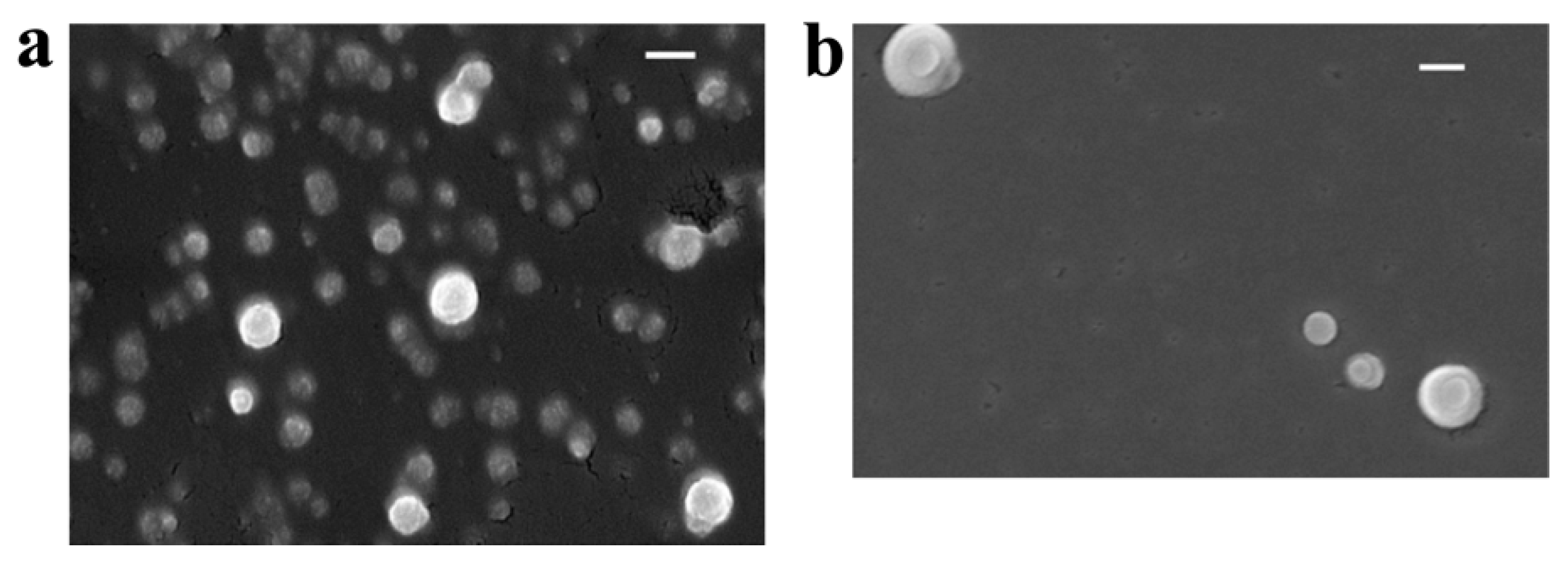
3.2.4. Field Emission Scanning Electron Microscopy (FESEM)

Images obtained using FESEM are pseudo-three-dimensional and capture the superficial layer and morphology of the NPs. Figure 3a shows that blank PEG-b-PLGA NPs were spherical and had a relatively smooth surface, similar to the PEG-b-PLGA NPs encapsulated with geraniin, Figure 3b. No porous forms or porous deposits were visible in either sample. The images also show that the NPs were well-dispersed.

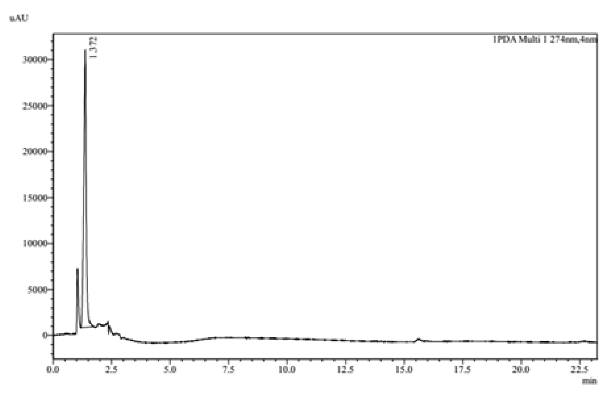
3.2.5. High Performance Liquid Chromatography (HPLC)

Reverse-phase HPLC was conducted to determine the encapsulation efficiency of geraniin into the NPs. Geraniin was detected at 274 nm with a retention time of 1.37 min and eluted with high velocity (Figure 4).
Hydrophilic phytochemicals were encapsulated in the hydrophobic region. Reverse-phase chromatography employs a polar mobile phase; thus, geraniin, a polar molecule, was used in this method. Hydrophobic molecules in the polar mobile phase tend to adsorb the hydrophobic stationary phase, and hydrophilic molecules in the mobile phase pass through the column and are eluted first. Thus, geraniin’s peak was eluted first. Late elution was checked by extending the run time. However, there were no other sharp peaks detected. The overall run time for this HPLC was 64 min.
The standard curve generated (Figure 5) shows that y = 731313x was obtained with an r2 value of 0.99. It is estimated that there was 0.30 mg/mL (30%) of geraniin in every 5 mg/mL of the encapsulated PEG-b-PLGA sample. The ratio of drug to polymer (1:10) was applied for encapsulation.

3.3. In Vitro Drug Release Study

The in vitro drug release study was conducted to determine the geraniin release time from the PEG-b-PLGA NPs. The standard curve (Figure 6) indicated that the geraniin was released at a fast rate with 38.51% ± 2.04% at 15 min from the PEG-b-PLGA NPs. Later, the amount of geraniin released by the NPs increased from 1 h at 58.56% ± 3.79% to 3 h at 79.67% ± 3.70% at a slower rate. The results suggested that the release profile was biphasic, where rapid burst release from the shell of the NPs occurred in the first 40 min, followed by sustained release. The drug release reached a plateau at 3 h.

3.4. In Vitro Cytotoxicity Study

Figure 7 shows that blank PEG-b-PLGA NPs were not cytotoxic to the CCD 841 CoN cells. Even at the highest concentration, 50 µg/mL, cell viability was reported at 83.2% ± 7.70. Thus, the PEG-b-PLGA polymer is a promising candidate in drug delivery systems because it is non-toxic, biodegradable, and biocompatible [30]. Furthermore, these results indicate that our modified double emulsion technique and the associated solvents used during the synthesis are suitable for the generation of PEG-b-PLGA NPs as a simple model system.
NPs encapsulated with geraniin showed some cytotoxic effects on CCD 841 CoN cells at concentrations of 25 and 50 µg/mL, which resulted in 75.74% ± 3.45 and 63.28% ± 13.42 cell viability, respectively. Based on the obtained data, the PEG-b-PLGA NPs and PEG-b-PLGA NPs encapsulated with geraniin are at a functional dosage up to 10 µg/mL for nutraceutical purposes, and a higher dosage can be used for cytotoxic purposes such as in cancer studies.

4. Discussion

Particle surface chemistry influences the initial interaction of this foreign material with the cells. Poly(lactic-co-glycolic acid) (PLGA) is a biocompatible and biodegradable copolymer that is extremely attractive and effective as a polymeric drug delivery carrier for clinical applications. For instance, when paclitaxel-loaded PLGA nanoparticles (NPs) were introduced in Hela cells to test their antiproliferative effects, the use of the NP system resulted in enhanced cell apoptosis compared to the use of the drug alone, possibly because of the sustained release of drugs inside the cancer cells [31]. Poly(ethylene glycol) (PEG) is a neutrally charged polymeric material that has been used widely to produce stealth nanocarriers. This hydrophilic material prevents the aggregation of NPs by forming hydrogen bonds with the solvent, and it increases the steric distance between NPs [32]. PEG also produces surface barrier layers that reduce the adsorption of opsonin’s present in the blood serum onto the NPs, thereby making the NPs “invisible” to phagocytic cells [33]. Therefore, coating the surface of PLGA with PEG, or “PEGylation,” is a commonly used approach for improving the efficiency of drug and gene delivery to target cells and tissues. Praveen and Sahoo (2008) [34] reported that polymeric NPs have promising potential in cancer therapy.
Particle size influences the distribution of carriers in vivo; the bioavailability, transport, and toxicity to the body; and the specificity of the carriers in the delivery systems [35]. In our study, the slightly larger size of the geraniin-encapsulated poly(ethylene glycol)-block-poly(lactic-co-glycolic acid) (PEG-b-PLGA) NPs could have been due to the encapsulation in the PLGA segment, which formed a larger inner (hydrophobic) layer of the NP. The particle must be small enough to enter vesicles, which range in size from 10 nm to 5 μm diameter. Oh and Park (2014) [36] suggested that polymeric NPs with sizes < 200 nm were taken up efficiently by cells. Thus, our data show that both types of synthesized PEG-b-PLGA NPs were at an optimal size for drug delivery and potentially for cellular uptake. The PDI value indicates monodispersity. When the particle size is distributed evenly, we would expect more NPs to be internalized into the cells once they have been exposed to the biological environment. A PDI value of ≤ 0.2 is most commonly deemed acceptable in practice for polymer-based NP materials [37]. Although our result did demonstrate a slightly higher PDI value, it is still considered to be in the acceptable range of 0.20 to 0.29.
Particle stability is shown by the zeta potential value, which is measured to identify the repulsion between particles; the higher the zeta potential value, the stronger the repulsion and more stable the NPs are. In our study, both groups of NPs were in a stable suspension, with zeta potential values of < −25 mV. The value of the PEG-b-PLGA NPs was due to the presence of an ether bond in the PEG structure. Alkoxide ions in the ether bond are negatively charged. These amphiphilic NPs have hydrophilic and hydrophobic terminals. The outer shell of the hydrophilic layer consists of PEG, and it has a negative charge. When it was encapsulated with geraniin, which is very hydrophilic, the zeta potential value decreased slightly. Presumably, the presence of oxygen atoms in the ester bonds of this structure contributed to additional negative charges for this NP. Zeta potential also is pH-dependent. NPs undergo a pH change from the acidic pH 2 in the stomach to pH 7 in the gastrointestinal tract. Particle charge may also influence the pathway of cellular uptake of the polymeric NPs, such as high surface charges, and large particle sizes were found to be phagocytosed by macrophages [38].
Particle shape in this study was spherical. The formation of small aggregates was noted in the TEM images, and this might be due to the charges of the particles. The electrostatic interaction between particles causes them to attract each other and aggregate. The Brownian motion also leads to NP aggregation [39]. However, individual PEG-b-PLGA NPs were also detected in the suspension. For TEM, the sample must be in a dry form before imaging. However, during the preparation of the sample, NPs need to be dispersed in water before observing. The long sample preparation time for TEM as compared to FESEM might explain the minimal irregularity of the outer shells of the NPs. Additionally, a spherical shape is considered to have a faster uptake rate in cells compared to other shapes [40]. The contact angle of the NPs on the cell membrane influences the rate of internalization [41]. Spherical, oblate-shaped particles are symmetric and have a large surface area, which allows them to be wrapped easily by cells, and they are viewed as the best option in receptor-ligand systems to reach the site of interest, as they follow the blood flow [24,42]. Thus, the shape we obtained in this study would allow the NPs to be mobile in the circulatory system and likely would contribute to an effective drug delivery system.
Particle content, based on our data, suggests that geraniin was encapsulated into the hydrophobic region of the polymeric NPs. This assumption is inferred primarily from past literature that reported geraniin (logP value = 3.4) and PLGA hydrophobicity [43,44]. However, the effect of the polar and charged group of geraniin has been shown to override the effect of geraniin hydrophobicity upon interaction with other chemicals at its interface [43]. Reverse-phase HPLC was used to quantify the encapsulation of geraniin into the NPs. This approach minimizes the risk of losing traces of geraniin from encapsulation and imparts accuracy of encapsulation as the NPs are small enough to pass the matrix in the column. Since geraniin is a polar compound due to the presence of carbonyl and hydroxyl groups in the structure, a polar solvent was used as the mobile phase to elute the polar compounds. Our HPLC setup and parameters were obtained from a previous study, which used reverse phase chromatography to separate the compounds from a P. watsonii extract [23]. In our study, negligible variability of retention time was seen in this chromatography analysis. The encapsulation efficiency of the NPs was calculated using the formula y = mx, which was obtained from the standard curve. The encapsulation efficiency was low, which could be because phytochemical extracts are hydrophobic but with polar and charged group activities, while the polymeric NPs are amphiphilic. Such characteristics of the phytochemicals will be encapsulated in the inner layer/aqueous compartment of the NPs [45], which results in low encapsulation due to steric interference of the drug-polymer interaction by the PEG-b-PLGA chains [44].
The content release was conducted at pH 7.4, which is the pH of the EMEM growth medium recommended for the growth of the cell model used (CCD 841 CoN cells). The release pattern showed an initial burst release of geraniin in the first 15 min, followed by a sustained release for subsequent hours. A possible explanation for this biphasic release pattern is that initially the dispersed geraniin molecules were located in the core of the hydrophobic chains; with increased geraniin loading, the geraniin entered the hydrophobic chains and the interfacial region between the hydrophobic and hydrophilic chains, which caused the many polymeric micelles to undergo an initial burst of drug release [4]. In our study, the polymer used to encapsulate geraniin is biodegradable, and therefore, the drug release mechanism might be from polymer erosion [46]. Biodegradable polymers degrade naturally within the body and release drugs at a constant rate [47]. The PLGA copolymer undergoes degradation by hydrolysis; as the water penetrates the polymer matrix, it disrupts the van der Waals forces and hydrogen bonds and causes cleavage of covalent bonds, thereby leading to degradation. Constant degradation occurs as the carboxylic end group autocatalyzes the degradation process. As water penetrates into the matrix, it hydrolyzes the PLGA copolymer into oligomeric and monomeric products. The encapsulated contents will be released by diffusion until the polymer is solubilized completely [48]. The study conducted on the release study was the proof-of-concept for the encapsulation of geraniin. As it was a combination of PEG and PLGA; therefore, the erosion would be different from PLGA only. Here, the results of our study supported the findings reported by Amin et al. (2017) [49], who reported that a hydrophilic drug encapsulated in PEG-b-PLGA NPs exhibited a biphasic drug release pattern. Thus, the biphasic drug release pattern appears to be common among encapsulated polymeric NPs.
Particle toxicity is also a concern in phytochemical and drug discovery. The cell viability test using MTT was employed to assess the percentage of viable cells after treatment with NPs. The highest percentage of viability was observed at the concentration of 1 µg/mL for both PEG-b-PLGA NPs and PEG-b-PLGA NPs encapsulated with geraniin. At the highest concentration (50 µg/mL), more than half (50%) of the cells were still viable for both groups. These results suggest that PEG-b-PLGA NPs are biodegradable, which may help overcome undesirable toxicity. PLGA polymers degrade into the biocompatible metabolites lactic acid and glycolic acid by hydrolysis [50]. The lactic acid is small enough to enter into the cells via the monocarboxylate transporter. It serves as an ab energy substrate and free radical scavenger for the cells. Research suggests that damage caused by free radicals leads to cell death [51]. Therefore, lactic acid prevents the cell from being damaged. In addition, lactic acid can promote cell proliferation [52]. Moreover, another study suggested that glycolic acid can induce cell proliferation [53]. Glycolic acid is an alpha-hydroxy acid that naturally occurs in sugarcane, and it is one of the components in PEG-b-PLGA that does not affect the normal functions of the cells.
PEG also is widely known to be non-reactive and safe to biological cells [54,55]. Song et al. (2012) [56] reported that PEG used as a surfactant in the formulation of a herbicide exhibited no cellular toxicity, even at high concentrations of 100 mM. Generally, PEG appears to have low toxicity to cells except at very high doses. Low toxicity of PEG may be due to the methoxy capping of PEG [57]. Additionally, Parnaud et al. (2001) [58] reported that PEG showed potent cancer chemopreventive activity, as it exhibited a dose-dependent inhibitory effect on colon cancer cell growth without any significant toxic effect on the normal colon cell line. These findings provide a reasonable explanation for the non-toxic effect of different concentrations of PEG-b-PLGA NPs on cell viability. Even at the highest concentration of geraniin-encapsulated NPs (50 µg/mL), cell viability was 61.9%, which means that there was no median lethal dose for the doses tested.
Geraniin is a plant compound that has antioxidant properties and is capable of scavenging reactive oxygen species [59]. The functional hydroxyl group, in its structure, mediates the antioxidant effects. However, there was a drop in the percentage of cell viability when cells were treated with geraniin-encapsulated NPs compared to those treated with blank NPs. This could be due to the higher molecular weight of geraniin (952.648 g/mol), which, together with its bulky molecules, might pose a steric hindrance to the donation of the phenolic hydrogen of the hydroxyl group to free radicals. The transfer of a hydrogen atom to a radical is what transforms it into a harmless molecule [60,61]. Geraniin might be slow at scavenging free radicals, allowing them to damage cells and cause some toxicity effects.
However, geraniin may be considered potentially safe for use in living cells at ≤ 1 µg/mL, as to date, no significant toxic effect has been reported in either in vitro or in vivo studies. In addition, geraniin can be tolerated at considerably high concentrations (up to 100 µM) without significantly affecting cell viability [62]. In this study, we demonstrated the first encapsulation of geraniin in PEG-b-PLGA NPs with low cytotoxicity in vitro and excellent drug release capacity. In brief, geraniin has proved to be a possible potent nutraceutical or pharmaceutical.
This research presents a proof-of-concept for the encapsulation of geraniin, which is also the concept for geraniin release. Other in-depth studies of PEG-b-PLGA NP release of different phytochemicals, including geraniin and its similar family tree, as well as the release model, can be useful for advances in nutraceutical applications.

5. Conclusions

In short, this research aimed to develop a modified double emulsion method specifically to create a simple poly(ethylene glycol)-block-poly(lactic-co-glycolic acid) (PEG-b-PLGA) nanoparticles (NPs) of ideal particle size, stability, and degradation that can entrap phytochemicals. We managed to (1) synthesize PEG-b-PLGA NPs that are <200 nm in size using the modified double emulsion method; (2) characterize the physicochemical properties and morphology of PEG-b-PLGA NPs; (3) measure the amount of geraniin encapsulated in PEG-b-PLGA NPs, and (4) evaluate the in vitro cytotoxicity of PEG-b-PLGA NPs and PEG-b-PLGA NPs encapsulated with geraniin to normal CCD 841 CoN colon epithelial cells.

Supplementary Materials

The following are available online at https://www.mdpi.com/2076-3417/10/14/4891/s1, Figure S1: Raw data of particle size analysis of blank and geraniin-loaded poly(ethylene glycol)-block-poly(lactic-co-glycolic acid) (PEG-b-PLGA) nanoparticles (NPs). Figure S2: Raw data of particle size analysis of geraniin loaded PEG-b-PLGA nanoparticles.

Author Contributions

Conceptualization, S.H.N., and I.B.S.J.; methodology, S.H.N., V.L., and Y.L.K.; software, K.D., S.J., and S.H.N.; validation, S.H.N., V.L., and Y.L.K.; formal analysis, K.D., S.J., and N.A.Y.; investigation, S.J., and N.A.Y.; resources, S.H.N. and I.B.S.J.; data curation, K.D., S.J., and N.A.Y; writing—original draft preparation, K.D.; writing—review and editing, K.D., S.H.N.; visualization, K.D., S.J., N.A.Y., and S.H.N.; supervision, S.H.N., V.L., Y.L.K.; project administration, S.H.N.; funding acquisition, S.H.N. and I.B.S.J. All authors have read and agreed to the published version of the manuscript.

Funding

This research was funded by Malaysian Agricultural Research and Development Institute (MARDI), MY grant number 304/CIPPT/650676.

Acknowledgments

K.D. and S.J. would like to acknowledge the financial support from Universiti Sains Malaysia Graduate Assistant Scholarship and Universiti Sains Malaysia Fellowship, respectively.

Conflicts of Interest

The authors declare no conflict of interest. The funders had no role in the design of the study; in the collection, analyses, or interpretation of data; in the writing of the manuscript, or in the decision to publish the results.

References

  1. Espanol, L.; Larrea, A.; Andreu, V.; Mendoza, G.; Arruebo, M.; Sebastian, V.; Auroraprado, M.S.; Kedorhackmann, E.R.M.; Santoro, M.I.R.M.; Santamaria, J. Dual encapsulation of hydrophobic and hydrophilic drugs in PLGA nanoparticles by a single-step method: Drug delivery and cytotoxicity assays. RSC Adv. 2016, 6, 111060–111069. [Google Scholar] [CrossRef] [Green Version]
  2. Danhier, F.; Ansorena, E.; Silva, J.M.; Coco, R.; le Breton, A.; Préat, V. PLGA-based nanoparticles: An overview of biomedical applications. J. Control. Release 2012. [Google Scholar] [CrossRef] [PubMed]
  3. Suk, J.S.; Xu, Q.; Kim, N.; Hanes, J.; Ensign, L.M. PEGylation as a strategy for improving nanoparticle-based drug and gene delivery. Adv. Drug Deliv. Rev. 2016. [Google Scholar] [CrossRef] [Green Version]
  4. Zhang, K.; Tang, X.; Zhang, J.; Lu, W.; Lin, X.; Zhang, Y.; Tian, B.; Yang, H.; He, H. PEG-PLGA copolymers: Their structure and structure-influenced drug delivery applications. J. Control. Release 2014. [Google Scholar] [CrossRef]
  5. Rafiei, P.; Haddadi, A. Docetaxel-loaded PLGA and PLGA-PEG nanoparticles for intravenous application: Pharmacokinetics and biodistribution profile. Int. J. Nanomed. 2017. [Google Scholar] [CrossRef] [PubMed] [Green Version]
  6. Lagoa, R.; Silva, J.; Rodrigues, J.R.; Bishayee, A. Advances in phytochemical delivery systems for improved anticancer activity. Biotechnol. Adv. 2020, 38, 107382. [Google Scholar] [CrossRef]
  7. Odeh, F.; Al-Jaber, H.; Khater, D. Nanoflora—How Nanotechnology Enhanced the Use of Active Phytochemicals. In Application of Nanotechnology in Drug Delivery; Sezer, A.D., Ed.; IntechOpen: London, UK, 2014. [Google Scholar]
  8. Tang, Y.Q.; Jaganath, I.B.; Sekaran, S.D. Phyllanthus spp. induces selective growth inhibition of PC-3 and mewo human cancer cells through modulation of cell cycle and induction of apoptosis. PLoS ONE 2010. [Google Scholar] [CrossRef]
  9. Bala, I.; Bhardwaj, V.; Hariharan, S.; Kumar, M.N.V.R. Analytical methods for assay of ellagic acid and its solubility studies. J. Pharm. Biomed. Anal. 2006. [Google Scholar] [CrossRef]
  10. Cheng, J.T.; Chang, S.S.; Hsu, F.L. Antihypertensive Action of Geraniin in Rats. J. Pharm. Pharmacol. 1994. [Google Scholar] [CrossRef]
  11. Konishi, Y.; Hitomi, Y.; Yoshioka, E. Intestinal Absorption of p-Coumaric and Gallic Acids in Rats after Oral Administration. J. Agric. Food Chem. 2004. [Google Scholar] [CrossRef]
  12. Romeo, J. Chemical Ecology and Phytochemistry of Forest Ecosystems: Proceedings of the Phytochemical Society of North America; Elsevier Science: Oxford, UK, 2005. [Google Scholar]
  13. Lee, J.; Tsai, C.; Kao, J.; Kao, M.; Tsai, S.; Chang, C.; Huang, L.; Kuo, S.; Lin, J.; Way, T. Geraniin-mediated apoptosis by cleavage of focal adhesion kinase through up-regulation of Fas ligand expression in human melanoma cells. Mol. Nutr. Food Res. 2008. [Google Scholar] [CrossRef] [PubMed]
  14. Yang, C.M.; Cheng, H.Y.; Lin, T.C.; Chiang, L.C.; Lin, C.C. The in vitro activity of geraniin and 1,3,4,6-tetra-O-galloyl-β-d-glucose isolated from Phyllanthus urinaria against herpes simplex virus type 1 and type 2 infection. J. Ethnopharmacol. 2007. [Google Scholar] [CrossRef] [PubMed]
  15. Londhe, J.S.; Devasagayam, T.P.A.; Foo, L.Y.; Shastry, P.; Ghaskadbi, S.S. Geraniin and amariin, ellagitannins from Phyllanthus amarus, protect liver cells against ethanol induced cytotoxicity. Fitoterapia 2012. [Google Scholar] [CrossRef] [PubMed]
  16. Palanisamy, U.D.; Ling, L.T.; Manaharan, T.; Appleton, D. Rapid isolation of geraniin from Nephelium lappaceum rind waste and its anti-hyperglycemic activity. Food Chem. 2011. [Google Scholar] [CrossRef]
  17. Chung, A.P.Y.S.; Ton, S.H.; Gurtu, S.; Palanisamy, U.D. Ellagitannin geraniin supplementation ameliorates metabolic risks in high-fat diet-induced obese Sprague Dawley rats. J. Funct. Foods 2014. [Google Scholar] [CrossRef]
  18. Tobío, M.; Gref, R.; Sánchez, A.; Langer, R.; Alonso, M.J. Stealth PLA-PEG nanoparticles as protein carriers for nasal administration. Pharm. Res. 1998. [Google Scholar] [CrossRef]
  19. Weissenboeck, A.; Bogner, E.; Wirth, M.; Gabor, F. Binding and uptake of wheat germ agglutinin-grafted PLGA-nanospheres by Caco-2 monolayers. Pharm. Res. 2004. [Google Scholar] [CrossRef]
  20. McCall, R.L.; Sirianni, R.W. PLGA nanoparticles formed by single- or double-emulsion with vitamin E-TPGS. J. Vis. Exp. 2013. [Google Scholar] [CrossRef] [Green Version]
  21. Sun, Y.P.; Li, X.Q.; Zhang, W.X.; Wang, H.P. A method for the preparation of stable dispersion of zero-valent iron nanoparticles. Colloids Surf. A Physicochem. Eng. Asp. 2007. [Google Scholar] [CrossRef]
  22. Feng, S.S.; Mu, L.; Win, K.Y.; Huang, G. Nanoparticles of biodegradable polymers for clinical administration of paclitaxel. Curr. Med. Chem. 2004, 11, 413–424. [Google Scholar] [CrossRef]
  23. Lee, S.H.; Tang, Y.; Rathkrishnan, A.; Wang, S.M.; Ong, K.C.; Manikam, R.; Payne, B.J.; Jaganath, I.B.; Sekaran, S.D. Effects of cocktail of four local Malaysian medicinal plants (Phyllanthus spp.) against dengue virus 2. BMC Complement. Altern. Med. 2013. [Google Scholar] [CrossRef] [PubMed] [Green Version]
  24. Li, Y.; Kröger, M.; Liu, W.K. Shape effect in cellular uptake of PEGylated nanoparticles: Comparison between sphere, rod, cube and disk. Nanoscale 2015. [Google Scholar] [CrossRef] [PubMed]
  25. Devasvaran, K.; Tan, J.J.; Ng, C.T.; Fong, L.Y.; Yong, Y.K. Malaysian Tualang Honey Inhibits Hydrogen Peroxide-Induced Endothelial Hyperpermeability. Oxid. Med. Cell. Longev. 2019. [Google Scholar] [CrossRef] [PubMed]
  26. Sarkar, K.; Meka, S.R.K.; Bagchi, A.; Krishna, N.S.; Ramachandra, S.G.; Madras, G.; Chatterjee, K. Polyester derived from recycled poly(ethylene terephthalate) waste for regenerative medicine. RSC Adv. 2014. [Google Scholar] [CrossRef]
  27. Win, K.Y.; Feng, S.S. Effects of particle size and surface coating on cellular uptake of polymeric nanoparticles for oral delivery of anticancer drugs. Biomaterials 2005. [Google Scholar] [CrossRef]
  28. Mahobia, S.; Bajpai, J.; Bajpai, A.K. An in-vitro investigation of swelling controlled delivery of insulin from egg albumin nanocarriers. Iran. J. Pharm. Res. 2016. [Google Scholar] [CrossRef]
  29. Alexis, F.; Pridgen, E.; Molnar, L.K.; Farokhzad, O.C. Factors affecting the clearance and biodistribution of polymeric nanoparticles. Mol. Pharm. 2008. [Google Scholar] [CrossRef] [Green Version]
  30. Paka, G.D.; Ramassamy, C. Optimization of Curcumin-Loaded PEG-PLGA Nanoparticles by GSH Functionalization: Investigation of the Internalization Pathway in Neuronal Cells. Mol. Pharm. 2017. [Google Scholar] [CrossRef]
  31. Parveen, S.; Misra, R.; Sahoo, S.K. Nanoparticles: A boon to drug delivery, therapeutics, diagnostics and imaging. Nanomed. Nanotechnol. Biol. Med. 2012. [Google Scholar] [CrossRef]
  32. Stiufiuc, R.; Iacovita, C.; Nicoara, R.; Stiufiuc, G.; Florea, A.; Achim, M.; Lucaciu, C.M. One-step synthesis of PEGylated gold nanoparticles with tunable surface charge. J. Nanomater. 2013. [Google Scholar] [CrossRef]
  33. Elendran, S.; Wang, L.W.; Prankerd, R.; Palanisamy, U.D. The physicochemical properties of geraniin, a potential antihyperglycemic agent. Pharm. Biol. 2015. [Google Scholar] [CrossRef] [PubMed]
  34. Parveen, S.; Sahoo, S.K. Polymeric nanoparticles for cancer therapy. J. Drug Target. 2008. [Google Scholar] [CrossRef]
  35. Singh, R.; Lillard, J.W. Nanoparticle-based targeted drug delivery. Exp. Mol. Pathol. 2009. [Google Scholar] [CrossRef] [PubMed] [Green Version]
  36. Oh, N.; Park, J.H. Endocytosis and exocytosis of nanoparticles in mammalian cells. Int. J. Nanomed. 2014. [Google Scholar] [CrossRef] [Green Version]
  37. Clarke, S. Development of Hierarchical Magnetic Nanocomposite Materials for Biomedical Applications. Ph.D. Thesis, Dublin City University, Dublin, Ireland, 2013. [Google Scholar]
  38. He, C.; Hu, Y.; Yin, L.; Tang, C.; Yin, C. Effects of particle size and surface charge on cellular uptake and biodistribution of polymeric nanoparticles. Biomaterials 2010. [Google Scholar] [CrossRef] [PubMed]
  39. Carpineti, M.; Ferri, F.; Giglio, M.; Paganini, E.; Perini, U. Salt-induced fast aggregation of polystyrene latex. Phys. Rev. A 1990. [Google Scholar] [CrossRef]
  40. Florez, L.V.; Herrmann, C.; Cramer, J.M.; Hauser, C.P.; Koynov, K.; Landfester, K.; Crespy, D.; Mailander, V. How shape influences uptake: Interactions of anisotropic polymer nanoparticles and human mesenchymal stem cells. Small 2012. [Google Scholar] [CrossRef]
  41. Parakhonskiy, B.; Zyuzin, M.V.; Yashchenok, A.M.; Carregalromero, S.; Rejman, J.; Mohwald, H.; Parak, W.J.; Skirtach, A.G. The influence of the size and aspect ratio of anisotropic, porous CaCO3 particles on their uptake by cells. J. Nanobiotechnol. 2015. [Google Scholar] [CrossRef] [Green Version]
  42. Chen, L.; Xiao, S.; Zhu, H.; Wang, L.; Liang, H. Shape-dependent internalization kinetics of nanoparticles by membranes. Soft Matter 2016. [Google Scholar] [CrossRef]
  43. Ahmad, S.A.A.; Palanisamy, U.D.; Tejo, B.A.; Chew, M.F.; Tham, H.W.; Hassan, S.S. Geraniin extracted from the rind of Nephelium lappaceum binds to dengue virus type-2 envelope protein and inhibits early stage of virus replication. Virol. J. 2017, 14, 229. [Google Scholar] [CrossRef] [Green Version]
  44. Makadia, H.K.; Siegel, S.J. Poly Lactic-co-Glycolic Acid (PLGA) as biodegradable controlled drug delivery carrier. Polymers 2011, 3, 1377–1397. [Google Scholar] [CrossRef] [PubMed]
  45. Ong, S.G.M.; Ming, L.C.; Lee, K.S.; Yuen, K.H. Influence of the encapsulation efficiency and size of liposome on the oral bioavailability of griseofulvin-loaded liposomes. Pharmaceutics 2016, 8, 25. [Google Scholar] [CrossRef] [PubMed]
  46. Kamaly, N.; Yameen, B.; Wu, J.; Farokhzad, O.C. Degradable controlled-release polymers and polymeric nanoparticles: Mechanisms of controlling drug release. Chem. Rev. 2016. [Google Scholar] [CrossRef] [PubMed] [Green Version]
  47. Von Burkersroda, F.; Schedl, L.; Göpferich, A. Why degradable polymers undergo surface erosion or bulk erosion. Biomaterials 2002. [Google Scholar] [CrossRef]
  48. Gentile, P.; Chiono, V.; Carmagnola, I.; Hatton, P.V. An Overview of Poly(lactic-co-glycolic) Acid (PLGA)-Based Biomaterials for Bone Tissue Engineering. Int. J. Mol. Sci 2014, 15, 3640–3659. [Google Scholar] [CrossRef]
  49. Amin, F.U.; Shah, S.A.; Badshah, H.; Khan, M.; Kim, M.O. Anthocyanins encapsulated by PLGA@PEG nanoparticles potentially improved its free radical scavenging capabilities via p38/JNK pathway against Aβ1-42-induced oxidative stress. J. Nanobiotechnol. 2017, 15. [Google Scholar] [CrossRef] [Green Version]
  50. Keles, H.; Naylor, A.; Clegg, F.; Sammon, C. Investigation of factors influencing the hydrolytic degradation of single PLGA microparticles. Polym. Degrad. Stab. 2015. [Google Scholar] [CrossRef] [Green Version]
  51. Silva, J.P.; Coutinho, O.P. Free radicals in the regulation of damage and cell death-basic mechanisms and prevention. Drug Discov. Ther. 2010, 4, 144. [Google Scholar]
  52. Lampe, K.J.; Namba, R.M.; Silverman, T.R.; Bjugstad, K.B.; Mahoney, M.J. Impact of lactic acid on cell proliferation and free radical-induced cell death in monolayer cultures of neural precursor cells. Biotechnol. Bioeng. 2009. [Google Scholar] [CrossRef] [Green Version]
  53. Kim, S.J.; Park, J.H.; Kim, D.H.; Won, Y.H.; Maibach, H.I. Increased in vivo collagen synthesis and in vitro cell proliferative effect of glycolic acid. Dermatol. Surg. 1998. [Google Scholar] [CrossRef]
  54. Alcantar, N.A.; Aydil, E.S.; Israelachvili, J.N. Polyethylene glycol-coated biocompatible surfaces. J. Biomed. Mater. Res. 2000. [Google Scholar] [CrossRef]
  55. Tong, R.; Gabrielson, N.P.; Fan, T.M.; Cheng, J. Polymeric nanomedicines based on poly(lactide) and poly(lactide-co- glycolide). Curr. Opin. Solid State Mater. Sci. 2012. [Google Scholar] [CrossRef] [Green Version]
  56. Song, H.Y.; Kim, Y.; Seok, S.J.; Gil, H.; Yang, J.O.; Lee, E.; Hong, S. Cellular toxicity of surfactants used as herbicide additives. J. Korean Med. Sci. 2012. [Google Scholar] [CrossRef] [Green Version]
  57. Webster, R.; Elliott, V.; Park, B.K.; Walker, D.; Hankin, M.; Taupin, P. PEG and PEG conjugates toxicity: Towards an understanding of the toxicity of PEG and its relevance to PEGylated biologicals. In PEGylated Protein Drugs: Basic Science and Clinical Applications; Veronese, F.M., Ed.; Berkhäuser: Basel, Switzerland, 2009. [Google Scholar]
  58. Parnaud, G.; Corpet, D.E.; Gamet-Payrastre, L. Cytostatic effect of polyethylene glycol on human colonic adenocarcinoma cells. Int. J. Cancer 2001. [Google Scholar] [CrossRef] [Green Version]
  59. Romera-Castillo, C.; Jaffé, R. Free radical scavenging (antioxidant activity) of natural dissolved organic matter. Mar. Chem. 2015. [Google Scholar] [CrossRef] [Green Version]
  60. Özyurt, H.; Luna, C.; Estévez, M. Redox chemistry of the molecular interactions between tea catechins and human serum proteins under simulated hyperglycemic conditions. Food Funct. 2016. [Google Scholar] [CrossRef] [Green Version]
  61. Wang, T.Y.; Li, Q.; Bi, K.S. Bioactive flavonoids in medicinal plants: Structure, activity and biological fate. Asian J. Pharm. Sci. 2018. [Google Scholar] [CrossRef] [PubMed]
  62. Miguel, O.G.; Calixto, J.B.; Santos, A.R.S.; Messana, I.; Ferrari, F.; Filho, V.C.; Pizzolatti, M.G.; Yunes, R.A. Chemical and preliminary analgesic evaluation of geraniin and furosin isolated from Phyllanthus sellowianus. Planta Med. 1996. [Google Scholar] [CrossRef]
Figure 1. W/O/W double emulsion method used to prepare nanoparticles (NPs) and typical structures of (a) blank and (b) geraniin encapsulated poly(ethylene glycol)-block-poly(lactic-co-glycolic acid) (PEG-b-PLGA) NPs. The percentage yield was calculated using a formula derived by McCall and Sirianni, 2013 [20]: % yield = [NPs obtained after synthesis/Total solid weight (PEG-b-PLGA + geraniin)] × 100.
Applsci 10 04891 g001
Figure 2. Representative TEM images of blank PEG-b-PLGA NPs and PEG-b-PLGA NPs encapsulated with geraniin at different image magnifications. (a) blank PEG-b-PLGA NPs, scale bar at 100 nm. (b) blank PEG-b-PLGA NPs, scale bar at 200 nm. (c) PEG-b-PLGA NPs encapsulated with geraniin, scale bar at 200 nm. (d) PEG-b-PLGA NPs encapsulated with geraniin, scale bar at 100 nm. Representative images were taken from one of three independent experiments.
Applsci 10 04891 g002
Figure 3. Representative Field Emission Scanning Electron Microscopy (FESEM) images of (a) blank PEG-b-PLGA NPs and (b) PEG-b-PLGA NPs encapsulated with geraniin. The scale bar is 200 nm.
Applsci 10 04891 g003
Figure 4. High Performance Liquid Chromatography (HPLC) chromatogram. Geraniin peak was detected at the retention time of 1.37 min at 274 nm by reverse-phase HPLC.
Applsci 10 04891 g004
Figure 5. Standard curve of geraniin by HPLC.
Applsci 10 04891 g005
Figure 6. In vitro percentage of geraniin release from PEG-b-PLGA NPs at pH 7.4. The graph shows the percentage of encapsulated geraniin released at different time points. Data are expressed in mean ± standard error of the mean from three independent experiments.
Applsci 10 04891 g006
Figure 7. CCD 841 CoN cells viability after treatment with blank PEG-b-PLGA NPs and PEG-b-PLGA NPs encapsulated with geraniin for 3 h. Data are expressed in mean ± standard error of the mean from three independent experiments. * p < 0.05 indicates a significant difference from the untreated control group.
Applsci 10 04891 g007

