Review

The Impact of Micro- and Nanoplastics on Aquatic Organisms: Mechanisms of Oxidative Stress and Implications for Human Health—A Review

by
Eugenio Geremia
1,
Maria Teresa Muscari Tomajoli
1,
Carola Murano
2,
Adriana Petito
3 and
Gianluca Fasciolo
3,*
1
International PhD Programme, UNESCO Chair “Environment, Resources and Sustainable Development”, Department of Science and Technology, Parthenope University of Naples, 80143 Naples, Italy
2
Department of Integrative Marine Ecology, Stazione Zoologica Anton Dohrn, 80121 Naples, Italy
3
Department of Biology, University of Naples Federico II, 80126 Naples, Italy
*
Author to whom correspondence should be addressed.
Environments 2023, 10(9), 161; https://doi.org/10.3390/environments10090161
Submission received: 9 July 2023 / Revised: 4 September 2023 / Accepted: 6 September 2023 / Published: 16 September 2023
(This article belongs to the Special Issue Plastic Contamination: Challenges and Solutions Volume II)

Abstract

:
In recent years, interest in the impact of environmental pollutants on the ecosystem has increased significantly, with particular attention being paid to the relationship between climate change and the aquatic world. This is because increasing pollution is causing fundamental changes to the welfare of animals and the marine ecosystem. A primary focus is on the impact of microplastics (MPs) and nanoplastics (NPs), as evidenced by our bibliometric network analysis (BNA). However, while research is focused on the accumulation of these pollutants in aquatic organisms, their effects on redox homeostasis are still seldom discussed despite the role played by reactive oxygen species and mitochondrial well-being in maintaining an optimal state of health. However, some scientific evidence suggests that the accumulation of MPs and NPs in organisms at the base of the trophic chain can cause a transfer of these substances towards more complex organisms, reaching humans through the consumption of aquatic fauna as food. Therefore, in this review, we have tried to discuss the effects of these substances on oxidative stress in aquatic organisms, even if studies in this regard are still scarce.

Graphical Abstract

1. Pollution and the Environment

The increase in the world population and, consequently, human activities, such as agricultural intensification, industrial development, and urbanization, have led to a sharp increase in waste production and environmental pollution [1,2]. Coastal, marine, and fluvial environments are constantly under pressure from anthropogenic release into the environment of heavy metals, hydrocarbons, pesticides, persistent organic pollutants (POPs), organophosphorus flame retardants (OPFRs), pharmaceuticals and personal care products (PPCPs), products of metallurgical processes, nanoparticles, plastics and microplastics, etc. [3,4,5,6,7,8]. Organisms living in the aquatic environment, such as plants and animals, can transfer pollutants to humans through bioaccumulation and trophic transfer, as well as exert harmful effects on them following their exposure [5,7]. In recent decades, there has been a rise in the emission of nanoparticles and heavy metals into the environment with the intensification of industrialization [9,10,11]. Nanoparticles are a group of materials that have a size between 1 and 1000 nm in diameter [12,13,14]. The nanoparticles are widespread in aquatic ecosystems through aerial deposition, effluent discharge, dumping, and run-off [11]. They can be divided into two groups: natural (i.e., desert dusts, aerosols, emissions from volcanic activities, etc.) and anthropogenic (i.e., metal oxides, drug production, burning fossil fuels, vehicle exhaust release, mining demolition emissions, etc.) [11,12,13]. One of the most widespread nanoparticles in the aquatic environment is titanium dioxide (TiO2), which the European Union reports as one of the main pollutants of surface waters, with a value of 2.2 μgL−1 [12]. Lovern and Klaper, showed an increase in the mortality rate of D. magna due to exposure to TiO2 [14]. Heavy metals also pose a serious threat to the aquatic environment. Twenty-three metals and metalloids out of seventy present in the environment are identified as heavy metals/trace metals, some of which are considered dangerous [9]. Heavy metals can be divided into two categories: essential and non-essential. The non-essential metals (aluminum (Al), cadmium (Cd), mercury (Hg), tin (Sn), lead (Pb), etc.) are elements that have no known biological function and can be considered toxic at high concentrations. Essential metals (copper (Cu), zinc (Zn), chromium (Cr), nickel (Ni), cobalt (Co), molybdenum (Mo), and iron (Fe)) play a specific role in the body’s metabolism and can be toxic both under metabolic deficiency conditions and at high concentrations [9,10,15]. Contaminants/heavy metals can have a negative impact on the physiology of aquatic organisms; fish especially are very sensitive to such changes [15]. The toxic effects of heavy metals on fish are numerous [16]. They enter the fish body via the gills, digestive tract, and body surface and accumulate in the liver, kidney, muscle, intestine, skin, and bones [16]. It is shown that in fish heavy metals can alter several physiological and biochemical processes, growth rates, mortality, and reproduction. Moreover, they can cause serious problems in the body by producing reactive oxygen species (ROS), which cause oxidative stress and DNA damage [17,18,19].
Another issue that has plagued the environment in recent decades is the spread of pharmaceuticals and personal care products (PPCPs) in aquatic environments. This group of pollutants includes several substances from different sources, mainly anti-inflammatory drugs, as well as antibiotics (antimicrobials or antibacterials), antiepileptics, and personal care products [20]. The increase in the consumption of PPCPs is caused by the average use in the use human population and the upward demand for animal protein [21,22,23,24]. These substances find their way into the environment, where they can negatively impact organisms in aquatic ecosystems. Antibiotics are the most widely used pharmaceuticals due to human and veterinary applications [25]. Environmental antibiotics are derived from different sources, such as wastewater treatment plants, hospitals, water from urban canals, agriculture (aquaculture, husbandry), and the pharmaceutical industry [26,27]. Recently, the use of antibiotics has increased with the rise in aquaculture; in fact, their use allows for the reduction in possible threats from microorganisms. However, on the other hand, antibiotics can promote the resistance of bacteria in aquaculture and lead to resistance even in wild fish populations [28]. They are continually discharged into the aquatic environment and are bioavailable for animals, crops, and aquatic plants [26,27,28]. Primary producers, like microalgae and decomposers, are more sensitive than crustaceans and fish to antibiotic agents. Consequently, antibiotics could negatively impact microbial processes and lead to variations in biogeochemical cycling and aquatic ecosystems [4,26].
Among the toxic pollutants with the greatest impact on the aquatic environment, we cannot fail to include pesticides [29,30,31,32]. Pesticides are important for the protection of crops from infection by many microorganisms and for eradicating pests and organisms, such as mice, rats, ticks, and mosquitoes, that are dangerous for the environment and the habitat of humans [30,32,33]. Among these, the best known and most discussed is DDT (1,1,1-trichloro-2,2-bis(4-chlorophenyl) ethane) [30,33]. However, due to its high toxicity, it has been banned in many Western countries since the 1970s [33]. Due to its ecotoxic effects, bioaccumulation, and environmental persistence, it was also considered by the international agreement of the Stockholm Convention 2001 (World Health Organization, 2011) as one of the world’s 12 restricted persistent organic pollutants [34]. Nevertheless, the World Health Organization controlling vector-borne diseases allowed it in certain tropical countries in 2006 [30]. DDT can land many issues for aquatic organisms. It is so hydrophobic that it is absorbed into organic sediment particles, where it can persist for many years. In the aquatic environment, it can accumulate in benthic organisms and subsequently be transferred to higher trophic levels through biomagnification [30]. Owens e Baer, 2000, revealed a dose-dependent increase in the lethality of the Japanese medaka (Oryzias latipes) exposed to DDT [35], while Ton et al., 2006, showed the teratogenic effects on zebrafish (Danio Rerio) larvae exposed to the organochlorine DDT [36].
Nowadays, new pollutants are causing harm in aquatic environments and beyond, namely micro- and nanoplastics [4,5,6,37,38]. Figure 1 shows how microplastic pollution has recently become one of the main topics in studies on the aquatic environmental stress. Figure 1 has been generated using the Scopus database and the software Vosviewer (version 1.6.19) which allows for the creation, viewing, and exploration of maps based on bibliometric network data [39]. Scopus database provided 3235 scientific documents published from 1976 to 2022 by selecting (Aquatic environment*) AND (“Oxidative stress” OR “ROS” OR “REDOX”) AND (“Pollution*” OR “Pollutant*”) as keywords.

2. Micro- and Nanoplastics

Since the 1950s, plastic production has soared to a value of 322 million tons in 2015, and projections indicate it could exceed one billion tons by 2050 [40]. Microplastic pollution is widespread in every environment, from the atmosphere to the ocean depths [41,42]. Plastic makes up about 80 to 85% of marine litter, and it is estimated that an average of 5 to 13 million tons of plastic enter the oceans each year [6,37]. For example, around 229,000 tons of plastic are released every year in the Mediterranean Sea [43], which appears to be one of the areas with the highest concentration of floating debris [44]. This is due to the characteristics of the Mediterranean Sea, such as its semi-enclosed conformation and the general anti-estuarine circulation, which favors the accumulation of floating material [44].

3. Characteristics and Sources

Plastic materials are made from a wide variety of synthetic or semisynthetic organic compounds, such as polyethylene (PE), polyethylene terephthalate (PET), polystyrene (PS), polypropylene (PP), and polyvinyl chloride (PVC) [37,38,45]. Due to low end-of-life recycling, PVC is a very abundant contaminant in the aquatic environment. It has been reported that only 3% of PVC is recycled, while 82% is discarded in landfills, and 15% is incinerated [45]. Plastic materials may persist in the environment thanks to their high ratio of aromatic compounds and consequent resistance to degradation by microorganisms [46]. Once released into the environment, plastic breaks down into smaller pieces called microplastics (MPs) and nanoplastics (NPs, <1 μm) due to the action of various environmental factors, such as ultraviolet (UV) radiation, mechanical abrasion, hydrolysis and biodegradation [5,6,37,47].
Microplastics can be divided into primary and secondary. These latter MPs, resulting from the fragmentation of larger plastic (e.g., plastic bags, food packaging, ropes, etc.), are tiny plastic particles with a size of less than 5 mm and are the main source of MPs in the aquatic environment [48,49]. Instead, primary MPs are not derived from the degradation of larger plastic objects but are manufactured directly in microscopic sizes, including products for industrial or domestic applications entering aquatic systems through wastewater [37,48,50]. Over time, microplastics can become even smaller, fragmenting and giving rise to nanoplastics, such as those recently detected in the seawater below the North Atlantic sub-tropical gyre [51].
Like microplastics, nanoplastics are also classified into primary and secondary sources. Primary NPs are plastic nanoparticles manufactured and released directly into the environment. Secondary NPs originate from fragments formed by the degradation of MPs, plastic litter, and bulk plastics. NPs have a high surface area-to-volume ratio, an important parameter when considering toxicity, and can enrich and transfer many hazardous pollutants to the environment [52,53]. These properties can promote processes such as dissolution, redox reactions, or the generation of ROS. Larger particles would not produce such biological effects even with the same chemical composition [54].
The pathways of microplastic from source to habitat can be different. The sources can be divided into four types: (I) large plastic waste, (II) medicines, (III) textiles, and (IV) cleaning products [50]. From these sources, they can enter aquatic habitats through wastewater treatment plants (WWTPs), which are considered the main route for MPs to enter freshwater, via runoff, through sewers and landfills, and then spread worldwide by wind and currents [5,37,47]. Activities directly related to the aquatic environment can also be sources of marine litter, such as in the case of aquaculture and fishing, which play a significant role in the increase in MPs in aquatic environments [55,56]. Plastic in aquaculture and fisheries are generally used to produce different products, such as cages or nets, buoys, gears, trawls, dredges, traps, and floats, and for different purposes, such as boat construction and maintenance [40,49]. In addition, ALDFGs (abandoned, lost, or otherwise discharged fishing gears) represent one of the main plastic marine litters in the world’s oceans [49,57,58,59].
In summary, the widespread use of microplastics is becoming a serious issue primarily for freshwater and marine environments due to their overwhelming presence on surface and deep waters, on beaches, and in seabed sediments with their consequent ingestion by the organisms and biomagnification or bioaccumulation [37,48]. The diversity of microplastics based on the shape and source of production has favored their diffusion, so much so that microplastic pollution has spread to the most remote corners of the planet and has led to the trophic transfer of micro- and nanoplastics in the marine food chain from plankton to fish [60,61].

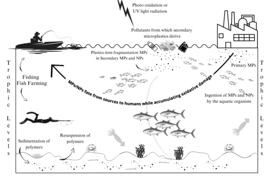
4. Environmental Life-Cycle of Plastic Debris

MPs and NPs are considered emerging contaminants of great concern, with unique characteristics that pose a real risk to aquatic life [62,63]. Once in the marine environment, MPs and NPs undergo numerous transformations, which determine both their availability and toxicity to marine organisms [64,65]. Abiotic components (pH, temperature, ionic strength, light intensity, and organic matter concentration) influence these transformations [66]. However, these transformations are mainly driven by the surface properties of MPs/NPs, which play an important role in determining their rapid changes and ecological impacts [66]. The hydrophobic surface of MPs/NPs attracts substances from the surrounding environment, including natural organic matter (NOM), nutrients, hydrophobic contaminants and heavy metals [67,68]. The interactions occurring with natural biomolecules and chemical substances present in the natural aquatic media lead to the formation of the so-called “eco-corona” [69]. Down the size scale, towards NPs, the absorption concept is translated to a finer level. NPs can adsorb the proteins present in biological fluids onto their surface, forming the “protein-corona”, i.e., the bridge at the bio-nano interface [70].
The photodegradation of micro- and nanoplastics can increase the amount of dissolved organic carbon (DOC) in the marine environment by releasing (among others) polybrominated diphenyl ethers, nonylphenol, and triclosan, which are additives added to plastics during production to improve their properties [71]. According to Romera-Castillo et al. (2016), microbes absorb DOC released from plastic, potentially impacting the carbon cycle and microbial ecology as a whole. However, the possible effects of increased DOC on biodegradation and marine biota remain largely unknown [72]. Micro- and nanoplastics can lead to the aggregation of dissolved organic matter (DOM) in particulate organic matter (POM) [73].
Among other materials, plastic represents an ideal substrate for rapid colonization by microorganisms such as bacteria, fungi, diatoms, and algae, which together form a biofilm called “Plastisphere” [74]. This consortium may involve an active interaction in which MPs can be utilized as a carbon source by microorganisms; otherwise, in the case of a passive interaction, this is based on various biochemical and/or physical processes [75]. Among the prokaryotes and eukaryotes that live in the plastisphere, species that degrade hydrocarbons stand out; however, fish and human pathogens are also commonly present [76,77,78]. The plastisphere formation has led to severe implications for the marine environment because it allows for not only the transport and accumulation of potential pathogenic strains (i.e., genus Arcobacter and Vibrio), but also the breakdown of plastic fragments by microbial enzymes [75]. Furthermore, the presence of biofilms can alter the properties of virgin polymers by changing their environmental fate (density and buoyancy) and increase bioavailability, appeal, and toxicity to polymers. Laboratory studies demonstrate that befouled plastic debris is ingested more than the corresponding virgin polymers and has magnified acute toxicity in various marine organisms [79,80,81]. One of the key aspects of the surface interactions is related to the behavior of MPs/NPs along the water column. The layered components of the eco-corona can stabilize or aggregate particles in the water column, increasing their nominal size and density and resulting in the vertical transport of MPs/NPs to the seabed [82]. In this case, MP resuspension processes may occur following natural phenomena such as strong winds or tidal currents. In this respect, bioturbators can play an important role in determining the fate of MPs/NPs, promoting their long-term burial in deeper sedimentary layers or the upward transport of particles buried in sediments [83]. In this context, microbial activity in the plastisphere is considered one of the most important mechanisms of the loss of floating particles, which, due to biofilm formation, will present a consequent positive sedimentation velocity. For this reason, the plastisphere is considered a potential explanation of the conspicuous disparity between the quantities of plastic potentially introduced into the marine environment and those found on the surface, generally referred to as the “missing 99%” surface plastic [84,85,86].

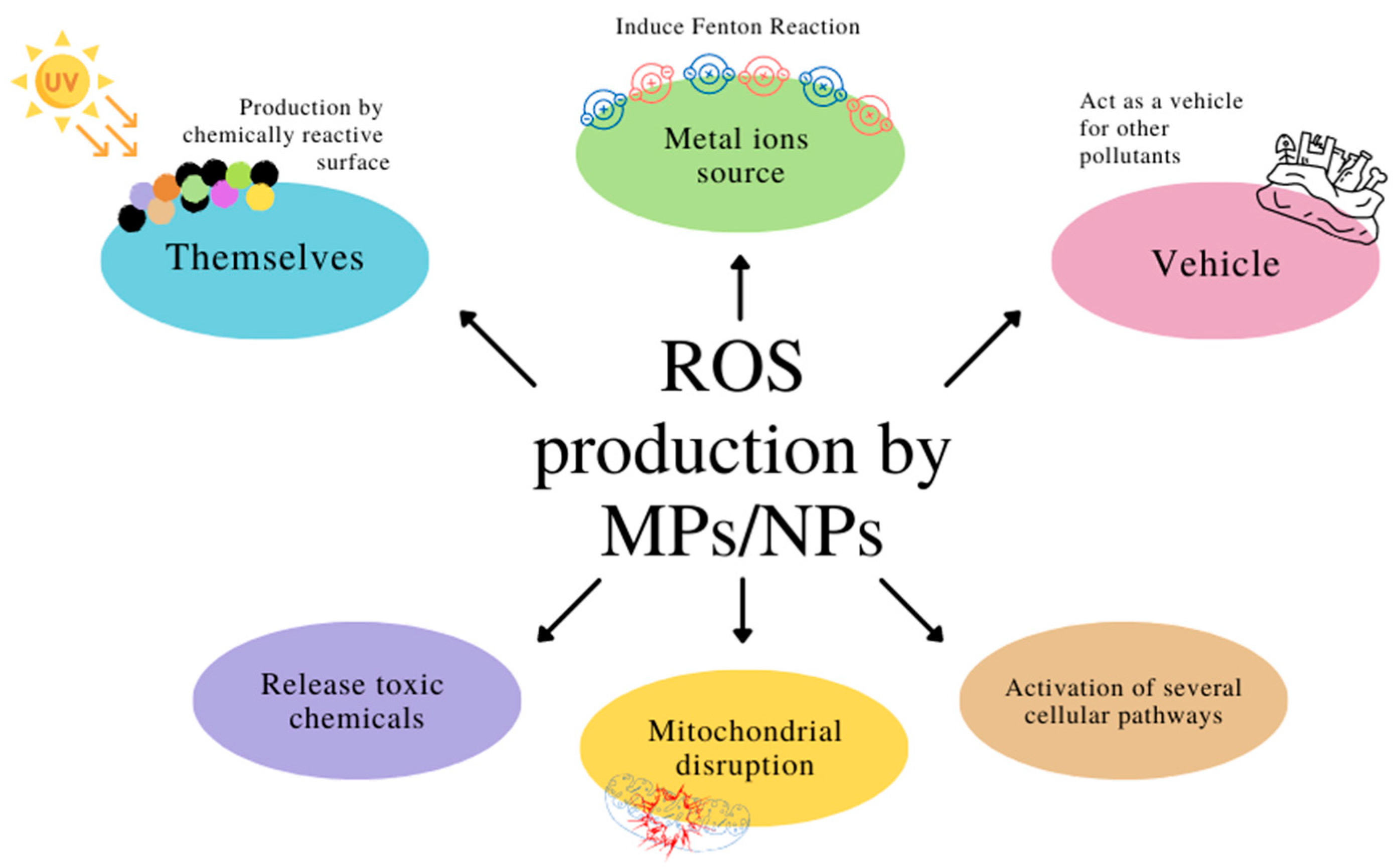
5. Reactive Oxygen Species and Micro- and Nanoplastics

A literature review based on the Adverse Outcome Pathways (AOP) approach revealed that MP/NPs share the formation of reactive oxygen species as an initial molecular event [87]. Micro- and nanoplastics can generate ROS through a variety of mechanisms (Figure 2). One mechanism is the production of ROS by the particles themselves. The surface of micro- and nanoplastics can be chemically reactive and catalyze the formation of ROS, such as superoxide anion (O2) and hydroxyl radical (•OH), through a process known as the Fenton reaction [88]. Fenton reaction is a chemical reaction that involves the reaction of hydrogen peroxide (H2O2) with a transition metal ion, such as iron or copper, to produce •OH and a hydroxyl anion (OH). Micro- and nanoplastics can act as a source of metal ions that can facilitate the Fenton reaction and produce ROS. Another mechanism through which they generate ROS is the adsorption of other pollutants onto the surface of the particles. MPs and NPs can act as a sink for other pollutants, including heavy metals, organic pollutants, and pesticides. The adsorption of these pollutants onto the surface of micro- and nanoplastics can lead to the formation of ROS through various chemical reactions [89]. For example, the adsorption of polycyclic aromatic hydrocarbons (PAHs) onto micro- and nanoplastics surfaces can lead to the formation of ROS through photochemical reactions under UV radiation [88,90].
Following the light irradiation of polystyrene microplastic suspensions, a significant correlation was observed between the production of organic EPFRs (Environmentally Persistent Free Radicals) and the production of ROS [91]. Micro- and nanoplastics can also release toxic chemicals such as phthalates, bisphenol A, and polycyclic aromatic hydrocarbons, which can induce ROS production [92]. It is possible that the effects induced by MPs and NPs, including ROS production, are size-dependent [54].
The generation of ROS in exposed organisms by micro- and nanoplastics involves several biomolecular mechanisms. One of the key mechanisms is the activation of NADPH oxidase (NOX), a complex enzyme responsible for the production of O2 [93]. Mice exposure to micro- and nanoplastics can activate NADPH oxidase through several pathways, including the activation of Toll-like receptor 4 (TLR4) and the aryl hydrocarbon receptor (AhR) [94]. Once activated, NADPH oxidase produces O2, which can be converted to other ROS, such as H2O2 and •OH. As reported by Fasciolo et al. [95,96], in rats, the increase in NOX activity, which increases ROS production, could be underlying several diseases in which ROS take part. This is one of the mechanisms by which MPs and NPs can affect human wellness. MPs and NPs themselves can affect redox homeostasis [87]. This is due to the ability of UV radiation and photo-oxidative and thermos-oxidative actions to alter their surface. [97,98]. Photo oxidation or UV light radiation can lead to the formation of free radicals on MP/NPs surfaces as primary products via the subtraction of a hydrogen atom from the macromolecular chain or the addition of a hydrogen atom to an unsaturated carbon chain group (crosslinking reaction) [99]. Once the free radicals are generated along the polymer chain, they can react with atmospheric oxygen and produce polymer peroxyl radicals with the further generation of secondary polymer alkyl radicals [87]. These weathering-induced extracellular free radicals could be one possible explanation for why a significant increase in ROS was observed after the cell entrance of the aged MPs [100].
Another biomolecular mechanism by which micro- and nanoplastics promote ROS production is of the disruption of mitochondrial function [17]. Exposure to micro- and nanoplastics can lead to mitochondrial dysfunction, which can result in the overproduction of ROS by the electron transport chain (ETC) [101]. The ETC is a series of protein complexes in the inner mitochondrial membrane that transfer electrons from electron donors to electron acceptors, generating a proton gradient that is used to produce ATP. ROS production by ETC is a normal physiological process that occurs as a byproduct of electron transfer. However, exposure to micro- and nanoplastics can disrupt the balance between ROS production and scavenging, leading to ROS overproduction. In addition, micro- and nanoplastics can also lead to the translocation of cytochrome c from mitochondria to the cytoplasm, which can trigger the activation of caspases and the induction of apoptosis [102,103]. The antioxidant defense system plays a critical role in protecting cells against oxidative stress induced by micro- and nanoplastics. Antioxidants scavenge ROS and prevent damage to cellular components, including lipids, proteins, and DNA. Exposure to micro- and nanoplastics can disrupt the antioxidant defense system by depleting antioxidants or impairing their function. For example, exposure to micro- and nanoplastics can lead to a decrease in the activity of antioxidant enzymes activity, such as superoxide dismutase (SOD), catalase (CAT), and glutathione peroxidase (GPX) [104]. Micro- and nanoplastics can also deplete the levels of non-enzymatic antioxidants, such as glutathione and ascorbic acid [105]. The depletion of antioxidants can cause ROS buildup and consequent oxidative damage to cellular components.

6. The Impact of Micro- and Nanoplastics on the Aquatic Environment

Pollution by human activities (agriculture, industrialization and urbanization) can take many forms, including air, water and soil pollution, electromagnetic pollution (radiation waste), and even noise pollution. These may contribute to health issues and decline the quality of life [106]. Water pollution by agricultural, municipal, and industrial sources have become a major concern for the well-being of humanity and biodiversity [107]. Anthropogenic activities on the aquatic ecosystem have direct and indirect effects that negatively affect biodiversity both in freshwater and sea [108,109,110]. Among the main threats to aquatic ecosystems and biodiversity is climate change. Climate change can impact marine and freshwater ecosystems in several ways: ocean warming; sea level rise; the loss of sea ice; a decrease in pH due to increased ocean surface acidity; an increased risk of diseases in marine biota; the loss of habitats such as Coral Reef; pollution; nutrient enrichment; hydrological modifications; the spread of invasive species; and increasing levels of UV light [110]. In addition to climate change, the production of plastic and its release into the environment is contributing to the loss of biodiversity and is becoming a serious threat to animal redox homeostasis (Table 1 and Table 2) and health (Table 3) and, subsequently, for human health [5].

6.1. MPs/NPs’ Effects on Phytoplankton

Normally, micro- and nanoplastics, due to their small size and low density compared to water, tend to float in the water column. When microplastics enter the aquatic environment, they are rapidly colonized by microorganisms and algae, typically within hours [111]. It has been highlighted that a microbial biofilm instantly colonizes plastics when exposed to the environment. It is estimated that between 1000 and 15,000 metric tons of microbial biomass are harbored on plastic marine debris [112]. However, the biomass accumulation due to biofouling can increase its density and cause it to sink [113]. Microalgae also can adhere to particles such as MPs/NPs. Many microalgae secrete polysaccharides if stressed due to decreased nutrients and light. Exopolysaccharides may coagulate to form sticky particles named transparent exopolymer particles (TEPs). TEP favors microalgae aggregation; these aggregates are the primary vehicle for transporting phytoplankton and debris towards the seabed [114]. Another sedimentation pathway consists of small MPs ingested by zooplankton and deposited within fecal pellets [115].
MPs and NPs show effects on each trophic level. Microalgae are the first level in the food chain and are indispensable for the marine ecosystem equilibrium. MPs seem to affect the well-being of microalgae. As has been reported, different sizes and concentrations of MPs and NPs can inhibit growth, reduce chlorophyll and photosynthesis, induce oxidative stress, cause changes in morphology, and promote the production of heteroaggregates [116,117].
Janak Raj Khatiwada (2023), evaluated the effect of PET microplastic (25 mgL−1, 50 mgL−1, 100 mgL−1 and 200 mgL−1) of Scenedesmus sp. for 24 days. PET showed the highest growth inhibition effect at 200 mg/L. Moreover, compared to the control group, microalgae exposed to microplastics showed a lower chlorophyll content, possibly due to blocking the pores of MPs for cellular respiration [118]. Cai Zhang (2017) carried out tests on Skeletonema costatum, demonstrating that microplastic (Micro-PVC, average diameter 1 μm) significantly inhibited the growth of microalgae and chlorophyll at 96 h under 50 mg/L treatment [119]. Microplastics may also affect microalgae lipid and fatty acid composition. Guschina et al. (2020) assessed the exposure of Chlorella sorokiniana to polystyrene microplastics (<70 μm, 60 mgL−1) for four weeks. The study showed that microplastic could alter the concentrations of essential fatty acids, which are necessary for algae’s lipid synthesis. Such changes could also have repercussions for food quality, growth, and stress resistance in primary consumers, and could affect potential propagation through trophic transfer [120]. On the other hand, microalgae can also produce effects on microplastics. Among these, it is possible to enumerate the different alterations of MP properties; in particular, their adsorption seems to be enhanced [111]. Wang et al., (2020) showed that the development of a biofilm altered the adsorption of copper Cu(II) and tetracycline (TC) by PE microplastic [121]. The biofilm can also camouflage the plastic particles and promote their ingestion by grazers, such as zooplanktonic organisms [111].

6.2. MPs/NPs in Sediments and Benthos

When micro- and nano plastics lose their ability to float, they can fall and settle on the sea bottom. This severe issue can be observed primarily in coastal shallow-water regions. However, these pollutants are not limited to the coastline; they have also been identified in deep-sea sediments with concentrations of up to 2000 particles m−2 at a depth of 5000 m [113,122]. Finally, these small-sized materials will find their pathway to the benthos, affecting some marine organisms. Several studies in the laboratory have investigated the ingestion of micro- and nanoplastics by benthic organisms.
Cole et al. (2020) have compared the toxicity of microplastics and nanoplastics on Mytilus edulis for 24 h or 7 days. Mussels were exposed to 500 ng mL−1 of 20 μm polystyrene microplastics, 10 × 30 μm polyamide microfibers, or 50 nm polystyrene nanoplastics. After 24 hours of exposure, there was a rise in SOD activity, but after 7 days, it returned to normal levels without negatively impacting health. Particle size, however, can influence sub-lethal toxicity because exposure to nanoscopic plastic raises the proportion of phagocytic hemocytes (indicating a heightened immune response) and leads to a significant increase in micronuclei formation [123]. The mussels are very important in these studies because they represent the prey of many intertidal species and serve as a food source for humans [124].
Sussarelli et al. (2016) assessed the impact of the polystyrene microspheres (micro-PS) on the physiology of the Pacific oyster (Crassostrea gigas). The organisms were exposed to virgin micro-PS (2 and 6 µm in diameter; 0.023 mgL−1) for 2 months during a reproductive cycle. Ingestion preference was shown for the 6 µm particles over 2 µm particles. After 2 months, the histological analysis only detected micro-PS particles in the stomach and intestine. Hyalinocytes and granulocytes in exposed oysters were larger than in controls. The total number of oocytes and the oocyte diameter were significantly lower in exposed females than in controls, while exposed males had a considerably lower sperm velocity. Finally, in progeny issued from exposed genitors than in progeny issued from control genitors, the larval growth and size were significantly slower [125].
Murray and Cowie (2011) assessed the capability of the Nephrops norvegicus, an ecologically and commercially important crustacean, to consume plastics. They evaluated the stomach microplastic content of shrimp through trawls in the north Clyde Sea area. Four tanks were set up, and the organisms were fed fish seeded with approximately 5 mm strands of blue polypropylene rope (ten filaments for one cm3 of fish). Fish without plastic seeds were provided to the animals in the control group. Nephrops were killed after 24 h, and new animals were placed in each tank. This experiment was repeated five times over a 2-week period. Nephrops with no plastic in their stomachs had a larger median carapace length than those with plastic. This is presumably due to the capacity of the larger animals to sort plastics from food or excreting plastic once ingested. This study demonstrates that N. norvegicus can passively accumulate plastic in the stomach via during feeding. There could be potential implications for human health because the Nephrops is a commercially fished species [126].

6.3. Trophic Levels: From Zooplankton to Main Fishes Consumed by Humans

Considering the enormous variety of fish and the environments in which they live [127], humans make use of fish in many ecosystem services [128,129]. Precisely in this regard, it must be considered that human activities are impacting fish biology and their redox homeostasis, on whose alteration micro- and nanoplastics play an increasingly decisive role.

6.3.1. MPs/NPs’ Effects on Zooplankton

Zooplankton is the second food chain level and represents a key trophic link in pelagic food webs. The role of zooplankton is vital in the aquatic environment, as they are primary consumers and include the juvenile stages of numerous commercially important species (e.g., the meroplankton) [130]. Cole et al. (2013) reported that the ingestion of polystyrene beads, already starts with the zooplankton, namely the copepod Centropages typicus, showing the relevance of the MPs in significantly reducing the consumption of algae. This study implies that marine microplastic debris can negatively impact health and zooplankton function [130]. Ziajahromi et al. (2017) investigated the effects of microplastics on the freshwater zooplankton Ceriodaphnia dubia. The acute (48 h) and chronic (8 d) effects of microplastic polyester fibers and 1–4 μm PE beads on zooplankton have been examined. This study demonstrated the microplastic fibers caused a 50% reduction in reproductive capacity at concentrations approximately six times higher than environmental concentrations. Unlike other studies, this study did not observe any ingested fibers. However, malformations have been observed in the carapace of organisms exposed to polyester fibers. However, malformations have been observed in the carapace of organisms exposed to polyester fibers. This demonstrates that the negative effects of microplastic fibers on exposed aquatic organisms can also include external physical damage and not only those resulting from ingestion [131]. Rehse et al. (2016) investigated the effects of 96 h exposure of PE-particles (size 1 μm) on Daphnia magna. The study showed that the particles are ingested and cause immobilization. The immobilization of daphnids increases with dose and time. EC50 was 57.43 mg L−1 [132].
Another species utilized to study the effects of MPs/NPs is the marine copepod Calanus helgolandicus. This organism is a keystone species that can constitute up to 90% of mesozooplankton biomass within marine waters throughout Europe and the northeast Atlantic. Calanus helgolandicus has a large size and high lipid content; its abundance makes it a vital prey species for the larvae of a number of commercially important fish. Cole et al. (2015) exposed Calanus helgolandicus to 20 μm polystyrene spheres and cultured algae for 24 h to evaluate ingestion rate and for 9 days to evaluate reproductive function. The results demonstrate that microplastics can alter the ingestion of algae and reduce the size of the eggs, probably due to a reduction in the ingested carbon biomass. Moreover, reducing ingested carbon could cause an energy depletion and the consequent death of the organism [133].
The effects of microplastics are not always so evident, as in the case of exposing the MPs to Pacific oyster larvae (Crassostrea gigas). The exposure has not shown any significant effects on the development or feeding capacity of the larvae. This could be because of the oyster’s more simplistic intestinal tract [134].
Zheng et al. (2020) suggested an interesting point of view. MPs ingested by copepods, the crustacean zooplankton that is the main prey of fish, are likely to be carried up the food chain, while those ingested by jellyfish, which have fewer predators than copepods, are more likely to be discharged into the marine environment and circulate in seawater or seabed sediments. In this regard, the distribution of MPs, in addition to depending on the chemical-physical characteristics already discussed, also depends on the different groups of zooplankton [135].

6.3.2. MPs/NPs’ Effects on Others Aquatic Trophic Levels

The effects of these pollutants on fish vary depending on the dose, the target organisms, and the interactions between the pollutants [136,137]. Nanoplastics have been reported to have a higher impact than microplastics [50,138]. However, this is not always true. As reported by Jiang et al. (2020), there are conditions in which microplastics can have a greater impact than nanoplastics [45]. These differences can be attributed to multiple factors, including the capacity of ingestion, the possibility of entering tissues and cells, and the type and shape of the polymer that can affect the particles’ interaction with other pollutants and chemical compounds [139,140,141].
Microplastics may not always have “negative” effects. As already stated, based on the shape and size of the polymer, microplastics can interact with other chemical pollutants. This capacity, which also depends on the dose, could mitigate the effect of chemical pollutants (antagonistic effect). If, on the one hand, MPs can reduce pollutants’ effects, on the other, they can act synergistically by enhancing its effects (agonist effect), which in the latter case is potentially more toxic than nanoplastics [142,143,144]. Environmental factors can also dictate the dose-dependent impacts of microplastics. For example, phenomena such as storms and cyclones can increase their concentration and thus induce an increment in dose in specific areas, leading to an increase in the absorption by aquatic organisms [50,145].
Neves et al. (2015) reported the presence of microplastics in 63.5% of benthic fish and 36.5% of pelagic fish species, with a total of 73 microplastics identified from fish stomach contents [146]. A study on rainbowfish (Melanotaenia fluviatilis) exposed to microbeads adsorbed with polybrominated diphenyl ethers (PBDEs) was monitored for 0, 21, 42, and 63 days. Exposed fish accumulated high concentrations of PBDEs (ca.115 pg g−1 ww per day) in tissue after ingestion [147]. Redondo-Hasselerharm et al. (2018) report that in Gammarus pulex exposed to sediment containing MPs (Micro-PS 20–500 μm for 28 d), the growth is reduced [148], as well as in Gammarus fossarum [149]. A study conducted on juvenile European perch (Perca fluviatilis) exposed to polystyrene microplastic particles (90–150 μm) over six months showed that the animals ingested and accumulated the polystyrene microplastics which resulted in reduced growth; delayed hatching; and impaired performance, nutrition, and behavior [150].
MPs and NPs also influence the olfactory senses, which increases susceptibility to being killed by predators [151,152]. The predator–prey relationship gradually increases the toxicity of these substances as they are transferred from organism to organism, accumulating [62,64,109,153]. About 18% of the main predators of the central Mediterranean, Swordfish (Xiphias gladius), bluefin tuna (Thunnus thymus), and albacore (Thunnus alalunga) ingested micro-, meso-, and macroplastic debris ranging in size from <5 mm to 5–25 mm to 25 mm, respectively, [154,155] found microplastics in 36.5% of the gastrointestinal tracts of pelagic and demersal fish. Tuna is the main consumed fish; in fact, much attention is being paid to the sustainability of its fishing by FAO and the UN [156]. Dias-Basantes et al. (2022) showed that canned tuna can provide an average of 692 ± 120 MPs/100 g in brine-soaked tuna and 442 ± 84 MPs/100 g in oil-soaked tuna, values that significantly exceed those reported in research on canned fish [157]. Di Giacinto et al. (2023) showed the presence of MPs (size < 10 μm), polymers (PET, polycarbonate (PC)), and additives (Bisphenol A (BPA) and p-phthalic acid (PTA)) in the muscle Thunnus Thynnus and Xiphias Gladius fished in the Mediterranean Sea [158]. Abihssira-García et al. (2020) report that MPs were also found in Atlantic Salmon (Salmo salar), the second main fish consumed by humans. They show that Atlantic salmon immune cells from blood, distal intestine, and head kidney can phagocytose MPs (1–5 μm) even at relatively low concentrations (low 0.05 mgL−1; medium 5 mgL−1; high 50 mgL−1 for 1, 24, 48 and 72 h) and that their mortality is affected by the time exposure and the microplastic type [159].
Another aspect concerning the trophic levels that should be considered is that the ingestion and accumulation of microplastics take place before maturity, impacting the number of available organisms. A study conducted on Artemia nauplii (the early stage of development that occurs after the cysts hatch), which were subjected to a high concentrations of microplastics (1.2 × 10⁶ particles per 20,000 nauplii), showed that they had ingested and accumulated microplastic particles ranging in size from 1 to 20 μm, in high concentrations; these particles were subsequently transferred to the zebrafish that fed on the nauplii [160]. Although not all particles would have been transferred from the nauplii to the zebra, as they were partially excreted, some were retained within the epithelial cells and intestinal villi. Another study on the absorption and effect of microplastics on zebrafish showed that most of the plastic particles (5 μm in diameter) had accumulated in the gills, intestines, and liver. Those with a larger diameter (20 μm) could accumulate only in the intestine and gills. Thus, the smaller particles (5 μm) are more toxic than the bigger ones (20 μm), causing inflammation and the accumulation of lipids in the fish liver, inducing oxidative stress, and altering the metabolic profiles of the fish liver, disturbing lipid and energy metabolism [161]. Accordingly, Jeong et al. (2016) showed that exposure to plastic beads (0.05, 0.5, 6 μm) in Brachionus koreanus was associated with increased oxidative stress and decreased growth rate, fecundity, lifespan, reproduction time, and body size, suggesting that the toxicities of MPs and NPs are size-dependent and that smaller plastics are more toxic than bigger ones [162]. This size dependency is probably correlated with the ability of smaller molecules to enter cellular compartments. Manabe et al. (2011) reported that smaller NPs were more easily ingested by medaka (Oryzias latipes) than the bigger ones and that NPs were excreted more slowly than the microplastics [163]. NPs negatively affect the liver health of medaka fish, leading to significant alterations in digestion, innate immune, and antioxidant-enzyme-based liver tissue damage [164] and inflammation [165]. However, microplastics are capable of damaging cellular organelles. In line with that, Felix et al. (2023) find that MPs in zebrafish can affect the center of the energy balance: mitochondria. After 21 days of exposure to a toxicological concentration of MPs, anxiety-like behavior arose, while 1 mg L−1 treatment showed a decrease in hepatic mitochondrial respiration and membrane potential (ΔΨ), both indices of suppression of mitochondrial respiratory chain [166]. Trevisan et al. (2019) also reported that NPs can cause mitochondrial energy disruption, a decline in energy efficiency, and differential mitochondrial uptake in developing zebrafish [167]. Mitochondria can also be a target for PAHs toxicity as its high lipid content facilitates PAH uptake [168]. Several studies reported that PAHs, such as benzo[a]pyrene (BaP), phenanthrene, and fluoranthene [169] or complex PAHs mixtures, can impair mitochondrial bioenergetics in embryonic or larval fish [170,171]. Gu et al. (2023) found that in sea cucumber (Apostichopus japonicus), exposure to polystyrene nanoplastics (PS-NPs) significantly inhibited the complex activities in the mitochondrial respiratory chain and affected the relative expression levels of mitochondrial apoptosis-related genes [172].

6.4. MPs/NPs’ Effects on the Redox Homeostasis of the Aquatic Organisms

A previously mentioned, MPs and NPs are able to affect the redox homeostasis. However, the effects reported in literature sometimes are conflicting, even in the same species. Probably, the differences found are related to age [173].

6.4.1. NPs

In zebrafish embryos, at 96 h post-fertilization (hpf), while exposed 50 nm (1 mgL−1) to PS-NPs, Bhagat et al. (2022) found that ROS production was increased without a significant change in the malondialdehyde (MDA) concentration. Instead, the antioxidant system was affected with decreased SOD and GR activity, increased CAT activity, and reduced glutathione content without significant change in GPX activity [174]. In 96 hpf zebrafish embryos exposed for 24 h to 50 nm and 1 µm PS-NPs (which are considered NPs by the authors) (10 mg/L), an increase in ROS content with both sizes was found [175].
In adult zebrafish exposed to 70 nm PS-NPs (0.5 ppm and 1.5 ppm) for 7 days, a significant increase in muscle (but not liver) ROS content was found only at the highest concentration [176]. According to these results, in adult zebrafish (F0) exposed to 42 nm PS-NPs added to the diet (10% of the food by mass) for one week, no difference in either genders was found in the liver GR, GPX, and CAT antioxidant enzymes [177]. Instead, a significant decrease was found in the GR activity of male muscle. In F1 96 hpf larvae from exposed maternal and co-parental groups displayed significantly lower total thiol levels (reduced forms of protein and non-protein thiols). GR activity was significantly reduced in these larvae, while the activities of GPX and CAT were not changed [177]. In another study, Aliakbarzadeh et al. (2023) exposed adult zebrafish for 45 days to PS-NPs of different sizes, 20–80 nm, at different concentrations (0.1 µgL−1, 1 µgL−1, 10 µgL−1, and 100 µgL−1). They found an NP-concentration-dependent decrease in CAT activity and total GSH content [178]. In another study conducted on the adult zebrafish exposed to PS-NPs (103–113 nm) for 14 days at 10 µgL−1 and 100 µgL−1, a reduction in gills’ antioxidant enzymes activity (SOD and total antioxidant capacity (TAC)) was found [179]. Although it may appear that in adult zebrafish, exposure to the different concentrations of PS-NPs reduces antioxidant capacity by promoting oxidative stress, in another study, it was shown that after 3 weeks of exposure to 70 nm PS-NPs at different concentrations (20 µg/L, 200 µg/L, and 2 mg/L), there is an increase in the activity of CAT and SOD only at the higher concentration (2 mg/L) [180].
Table 1. Effects of PS-NPs on redox homeostasis of zebrafish. ROS: reactive oxygen species; CAT: catalase; SOD: superoxide dismutase; GR: glutathione reductase; NS: No significative difference compared to control; t-GSH: total glutathione content; TAC: total antioxidant capacity; hpf: hours post-fertilization (↑ increase; ↓ decrease).
Redox Homeostasis in Danio Rerio Exposed to PS-NPs
AuthorAgeExpositionTissueResults
SizeConcentrationTime
Bhagat et al. [174]96 hpf50 nm1 mgL−1~2–96 hpf/↑ROS
↑CAT
↓SOD
↓GR
Sendra et al. [175] 96 hpf50 nm, 1 µm10 mgL−124 h/↑ROS
Pitt et al. [177]96 hpf42 nm10% add. to diet1 weekCo-parental↓Thiols
↓GR
Sarasamma et al. [176]Adult70 nm0.5 ppm, 1.5 ppm1 weekMuscle
Liver
↑ROS
NS
Pitt et al. [177]Adult42 nm10% add. to diet1 weekMale muscle↓GR
Aliakbarzadeh et al. [178]Adult20–80 nm0.1 µgL−1, 1 gL−1, 10 gL−1, 100 gL−145 daysGut↓CAT
↓t-GSH
Umamaheswari et al. [179]Adult103–113 nm5 mgL−1, 10 mgL−114 daysGills↓SOD (5 mgL−1)
↓TAC (10 mgL−1)
Lu et al. [180]Adult70 nm20 µgL−1, 200 µgL−1, 2 mgL−13 weeksLiver↑SOD (2 mgL−1)
↑CAT (2 mgL−1)

6.4.2. MPs

In 96 hpf zebrafish embryos exposed to PS-MPs 1–5 µm (2 mgL−1), the redox homeostasis results unaffected [181]. The lack of effects of PS-MPs on the redox homeostasis of zebrafish embryos was also highlighted in another work, in which, following seven days of exposure of 6dpf (days post fertilization) zebrafish to 5 μm PS-MPs (50 ngmL−1), no changes were found in the concentration of MDA and the activity of SOD, CAT and GPX [182]. Similar results were found in 30dpf zebrafish exposed to 5 μm PS-MPs (500 μgL−1) for 25 days [183]. However, while in 96hpf zebrafish, the activity of antioxidant enzymes was not affected by MPs exposition, in 30 dpf zebrafish, CAT activity was decreased [183], reinforcing the hypothesis that the MP-induced impairment of redox homeostasis is age-related. On the other hand, Guimarães et al. (2021) found that in juvenile zebrafish exposed to two different concentrations of PS-MPs (4 × 10⁴ and 4 × 10⁶ particles/m3) for five days, an increase in thiobarbituric acid reactive species (TBARS) following both treatments. However, they found no increase in the hydrogen peroxide concentration but verified an increase in the SOD and CAT activity enzyme and in the total glutathione concentration, without difference in the reduced glutathione concentration [173].
CAT and SOD were also increased in the gut tissue of adult zebrafish exposed to PS-MPs (5 µm) for 21 days at two different concentrations (50 µgL−1 and 500 µgL−1) [184]. Another work conducted on adult zebrafish exposed to PS-MPs 9–10 µm (10 mg/L) for 4 or 8 days reports an interesting view of the effect exerted by MPs on the liver [185]. They found that after four days of exposition to MPs, the activity of the SOD, CAT, and GPX enzymes was increased, while MDA concentration was unaffected. After eight days of exposition, SOD activity returned to the control level, while CAT and GPX activity remained higher, and MDA content decreased compared with the control. Accordingly, Lu et al. (2016) found that after three weeks of exposure of adult zebrafish to PS-MPs (5 µm) at different concentrations (20 μgL−1, 200 μgL−1, and 2 mgL−1), SOD activity increased at all concentrations, while CAT activity increased with 200 μgL−1 and 2 mgL−1 treatment [180].
Table 2. Effects of PS-NPs on redox homeostasis of zebrafish. CAT: catalase; NS: no significative difference compared to control; TBARS: thiobarbituric acid reactive substances; SOD: superoxide dismutase; t-GSH: total glutathione content; GPX: glutathione peroxidase; MDA: malondialdehyde; hpf: hours post-fertilization; dpf: days post-fertilization (↑ increase; ↓ decrease).
Redox Homeostasis in Danio Rerio Exposed to PS-MPs
AuthorAgeExpositionTissueResults
SizeConcentrationTime
Santos et al. [181]96 hpf1,5 µm2 mgL−1//NS
Yang et al. [182]6 dpf5 μm50 ngmL−11 week/NS
Chen et al. [183]30 dpf5 μm500 μgL−125 days/↓CAT
Guimarães et al. [173]Juvenile~17 µm4 × 104, 4 × 10⁶ particles/m35 days/↑TBARS
↑CAT
↑SOD
↑t-GSH
Qiao et al. [184]Adult5 µm50 μgL−1, 500 μgL−121 daysGut↑CAT
↑SOD
Li et al. [185]Adult9, 10 µm10 mgL−14 days
8 days
Liver4 d
↑SOD
↑CAT
↑GPX
NS MDA
8 d
NS SOD
↑CAT
↑GPX
↓MDA
Lu et al. [180]Adult5 µm20 μgL−1, 200 μgL−1, 2 mgL−13 weeksLiver↑SOD
↑CAT (200 μgL−1, 2 mgL−1)
Table 3. PET: polyethylene terephthalate; PVC: polyvinyl chloride; PS: polystyrene; PA: polyamide; PP: polypropylene; PE: polyethylene PHB: polyhydroxybutyrate; PMMA: poly(methyl methacrylate); PLA: poly(l-lactide).
AuthorsSpeciesType of MPs/NPsExposition TimeEffects
Khatiwada et al. [118]Scenedesmus sp.Micro-PET24 daysNegative influence on the growth of microalgae and eventually reducing chlorophyll.
Zhang et al. [119]Skeletonema costatumMicro-PVC96 hGrowth inhibition and chlorophyll.
Guschina et al. [120]Chlorella sorokinianaMicro-PS <70 μm4 weeks Alteration of the concentrations of fatty acid molecules and lipid synthesis.
Cole et al. [123]Mytilus edulisMicro-PS 20 μm/Microfibers-PA 10 × 30 μm/Nano-PS 50 nm24 h
7 days
Higher SOD activity after 24 h exposure. SOD activity returned to normal values after 7 days. Nanoplastics increased the proportion of phagocytic hemocytes and resulted in a marked increase in micronuclei formation.
Sussarellu et al. [125]Crassostrea gigasMicro-PS 2–6 µm 2 monthsExposed oysters, hyalinocytes, and granulocytes > control. Exposed oocytes and oocyte diameter < control. Exposed sperm velocity < control. Exposed Progeny size and growth < control.
Murray and Cowie et al. [126]Nephrops norvegicusPP24 h each tank five times
2 weeks total
Plastic in stomachs; potential implications for human health.
Cole et al. [130]Centropages typicusMicro-PS 7.3 μm 24 hDecreased algal feeding and negative impact upon health and zooplankton function.
Ziajahromi et al. [131]Ceriodaphnia dubiaMicro-PS/PE 1–4 μm48 h
8 days
50% reduction in reproductive and malformations in the carapace.
Rehse et al. [132]Daphnia magnaMicro-PE 1 μm96 hImmobilization and EC50 of 1-μm was 57.43 mgL−1 after 96 h.
Cole et al. [133]Calanus helgolandicusMicro-PS 20 μm24 h
9 days
Decrease in microalgae ingestion and reduction in egg size.
Redondo-Hasselerharm et al. [148]Gammarus PulexMicro-PS 20–500 μm28 daysGrowth decreased.
Straub et al. [149]Gammarus FossarumMicro-PHB PMMA 32–250 μm 28 daysGrowth decreased.
Kardgar et al. [150]Perca fluviatilisPLA 90–150 μmOver 6 monthsReduced growth; hindered hatching; and impaired their performance, nutrition, and behavior.
Abihssira-García et al. [159]Salmo salarMicro PS/PE 1–5 μm1, 24, 48, and 72 hImmune cells can phagocytose MPs and the mortality of the blood cells, distal intestine, and head kidney is affected by the time exposure, and the impact is dependent on the microplastic type.
Lu et al. [161]Danio RerioMicro-PS 5 μm–70 nm1 weekIncreased inflammation, oxidative stress, altered metabolic liver profile.
Jeong et al. [162]Brachionus koreanusMicro-PS 0.05–0.5–6 μm12 daysIncreased oxidative stress and decreased growth rate, fecundity, lifespan, reproduction time, and body size.
Zhou et al. [164]Oryzias latipesNano-PS 100 nm3 monthsLiver health impaired.
Felix et al. [166]Danio RerioMicro-PP 200 μm21 daysDecreased mitochondrial health, anxiety-like behavior, and increased oxidative stress.
Trevisan et al. [167]Danio RerioNano-PS 44 nm 24–48–96 h post-fertilizationMitochondrial energy disruption, a decline in energy efficiency, and differential mitochondrial uptake.
Gu et al. [172]Apostichopus japonicusNano-PS 100–200 nm20 daysInhibits the complex activities in the mitochondrial respiratory chain and affect the relative expression levels of mitochondrial apoptosis-related genes.
Li et al. [186]Chlamydomonas reinhardtiiMicro-PS10 daysMicro-PS had negative effects on growth and algal photosynthesis.
Besseling et al. [187]Arenicola marinaMicro-PS 400–1300 μm28 daysAs plastic is ingested by A. marina, its predators will be exposed to plastic as well. Weight loss.
Wang X et al. [188]Mytilus coruscusMicro-PS 2 μm14 days plus 7-day recovery acclimationInhibition of digestive enzymes.

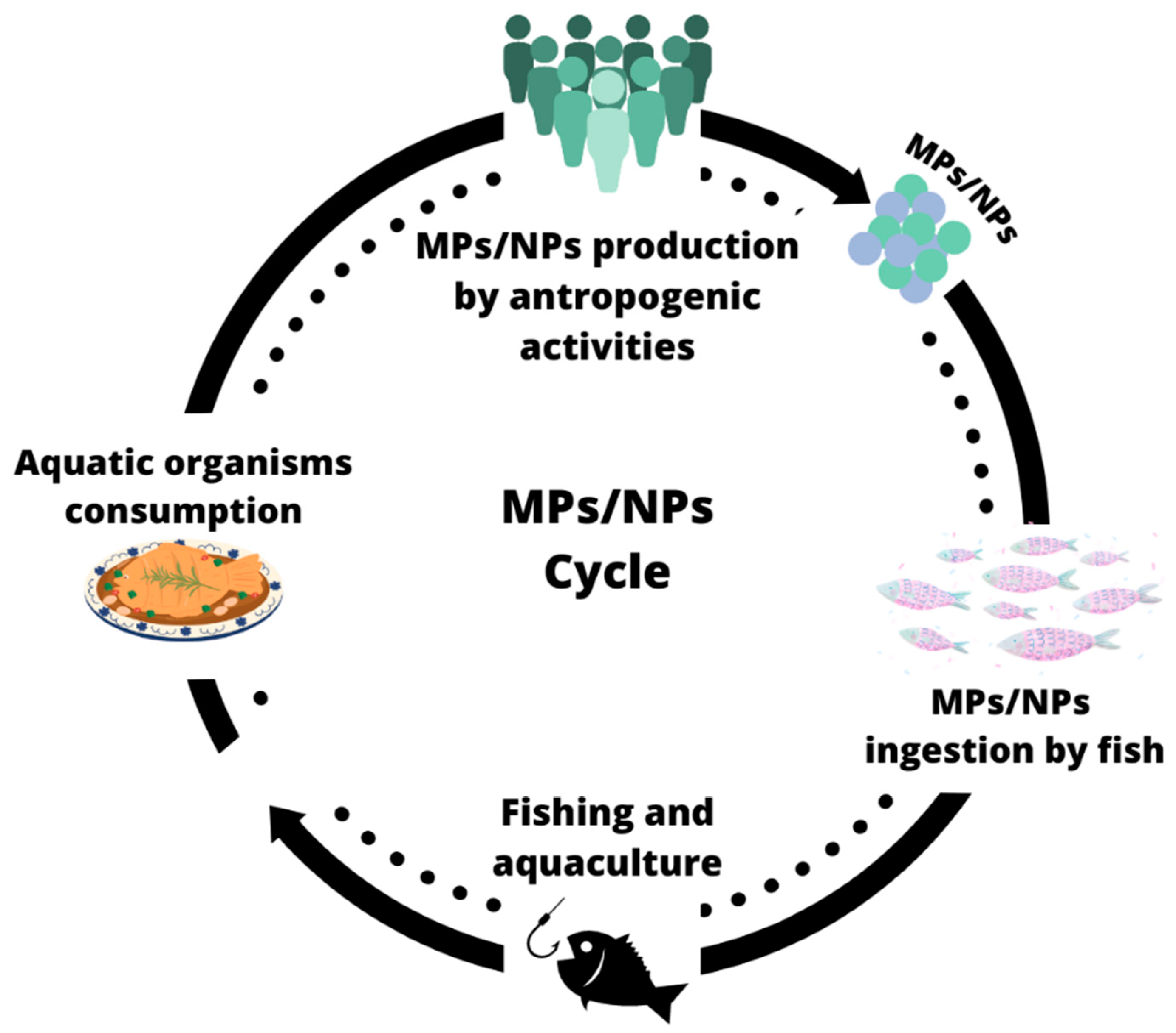
7. Ingestion of MPs/NPs from Aquatic Food by Human

Total aquaculture and fisheries production reached a record value of 214 million tonnes in 2020, 178 million tonnes of which were accounted for by aquatic animals and 36 million tonnes by algae. These activities generated about USD 424 billion in revenue. Aquatic food furnishes about 17% of animal proteins globally; this figure rises to over 50% in several countries, mainly from Asia and Africa. Primary production alone employs 58.5 million people, 21% of whom are women. In 2019, global aquatic food available for human consumption was estimated at 158 million tonnes [189]. Asia accounted for 72% of the total, while its population represented 60% of the world population. As aquatic food consumption expands worldwide, humans are inevitably exposed to microplastics at some level. Van Cawenberghe and Janssen (2014), estimated that in Europe, mollusks consumers ingest up to 11,000 microplastics per year [190]. In the case of the annual consumption of shrimps, the amount is 175 microplastic particles per person per year, assuming a scenario in which 90% of microplastics are removed [191]. Similarly, the consumption of sea urchins Paracentrotus lividus, assuming an average consumption by humans of approximately 1 kg of sea urchin gonads, could deliver 166–207 items/per year/per person [42]. The evidence of genetic alterations in mollusks associated with the presence of micro- and nanoplastics in the food web indicates that potential genetic alterations could also occur in humans due to the consumption of seafood contaminated with plastic particles [192,193]. The problem increases where it is customary to consume raw or dried fish, since the transfer of microplastics is greater, as in the transfer of MPs accumulated in the intestine and the lymphatic system [194]. The transport mechanism of MPs and NPs across physiological barriers is influenced by the para-physiological condition. In fact, it has been observed that there is an increased intestinal permeability in patients with inflammatory diseases of the colon related to an increased transport of MPs [194]. High concentrations of MPs/NPs are usually highly cytotoxic, usually via ROS production and pro-inflammatory processes. MPs/NPs’ toxicity is assessed by the response of intestinal lipid peroxidation biomarkers as an indication of the feedback leading to oxidative stress, inflammation, epithelial barrier integrity, and changes in the intestinal microbiota [195,196]. In the immortalized cell line of human colorectal adenocarcinoma cells (Caco-2), it has been shown that PS-MPs (0.1 µm and 5 µm, 1–200 µg/mL) can disrupt the mitochondrial membrane potential and inhibit ATP-binding cassette (ABC) transporter activity [197]. In addition to local damage in the intestine, it has been reported, in mammalian systems models, that microplastics that have specific characteristics (resistance to chemical degradation, resistance to mechanical clearance, and biopersistence) can translocate across living cells, such as M cells or dendritic cells, to the lymphatic and/or circulatory system, accumulate in secondary organs, and impact the immune system and cell health [198]. Buyan (2022) also reports that MPs can be transferred in distal tissues via the circulatory system, causing inflammation reactions, cell blood cytotoxicity, vascular swelling, obstruction, and respiratory high blood pressure [199]. The close link between MPs and NPs, mitochondria and ROS has also been demonstrated in human lung epithelial BEAS-2B cells, on which they have a cytotoxic and inflammatory effect related to an increase in reactive oxygen species [200]. Another study has demonstrated that the accumulation of NPs in lysosomes played a central role in the observed cell death, leading to the swelling of the lysosomes and the release of cathepsins into the cytosol, which ultimately propagated the damage to the mitochondria with the subsequent activation of apoptosis. This process was accompanied and sustained by other events, such as increasing ROS levels and autophagy [201]. These effects are due to the ability of internalized NPs to impair the function of mitochondria and induce the monomerization of BAX on the mitochondrial surface, leading to the influx of calcium and the outflux of mitochondrial cytochrome c, which would result in a reduction in the membrane potential, insufficient ATP production, and the activation of downstream caspase-9 and caspase-3, eventually initiating the mitochondrial apoptotic pathway [202].
In summary, the micro- and nanoplastics released into the environment, through the consumption of food return to humans (Figure 3), and once internalized, MPs and NPs can act in multiple districts and levels, representing a key factor in human health. In fact, Wick et al. (2010) report that nanoparticles can pass the placental barrier in a perfusion model of the human placenta [203], while Lehner et al. (2019) report that NPs can permeate the blood–brain barrier [204], making it critical and urgent to deepen studies. Following ingestion, MPs/NPs access the bloodstream and, following the translocation process, reach different organs and tissues, triggering toxicity pathways, thereby including inflammation and oxidative stress.

8. Future Studies and Perspectives

The production and accumulation of plastic debris in aquatic environments has become a major concern, with microplastics and nanoplastics posing a significant risk to the ecosystem. Studies on the ecotoxicities and effects of microplastics and nanoplastics on aquatic organisms have increased since the 1990s, and many questions about their fate and invisible effects remain incomplete. The association of metal ions with plastic particles in aquatic systems alters their potential bioavailability and may trigger toxic effects. It is crucial to conduct more realistic experiments to better assess the risk of micro- and nanoplastics in aquatic environments and to establish new standardized methods to isolate, identify, and quantify nanoplastics. The trophic transfer of micro- and nanoplastics in complex food chains, particularly in brackish water environments, must be further investigated. It is also essential to thoroughly assess the toxicity of biodegradable plastics, such as polyhydroxybutyrate (PHB), to ensure the necessary health and safety assessment before substituting them for non-biodegradable plastics [205,206]. Future studies on micro- and nanoplastics should focus on urgent topics, such as the interactions between the surfaces of micro- and nanoplastics and the environment, trophic transfer, generational effects, and the long-term effects of these small plastics on aquatic organisms. To achieve this goal, it would be helpful to use plastic particles sampled in natural environments, characterize their composition, pollutants, and additive content, and analyze the effects of plastic particle contamination at the ecosystem level. As indicated by Prüst et al. (2020), it should be considered that there are significant differences between the most-used model species (such as fish and bivalves) and mammals, including humans with respect to physiology and routes of exposure (e.g., exposure via water (gills) vs. oral uptake (gut) or inhalation (lungs)). Such differences in exposure pathways may affect uptake and/or distribution, explaining the diversity in the observed effects [193]. One solution could be to start using alternative materials. As mentioned in the previous chapters, ALDFGs are a constantly increasing problem in aquatic environments. Using bio-based and/or biodegradable polymers may reduce plastic waste and MPs entering aquatic environments. The bio-products include different materials, like polylactic acid (PLA) or polybutylene succinate (PBS) (biodegradable in industrial composting conditions at temperatures higher than 50 °C), polystarch or cellulose composites, and polyhydroxyalkanoate (PHA) polyesters of natural origin, accumulated in the form of intracellular granules by a wide variety of bacterial strains during unstable growth circumstances [207,208]. PHB belongs to the family of PHA, and its accumulation has been demonstrated in several cyanobacteria [1,2,209]. PHB is a biodegradable polymer that is renewable and can be an alternative to petroleum-based plastics [210]. The main issue in producing these polymers is the high cost of the nutrient source. The price of the raw material, mainly the carbon source and the biopolymer recovery process, contributes to the high PHA production cost [211]. The cultivation of source organisms on wastewater can help overcome this limit [1,2].
Aquaculture wastewater could be a viable alternative for low-cost PHB production. Arthrospira platensis (Spirulina) is an important microorganism that accumulates PHB [210] and, in addition to growing on aquaculture wastewater for bioremediation, can also be a viable alternative in the partial replacement of fishmeal in aquafeed, where overfishing of fish stock and impacts of aquaculture are becoming another issue for our oceans [212]. Furthermore, Harding et al. (2007) evaluated through life cycle assessment (LCA) the cradle-to-gate impact of PHB production compared to PP and PE production. The results showed that PHB production is more beneficial than PP, while PE production has lower environmental costs than PHB in acidification and eutrophication. For the impacts in all other categories, PHB performs better than PE. The effects of polymer disposal were not included in this study. The disposing of polymers different from PHB is expected to have additional negative environmental impacts on their life cycles, giving PHB further environmental benefit [213]. However, the utilization of these materials should be limited in aquatic environments due to the little data available on their functionality or durability [207].

9. Conclusions

In this review, we wanted to collect information about the impact of micro- and nanoplastics on the food chain, particularly on redox homeostasis. In this regard, we have focused on oxidative stress regarding the effect of a single pollutant, polystyrene, at different exposure times, concentrations, tissues, and evolutionary stages. The effects of micro- and nanoplastics reported in the literature on redox homeostasis appear varied and sometimes conflicting. NP effects are quite contrasting. Contrarily, MP effects appear to be somewhat more consistent. This difference is probably due to the ability of NPs to enter cells more than MPs and to be ejected more slowly. The only certainty is the ability of nanoplastics and microplastics to affect redox homeostasis and mitochondrial well-being, acting at multiple levels and in several tissues directly or as vectors to other pollutants. The ability of MPs/NPs to cross almost any barrier, such as the blood–brain barrier, or the ability to reach the placenta, makes it urgent to find solutions that can be implemented in the shortest possible time. A start could be represented by the use of bio-based and/or biodegradable polymers in the perspective of the circular economy to minimize the impact of MPs and NPs on the environment and the health of both aquatic organisms and humans.

Author Contributions

Conceptualization, writing—original draft preparation, G.F. and E.G.; writing—review and editing, G.F., E.G., A.P., M.T.M.T. and C.M. All authors have read and agreed to the published version of the manuscript.

Funding

This research received no external funding.

Acknowledgments

The authors gratefully acknowledge the support of Claudio Agnisola ([email protected]) for the support with English editing.

Conflicts of Interest

The authors declare no conflict of interest.

References

  1. Geremia, E.; Ripa, M.; Catone, C.M.; Ulgiati, S. A Review about Microalgae Wastewater Treatment for Bioremediation and Biomass Production—A New Challenge for Europe. Environments 2021, 8, 136. [Google Scholar] [CrossRef]
  2. Catone, C.M.; Ripa, M.; Geremia, E.; Ulgiati, S. Bio-Products from Algae-Based Biorefinery on Wastewater: A Review. J. Environ. Manag. 2021, 293, 112792. [Google Scholar] [CrossRef] [PubMed]
  3. Hou, R.; Xu, Y.; Wang, Z. Review of OPFRs in Animals and Humans: Absorption, Bioaccumulation, Metabolism, and Internal Exposure Research. Chemosphere 2016, 153, 78–90. [Google Scholar] [CrossRef] [PubMed]
  4. Kovalakova, P.; Cizmas, L.; McDonald, T.J.; Marsalek, B.; Feng, M.; Sharma, V.K. Occurrence and Toxicity of Antibiotics in the Aquatic Environment: A Review. Chemosphere 2020, 251, 126351. [Google Scholar] [CrossRef] [PubMed]
  5. Li, C.; Busquets, R.; Campos, L.C. Assessment of Microplastics in Freshwater Systems: A Review. Sci. Total Environ. 2020, 707, 135578. [Google Scholar] [CrossRef]
  6. Elizalde-Velázquez, G.A.; Gómez-Oliván, L.M. Microplastics in Aquatic Environments: A Review on Occurrence, Distribution, Toxic Effects, and Implications for Human Health. Sci. Total Environ. 2021, 780, 146551. [Google Scholar] [CrossRef]
  7. Bilal, M.; Rasheed, T.; Sosa-Hernández, J.E.; Raza, A.; Nabeel, F.; Iqbal, H.M.N. Biosorption: An Interplay between Marine Algae and Potentially Toxic Elements—A Review. Mar. Drugs 2018, 16, 65. [Google Scholar] [CrossRef]
  8. Saravanan, A.; Kumar, P.S.; Hemavathy, R.V.; Jeevanantham, S.; Harikumar, P.; Priyanka, G.; Devakirubai, D.R.A. A Comprehensive Review on Sources, Analysis and Toxicity of Environmental Pollutants and Its Removal Methods from Water Environment. Sci. Total Environ. 2022, 812, 152456. [Google Scholar] [CrossRef]
  9. Shahjahan, M.; Taslima, K.; Rahman, M.S.; Al-Emran, M.; Alam, S.I.; Faggio, C. Effects of Heavy Metals on Fish Physiology—A Review. Chemosphere 2022, 300, 134519. [Google Scholar] [CrossRef]
  10. Sfakianakis, D.G.; Renieri, E.; Kentouri, M.; Tsatsakis, A.M. Effect of Heavy Metals on Fish Larvae Deformities: A Review. Environ. Res. 2015, 137, 246–255. [Google Scholar] [CrossRef]
  11. Kahlon, S.K.; Sharma, G.; Julka, J.M.; Kumar, A.; Sharma, S.; Stadler, F.J. Impact of Heavy Metals and Nanoparticles on Aquatic Biota. Environ. Chem. Lett. 2018, 16, 919–946. [Google Scholar] [CrossRef]
  12. Turan, N.B.; Erkan, H.S.; Engin, G.O.; Bilgili, M.S. Nanoparticles in the Aquatic Environment: Usage, Properties, Transformation and Toxicity—A Review. Process Saf. Environ. Prot. 2019, 130, 238–249. [Google Scholar] [CrossRef]
  13. Nowack, B.; Bucheli, T.D. Occurrence, Behavior and Effects of Nanoparticles in the Environment. Environ. Pollut. 2007, 150, 5–22. [Google Scholar] [CrossRef] [PubMed]
  14. Lovern, S.B.; Klaper, R. Daphnia Magna Mortality When Exposed to Titanium Dioxide and Fullerene (C60) Nanoparticles. Environ. Toxicol. Chem. 2006, 25, 1132–1137. [Google Scholar] [CrossRef] [PubMed]
  15. Bosch, A.C.; O’Neill, B.; Sigge, G.O.; Kerwath, S.E.; Hoffman, L.C. Heavy Metals in Marine Fish Meat and Consumer Health: A Review. J. Sci. Food Agric. 2016, 96, 32–48. [Google Scholar] [CrossRef] [PubMed]
  16. Napolitano, G.; Capriello, T.; Venditti, P.; Fasciolo, G.; La Pietra, A.; Trifuoggi, M.; Giarra, A.; Agnisola, C.; Ferrandino, I. Aluminum Induces a Stress Response in Zebrafish Gills by Influencing Metabolic Parameters, Morphology, and Redox Homeostasis. Comp. Biochem. Physiol. Part C Toxicol. Pharmacol. 2023, 271, 109633. [Google Scholar] [CrossRef] [PubMed]
  17. Napolitano, G.; Fasciolo, G.; Venditti, P. Mitochondrial Management of Reactive Oxygen Species. Antioxidants 2021, 10, 1824. [Google Scholar] [CrossRef]
  18. Javed, M.; Usmani, N. An Overview of the Adverse Effects of Heavy Metal Contamination on Fish Health. Proc. Natl. Acad. Sci. India Sect. B-Biol. Sci. 2019, 89, 389–403. [Google Scholar] [CrossRef]
  19. Afshan, S.; Ali, S.; Shaista Ameen, U.; Farid, M.; Aslam Bharwana, S.; Hannan, F.; Ahmad, R.; Pakistan, F. Effect of Different Heavy Metal Pollution on Fish. Res. J. Chem. Environ. Sci. 2014, 2, 74–79. [Google Scholar]
  20. Fogliano, C.; Motta, C.M.; Venditti, P.; Fasciolo, G.; Napolitano, G.; Avallone, B.; Carotenuto, R. Environmental Concentrations of a Delorazepam-Based Drug Impact on Embryonic Development of Non-Target Xenopus Laevis. Aquat. Toxicol. 2022, 250, 106244. [Google Scholar] [CrossRef]
  21. Gothwal, R.; Shashidhar, T. Antibiotic Pollution in the Environment: A Review. Clean 2015, 43, 479–489. [Google Scholar] [CrossRef]
  22. Liu, J.L.; Wong, M.H. Pharmaceuticals and Personal Care Products (PPCPs): A Review on Environmental Contamination in China. Environ. Int. 2013, 59, 208–224. [Google Scholar] [CrossRef] [PubMed]
  23. Rasheed, T.; Bilal, M.; Nabeel, F.; Adeel, M.; Iqbal, H.M.N. Environmentally-Related Contaminants of High Concern: Potential Sources and Analytical Modalities for Detection, Quantification, and Treatment. Environ. Int. 2019, 122, 52–66. [Google Scholar] [CrossRef] [PubMed]
  24. Van Boeckel, T.P.; Brower, C.; Gilbert, M.; Grenfell, B.T.; Levin, S.A.; Robinson, T.P.; Teillant, A.; Laxminarayan, R. Global Trends in Antimicrobial Use in Food Animals. Proc. Natl. Acad. Sci. USA 2015, 112, 5649–5654. [Google Scholar] [CrossRef] [PubMed]
  25. Yang, L.H.; Ying, G.G.; Su, H.C.; Stauber, J.L.; Adams, M.S.; Binet, M.T. Growth-Inhibiting Effects of 12 Antibacterial Agents and Their Mixtures on the Freshwater Microalga Pseudokirchneriella Subcapitata. Environ. Toxicol. Chem. 2008, 27, 1201–1208. [Google Scholar] [CrossRef]
  26. Binh, V.N.; Dang, N.; Anh, N.T.K.; Ky, L.X.; Thai, P.K. Antibiotics in the Aquatic Environment of Vietnam: Sources, Concentrations, Risk and Control Strategy. Chemosphere 2018, 197, 438–450. [Google Scholar] [CrossRef]
  27. Yi, X.; Lin, C.; Ong, E.J.L.; Wang, M.; Zhou, Z. Occurrence and Distribution of Trace Levels of Antibiotics in Surface Waters and Soils Driven by Non-Point Source Pollution and Anthropogenic Pressure. Chemosphere 2019, 216, 213–223. [Google Scholar] [CrossRef]
  28. Moore, M.N. Do Nanoparticles Present Ecotoxicological Risks for the Health of the Aquatic Environment? Environ. Int. 2006, 32, 967–976. [Google Scholar] [CrossRef]
  29. Umulisa, V.; Kalisa, D.; Skutlarek, D.; Reichert, B. First Evaluation of DDT (Dichlorodiphenyltrichloroethane) Residues and Other Persistence Organic Pollutants in Soils of Rwanda: Nyabarongo Urban versus Rural Wetlands. Ecotoxicol. Environ. Saf. 2020, 197, 110574. [Google Scholar] [CrossRef]
  30. Mansouri, A.; Cregut, M.; Abbes, C.; Durand, M.J.; Landoulsi, A.; Thouand, G. The Environmental Issues of DDT Pollution and Bioremediation: A Multidisciplinary Review. Appl. Biochem. Biotechnol. 2016, 181, 309–339. [Google Scholar] [CrossRef]
  31. Teklu, B.M.; Haileslassie, A.; Mekuria, W. Pesticides as Water Pollutants and Level of Risks to Environment and People: An Example from Central Rift Valley of Ethiopia. Environ. Dev. Sustain. 2022, 24, 5275–5294. [Google Scholar] [CrossRef]
  32. Islam, M.A.; Amin, S.M.N.; Rahman, M.A.; Juraimi, A.S.; Uddin, M.K.; Brown, C.L.; Arshad, A. Chronic Effects of Organic Pesticides on the Aquatic Environment and Human Health: A Review. Environ. Nanotechnol. Monit. Manag. 2022, 18, 100740. [Google Scholar] [CrossRef]
  33. Thuy, T.T. Effects of DDT on Environment and Human Health. J. Educ. Soc. Sci. 2015, 2, 108–114. [Google Scholar]
  34. Who Global Malaria Programme. The Use of DDT in Malaria Vector Control WHO Position Statement; World Health Organization: Basel, Switzerland, 2011. [Google Scholar]
  35. Owens, K.D.; Baer, K.N. Modifications of the Topical Japanese Medaka (Oryzias latipes) Embryo Larval Assay for Assessing Developmental Toxicity of Pentachlorophenol and p, P′-Dichlorodiphenyltrichloroethane. Ecotoxicol. Environ. Saf. 2000, 47, 87–95. [Google Scholar] [CrossRef] [PubMed]
  36. Ton, C.; Lin, Y.; Willett, C. Zebrafish as a Model for Developmental Neurotoxicity Testing. Birth Defects Res. A Clin. Mol. Teratol. 2006, 76, 553–567. [Google Scholar] [CrossRef]
  37. Auta, H.S.; Emenike, C.U.; Fauziah, S.H. Distribution and Importance of Microplastics in the Marine Environment: A Review of the Sources, Fate, Effects, and Potential Solutions. Environ. Int. 2017, 102, 165–176. [Google Scholar] [CrossRef]
  38. Lu, H.C.; Ziajahromi, S.; Neale, P.A.; Leusch, F.D.L. A Systematic Review of Freshwater Microplastics in Water and Sediments: Recommendations for Harmonisation to Enhance Future Study Comparisons. Sci. Total Environ. 2021, 781, 146693. [Google Scholar] [CrossRef]
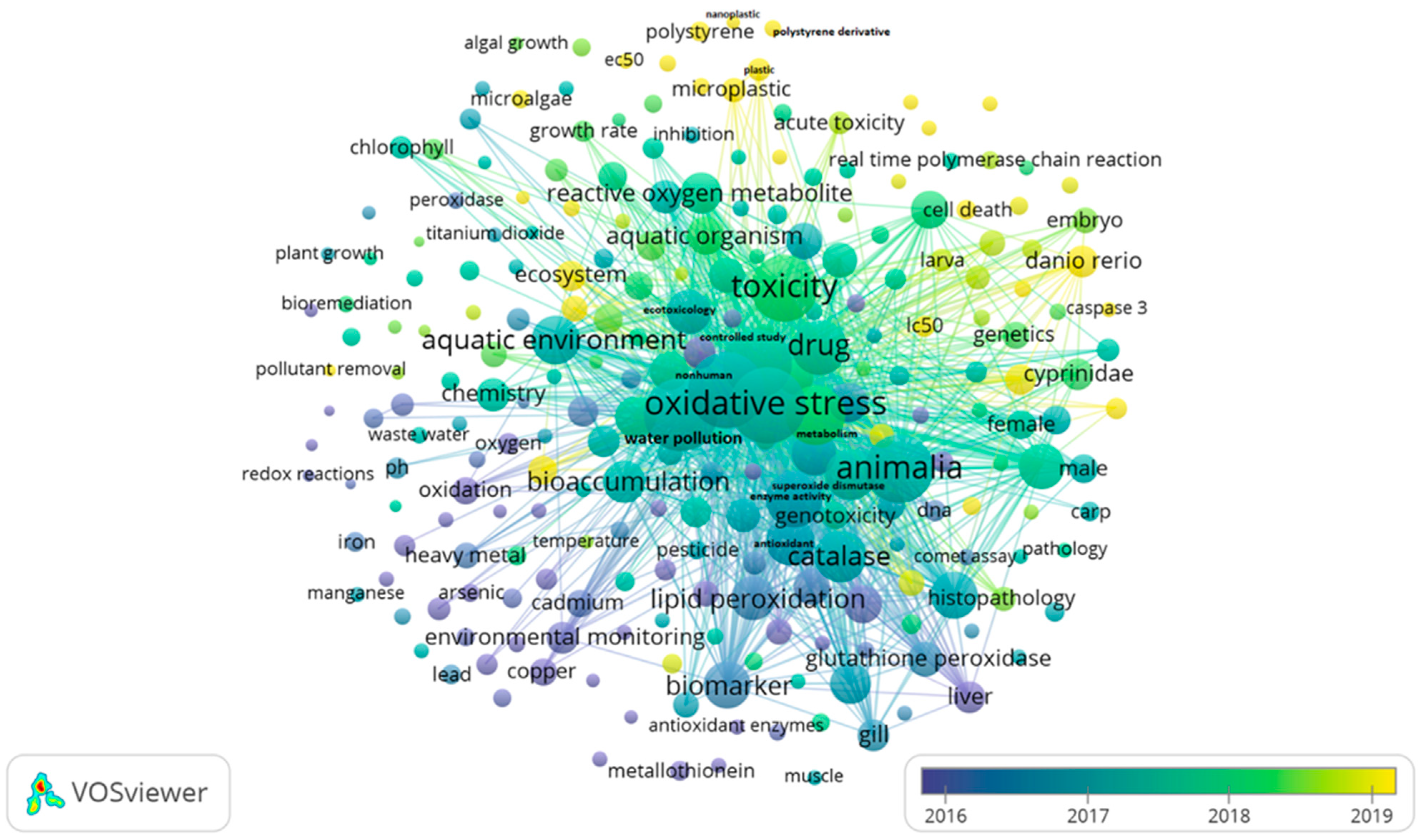
  39. VOSviewer—Visualizing Scientific Landscapes. Available online: https://www.vosviewer.com/ (accessed on 1 August 2023).
  40. Lusher, A.; Hollman, P.; Mendoza-Hill, J. Microplastics in Fisheries and Aquaculture Status of Knowledge on Their Occurrence and Implications for Aquatic Organisms and Food Safety; FAO: Rome, Italy, 2017. [Google Scholar]
  41. Zhao, S.; Zettler, E.R.; Bos, R.P.; Lin, P.; Amaral-Zettler, L.A.; Mincer, T.J. Large Quantities of Small Microplastics Permeate the Surface Ocean to Abyssal Depths in the South Atlantic Gyre. Glob. Chang. Biol. 2022, 28, 2991–3006. [Google Scholar] [CrossRef]
  42. Murano, C.; Vaccari, L.; Casotti, R.; Corsi, I.; Palumbo, A. Occurrence of Microfibres in Wild Specimens of Adult Sea Urchin Paracentrotus lividus (Lamarck, 1816) from a Coastal Area of the Central Mediterranean Sea. Mar. Pollut. Bull. 2022, 176, 113448. [Google Scholar] [CrossRef]
  43. Murano, C.; Nonnis, S.; Scalvini, F.G.; Maffioli, E.; Corsi, I.; Tedeschi, G.; Palumbo, A. Response to Microplastic Exposure: An Exploration into the Sea Urchin Immune Cell Proteome. Environ. Pollut. 2023, 320, 121062. [Google Scholar] [CrossRef]
  44. Macias, D.; Cózar, A.; Garcia-Gorriz, E.; González-Fernández, D.; Stips, A. Surface Water Circulation Develops Seasonally Changing Patterns of Floating Litter Accumulation in the Mediterranean Sea. A Modelling Approach. Mar. Pollut. Bull. 2019, 149, 110619. [Google Scholar] [CrossRef]
  45. Jiang, B.; Kauffman, A.E.; Li, L.; McFee, W.; Cai, B.; Weinstein, J.; Lead, J.R.; Chatterjee, S.; Scott, G.I.; Xiao, S. Health Impacts of Environmental Contamination of Micro- and Nanoplastics: A Review. Environ. Health Prev. Med. 2020, 25, 29. [Google Scholar] [CrossRef] [PubMed]
  46. Yoshida, S.; Hiraga, K.; Takehana, T.; Taniguchi, I.; Yamaji, H.; Maeda, Y.; Toyohara, K.; Miyamoto, K.; Kimura, Y.; Oda, K. A Bacterium That Degrades and Assimilates Poly(Ethylene Terephthalate). Science 2016, 351, 1196–1199. [Google Scholar] [CrossRef] [PubMed]
  47. Okoffo, E.D.; O’Brien, S.; O’Brien, J.W.; Tscharke, B.J.; Thomas, K.V. Wastewater Treatment Plants as a Source of Plastics in the Environment: A Review of Occurrence, Methods for Identification, Quantification and Fate. Environ. Sci. Water Res. Technol. 2019, 5, 1908–1931. [Google Scholar] [CrossRef]
  48. Jiang, J.Q. Occurrence of Microplastics and Its Pollution in the Environment: A Review. Sustain. Prod. Consum. 2018, 13, 16–23. [Google Scholar] [CrossRef]
  49. FAO. Microplastics in Fisheries and Aquaculture: What Do We Know? Should We Be Worried? FAO: Rome, Italy, 2019. [Google Scholar]
  50. Bergmann, M.; Gutow, L.; Klages, M. Marine Anthropogenic Litter; Springer Nature: Berlin/Heidelberg, Germany, 2015; pp. 1–447. [Google Scholar] [CrossRef]
  51. Murano, C.; Agnisola, C.; Caramiello, D.; Castellano, I.; Casotti, R.; Corsi, I.; Palumbo, A. How Sea Urchins Face Microplastics: Uptake, Tissue Distribution and Immune System Response. Environ. Pollut. 2020, 264, 114685. [Google Scholar] [CrossRef]
  52. Junaid, M.; Wang, J. Interaction of Nanoplastics with Extracellular Polymeric Substances (EPS) in the Aquatic Environment: A Special Reference to Eco-Corona Formation and Associated Impacts. Water Res. 2021, 201, 117319. [Google Scholar] [CrossRef]
  53. Wang, J.; Zhao, X.; Wu, F.; Niu, L.; Tang, Z.; Liang, W.; Zhao, T.; Fang, M.; Wang, H.; Wang, X. Characterization, Occurrence, Environmental Behaviors, and Risks of Nanoplastics in the Aquatic Environment: Current Status and Future Perspectives. Fundam. Res. 2021, 1, 317–328. [Google Scholar] [CrossRef]
  54. Auffan, M.; Rose, J.; Bottero, J.Y.; Lowry, G.V.; Jolivet, J.P.; Wiesner, M.R. Towards a Definition of Inorganic Nanoparticles from an Environmental, Health and Safety Perspective. Nat. Nanotechnol. 2009, 4, 634–641. [Google Scholar] [CrossRef]
  55. Xiong, X.; Liu, Q.; Chen, X.; Wang, R.; Duan, M.; Wu, C. Occurrence of Microplastic in the Water of Different Types of Aquaculture Ponds in an Important Lakeside Freshwater Aquaculture Area of China. Chemosphere 2021, 282, 131126. [Google Scholar] [CrossRef]
  56. Wu, F.; Wang, Y.; Leung, J.Y.S.; Huang, W.; Zeng, J.; Tang, Y.; Chen, J.; Shi, A.; Yu, X.; Xu, X.; et al. Accumulation of Microplastics in Typical Commercial Aquatic Species: A Case Study at a Productive Aquaculture Site in China. Sci. Total Environ. 2020, 708, 135432. [Google Scholar] [CrossRef]
  57. Enrichetti, F.; Bavestrello, G.; Betti, F.; Rindi, F.; Tregrosso, A.; Bo, M. Fate of Lost Fishing Gears: Experimental Evidence of Biofouling Colonization Patterns from the Northwestern Mediterranean Sea. Environ. Pollut. 2021, 268, 115746. [Google Scholar] [CrossRef]
  58. Gajanur, A.R.; Jaafar, Z. Abandoned, Lost, or Discarded Fishing Gear at Urban Coastlines. Mar. Pollut. Bull. 2022, 175, 113341. [Google Scholar] [CrossRef]
  59. Gajanur, A.R.; Jaafar, Z. Behaviour of Stranded Abandoned Plastic Fishing Nets in the Equatorial Tropics. Nat. Singap. 2023, 16, e2023034. [Google Scholar] [CrossRef]
  60. Hale, R.C.; Seeley, M.E.; La Guardia, M.J.; Mai, L.; Zeng, E.Y. A Global Perspective on Microplastics. J. Geophys. Res. Ocean. 2020, 125, e2018JC014719. [Google Scholar] [CrossRef]
  61. Lehtiniemi, M.; Hartikainen, S.; Näkki, P.; Engström-Öst, J.; Koistinen, A.; Setälä, O. Size Matters More than Shape: Ingestion of Primary and Secondary Microplastics by Small Predators. Food Webs 2018, 17, e00097. [Google Scholar] [CrossRef]
  62. Murano, C.; Palumbo, A.; Leone, S. Toxicological Impacts of Microplastics: Effects on Levels of Cellular Thiols in Mytilus Galloprovincialis. Environ. Toxicol. Chem. 2023, 42, 1607–1613. [Google Scholar] [CrossRef]
  63. Aragaw, T.A.; Mekonnen, B.A.; Aragaw, T.A.; Mekonnen, B.A.; Mekonnen Bahir, B.A. Distribution and Impact of Microplastics in the Aquatic Systems: A Review of Ecotoxicological Effects on Biota. In Microplastic Pollution. Sustainable Textiles: Production, Processing, Manufacturing & Chemistry; Springer: Berlin/Heidelberg, Germany, 2021; pp. 65–104. [Google Scholar] [CrossRef]
  64. Andrady, A.L. Microplastics in the Marine Environment. Mar. Pollut. Bull. 2011, 62, 1596–1605. [Google Scholar] [CrossRef] [PubMed]
  65. Alimi, O.S.; Farner Budarz, J.; Hernandez, L.M.; Tufenkji, N. Microplastics and Nanoplastics in Aquatic Environments: Aggregation, Deposition, and Enhanced Contaminant Transport. Environ. Sci. Technol. 2018, 52, 1704–1724. [Google Scholar] [CrossRef] [PubMed]
  66. Sharma, V.K.; Ma, X.; Guo, B.; Zhang, K. Environmental Factors-Mediated Behavior of Microplastics and Nanoplastics in Water: A Review. Chemosphere 2021, 271, 129597. [Google Scholar] [CrossRef]
  67. Velzeboer, I.; Kwadijk, C.J.A.F.; Koelmans, A.A. Strong Sorption of PCBs to Nanoplastics, Microplastics, Carbon Nanotubes, and Fullerenes. Environ. Sci. Technol. 2014, 48, 4869–4876. [Google Scholar] [CrossRef] [PubMed]
  68. Cao, J.; Yang, Q.; Jiang, J.; Dalu, T.; Kadushkin, A.; Singh, J.; Fakhrullin, R.; Wang, F.; Cai, X.; Li, R. Coronas of Micro/Nano Plastics: A Key Determinant in Their Risk Assessments. Part. Fibre Toxicol. 2022, 19, 55. [Google Scholar] [CrossRef] [PubMed]
  69. Canesi, L.; Corsi, I. Effects of Nanomaterials on Marine Invertebrates. Sci. Total Environ. 2016, 565, 933–940. [Google Scholar] [CrossRef]
  70. Corsi, I.; Bergami, E.; Grassi, G. Behavior and Bio-Interactions of Anthropogenic Particles in Marine Environment for a More Realistic Ecological Risk Assessment. Front. Environ. Sci. 2020, 8, 524966. [Google Scholar] [CrossRef]
  71. Yu, F.; Yang, C.; Zhu, Z.; Bai, X.; Ma, J. Adsorption Behavior of Organic Pollutants and Metals on Micro/Nanoplastics in the Aquatic Environment. Sci. Total Environ. 2019, 694, 133643. [Google Scholar] [CrossRef] [PubMed]
  72. Romera-Castillo, C.; Letscher, R.T.; Hansell, D.A. New Nutrients Exert Fundamental Control on Dissolved Organic Carbon Accumulation in the Surface Atlantic Ocean. Proc. Natl. Acad. Sci. USA 2016, 113, 10497–10502. [Google Scholar] [CrossRef] [PubMed]
  73. Chen, C.S.; Le, C.; Chiu, M.H.; Chin, W.C. The Impact of Nanoplastics on Marine Dissolved Organic Matter Assembly. Sci. Total Environ. 2018, 634, 316–320. [Google Scholar] [CrossRef]
  74. Zettler, E.R.; Mincer, T.J.; Amaral-Zettler, L.A. Life in the “Plastisphere”: Microbial Communities on Plastic Marine Debris. Environ. Sci. Technol. 2013, 47, 7137–7146. [Google Scholar] [CrossRef]
  75. Sooriyakumar, P.; Bolan, N.; Kumar, M.; Singh, L.; Yu, Y.; Li, Y.; Weralupitiya, C.; Vithanage, M.; Ramanayaka, S.; Sarkar, B.; et al. Biofilm Formation and Its Implications on the Properties and Fate of Microplastics in Aquatic Environments: A Review. J. Hazard. Mater. Adv. 2022, 6, 100077. [Google Scholar] [CrossRef]
  76. Oberbeckmann, S.; Labrenz, M. Marine Microbial Assemblages on Microplastics: Diversity, Adaptation, and Role in Degradation. Annu. Rev. Mar. Sci. 2020, 12, 209–232. [Google Scholar] [CrossRef]
  77. Amaral-Zettler, L.A.; Zettler, E.R.; Mincer, T.J. Ecology of the Plastisphere. Nat. Rev. Microbiol. 2020, 18, 139–151. [Google Scholar] [CrossRef] [PubMed]
  78. Kirstein, I.V.; Kirmizi, S.; Wichels, A.; Garin-Fernandez, A.; Erler, R.; Löder, M.; Gerdts, G. Dangerous Hitchhikers? Evidence for Potentially Pathogenic Vibrio Spp. on Microplastic Particles. Mar. Environ. Res. 2016, 120, 1–8. [Google Scholar] [CrossRef] [PubMed]
  79. Vroom, R.J.E.; Koelmans, A.A.; Besseling, E.; Halsband, C. Aging of Microplastics Promotes Their Ingestion by Marine Zooplankton. Environ. Pollut. 2017, 231, 987–996. [Google Scholar] [CrossRef] [PubMed]
  80. Porter, A.; Smith, K.E.; Lewis, C. The Sea Urchin Paracentrotus Lividus as a Bioeroder of Plastic. Sci. Total Environ. 2019, 693, 133621. [Google Scholar] [CrossRef]
  81. Casotti, R.; Palumbo, A.; Murano, C.; Donnarumma, V.; Corsi, I. Impact of Microbial Colonization of Polystyrene Microbeads on the Toxicological Responses in the Sea Urchin Paracentrotus Lividus. Environ. Sci. Technol. 2021, 55, 7990–8000. [Google Scholar] [CrossRef]
  82. Galloway, T.S.; Cole, M.; Lewis, C. Interactions of Microplastic Debris throughout the Marine Ecosystem. Nat. Ecol. Evol. 2017, 1, 116. [Google Scholar] [CrossRef]
  83. Bulleri, F.; Ravaglioli, C.; Anselmi, S.; Renzi, M. The Sea Cucumber Holothuria Tubulosa Does Not Reduce the Size of Microplastics but Enhances Their Resuspension in the Water Column. Sci. Total Environ. 2021, 781, 146650. [Google Scholar] [CrossRef]
  84. Choy, C.A.; Robison, B.H.; Gagne, T.O.; Erwin, B.; Firl, E.; Halden, R.U.; Hamilton, J.A.; Katija, K.; Lisin, S.E.; Rolsky, C.; et al. The Vertical Distribution and Biological Transport of Marine Microplastics across the Epipelagic and Mesopelagic Water Column. Sci. Rep. 2019, 9, 7843. [Google Scholar] [CrossRef]
  85. Kooi, M.; Van Nes, E.H.; Scheffer, M.; Koelmans, A.A. Ups and Downs in the Ocean: Effects of Biofouling on Vertical Transport of Microplastics. Environ. Sci. Technol. 2017, 51, 7963–7971. [Google Scholar] [CrossRef]
  86. Cózar, A.; Echevarría, F.; González-Gordillo, J.I.; Irigoien, X.; Úbeda, B.; Hernández-León, S.; Palma, Á.T.; Navarro, S.; García-de-Lomas, J.; Ruiz, A.; et al. Plastic Debris in the Open Ocean. Proc. Natl. Acad. Sci. USA 2014, 111, 10239–10244. [Google Scholar] [CrossRef]
  87. Hu, M.; Palić, D. Micro- and Nano-Plastics Activation of Oxidative and Inflammatory Adverse Outcome Pathways. Redox Biol. 2020, 37, 101620. [Google Scholar] [CrossRef] [PubMed]
  88. Napolitano, G.; Fasciolo, G.; Venditti, P. The Ambiguous Aspects of Oxygen. Oxygen 2022, 2, 382–409. [Google Scholar] [CrossRef]
  89. Rai, P.K.; Sonne, C.; Brown, R.J.C.; Younis, S.A.; Kim, K.H. Adsorption of Environmental Contaminants on Micro- and Nano-Scale Plastic Polymers and the Influence of Weathering Processes on Their Adsorptive Attributes. J. Hazard. Mater. 2022, 427, 127903. [Google Scholar] [CrossRef] [PubMed]
  90. Duan, J.; Li, Y.; Gao, J.; Cao, R.; Shang, E.; Zhang, W. ROS-Mediated Photoaging Pathways of Nano- and Micro-Plastic Particles under UV Irradiation. Water Res. 2022, 216, 118320. [Google Scholar] [CrossRef] [PubMed]
  91. Zhu, K.; Jia, H.; Sun, Y.; Dai, Y.; Zhang, C.; Guo, X.; Wang, T.; Zhu, L. Long-Term Phototransformation of Microplastics under Simulated Sunlight Irradiation in Aquatic Environments: Roles of Reactive Oxygen Species. Water Res. 2020, 173, 115564. [Google Scholar] [CrossRef]
  92. Urli, S.; Corte Pause, F.; Crociati, M.; Baufeld, A.; Monaci, M.; Stradaioli, G. Impact of Microplastics and Nanoplastics on Livestock Health: An Emerging Risk for Reproductive Efficiency. Animals 2023, 13, 1132. [Google Scholar] [CrossRef]
  93. Riesbeck, S. Do Microplastics Induce Oxidative Stress in Marine Invertebrates? Available online: https://epic.awi.de/id/eprint/48395/ (accessed on 21 October 2022).
  94. Wu, H.; Xu, T.; Chen, T.; Liu, J.; Xu, S. Oxidative Stress Mediated by the TLR4/NOX2 Signalling Axis Is Involved in Polystyrene Microplastic-Induced Uterine Fibrosis in Mice. Sci. Total Environ. 2022, 838, 155825. [Google Scholar] [CrossRef]
  95. Fasciolo, G.; Napolitano, G.; Aprile, M.; Cataldi, S.; Costa, V.; Tomajoli, M.T.M.; Lombardi, A.; Di Meo, S.; Venditti, P. Muscle Oxidative Stress Plays a Role in Hyperthyroidism-Linked Insulin Resistance. Antioxidants 2023, 12, 592. [Google Scholar] [CrossRef]
  96. Fasciolo, G.; Napolitano, G.; Aprile, M.; Cataldi, S.; Costa, V.; Ciccodicola, A.; Di Meo, S.; Venditti, P. Hepatic Insulin Resistance in Hyperthyroid Rat Liver: Vitamin E Supplementation Highlights a Possible Role of ROS. Antioxidants 2022, 11, 1295. [Google Scholar] [CrossRef]
  97. Jahnke, A.; Arp, H.P.H.; Escher, B.I.; Gewert, B.; Gorokhova, E.; Kühnel, D.; Ogonowski, M.; Potthoff, A.; Rummel, C.; Schmitt-Jansen, M.; et al. Reducing Uncertainty and Confronting Ignorance about the Possible Impacts of Weathering Plastic in the Marine Environment. Environ. Sci. Technol. Lett. 2017, 4, 85–90. [Google Scholar] [CrossRef]
  98. Celina, M.C. Review of Polymer Oxidation and Its Relationship with Materials Performance and Lifetime Prediction. Polym. Degrad. Stab. 2013, 98, 2419–2429. [Google Scholar] [CrossRef]
  99. Yousif, E.; Haddad, R. Photodegradation and Photostabilization of Polymers, Especially Polystyrene: Review. SpringerPlus 2013, 2, 398. [Google Scholar] [CrossRef] [PubMed]
  100. Pannetier, P.; Cachot, J.; Clérandeau, C.; Faure, F.; Van Arkel, K.; de Alencastro, L.F.; Levasseur, C.; Sciacca, F.; Bourgeois, J.P.; Morin, B. Toxicity Assessment of Pollutants Sorbed on Environmental Sample Microplastics Collected on Beaches: Part I-Adverse Effects on Fish Cell Line. Environ. Pollut. 2019, 248, 1088–1097. [Google Scholar] [CrossRef]
  101. Lee, S.E.; Yi, Y.; Moon, S.; Yoon, H.; Park, Y.S. Impact of Micro- and Nanoplastics on Mitochondria. Metabolites 2022, 12, 897. [Google Scholar] [CrossRef] [PubMed]
  102. Balk, J.; Leaver, C.J.; McCabe, P.F. Translocation of Cytochrome c from the Mitochondria to the Cytosol Occurs during Heat-Induced Programmed Cell Death in Cucumber Plants. FEBS Lett. 1999, 463, 151–154. [Google Scholar] [CrossRef] [PubMed]
  103. Tang, Q.; Li, T.; Chen, K.; Deng, X.; Zhang, Q.; Tang, H.; Shi, Z.; Zhu, T.; Zhu, J. PS-NPs Induced Neurotoxic Effects in SHSY-5Y Cells via Autophagy Activation and Mitochondrial Dysfunction. Brain Sci. 2022, 12, 952. [Google Scholar] [CrossRef]
  104. Li, Y.; Liu, Z.; Li, M.; Jiang, Q.; Wu, D.; Huang, Y.; Jiao, Y.; Zhang, M.; Zhao, Y. Effects of Nanoplastics on Antioxidant and Immune Enzyme Activities and Related Gene Expression in Juvenile Macrobrachium nipponense. J. Hazard. Mater. 2020, 398, 122990. [Google Scholar] [CrossRef]
  105. Ferrante, M.C.; Monnolo, A.; Del Piano, F.; Raso, G.M.; Meli, R. The Pressing Issue of Micro- and Nanoplastic Contamination: Profiling the Reproductive Alterations Mediated by Oxidative Stress. Antioxidants 2022, 11, 193. [Google Scholar] [CrossRef]
  106. Arihilam, N.H.; Arihilam, E.C. Impact and Control of Anthropogenic Pollution on the Ecosystem—A Review. J. Biosci. Biotechnol. Discov. 2019, 4, 2536–7064. [Google Scholar] [CrossRef]
  107. Bukola, D.; Zaid, A.; Olalekan, E.I.; Falilu, A. Consequences of Anthropogenic Activities on Fish and the Aquatic Environment. Poult. Fish. Wildl. Sci. 2015, 3, 2. [Google Scholar] [CrossRef]
  108. Gray, J.S. Marine Biodiversity: Patterns, Threats and Conservation Needs. Available online: https://www.sciencebase.gov/catalog/item/50539dc6e4b097cd4fce79f5 (accessed on 4 July 2023).
  109. Dudgeon, D.; Arthington, A.H.; Gessner, M.O.; Kawabata, Z.I.; Knowler, D.J.; Lévêque, C.; Naiman, R.J.; Prieur-Richard, A.H.; Soto, D.; Stiassny, M.L.J.; et al. Freshwater Biodiversity: Importance, Threats, Status and Conservation Challenges. Biol. Rev. Camb. Philos. Soc. 2006, 81, 163–182. [Google Scholar] [CrossRef]
  110. Prakash, S. Impact of Climate Change on Aquatic Ecosystem and Its Biodiversity: An Overview. Available online: http://ijbi.org.in/papers/10.%20IJBI%20Dec%202021%20Dr%20Sadguru.pdf (accessed on 2 July 2023).
  111. Nava, V.; Leoni, B. A Critical Review of Interactions between Microplastics, Microalgae and Aquatic Ecosystem Function. Water Res. 2021, 188, 116476. [Google Scholar] [CrossRef] [PubMed]
  112. Karapanagioti, H.H. Hazardous Chemicals Associated with Plastics in the Marine Environment; Springer International Publishing: Berlin/Heidelberg, Germany, 2019. [Google Scholar]
  113. Van Cauwenberghe, L.; Devriese, L.; Galgani, F.; Robbens, J.; Janssen, C.R. Microplastics in Sediments: A Review of Techniques, Occurrence and Effects. Mar. Environ. Res. 2015, 111, 5–17. [Google Scholar] [CrossRef] [PubMed]
  114. Long, M.; Moriceau, B.; Gallinari, M.; Lambert, C.; Huvet, A.; Raffray, J.; Soudant, P. Interactions between Microplastics and Phytoplankton Aggregates: Impact on Their Respective Fates. Mar. Chem. 2015, 175, 39–46. [Google Scholar] [CrossRef]
  115. Wang, Z.; Su, B.; Xu, X.; Di, D.; Huang, H.; Mei, K.; Dahlgren, R.A.; Zhang, M.; Shang, X. Preferential Accumulation of Small (<300 μm) Microplastics in the Sediments of a Coastal Plain River Network in Eastern China. Water Res. 2018, 144, 393–401. [Google Scholar] [CrossRef]
  116. Wang, S.; Liu, M.; Wang, J.; Huang, J.; Wang, J. Polystyrene Nanoplastics Cause Growth Inhibition, Morphological Damage and Physiological Disturbance in the Marine Microalga Platymonas helgolandica. Mar. Pollut. Bull. 2020, 158, 111403. [Google Scholar] [CrossRef]
  117. Prata, J.C.; da Costa, J.P.; Lopes, I.; Duarte, A.C.; Rocha-Santos, T. Effects of Microplastics on Microalgae Populations: A Critical Review. Sci. Total Environ. 2019, 665, 400–405. [Google Scholar] [CrossRef]
  118. Khatiwada, J.R.; Madsen, C.; Warwick, C.; Shrestha, S.; Chio, C.; Qin, W. Interaction between Polyethylene Terephthalate (PET) Microplastic and Microalgae (Scenedesmus spp.): Effect on the Growth, Chlorophyll Content, and Hetero-Aggregation. Environ. Adv. 2023, 13, 100399. [Google Scholar] [CrossRef]
  119. Zhang, C.; Chen, X.; Wang, J.; Tan, L. Toxic Effects of Microplastic on Marine Microalgae Skeletonema Costatum: Interactions between Microplastic and Algae. Environ. Pollut. 2017, 220, 1282–1288. [Google Scholar] [CrossRef]
  120. Guschina, I.A.; Hayes, A.J.; Ormerod, S.J. Polystyrene Microplastics Decrease Accumulation of Essential Fatty Acids in Common Freshwater Algae. Environ. Pollut. 2020, 263, 114425. [Google Scholar] [CrossRef]
  121. Wang, Y.; Wang, X.; Li, Y.; Li, J.; Wang, F.; Xia, S.; Zhao, J. Biofilm Alters Tetracycline and Copper Adsorption Behaviors onto Polyethylene Microplastics. Chem. Eng. J. 2020, 392, 123808. [Google Scholar] [CrossRef]
  122. Wu, P.; Huang, J.; Zheng, Y.; Yang, Y.; Zhang, Y.; He, F.; Chen, H.; Quan, G.; Yan, J.; Li, T.; et al. Environmental Occurrences, Fate, and Impacts of Microplastics. Ecotoxicol. Environ. Saf. 2019, 184, 109612. [Google Scholar] [CrossRef] [PubMed]
  123. Cole, M.; Liddle, C.; Consolandi, G.; Drago, C.; Hird, C.; Lindeque, P.K.; Galloway, T.S. Microplastics, Microfibres and Nanoplastics Cause Variable Sub-Lethal Responses in Mussels (Mytilus spp.). Mar. Pollut. Bull. 2020, 160, 111552. [Google Scholar] [CrossRef] [PubMed]
  124. Wegner, A.; Besseling, E.; Foekema, E.M.; Kamermans, P.; Koelmans, A.A. Effects of Nanopolystyrene on the Feeding Behavior of the Blue Mussel (Mytilus Edulis L.). Environ. Toxicol. Chem. 2012, 31, 2490–2497. [Google Scholar] [CrossRef] [PubMed]
  125. Sussarellu, R.; Suquet, M.; Thomas, Y.; Lambert, C.; Fabioux, C.; Pernet, M.E.J.; Le Goïc, N.; Quillien, V.; Mingant, C.; Epelboin, Y.; et al. Oyster Reproduction Is Affected by Exposure to Polystyrene Microplastics. Proc. Natl. Acad. Sci. USA 2016, 113, 2430–2435. [Google Scholar] [CrossRef]
  126. Murray, F.; Cowie, P.R. Plastic Contamination in the Decapod Crustacean Nephrops Norvegicus (Linnaeus, 1758). Mar. Pollut. Bull. 2011, 62, 1207–1217. [Google Scholar] [CrossRef]
  127. Helfman, G.S.; Collette, B.B.; Facey, D.E.; Bowen, B.W. The Diversity of Fishes: Biology, Evolution, and Ecology; Helfman, G.S., Collette, B.B., Facey, D.E., Bowen, B.W., Eds.; John Wiley & Sons: Hoboken, NJ, USA, 2019; Available online: https://books.google.it/books?hl=it&lr=&id=FyehAR6hsUUC&oi=fnd&pg=PR7&dq=The+diversity+of+fishes:+Biology,+evolution,+and+ecology.+&ots=lqBp8z5KZy&sig=Cf4pwwIp65J_-_BkdqMIQ6Ir48A&redir_esc=y#v=onepage&q=The%20diversity%20of%20fishes%3A%20Biology%2C%20evolution%2C%20and%20ecology.&f=false (accessed on 4 July 2023).
  128. Holmlund, C.M.; Hammer, M. Ecosystem Services Generated by Fish Populations. Ecol. Econ. 1999, 29, 253–268. [Google Scholar] [CrossRef]
  129. Lynch, A.J.; Myers, B.J.E.; Chu, C.; Eby, L.A.; Falke, J.A.; Kovach, R.P.; Krabbenhoft, T.J.; Kwak, T.J.; Lyons, J.; Paukert, C.P.; et al. Climate Change Effects on North American Inland Fish Populations and Assemblages. Fisheries 2016, 41, 346–361. [Google Scholar] [CrossRef]
  130. Cole, M.; Lindeque, P.; Fileman, E.; Halsband, C.; Goodhead, R.; Moger, J.; Galloway, T.S. Microplastic Ingestion by Zooplankton. Environ. Sci. Technol. 2013, 47, 6646–6655. [Google Scholar] [CrossRef]
  131. Ziajahromi, S.; Kumar, A.; Neale, P.A.; Leusch, F.D.L. Impact of Microplastic Beads and Fibers on Waterflea (Ceriodaphnia dubia) Survival, Growth, and Reproduction: Implications of Single and Mixture Exposures. Environ. Sci. Technol. 2017, 51, 13397–13406. [Google Scholar] [CrossRef]
  132. Rehse, S.; Kloas, W.; Zarfl, C. Short-Term Exposure with High Concentrations of Pristine Microplastic Particles Leads to Immobilisation of Daphnia magna. Chemosphere 2016, 153, 91–99. [Google Scholar] [CrossRef]
  133. Cole, M.; Lindeque, P.; Fileman, E.; Halsband, C.; Galloway, T.S. The Impact of Polystyrene Microplastics on Feeding, Function and Fecundity in the Marine Copepod Calanus helgolandicus. Environ. Sci. Technol. 2015, 49, 1130–1137. [Google Scholar] [CrossRef]
  134. Cole, M.; Galloway, T.S. Ingestion of Nanoplastics and Microplastics by Pacific Oyster Larvae. Environ. Sci. Technol. 2015, 49, 14625–14632. [Google Scholar] [CrossRef]
  135. Zheng, S.; Zhao, Y.; Liangwei, W.; Liang, J.; Liu, T.; Zhu, M.; Li, Q.; Sun, X. Characteristics of Microplastics Ingested by Zooplankton from the Bohai Sea, China. Sci. Total Environ. 2020, 713, 136357. [Google Scholar] [CrossRef]
  136. Karami, A.; Romano, N.; Galloway, T.; Hamzah, H. Virgin Microplastics Cause Toxicity and Modulate the Impacts of Phenanthrene on Biomarker Responses in African Catfish (Clarias gariepinus). Environ. Res. 2016, 151, 58–70. [Google Scholar] [CrossRef]
  137. Yang, Y.; Liu, G.; Song, W.; Ye, C.; Lin, H.; Li, Z.; Liu, W. Plastics in the Marine Environment Are Reservoirs for Antibiotic and Metal Resistance Genes. Environ. Int. 2019, 123, 79–86. [Google Scholar] [CrossRef]
  138. Gigault, J.; Ter Halle, A.; Baudrimont, M.; Pascal, P.-Y.; Gauffre, F.; Phi, T.-L.; El Hadri, H.; Grassl, B.; Ephanie Reynaud, S. Current Opinion: What Is a Nanoplastic? *. Environ. Pollut. 2018, 235, 1030–1034. [Google Scholar] [CrossRef]
  139. Bucci, K.; Tulio, M.; Rochman, C.M. What Is Known and Unknown about the Effects of Plastic Pollution: A Meta-Analysis and Systematic Review. Ecol. Appl. 2020, 30, e02044. [Google Scholar] [CrossRef]
  140. Kögel, T.; Bjorøy, Ø.; Toto, B.; Bienfait, A.M.; Sanden, M. Micro- and Nanoplastic Toxicity on Aquatic Life: Determining Factors. Sci. Total Environ. 2020, 709, 136050. [Google Scholar] [CrossRef]
  141. Lozano, Y.M.; Rillig, M.C. Effects of Microplastic Fibers and Drought on Plant Communities. Environ. Sci. Technol. 2020, 54, 6166–6173. [Google Scholar] [CrossRef]
  142. Felten, V.; Toumi, H.; Masfaraud, J.F.; Billoir, E.; Camara, B.I.; Férard, J.F. Microplastics Enhance Daphnia Magna Sensitivity to the Pyrethroid Insecticide Deltamethrin: Effects on Life History Traits. Sci. Total Environ. 2020, 714, 136567. [Google Scholar] [CrossRef]
  143. Agathokleous, E.; Iavicoli, I.; Barceló, D.; Calabrese, E.J. Micro/Nanoplastics Effects on Organisms: A Review Focusing on ‘Dose’. J. Hazard. Mater. 2021, 417, 126084. [Google Scholar] [CrossRef]
  144. Routti, H.; Harju, M.; Lühmann, K.; Aars, J.; Ask, A.; Goksøyr, A.; Kovacs, K.M.; Lydersen, C. Concentrations and Endocrine Disruptive Potential of Phthalates in Marine Mammals from the Norwegian Arctic. Environ. Int. 2021, 152, 106458. [Google Scholar] [CrossRef] [PubMed]
  145. Lo, H.S.; Lee, Y.K.; Po, B.H.K.; Wong, L.C.; Xu, X.; Wong, C.F.; Wong, C.Y.; Tam, N.F.Y.; Cheung, S.G. Impacts of Typhoon Mangkhut in 2018 on the Deposition of Marine Debris and Microplastics on Beaches in Hong Kong. Sci. Total Environ. 2020, 716, 137172. [Google Scholar] [CrossRef] [PubMed]
  146. Neves, D.; Sobral, P.; Ferreira, J.L.; Pereira, T. Ingestion of Microplastics by Commercial Fish off the Portuguese Coast. Mar. Pollut. Bull. 2015, 101, 119–126. [Google Scholar] [CrossRef]
  147. Wardrop, P.; Shimeta, J.; Nugegoda, D.; Morrison, P.D.; Miranda, A.; Tang, M.; Clarke, B.O. Chemical Pollutants Sorbed to Ingested Microbeads from Personal Care Products Accumulate in Fish. Environ. Sci. Technol. 2016, 50, 4037–4044. [Google Scholar] [CrossRef]
  148. Redondo-Hasselerharm, P.E.; Falahudin, D.; Peeters, E.T.H.M.; Koelmans, A.A. Microplastic Effect Thresholds for Freshwater Benthic Macroinvertebrates. Environ. Sci. Technol. 2018, 52, 2278–2286. [Google Scholar] [CrossRef]
  149. Straub, S.; Hirsch, P.E.; Burkhardt-Holm, P. Biodegradable and Petroleum-Based Microplastics Do Not Differ in Their Ingestion and Excretion but in Their Biological Effects in a Freshwater Invertebrate Gammarus Fossarum. Int. J. Environ. Res. Public Health 2017, 14, 774. [Google Scholar] [CrossRef]
  150. König Kardgar, A.; Ghosh, D.; Sturve, J.; Agarwal, S.; Carney Almroth, B. Chronic Poly(l-Lactide) (PLA)- Microplastic Ingestion Affects Social Behavior of Juvenile European Perch (Perca fluviatilis). Sci. Total Environ. 2023, 881, 163425. [Google Scholar] [CrossRef]
  151. Moore, C.J.; Lattin, G.L.; Zellers, A.F. Working Our Way Upstream: A Snapshot of Land-Based Contributions of Plastic and Other Trash to Coastal Waters and Beaches of Southern California. In Proceedings of the Plastic Debris Rivers to Sea Conference, Algalita Marine Research Foundation, Long Beach, CA, USA, 8 September 2005. [Google Scholar]
  152. Draper, A.M.; Weissburg, M.J. Impacts of Global Warming and Elevated CO2 on Sensory Behavior in Predator-Prey Interactions: A Review and Synthesis. Front. Ecol. Evol. 2019, 7, 428445. [Google Scholar] [CrossRef]
  153. Wang, J.; Tan, Z.; Peng, J.; Qiu, Q.; Li, M. The Behaviors of Microplastics in the Marine Environment. Mar. Environ. Res. 2016, 113, 7–17. [Google Scholar] [CrossRef]
  154. Lusher, A.L.; McHugh, M.; Thompson, R.C. Occurrence of Microplastics in the Gastrointestinal Tract of Pelagic and Demersal Fish from the English Channel. Mar. Pollut. Bull. 2013, 67, 94–99. [Google Scholar] [CrossRef] [PubMed]
  155. Romeo, T.; Pietro, B.; Pedà, C.; Consoli, P.; Andaloro, F.; Fossi, M.C. First Evidence of Presence of Plastic Debris in Stomach of Large Pelagic Fish in the Mediterranean Sea. Mar. Pollut. Bull. 2015, 95, 358–361. [Google Scholar] [CrossRef] [PubMed]
  156. United Nations Tuna Day|United Nations. Available online: https://www.un.org/en/observances/tuna-day (accessed on 6 July 2023).
  157. Diaz-Basantes, M.F.; Nacimba-Aguirre, D.; Conesa, J.A.; Fullana, A. Presence of Microplastics in Commercial Canned Tuna. Food Chem. 2022, 385, 132721. [Google Scholar] [CrossRef] [PubMed]
  158. Di Giacinto, F.; Di Renzo, L.; Mascilongo, G.; Notarstefano, V.; Gioacchini, G.; Giorgini, E.; Bogdanović, T.; Petričević, S.; Listeš, E.; Brkljača, M.; et al. Detection of Microplastics, Polymers and Additives in Edible Muscle of Swordfish (Xiphias gladius) and Bluefin Tuna (Thunnus thynnus) Caught in the Mediterranean Sea. J. Sea Res. 2023, 192, 102359. [Google Scholar] [CrossRef]
  159. Abihssira-García, I.S.; Park, Y.; Kiron, V.; Olsvik, P.A. Fluorescent Microplastic Uptake by Immune Cells of Atlantic Salmon (Salmo salar L.). Front. Environ. Sci. 2020, 8, 560206. [Google Scholar] [CrossRef]
  160. Batel, A.; Linti, F.; Scherer, M.; Erdinger, L.; Braunbeck, T. Transfer of Benzo[a]Pyrene from Microplastics to Artemia Nauplii and Further to Zebrafish via a Trophic Food Web Experiment: CYP1A Induction and Visual Tracking of Persistent Organic Pollutants. Environ. Toxicol. Chem. 2016, 35, 1656–1666. [Google Scholar] [CrossRef]
  161. Lu, Y.; Zhang, Y.; Deng, Y.; Jiang, W.; Zhao, Y.; Geng, J.; Ding, L.; Ren, H. Response to Comment on “Uptake and Accumulation of Polystyrene Microplastics in Zebrafish (Danio rerio) and Toxic Effects in Liver”. Environ. Sci. Technol. 2016, 50, 12523–12524. [Google Scholar] [CrossRef]
  162. Jeong, C.B.; Won, E.J.; Kang, H.M.; Lee, M.C.; Hwang, D.S.; Hwang, U.K.; Zhou, B.; Souissi, S.; Lee, S.J.; Lee, J.S. Microplastic Size-Dependent Toxicity, Oxidative Stress Induction, and p-JNK and p-P38 Activation in the Monogonont Rotifer (Brachionus koreanus). Environ. Sci. Technol. 2016, 50, 8849–8857. [Google Scholar] [CrossRef]
  163. Manabe, M.; Tatarazako, N.; Kinoshita, M. Uptake, Excretion and Toxicity of Nano-Sized Latex Particles on Medaka (Oryzias latipes) Embryos and Larvae. Aquat. Toxicol. 2011, 105, 576–581. [Google Scholar] [CrossRef]
  164. Zhou, Y.; Zhao, L.; Xu, H.; Xu, E.G.; Li, M.; Wang, Y. Long-Term Exposure to Polystyrene Nanoplastics Impairs the Liver Health of Medaka. Water 2022, 14, 2767. [Google Scholar] [CrossRef]
  165. Rochman, C.M.; Hoh, E.; Kurobe, T.; Teh, S.J. Ingested Plastic Transfers Hazardous Chemicals to Fish and Induces Hepatic Stress. Sci. Rep. 2013, 3, 3263. [Google Scholar] [CrossRef] [PubMed]
  166. Félix, L.; Carreira, P.; Peixoto, F. Effects of Chronic Exposure of Naturally Weathered Microplastics on Oxidative Stress Level, Behaviour, and Mitochondrial Function of Adult Zebrafish (Danio rerio). Chemosphere 2023, 310, 136895. [Google Scholar] [CrossRef] [PubMed]
  167. Trevisan, R.; Voy, C.; Chen, S.; Di Giulio, R.T. Nanoplastics Decrease the Toxicity of a Complex PAH Mixture but Impair Mitochondrial Energy Production in Developing Zebrafish. Environ. Sci. Technol. 2019, 53, 8405–8415. [Google Scholar] [CrossRef] [PubMed]
  168. Meyer, J.N.; Leung, M.C.K.; Rooney, J.P.; Sendoel, A.; Hengartner, M.O.; Kisby, G.E.; Bess, A.S. Mitochondria as a Target of Environmental Toxicants. Toxicol. Sci. 2013, 134, 1–17. [Google Scholar] [CrossRef] [PubMed]
  169. Raftery, T.D.; Jayasundara, N.; Di Giulio, R.T. A Bioenergetics Assay for Studying the Effects of Environmental Stressors on Mitochondrial Function in Vivo in Zebrafish Larvae. Comp. Biochem. Physiol. C Toxicol. Pharmacol. 2017, 192, 23–32. [Google Scholar] [CrossRef] [PubMed]
  170. Lindberg, C.D.; Jayasundara, N.; Kozal, J.S.; Leuthner, T.C.; Di Giulio, R.T. Resistance to Polycyclic Aromatic Hydrocarbon Toxicity and Associated Bioenergetic Consequences in a Population of Fundulus Heteroclitus. Ecotoxicology 2017, 26, 435–448. [Google Scholar] [CrossRef]
  171. Massarsky, A.; Jayasundara, N.; Bailey, J.M.; Oliveri, A.N.; Levin, E.D.; Prasad, G.L.; Di Giulio, R.T. Teratogenic, Bioenergetic, and Behavioral Effects of Exposure to Total Particulate Matter on Early Development of Zebrafish (Danio rerio) Are Not Mimicked by Nicotine. Neurotoxicol. Teratol. 2015, 51, 77–88. [Google Scholar] [CrossRef]
  172. Gu, Y.; Xu, D.; Liu, J.; Chen, Y.; Wang, J.; Song, Y.; Sun, B.; Xia, B. Bioaccumulation of Functionalized Polystyrene Nanoplastics in Sea Cucumber Apostichopus Japonicus (Selenka, 1867) and Their Toxic Effects on Oxidative Stress, Energy Metabolism and Mitochondrial Pathway. Environ. Pollut. 2023, 319, 121015. [Google Scholar] [CrossRef]
  173. Guimarães, A.T.B.; Charlie-Silva, I.; Malafaia, G. Toxic Effects of Naturally-Aged Microplastics on Zebrafish Juveniles: A More Realistic Approach to Plastic Pollution in Freshwater Ecosystems. J. Hazard. Mater. 2021, 407, 124833. [Google Scholar] [CrossRef]
  174. Bhagat, J.; Zang, L.; Kaneco, S.; Nishimura, N.; Shimada, Y. Combined Exposure to Nanoplastics and Metal Oxide Nanoparticles Inhibits Efflux Pumps and Causes Oxidative Stress in Zebrafish Embryos. Sci. Total Environ. 2022, 835, 155436. [Google Scholar] [CrossRef]
  175. Sendra, M.; Pereiro, P.; Yeste, M.P.; Mercado, L.; Figueras, A.; Novoa, B. Size Matters: Zebrafish (Danio rerio) as a Model to Study Toxicity of Nanoplastics from Cells to the Whole Organism. Environ. Pollut. 2021, 268, 115769. [Google Scholar] [CrossRef] [PubMed]
  176. Sarasamma, S.; Audira, G.; Siregar, P.; Malhotra, N.; Lai, Y.H.; Liang, S.T.; Chen, J.R.; Chen, K.H.C.; Hsiao, C. Der Nanoplastics Cause Neurobehavioral Impairments, Reproductive and Oxidative Damages, and Biomarker Responses in Zebrafish: Throwing up Alarms of Wide Spread Health Risk of Exposure. Int. J. Mol. Sci. 2020, 21, 1410. [Google Scholar] [CrossRef] [PubMed]
  177. Pitt, J.A.; Trevisan, R.; Massarsky, A.; Kozal, J.S.; Levin, E.D.; Di Giulio, R.T. Maternal Transfer of Nanoplastics to Offspring in Zebrafish (Danio rerio): A Case Study with Nanopolystyrene. Sci. Total Environ. 2018, 643, 324–334. [Google Scholar] [CrossRef] [PubMed]
  178. Aliakbarzadeh, F.; Rafiee, M.; Khodagholi, F.; Khorramizadeh, M.R.; Manouchehri, H.; Eslami, A.; Sayehmiri, F.; Mohseni-Bandpei, A. Adverse Effects of Polystyrene Nanoplastic and Its Binary Mixtures with Nonylphenol on Zebrafish Nervous System: From Oxidative Stress to Impaired Neurotransmitter System. Environ. Pollut. 2023, 317, 120587. [Google Scholar] [CrossRef]
  179. Umamaheswari, S.; Priyadarshinee, S.; Kadirvelu, K.; Ramesh, M. Polystyrene Microplastics Induce Apoptosis via ROS-Mediated P53 Signaling Pathway in Zebrafish. Chem. Biol. Interact. 2021, 345, 109550. [Google Scholar] [CrossRef]
  180. Lu, Y.; Zhang, Y.; Deng, Y.; Jiang, W.; Zhao, Y.; Geng, J.; Ding, L.; Ren, H. Uptake and Accumulation of Polystyrene Microplastics in Zebrafish (Danio rerio) and Toxic Effects in Liver. Environ. Sci. Technol. 2016, 50, 4054–4060. [Google Scholar] [CrossRef]
  181. Santos, D.; Félix, L.; Luzio, A.; Parra, S.; Cabecinha, E.; Bellas, J.; Monteiro, S.M. Toxicological Effects Induced on Early Life Stages of Zebrafish (Danio rerio) after an Acute Exposure to Microplastics Alone or Co-Exposed with Copper. Chemosphere 2020, 261, 127748. [Google Scholar] [CrossRef]
  182. Yang, H.; Lai, H.; Huang, J.; Sun, L.; Mennigen, J.A.; Wang, Q.; Liu, Y.; Jin, Y.; Tu, W. Polystyrene Microplastics Decrease F–53B Bioaccumulation but Induce Inflammatory Stress in Larval Zebrafish. Chemosphere 2020, 255, 127040. [Google Scholar] [CrossRef]
  183. Chen, X.; Peng, L.B.; Wang, D.; Zhu, Q.L.; Zheng, J.L. Combined Effects of Polystyrene Microplastics and Cadmium on Oxidative Stress, Apoptosis, and GH/IGF Axis in Zebrafish Early Life Stages. Sci. Total Environ. 2022, 813, 152514. [Google Scholar] [CrossRef]
  184. Qiao, R.; Sheng, C.; Lu, Y.; Zhang, Y.; Ren, H.; Lemos, B. Microplastics Induce Intestinal Inflammation, Oxidative Stress, and Disorders of Metabolome and Microbiome in Zebrafish. Sci. Total Environ. 2019, 662, 246–253. [Google Scholar] [CrossRef] [PubMed]
  185. Li, C.; Yuan, S.; Zhou, Y.; Li, X.; Duan, L.; Huang, L.; Zhou, X.; Ma, Y.; Pang, S. Microplastics Reduce the Bioaccumulation and Oxidative Stress Damage of Triazole Fungicides in Fish. Sci. Total Environ. 2022, 806, 151475. [Google Scholar] [CrossRef] [PubMed]
  186. Li, S.; Wang, P.; Zhang, C.; Zhou, X.; Yin, Z.; Hu, T.; Hu, D.; Liu, C.; Zhu, L. Influence of Polystyrene Microplastics on the Growth, Photosynthetic Efficiency and Aggregation of Freshwater Microalgae Chlamydomonas Reinhardtii. Sci. Total Environ. 2020, 714, 136767. [Google Scholar] [CrossRef] [PubMed]
  187. Besseling, E.; Wegner, A.; Foekema, E.M.; Van Den Heuvel-Greve, M.J.; Koelmans, A.A. Effects of Microplastic on Fitness and PCB Bioaccumulation by the Lugworm Arenicola Marina (L.). Environ. Sci. Technol. 2013, 47, 593–600. [Google Scholar] [CrossRef]
  188. Wang, X.; Huang, W.; Wei, S.; Shang, Y.; Gu, H.; Wu, F.; Lan, Z.; Hu, M.; Shi, H.; Wang, Y. Microplastics Impair Digestive Performance but Show Little Effects on Antioxidant Activity in Mussels under Low PH Conditions. Environ. Pollut. 2020, 258, 113691. [Google Scholar] [CrossRef]
  189. FAO. The State of World Fisheries and Aquaculture 2022; FAO: Rome, Italy, 2022. [Google Scholar] [CrossRef]
  190. Van Cauwenberghe, L.; Janssen, C.R. Microplastics in Bivalves Cultured for Human Consumption. Environ. Pollut. 2014, 193, 65–70. [Google Scholar] [CrossRef]
  191. Devriese, L.I.; van der Meulen, M.D.; Maes, T.; Bekaert, K.; Paul-Pont, I.; Frère, L.; Robbens, J.; Vethaak, A.D. Microplastic Contamination in Brown Shrimp (Crangon Crangon, Linnaeus 1758) from Coastal Waters of the Southern North Sea and Channel Area. Mar. Pollut. Bull. 2015, 98, 179–187. [Google Scholar] [CrossRef]
  192. Al-Thawadi, S. Microplastics and Nanoplastics in Aquatic Environments: Challenges and Threats to Aquatic Organisms. Arab. J. Sci. Eng. 2020, 45, 4419–4440. [Google Scholar] [CrossRef]
  193. Prüst, M.; Meijer, J.; Westerink, R.H.S. The Plastic Brain: Neurotoxicity of Micro- and Nanoplastics. Part. Fibre Toxicol. 2020, 17, 24. [Google Scholar] [CrossRef]
  194. Greven, A.C.; Merk, T.; Karagöz, F.; Mohr, K.; Klapper, M.; Jovanović, B.; Palić, D. Polycarbonate and Polystyrene Nanoplastic Particles Act as Stressors to the Innate Immune System of Fathead Minnow (Pimephales promelas). Environ. Toxicol. Chem. 2016, 35, 3093–3100. [Google Scholar] [CrossRef]
  195. Mattsson, K.; Ekvall, M.T.; Hansson, L.A.; Linse, S.; Malmendal, A.; Cedervall, T. Altered Behavior, Physiology, and Metabolism in Fish Exposed to Polystyrene Nanoparticles. Environ. Sci. Technol. 2015, 49, 553–561. [Google Scholar] [CrossRef] [PubMed]
  196. Chen, Q.; Yin, D.; Jia, Y.; Schiwy, S.; Legradi, J.; Yang, S.; Hollert, H. Enhanced Uptake of BPA in the Presence of Nanoplastics Can Lead to Neurotoxic Effects in Adult Zebrafish. Sci. Total Environ. 2017, 609, 1312–1321. [Google Scholar] [CrossRef] [PubMed]
  197. Wu, B.; Wu, X.; Liu, S.; Wang, Z.; Chen, L. Size-Dependent Effects of Polystyrene Microplastics on Cytotoxicity and Efflux Pump Inhibition in Human Caco-2 cells. Chemosphere 2019, 221, 333–341. [Google Scholar] [CrossRef] [PubMed]
  198. Wright, S.L.; Kelly, F.J. Plastic and Human Health: A Micro Issue? Environ. Sci. Technol. 2017, 51, 6634–6647. [Google Scholar] [CrossRef]
  199. Bhuyan, M.S. Effects of Microplastics on Fish and in Human Health. Front. Environ. Sci. 2022, 10, 827289. [Google Scholar] [CrossRef]
  200. Dong, C.D.; Chen, C.W.; Chen, Y.C.; Chen, H.H.; Lee, J.S.; Lin, C.H. Polystyrene Microplastic Particles: In Vitro Pulmonary Toxicity Assessment. J. Hazard. Mater. 2020, 385, 121575. [Google Scholar] [CrossRef]
  201. Wang, F.; Bexiga, M.G.; Anguissola, S.; Boya, P.; Simpson, J.C.; Salvati, A.; Dawson, K.A. Time Resolved Study of Cell Death Mechanisms Induced by Amine-Modified Polystyrene Nanoparticles. Nanoscale 2013, 5, 10868–10876. [Google Scholar] [CrossRef]
  202. Xu, M.; Halimu, G.; Zhang, Q.; Song, Y.; Fu, X.; Li, Y.; Li, Y.; Zhang, H. Internalization and Toxicity: A Preliminary Study of Effects of Nanoplastic Particles on Human Lung Epithelial Cell. Sci. Total Environ. 2019, 694, 133794. [Google Scholar] [CrossRef]
  203. Wick, P.; Malek, A.; Manser, P.; Meili, D.; Maeder-Althaus, X.; Diener, L.; Diener, P.A.; Zisch, A.; Krug, H.F.; Von Mandach, U. Barrier Capacity of Human Placenta for Nanosized Materials. Environ. Health Perspect. 2010, 118, 432–436. [Google Scholar] [CrossRef]
  204. Lehner, R.; Weder, C.; Petri-Fink, A.; Rothen-Rutishauser, B. Emergence of Nanoplastic in the Environment and Possible Impact on Human Health. Environ. Sci. Technol. 2019, 53, 1748–1765. [Google Scholar] [CrossRef]
  205. González-Pleiter, M.; Tamayo-Belda, M.; Pulido-Reyes, G.; Amariei, G.; Leganés, F.; Rosal, R.; Fernández-Piñas, F. Secondary Nanoplastics Released from a Biodegradable Microplastic Severely Impact Freshwater Environments. Environ. Sci. Nano 2019, 6, 1382–1392. [Google Scholar] [CrossRef]
  206. Zhu, J.; Wang, C. Biodegradable Plastics: Green Hope or Greenwashing? Mar. Pollut. Bull. 2020, 161, 111774. [Google Scholar] [CrossRef]
  207. Arantzamendi, L.; Andrés, M.; Basurko, O.C.; Suárez, M.J. Circular and Lower Impact Mussel and Seaweed Aquaculture by a Shift towards Bio-Based Ropes. Rev. Aquac. 2023, 15, 1010–1019. [Google Scholar] [CrossRef]
  208. Sandhya, M.; Aravind, J.; Kanmani, P. Production of Polyhydroxyalkanoates from Ralstonia Eutropha Using Paddy Straw as Cheap Substrate. Int. J. Environ. Sci. Technol. 2013, 10, 47–54. [Google Scholar] [CrossRef]
  209. Narayan Bharti, S.S.G. Need for Bioplastics and Role of Biopolymer PHB: A Short Review. Artic. J. Pet. Environ. Biotechnol. 2016, 7, 2. [Google Scholar] [CrossRef]
  210. Maheswari, N.U.; Ahilandeswari, K. Production of Bioplastic Using Spirulina Platensis and Comparison with Commercial Plastic. Res. Environ. Life Sci. 2011, 4, 133–136. [Google Scholar]
  211. Urtuvia, V.; Villegas, P.; González, M.; Seeger, M. Bacterial Production of the Biodegradable Plastics Polyhydroxyalkanoates. Int. J. Biol. Macromol. 2014, 70, 208–213. [Google Scholar] [CrossRef]
  212. Napolitano, G.; Venditti, P.; Agnisola, C.; Quartucci, S.; Fasciolo, G.; Muscari Tomajoli, M.T.; Geremia, E.; Catone, C.M.; Ulgiati, S. Towards Sustainable Aquaculture Systems: Biological and Environmental Impact of Replacing Fishmeal with Arthrospira Platensis (Nordstedt) (Spirulina). J. Clean Prod. 2022, 374, 133978. [Google Scholar] [CrossRef]
  213. Harding, K.G.; Dennis, J.S.; von Blottnitz, H.; Harrison, S.T.L. Environmental Analysis of Plastic Production Processes: Comparing Petroleum-Based Polypropylene and Polyethylene with Biologically-Based Poly-β-Hydroxybutyric Acid Using Life Cycle Analysis. J. Biotechnol. 2007, 130, 57–66. [Google Scholar] [CrossRef]
Figure 1. Bibliometric network analysis.
Environments 10 00161 g001
Figure 2. ROS production by MPs and NPs.
Environments 10 00161 g002
Figure 3. MPs/NPs cycle. Micro- and nanoplastics produced by anthropogenic activities end up in the water and, by the consumption of aquatic organisms as food, return to human.
Environments 10 00161 g003
Disclaimer/Publisher’s Note: The statements, opinions and data contained in all publications are solely those of the individual author(s) and contributor(s) and not of MDPI and/or the editor(s). MDPI and/or the editor(s) disclaim responsibility for any injury to people or property resulting from any ideas, methods, instructions or products referred to in the content.

