Article

Characterization of Two Gonadal Genes, zar1 and wt1b, in Hermaphroditic Fish Asian Seabass (Lates calcarifer)

1
Integrative Science Center of Germplasm Creation in Western China (CHONGQING) Science City, College of Fisheries, Southwest University, Chongqing 402460, China
2
Key Laboratory of Freshwater Fish Reproduction and Development, Chongqing 400715, China
3
Key Laboratory of Aquatic Sciences of Chongqing, Ministry of Education, Chongqing 400715, China
*
Authors to whom correspondence should be addressed.
Animals 2024, 14(3), 508; https://doi.org/10.3390/ani14030508
Submission received: 23 November 2023 / Revised: 29 January 2024 / Accepted: 30 January 2024 / Published: 3 February 2024

Abstract

:

Simple Summary

Gonadal development and gametogenesis are key processes for fish genetic breeding. Particularly, reproductive development-related genes such as zygote arrest-1 (Zar1) and Wilms’ tumor 1 (Wt1) play an important role in oogenesis, with the latter also involved in testicular development and gender differentiation. Here, the cDNA fragments of Lczar1 and Lcwt1b were identified in Asian seabass (Lates calcarifer), a valuable hermaphrodite fish for studying sex reverse. Furthermore, their mRNA distributions in the cells of gonadal tissues were detected. Lczar1 mRNA was exclusively expressed in the ovary, while Lcwt1b mRNA was majorly expressed in the gonads in a higher amount in the testis than in the ovary. Specifically, Lczar1 mRNA was mainly concentrated in oogonia and oocytes at early stages in the ovary, but was undetectable in the testis. Lcwt1b mRNA was localized not only in gonadal somatic cells (testis and ovary), but also in female and male germ cells at the early developmental stages. Our study indicated that Lczar1 and Lcwt1b possibly play roles in gonadal development. Therefore, the findings of this study will provide a basis for clarifying the mechanism of Lczar1 and Lcwt1b in regulating germ cell development and the sex reverse of Asian seabass and even other hermaphroditic species.

Abstract

Zygote arrest-1 (Zar1) and Wilms’ tumor 1 (Wt1) play an important role in oogenesis, with the latter also involved in testicular development and gender differentiation. Here, Lczar1 and Lcwt1b were identified in Asian seabass (Lates calcarifer), a hermaphrodite fish, as the valuable model for studying sex differentiation. The cloned cDNA fragments of Lczar1 were 1192 bp, encoding 336 amino acids, and contained a zinc-binding domain, while those of Lcwt1b cDNA were 1521 bp, encoding a peptide of 423 amino acids with a Zn finger domain belonging to Wt1b family. RT-qPCR analysis showed that Lczar1 mRNA was exclusively expressed in the ovary, while Lcwt1b mRNA was majorly expressed in the gonads in a higher amount in the testis than in the ovary. In situ hybridization results showed that Lczar1 mRNA was mainly concentrated in oogonia and oocytes at early stages in the ovary, but were undetectable in the testis. Lcwt1b mRNA was localized not only in gonadal somatic cells (the testis and ovary), but also in female and male germ cells in the early developmental stages, such as those of previtellogenic oocytes, spermatogonia, spermatocytes and spermatids. These results indicated that Lczar1 and Lcwt1b possibly play roles in gonadal development. Therefore, the findings of this study will provide a basis for clarifying the mechanism of Lczar1 and Lcwt1b in regulating germ cell development and the sex reversal of Asian seabass and even other hermaphroditic species.

1. Introduction

Gonadal development and gametogenesis are key processes for fish reproduction and breeding. Among them, reproductive development-related genes and auxiliary factors play essential roles in gonadal development and gametogenesis [1,2,3]. The development and differentiation of gonads are highly similar in animals, including the specification and migration of primordial germ cells, the differentiation of germ cells, and the formation of gametes [4]. Although it is reported that a series of genes are involved in the process of gonadal differentiation and development, only some of them have been identified so far, such as nanos3, vasa (Ddx4, DEAD box polypeptide 4), dazl (deleted in azoospermia-like genes), and piwi (a P-element-induced wimpy testis) [5,6,7]. In addition, some genes crucial for sex differentiation, including cyp19a1a (cytochrome P450, family 19, subfamily A, polypeptide 1a), foxl2 (forkhead-box protein L2), rspo1 (R-spondin 1), dmrt1 (dsxand mab-3-related transcription factor 1), amh (anti-Mullerian hormone), and sox9 (SRY box 9), usually participate in the process of sex reversal [8]. Recently, other genes like zar1 (zygote arrest-1) and wt1 (Wilms’ tumor 1) were also discovered to be closely related to gonadal development [9,10].
Zar1 is an oocyte-specific and maternal gene that functions in the oocyte-to-embryo transition in mouse [11]. Zar1 protein has a conserved C12 structure (i.e., a zinc finger domain) that may be related to transcriptional regulation [10,12]. Zar1 was first identified in a mouse (Mus musculus) [11]. Subsequently, zar1 has been identified in more than ten species, including rats (Rattus norvegicus) [12], cattle (Bos taurus) [13], pigs (Sus scrofa) [14], rabbits (Leporidae) [15], chickens (Gallus gallus) [16], pufferfish (Tetraodontidae) [12], African Xenopus (Xenopus laevis) [17], zebrafish (Danio rerio) [18], etc. Zar1 has conserved ovarian localization [19], and it may also have some non-ovarian functions, because it is expressed in some non-ovarian organs, such as the lung, spleen, kidney and heart [13,14,15,20,21]; the zar1 gene is also expressed in cancer cells [22,23,24]. In mice, the deletion of the zar1 gene has no effect on growth and development, but can lead to an imbalance of the transcriptome of oocytes and the cessation of the development of zygotes in the two-cell stage, resulting in the infertility of female mice [19]. Furthermore, the loss of zar1 expression in female zebrafish leads to early oocyte apoptosis, the masculinization of gonads, and sex reversal from female to male [18]. Meanwhile, in Ussuri catfish (Pseudobagrus ussuriensis), it is found that the zar1 gene may be involved in oogenesis [25]. In Xenopus laevis, zar1 is expressed throughout oogenesis, and its expression level is stable during oocyte maturation [17]. In rainbow trout, zar1 may play an important role in oocyte development, meiotic control, and early embryonic development [26]. Zar1 is expressed in the ovary and embryonic stage and functions in the oogenesis and embryonic development of the Spotted scat (Scatophagus argus) [27]. Japanese eel zar1 began to play a role after ovarian differentiation, which may function in oogenesis [28]. These results indicate that zar1 is majorly crucial for early oogenesis.
Wt1 was reported in earlier studies on tumors, and was recently found to be involved in animal gonadal development [29,30,31]. Recent studies verify that wt1 regulates gonadal development at various stages [9,32,33,34,35,36]. The Wt1 protein contains a conserved zinc finger structure in the C-terminal, which is responsible for gonadal development [29,30,31]. There is only one Wt1 homologue in tetrapod species, while there are two paralogs in teleost fish, namely Wt1a and Wt1b [37]. Especially, Wt1 (tetrapod Wt1; fish Wt1a and Wt1b) includes the variants of Wt1 (+KTS) and Wt1 (-KTS), being different in three amino acids (aa), e.g., KTS (lysine, threonine, and serine) [38,39,40]. In animals lacking the Wt1 (+KTS) variant, podocyte function was disturbed and sex reversal occurred, while +KTS variants were considered important regulators of the Sry gene in the sex determination pathway [30,39]. In mice, Wt1 plays a crucial role in spermatogenesis [41,42]; the deletion of the wt1 gene led to kidney necrosis, gonadal dysplasia, and sex cell loss [29,42,43]. In medaka, the knockdown of wt1 led to the abnormal development of the forekidney, and a great reduction in the number of primordial germ cells [44]. The decreased expression levels of wt1a and wt1b in zebrafish resulted in defects in the development of the kidney and pronephros, as well as embryonic glomerular and tubular cysts [38]. Although those studies have shown that zar1 and wt1 are essential for gonadal differentiation and development, their expressions profiles vary in different species [10,18,25,41,42], and their potential specific functions also need to be further addressed.
Asian seabass (Lates calcarifer) is a proandrous hermaphrodite and tropic species [45], responds to exogenous hormone stimulation [46], and has faster growth rate in the young stage [47]. These characteristics have made the Asian seabass a valuable model for investigating sex reversal in vertebrates. However, the mechanism of sex reversal initiation is still unclear. In Asian seabass, several genes related to sex reversal, such as vasa, dmrt1, and cyp19a1a, have been identified and well studied [48,49,50]. Based on the previous study, zar1 was found to be exclusively expressed in the ovary while wt1b was predominantly involved in testicular development and sex differentiation; thus, investigations on zar1 and wt1b will facilitate a demonstration of the molecular mechanism behind sex reversal in fish species. In this study, we aimed to characterize the cDNAs and aa sequences of Lczar1 and Lcwt1b genes in Asian seabass, and then analyzed their expression patterns in gonadal cells during gametogenesis. Thus, our research may be helpful for further studies on the mechanism of gonadal development and germ cell development in sex reversal hermaphroditic fish.

2. Materials and Methods

2.1. Fish and Ethics

Animal experiments in this study were performed under the guidelines and approval of the Institutional Animal Care and use Committee of Southwest University (No. IACUC-20210111-01). The sexually mature Asian seabass used in the experiment were collected from a fish farm in Hainan Haikou. The fish were anesthetized with 30 mg/L of eugenol (Solarbio, Beijing, China) before being sacrificed. A panel of tissue samples were dissected as follows: brain, heart, kidney, liver, intestine, spleen, testis, and ovary. These tissues were used for RNA extraction, frozen sections, and paraffin sections.

2.2. RNA Extraction and Reverse Transcription

The total RNA was extracted from the tissues using Trizol (invitrogen, Singapore), and genomic DNA contamination was removed using DNase. The quantity and purity of RNA was examined using a NanoQ TM micro spectrophotometer, and the integrity of RNA was examined via 1% agarose gel electrophoresis. The ratio of 260/280 was 2.04 to 2.22, and the ratio of 260/230 was 1.94 to 2.46, indicating that the RNA was pure. We used the PrimeScriptTM RT reagent kit with gDNA Eraser (Takara, Dalian, China) (a reverse transcription kit) to reverse transcribe total RNA to synthesize the first strand of cDNA.

2.3. Cloning of cDNA Fragments

According to the predicted cDNA sequences of Lczar1 (XM_018669829.1) and Lcwt1b (XM_018668041.2) in the NCBI database, specific primers pairs (Lczar1-CDS; Lcwt1b-CDS) were designed (Table 1) and synthesized by Tsingke Biotechnology Co., Ltd. PrimeSTAR® Max DNA Polymerase was used for PCR. The amplification reaction conditions were 98 °C for 30 s, 60 °C for 30 s, and 72 °C for 60 s in 30 cycles, and 72 °C for 5 min. PCR products were detected via electrophoresis in 1% agarose gel; DNA was recovered with the Tiangen gum recovery kit and ligated into a pGEM-T-easy vector, and then the recombinant plasmid was transformed into Top10 chemically competent cells. After the screening of colonies, the positive ones were picked up and sequenced by the company (Tsingke Biotechnology Co., Ltd., Beijing, China).

2.4. Sequence and Phylogenetic Analysis

The open reading frames (ORFs) of Lczar1 and Lcwt1b were predicted using ORF Finder (https://www.ncbi.nlm.nih.gov/orffinder/, accessed 20 March 2023), and the aa sequences were deduced using DNAMAN 6.0 software. Likewise, the conserved motifs of LcZar1 and LcWt1b were searched for using a website tool (https://www.ncbi.nlm.nih.gov/Structure/cdd/wrpsb.cgi, accessed 21 March 2023). The multiple alignment analysis of protein sequences was performed using Vector NTI 11.5 software. Phylogenetic trees were constructed by MEGA 7.0 software through the neighbor-joining (NJ) method, and the bootstrap value was set to 1000.

2.5. Expression Analysis of Lczar1 and Lcwt1b in Tissues via RT-qPCR

To determine the tissue expression profiles of Lczar1 and Lcwt1b, RT-qPCR was performed with specific primers (Table 1) and Lcβ-actin (an internal control) primers (Table 1). RT-qPCR was run using Bio-Rad CFX96 Real-Time PCR Detection System (Bio-Rad, Hercules, CA, USA) with a program of 95 °C for 30 s, 95 °C for 5 s, and 60 °C for 34 s in 39 cycles, and 95 °C for 15 s, 65 °C for 5 s, and 95 °C for 15 s. The relative mRNA expression levels were calculated using the 2−∆∆CT method, and the control was set as heart tissue. Statistical analysis was implemented via one-way ANOVA and Tukey in SPSS 20.0. All statistics were carried out using GraphPad Prism version 9.0 (GraphPad Software, San Diego, CA, USA).

2.6. In Situ Hybridization

In situ hybridization (ISH) of cryostat sections was performed as previously described [4] to detect the cellular distribution of Lczar1 and Lcwt1b mRNA. Gonad tissues were fixed with 4% PFA overnight, dehydrated with gradient methanol in PBS, and stored at −20 °C until use. The samples were embedded in O.T.C. (Tissue-Tek, Torrance, CA, USA), and then sectioned using a Thermo Scientific HM325 microtome (Thermo Fisher Scientific, Waltham, MA, USA) with a thickness of 5 μm. Plasmids containing fragments of Lczar1 and Lcwt1b were digested and linearized with restriction endonuclease (NcoI, NdeI, SacII, and SalI) (Takara, Dalian, China) for preparing antisense and sense RNA probes. Probes were synthesized using the DIG RNA labeling kit (Roche, Mannheim, Germany). The signals were developed with BCIP/NBT substrates on sections, and propidium iodide (PI) was used for nucleus staining. The images were taken by a Zeiss Axiovert upright microscope (EK 130x85 Axiovert 200, ZEISS Group, Oberkochen, Germany) and photographed using a Zeiss digital camera (Axiocam 506 color, ZEISS Group, Oberkochen, Germany).

3. Results

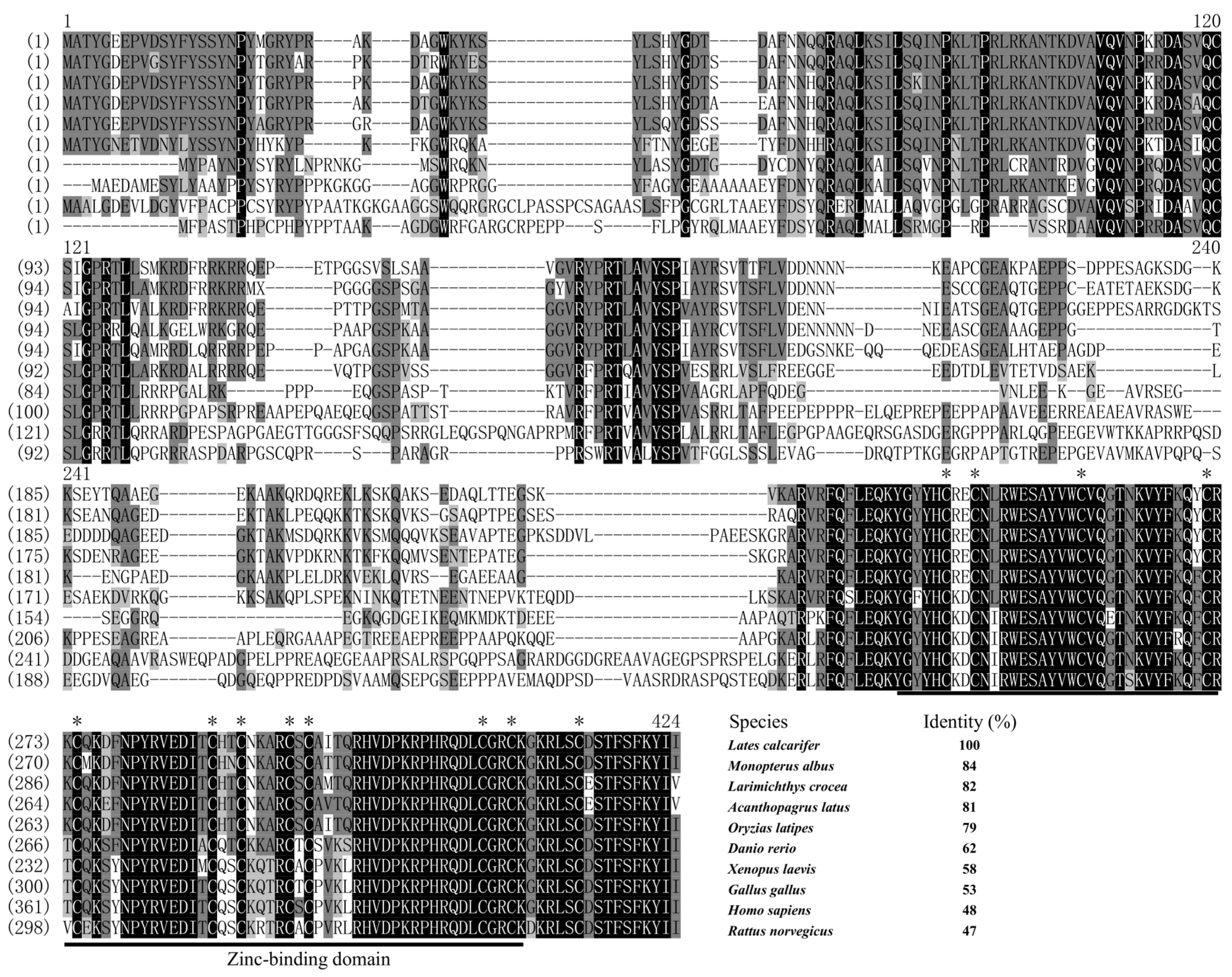
3.1. Sequence Alignment and Phylogenetic Analysis of zar1 and wt1b cDNA

A partial fragment of 1192 bp was isolated in Asian seabass via RT-PCR, and shared 99.08% identity with zar1 (XM_018669829.1) deposited in the public database (NCBI). The cloned zar1 cDNA fragment contains a partial 5′ UTR of 16 bp, an ORF of 1011 bp, and a partial 3′ UTR of 165 bp (Supplementary Materials: Figure S1A), encoding a peptide of 336 aa with a zinc-binding domain (Supplementary Materials: Figure S1A). Likewise, a cDNA fragment of 1521 bp was obtained via RT-PCR and shared 99.87% identity with wt1b (XM_018668041.2) retrieved from GenBank of NCBI. The isolated wt1b cDNA fragment was composed of a partial 23 bp 5’ UTR, a partial 226 bp 3’ UTR, and a 1272 bp ORF encoding a peptide of 423 aa with a Zn finger domain (Supplementary Materials: Figure S1B).
Multiple alignment of the Zar1 protein sequences revealed that LcZar1 shared a high identity of 84% with Monopterus albus, and a moderate identity of 62% with Danio rerio. However, LcZar1 shared a lower identity of 47% with Rattus norvegicus (Figure 1). Furthermore, the results showed a highly conserved zinc-binding domain in the C-terminal among LcZar1 and its homologs (Figure 1). The Wt1 protein sequence alignment analysis revealed that LcWt1b shared a high identity of 95% with Acanthopagrus latus Wt1b and a moderate identity of 86% with Oryzias latipes Wt1b (Figure 2), showing that Wt1 protein has a conserved zinc finger structure (Figure 2).
Phylogenetic analysis indicated that LcZar1 was clustered into a single clade with fish homologs, in contrast to other species (Figure 3A). LcWt1b was clustered with Wt1b homologs into a branch, while Wt1a homologs of fish and tetrapods were clustered into another branch (Figure 3B).

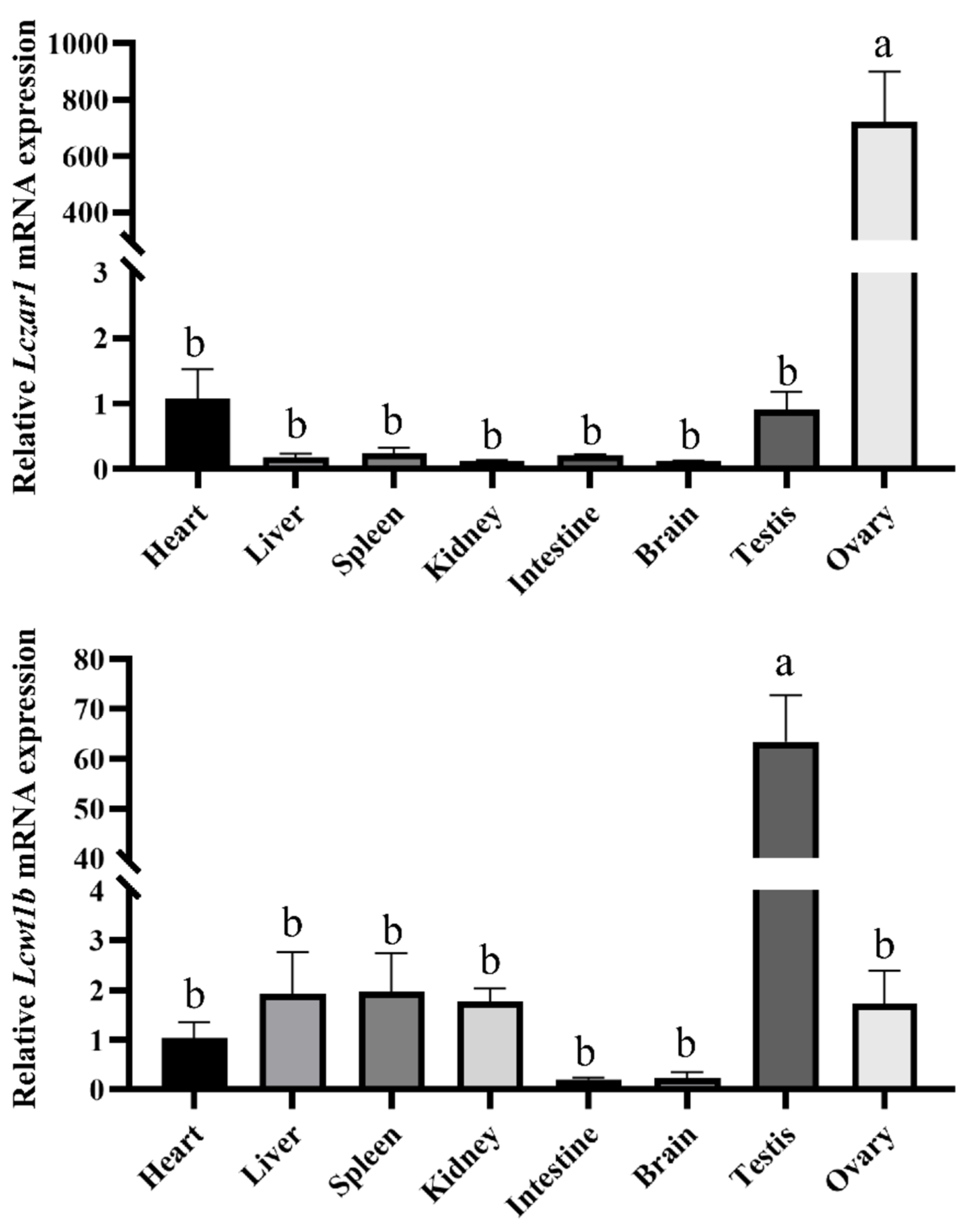
3.2. The mRNA Expression Profiles of Lczar1 and Lcwt1b

In order to examine the expression levels of Lczar1 and Lcwt1b in different tissues of seabass, RT-qPCR (real-time quantitative PCR) was conducted. As shown in Figure 4A, Lczar1 was significantly highly expressed in the ovary, in a concentration over 700 times higher than that in the heart and testis, but was barely detected in other tissues examined in this study (Figure 4A). However, Lcwt1b was significantly expressed highly in the testis, and the expression level of Lcwt1b in the testis was about 60 times higher than that in the ovary. Furthermore, Lcwt1b was also partially detected in other tissues including the heart, liver, spleen, and kidney examined in this study, but was scarcely detected in the intestine and brain (Figure 4B).

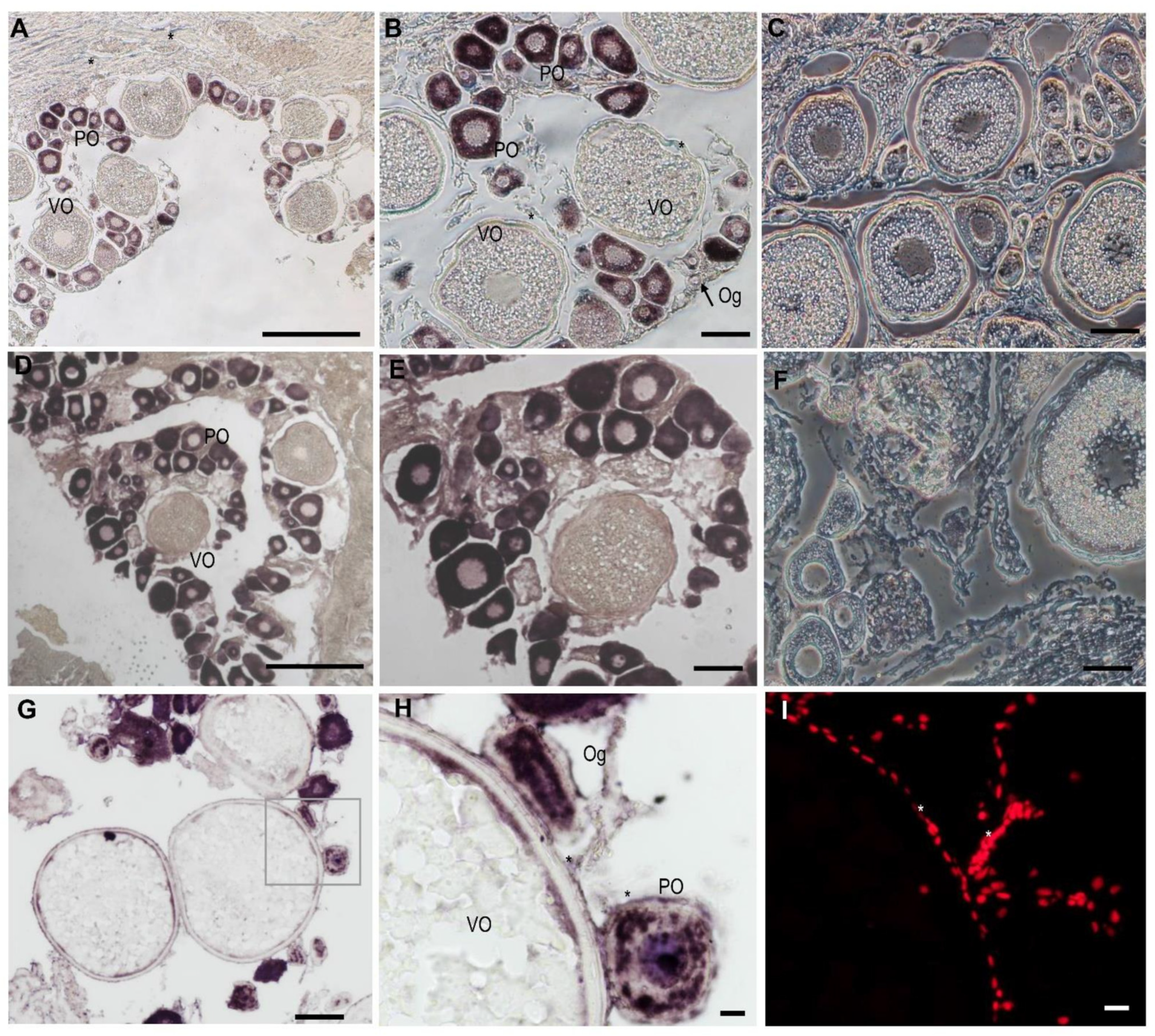
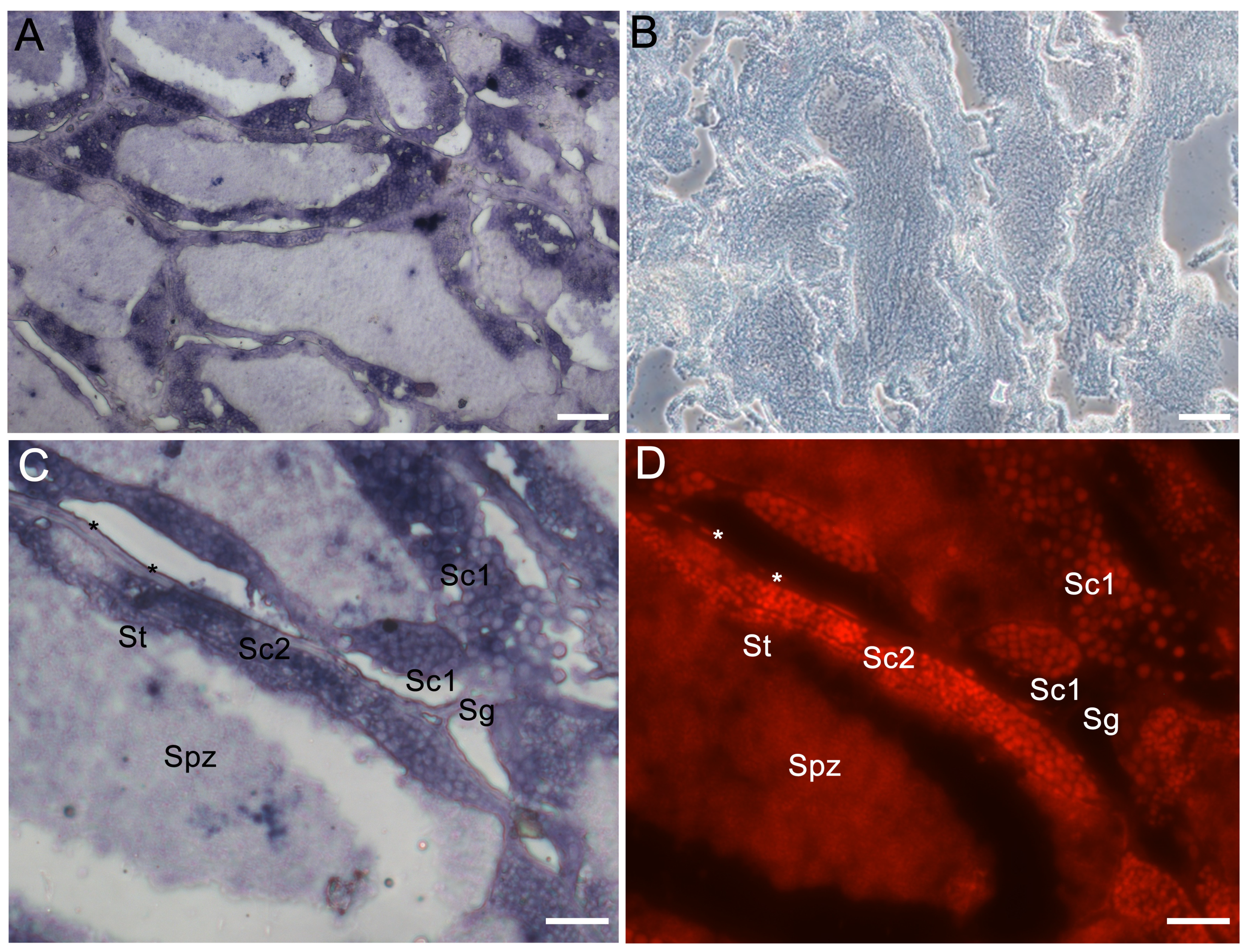
3.3. The Cellular Localization of Lczar1 and Lcwt1b in Gonads

In order to detect the cellular distribution of the Lczar1 and Lcwt1b mRNA in the gonad tissues, chemical ISH was performed. The results showed that the signal of Lczar1 mRNA was obviously observed in oogonia and oocytes at the primary growth stage, but scarcely detected in oocytes at the vitellogenic stage (Figure 5A,B), and there was no signal in the ovary detected via the Lczar1 sense riboprobe (Figure 5C). In particular, the Lczar1 mRNA signal was detected to be the strongest in oocytes at the primary growth stage, moderate in oogonia, and almost undetectable in oocytes at the vitellogenic stage (Figure 5A,B). Moreover, the cellular distribution of Lcwt1b mRNA in the ovary showed a different pattern from that of Lczar1 mRNA. The Lcwt1b mRNA signal was detected in oogonia and oocytes at the primary growth stage and as well as in some somatic cells (Figure 5D,E,G–I), and there was no signal in the ovary detected by the sense riboprobes of Lcwt1b (Figure 5F). Particularly, the signals in oogonia and primary growth stage oocytes were much stronger, but almost no signal was detected in vitellogenic-stage oocytes (Figure 5D,E,G,H).
In the testis, the sense and antisense probe of Lczar1 did not detect any signal in spermatogonia, spermatocytes, or spermatids (data not shown). However, the Lcwt1b mRNA signals detected by the antisense probe were mainly localized in spermatogonia, spermatocytes, spermatids, and some somatic cells, such as Sertoli cells, Leydig cells, and so on (Figure 6A,C,D), while the sense probe of Lcwt1b mRNA did not detect any signal in the testis (Figure 6B).

4. Discussion

In this study, LcZar1 protein had a conserved zinc-binding domain and LcWt1b protein had a conserved zinc finger structure, similarly to their homologs in different species, indicating that Zar1 and Wt1 are evolutionarily conserved. Additionally, sequence alignment showed that LcZar1 and LcWt1b had the highest homology with the orthologs in fish, and the highly conserved domain of Zar1 was the C12 structure, being essential for transcriptional regulation [10,11,51]. There is only one isoform, Wt1, in tetrapod vertebrates, while two are identified in teleost fish, namely Wt1a and Wt1b. Moreover, LcWt1b was clustered with the Wt1b homologs from other species, but obviously separated from the branch of fish and tetrapods’ Wt1a homologs, suggesting that LcWt1b is a member of the Wt1b family.
RT-qPCR analysis showed that both Lczar1 and Lcwt1b were primarily expressed in the gonads, but exhibited different expression profiles in adult tissues of Asian seabass. Lczar1 was expressed extremely highly in the ovary, which is consistent with previous studies [52], suggesting that Lczar1 possibly functions majorly in the ovary of the Asian seabass. In eel and Spotted scat, it was found that zar1 was strongly expressed in the ovary, but undetectable in other tissues [27,53]. In Japanese eel, zar1 was also mainly expressed in the ovary [28]. In rainbow trout, zar1 was predominantly expressed in the ovary and testis, with slight expression in other somatic tissues [26,54]. However, in frogs, zar1 is highly expressed not only in the ovaries, but also in the lungs and muscles [12]. In mice, zar1 is specifically expressed in the ovary [11,12,19]. In bovine tissues, zar1 is expressed not only in the ovary, but also in the heart, muscle, and testis [13,20]. Some studies also showed that zar1 is expressed in the hypothalamus part of the brain [14]. In pig tissues, zar1 is expressed not only in the ovary, but also in the testis, pituitary gland, and part of the hypothalamus [14]. In New Zealand rabbits, zar1 is expressed in the lung, heart, liver, spleen, kidney, ovary, and uterus, and has the highest expression in the lung [15]. In addition, studies have shown that methylation of zar1 is associated with cancer [22,23,24]. These studies show that zar1 is mainly expressed in the gonads of fish, while in higher animals, it shows different expression patterns. It is indicated that the spatial and temporary expressions of zar1 are divergent among species and may have species-specific functions. Unlike zar1, in this study, Lcwt1b was significantly highly expressed in the testis. The expression of Lcwt1b in the testis was about 30–60 times higher than that in the ovary, heart, liver, spleen, and kidney, but Lcwt1b mRNA was scarcely detected in the tissues of the intestine and brain, implying that Lcwt1b plays a more important role in the testis than in the ovary. As a male precocious hermaphrodite fish, it is suggested that in Asian seabass, Lcwt1b may play a role in the process of sex reversal. Wt1 was found to be related to tumors in earlier studies, but recent studies verify that it also functions in the development of animal gonads. In mice, wt1 is strongly expressed in the ovary and testis, but also in the heart, spleen, kidney, muscle, eyes, and gills [37]. In zebrafish, wt1a and wt1b are strongly expressed in the ovary, testis, spleen, and kidney, and also in the heart, skin, and muscle; in addition, wt1a is weakly expressed in the liver, and wt1b is weakly expressed in eyes and gills [37]. In medaka adult tissues, Wt1a and Wt1b showed an overlapping expression in the heart, kidney, spleen, testis, and ovary [44]. It was found that wt1a was expressed in the kidney, ovary, and spleen of adult eels [55]. In our present study, the expression profile of Lcwt1a and Lcwt1b were examined in a panel of adult fish tissues via RT-qPCR, and the results showed that Lcwt1a mRNA was widely expressed in the kidney, spleen, ovary, and testis, while Lcwt1b was expressed mainly in the testis and then the ovary. Thus, here, we just focused on investigating the Lcwt1b gene in Asian seabass. Additionally, Lcwt1b showed a different expression pattern in adult tissue from those of other species, which may be due to the Asian seabass being a male precocious hermaphrodite fish; however, to address these issues, more extensive investigations on Lcwt1b gene will be carried out in our future work. In other words, wtla and wt1b are genes that are paralogous with each other, and their expression patterns showed some divergences but also some consistency in different teleosts, which suggests that they may both both be involved in the development of gonads and other organs in different fish species with various roles.
ISH showed that the signal of Lczar1 was obvious in oogonia and primary-growth-stage oocytes, and was almost undetectable in vitellogenic-stage oocytes. Specifically, the signal was strongest in primary-growth-stage oocytes. It is indicated that Lczar1 may play a crucial role in the early stage of oogenesis. The expression profile of Lczar1 is similar to that in the early oocytes of zebrafish [18]. In the frog ovary, zar1 is also detected in early oocytes [12]. In mice, zar1 mRNA is detected in the early primary follicle through the antral follicle stages, but not in primordial follicles [11]. In Xenopus laevis, the expression level of zar1 reaches the highest level in oocytes of stage I to III, and then decreases in stages IV to VI [17]. In rainbow trout, zar1 is mainly expressed in metaphase II oocytes [26]. At the same time, in a study of spotted scat, it was found that the expression of zar1 gradually increases with the development of the ovary, reaching its highest expression in Phase IV ovaries [27]. In Japanese eel, the expression of zar1 gradually increases after ovarian differentiation, and is relatively high in Phase III–V ovaries [28]. These studies show that zar1 in different species is expressed in early oocytes, including monoecious fish such as Asian seabass, which reveals that zar1 plays a vital role in early oogenesis.
On the contrary, Lcwt1b was predominantly expressed in the testis. Particularly, Lcwt1b was expressed in the somatic cells and germ cells of the ovary and testis, respectively. In mice, it was found that the expression of wt1b was detected in the embryonic mesoderm (such as the genitourinary ridge, gonad, and mesonephros) [56]. In mouse gonads, wt1 is expressed in follicular granulosa cells, ovarian epithelial cell layer and oviduct fimbria, and also in Sertoli cells (SCs) in testicular convoluted seminiferous tubules [57]. In humans, wt1 is only expressed by SCs in the adult testicular seminiferous epithelium [58], but also by endothelial cells of micro vessels and mesenchymal cells in fetal and neonatal testicular stroma [59]. In the testis of adult Medaka, wt1a expression is detected in somatic cells probably in SCs, whereas wt1b mRNA transcripts are restricted to the somatic cells of the testis periphery, being either germ cell-supporting cells or SCs [44]. Recently, Chen et al. believed that granulosa cells in the ovary and SCs in the testis are differentiated from the somatic cells of the genital ridge under the guidance of wt1 [9]. As a female homozygous fish, Lcwt1b is expressed in somatic cells of testis and ovary, indicating that it may play an imperative role in the whole life cycle of Asian seabass, but the mechanism behind this in the process of sex reversal of Asian seabass is unclear. In addition, Lcwt1b was also expressed in oogonia and primary-growth-stage oocytes in the ovary, and in spermatocytes and spermatids in the testis, but the expression of Lcwt1b gradually decreased with the development of gametogenesis. Therefore, Lczar1 and Lcwt1b may work in the early stage of oogenesis and spermatogenesis.

5. Conclusions

In summary, Lczar1 and Lcwt1b were predominantly expressed in gonads and speculated to function in the gametogenesis of the androgynous fish Asian seabass. Our findings will lay a foundation for studying the molecular mechanisms of germ cell development, gametogenesis, and sex reversal in hermaphroditic fish.

Supplementary Materials

The following supporting information can be downloaded at: https://www.mdpi.com/article/10.3390/ani14030508/s1. Figure S1. The cDNA sequences and predicted peptides of Lczar1 (A) and Lcwt1b (B).

Author Contributions

H.X. and K.C. designed and supervised this study, H.C., H.Z., W.B. and R.C. conducted the experiment, L.L. and X.Z. analyzed the data. Y.L. and K.C. wrote the original manuscript. H.X. and K.C. edited the manuscript. All authors have read and agreed to the published version of the manuscript.

Funding

This study was supported by the National Key Research and Development Program of China [grant number 2022YFD2400902]; the National Natural Science Foundation of China [grant number 31873035]; Fundamental Research Funds for the Central Universities [grant number SWU020014]; Natural Science Foundation of Chongqing City [grant number CSTB2023NSCQ-MSX0325].

Institutional Review Board Statement

This article does not contain any studies with human participants. All animals used in this study were purchased from market, and the experimental protocols adhered to the Guidelines of Animal Care and approved by the Animal Management and Ethics Committee of Southwest University.

Informed Consent Statement

Not applicable.

Data Availability Statement

The data that support the findings of this study are included in the manuscript or Supplementary Materials.

Conflicts of Interest

All authors declare that they have no conflicts of interests.

References

  1. Dodson, A.E.; Kennedy, S. Phase separation in germ cells and development. Dev. Cell 2020, 55, 4–17. [Google Scholar] [CrossRef] [PubMed]
  2. Robles, V.; Riesco, M.F.; Psenicka, M.; Saito, T.; Valcarce, D.G.; Cabrita, E.; Herráez, P. Biology of teleost primordial germ cells (PGCs) and spermatogonia: Biotechnological applications. Aquaculture 2017, 472, 4–20. [Google Scholar] [CrossRef]
  3. Williamson, A.; Lehmann, R. Germ cell development in Drosophila. Annu. Rev. Cell Dev. Biol. 1996, 12, 365–391. [Google Scholar] [CrossRef] [PubMed]
  4. Zhong, C.; Liu, M.; Tao, Y.; Wu, X.; Yang, Y.; Wang, T.; Meng, Z.; Xu, H.; Liu, X. Pou5f1 and Nanog are reliable germ cell-specific genes in gonad of a protogynous hermaphroditic fish, orange-spotted grouper (Epinephelus coioides). Genes 2022, 13, 79. [Google Scholar] [CrossRef] [PubMed]
  5. Qu, L.; Wu, X.; Liu, M.; Zhong, C.; Xu, H.; Li, S.; Lin, H.; Liu, X. Identification and characterization of germ cell genes vasa and dazl in a protogynous hermaphrodite fish, orange-spotted grouper (Epinephelus coioides). Gene Expr. Patterns 2020, 35, 119095. [Google Scholar] [CrossRef] [PubMed]
  6. Sun, Z.-H.; Wang, Y.; Lu, W.-J.; Li, Z.; Liu, X.-C.; Li, S.-S.; Zhou, L.; Gui, J.-F. Divergent expression patterns and function implications of four nanos genes in a hermaphroditic fish, Epinephelus coioides. Int. J. Mol. Sci. 2017, 18, 685. [Google Scholar] [CrossRef] [PubMed]
  7. Zhao, H.; Duan, J.; Cheng, N.; Nagahama, Y. Specific expression of Olpiwi1 and Olpiwi2 in medaka (Oryzias latipes) germ cells. Biochem. Biophys. Res. Commun. 2012, 418, 592–597. [Google Scholar] [CrossRef]
  8. Li, X.-Y.; Mei, J.; Ge, C.-T.; Liu, X.-L.; Gui, J.-F. Sex determination mechanisms and sex control approaches in aquaculture animals. Sci. China Life Sci. 2022, 65, 1091–1122. [Google Scholar] [CrossRef]
  9. Chen, M.; Cen, C.; Wang, N.; Shen, Z.; Wang, M.; Liu, B.; Li, J.; Cui, X.; Wang, Y.; Gao, F. The functions of Wt1 in mouse gonad development and somatic cells differentiation. Biol. Reprod. 2022, 107, 269–274. [Google Scholar] [CrossRef]
  10. Wu, Y.-K.; Fan, H.-Y. Revisiting ZAR proteins: The understudied regulator of female fertility and beyond. Cell. Mol. Life Sci. 2022, 79, 92. [Google Scholar] [CrossRef]
  11. Wu, X.M.; Viveiros, M.M.; Eppig, J.J.; Bai, Y.C.; Fitzpatrick, S.L.; Matzuk, M.M. Zygote arrest 1 (Zar1) is a novel maternal-effect gene critical for the oocyte-to-embryo transition. Nat. Genet. 2003, 33, 187–191. [Google Scholar] [CrossRef]
  12. Wu, X.; Wang, P.; Brown, C.A.; Zilinski, C.A.; Matzuk, M.M. Zygote arrest 1 (Zar1) is an evolutionarily conserved gene expressed in vertebrate ovaries. Biol. Reprod. 2003, 69, 861–867. [Google Scholar] [CrossRef]
  13. Brevini, T.A.L.; Cillo, F.; Colleoni, S.; Lazzari, G.; Galli, C.; Gandolfi, F. Expression pattern of the maternal factor zygote arrest 1 (Zar1) in bovine tissues, oocytes, and embryos. Mol. Reprod. Dev. 2004, 69, 375–380. [Google Scholar] [CrossRef]
  14. Uzbekova, S.; Roy-Sabau, M.; Dalbiès-Tran, R.; Perreau, C.; Papillier, P.; Mompart, F.; Thelie, A.; Pennetier, S.; Cognie, J.; Cadoret, V.; et al. Zygote arrest 1 gene in pig, cattle and human: Evidence of different transcript variants in male and female germ cells. Reprod. Biol. Endocrinol. 2006, 4, 12. [Google Scholar] [CrossRef]
  15. Wang, D.A.N.; Xie, S.-Y.; Zhang, W.E.I.; Sun, C.-X.; Huang, T.A.O.; Wang, A.-S.; Han, X.-L.; Sun, G.-R.; Li, M. Cloning and expression analysis of zygote arrest 1 (Zar1) in New Zealand white rabbits. J. Genet. 2017, 96, 3–8. [Google Scholar] [CrossRef] [PubMed]
  16. Michailidis, G.; Argiriou, A.; Avdi, M. Expression of chicken zygote arrest 1 (Zar1) and Zar1-like genes during sexual maturation and embryogenesis. Vet. Res. Commun. 2010, 34, 173–184. [Google Scholar] [CrossRef] [PubMed]
  17. Yamamoto, T.M.; Cook, J.M.; Kotter, C.V.; Khat, T.; Silva, K.D.; Ferreyros, M.; Holt, J.W.; Knight, J.D.; Charlesworth, A. Zar1 represses translation in Xenopus oocytes and binds to the TCS in maternal mRNAs with different characteristics than Zar2. Biochim. Biophys. Acta (BBA) Gene Regul. Mech. 2013, 1829, 1034–1046. [Google Scholar] [CrossRef] [PubMed]
  18. Miao, L.; Yuan, Y.; Cheng, F.; Fang, J.; Zhou, F.; Ma, W.; Jiang, Y.; Huang, X.; Wang, Y.; Shan, L.; et al. Translation repression by maternal RNA binding protein zar1 is essential for early oogenesis in zebrafish. Development 2017, 144, 128–138. [Google Scholar] [CrossRef] [PubMed]
  19. Rong, Y.; Ji, S.-Y.; Zhu, Y.-Z.; Wu, Y.-W.; Shen, L.; Fan, H.-Y. ZAR1 and ZAR2 are required for oocyte meiotic maturation by regulating the maternal transcriptome and mRNA translational activation. Nucleic Acids Res. 2019, 47, 11387–11402. [Google Scholar] [CrossRef] [PubMed]
  20. Pennetier, S.; Uzbekova, S.; Perreau, C.; Papillier, P.; Mermillod, P.; Dalbiès-Tran, R. Spatio-temporal expression of the germ cell marker genes mater, zar1, gdf9, bmp15,and vasa in adult bovine tissues, oocytes, and preimplantation embryos1. Biol. Reprod. 2004, 71, 1359–1366. [Google Scholar] [CrossRef] [PubMed]
  21. Sangiorgio, L.; Strumbo, B.; Brevini, T.A.L.; Ronchi, S.; Simonic, T. A putative protein structurally related to zygote arrest 1 (Zar1), Zar1-like, is encoded by a novel gene conserved in the vertebrate lineage. Comp. Biochem. Physiol. Part B Biochem. Mol. Biol. 2008, 150, 233–239. [Google Scholar] [CrossRef]
  22. Richter, A.M.; Kiehl, S.; Köger, N.; Breuer, J.; Stiewe, T.; Dammann, R.H. ZAR1 is a novel epigenetically inactivated tumour suppressor in lung cancer. Clin. Epigenetics 2017, 9, 60. [Google Scholar] [CrossRef]
  23. Shinojima, Y.; Terui, T.; Hara, H.; Kimura, M.; Igarashi, J.; Wang, X.; Kawashima, H.; Kobayashi, Y.; Muroi, S.; Hayakawa, S.; et al. Identification and analysis of an early diagnostic marker for malignant melanoma: ZAR1 intra-genic differential methylation. J. Dermatol. Sci. 2010, 59, 98–106. [Google Scholar] [CrossRef]
  24. Watanabe, T.; Yachi, K.; Ohta, T.; Fukushima, T.; Yoshino, A.; Katayama, Y.; Shinojima, Y.; Terui, T.; Nagase, H. Aberrant hypermethylation of non-promoter zygote arrest 1 (Zar1) in human brain tumors. Neurol. Med. Chir. 2010, 50, 1062–1069. [Google Scholar] [CrossRef]
  25. Pan, Z.; Zhu, C.; Chang, G.; Wu, N.; Ding, H.; Wang, H. Differential expression analysis and identification of sex-related genes by gonad transcriptome sequencing in estradiol-treated and non-treated Ussuri catfish Pseudobagrus ussuriensis. Fish Physiol. Biochem. 2021, 47, 565–581. [Google Scholar] [CrossRef] [PubMed]
  26. Bobe, J.; Nguyen, T.; Mahé, S.; Monget, P. In silico identification and molecular characterization of genes predominantly expressed in the fish oocyte. BMC Genom. 2008, 9, 499. [Google Scholar] [CrossRef]
  27. He, F.; Jiang, D.; Chen, H.; Deng, S.; Wu, T.; Tian, C.; Shi, H.; Zhu, C.; LI, G. cDNA Cloning and mRNA expression analysis of zar1 in spotted scat (Scatophagus argus). J. Guangdong Ocean Univ. 2019, 39, 1–7. [Google Scholar]
  28. Horiuchi, M.; Hagihara, S.; Kume, M.; Chushi, D.; Hasegawa, Y.; Itakura, H.; Yamashita, Y.; Adachi, S.; Ijiri, S. Morphological and molecular gonadal sex differentiation in the wild Japanese eel Anguilla japonica. Cells 2022, 11, 1554. [Google Scholar] [CrossRef]
  29. Natoli, T.A.; Alberta, J.A.; Bortvin, A.; Taglienti, M.E.; Menke, D.B.; Loring, J.; Jaenisch, R.; Page, D.C.; Housman, D.E.; Kreidberg, J.A. Wt1 functions in the development of germ cells in addition to somatic cell lineages of the testis. Dev. Biol. 2004, 268, 429–440. [Google Scholar] [CrossRef] [PubMed]
  30. Hammes, A.; Guo, J.K.; Lutsch, G.; Leheste, J.R.; Landrock, D.; Ziegler, U.; Gubler, M.C.; Schedl, A. Two splice variants of the Wilms’ tumor 1 gene have distinct functions during sex determination and nephron formation. Cell 2001, 106, 319–329. [Google Scholar] [CrossRef]
  31. Call, K.M.; Glaser, T.; Ito, C.Y.; Buckler, A.J.; Pelletier, J.; Haber, D.A.; Rose, E.A.; Kral, A.; Yeger, H.; Lewis, W.H.; et al. Isolation and characterization of a zinc finger polypeptide gene at the human chromosome 11 Wilms’ tumor locus. Cell 1990, 63, 509–520. [Google Scholar] [CrossRef]
  32. Cen, C.; Chen, M.; Zhou, J.; Zhang, L.; Duo, S.; Jiang, L.; Hou, X.; Gao, F. Inactivation of Wt1 causes pre-granulosa cell to steroidogenic cell transformation and defect of ovary development. Biol. Reprod. 2020, 103, 60–69. [Google Scholar] [CrossRef]
  33. Chen, M.; Zhang, L.; Cui, X.; Lin, X.; Li, Y.; Wang, Y.; Wang, Y.; Qin, Y.; Chen, D.; Han, C.; et al. Wt1 directs the lineage specification of sertoli and granulosa cells by repressing Sf1 expression. Development 2017, 144, 44–53. [Google Scholar] [CrossRef]
  34. Gao, F.; Zhang, J.; Wang, X.; Yang, J.; Chen, D.; Huff, V.; Liu, Y.X. Wt1 functions in ovarian follicle development by regulating granulosa cell differentiation. Hum. Mol. Genet. 2014, 23, 333–341. [Google Scholar] [CrossRef]
  35. Wang, X.N.; Li, Z.S.; Ren, Y.; Jiang, T.; Wang, Y.Q.; Chen, M.; Zhang, J.; Hao, J.X.; Wang, Y.B.; Sha, R.N.; et al. The Wilms tumor gene, Wt1, is critical for mouse spermatogenesis via regulation of sertoli cell polarity and is associated with non-obstructive azoospermia in humans. PLoS Genet. 2013, 9, e1003645. [Google Scholar] [CrossRef]
  36. Zhang, L.; Chen, M.; Wen, Q.; Li, Y.; Wang, Y.; Wang, Y.; Qin, Y.; Cui, X.; Yang, L.; Huff, V.; et al. Reprogramming of Sertoli cells to fetal-like Leydig cells by Wt1 ablation. Proc. Natl. Acad. Sci. USA 2015, 112, 4003–4008. [Google Scholar] [CrossRef] [PubMed]
  37. Bollig, F.; Mehringer, R.; Perner, B.; Hartung, C.; Schäfer, M.; Schartl, M.; Volff, J.N.; Winkler, C.; Englert, C. Identification and comparative expression analysis of a second wt1 gene in zebrafish. Dev. Dyn. 2006, 235, 554–561. [Google Scholar] [CrossRef] [PubMed]
  38. Hsu, H.-J.; Lin, G.; Chung, B.-c. Parallel early development of zebrafish interrenal glands and pronephros:differential control bywt1andff1b. Development 2003, 130, 2107–2116. [Google Scholar] [CrossRef] [PubMed]
  39. Wagner, K.-D.; Wagner, N.; Schedl, A. The complex life of WT1. J. Cell Sci. 2003, 116, 1653–1658. [Google Scholar] [CrossRef] [PubMed]
  40. Wang, X.; Meng, K.; Wang, Y.; Zhao, Y.; Lin, F.; Liu, X.; Zhang, Y.; Quan, F. Wilms’ tumor (WT1) (±KTS) variants decreases the progesterone secretion of bovine ovarian theca cells. Domest. Anim. Endocrinol. 2021, 74, 106521. [Google Scholar] [CrossRef] [PubMed]
  41. Kirschner, K.M.; Scholz, H. WT1 in adipose tissue: From development to adult physiology. Front. Cell Dev. Biol. 2022, 10, 854120. [Google Scholar] [CrossRef] [PubMed]
  42. Rao, M.K.; Pham, J.; Imam, J.S.; MacLean, J.A.; Murali, D.; Furuta, Y.; Sinha-Hikim, A.P.; Wilkinson, M.F. Tissue-specific RNAi reveals that WT1 expression in nurse cells controls germ cell survival and spermatogenesis. Genes Dev. 2006, 20, 147–152. [Google Scholar] [CrossRef] [PubMed]
  43. Moore, A.W.; McInnes, L.; Kreidberg, J.; Hastie, N.D.; Schedl, A. YAC complementation shows a requirement for Wt1 in the development of epicardium, adrenal gland and throughout nephrogenesis. Development 1999, 126, 1845–1857. [Google Scholar] [CrossRef] [PubMed]
  44. Klüver, N.; Herpin, A.; Braasch, I.; Driessle, J.; Schartl, M. Regulatory back-up circuit of medaka Wt1 co-orthologs ensures PGC maintenance. Dev. Biol. 2009, 325, 179–188. [Google Scholar] [CrossRef] [PubMed]
  45. Athauda, S.; Anderson, T. Effect of temperature and salinity on sex inversion in Asian seabass (Lates calcarifer): Relationship with plasma sex steroids concentration and aromatase activity of gonad and brain. Aquac. Res. 2014, 45, 787–797. [Google Scholar] [CrossRef]
  46. Banh, Q.Q.T.; Guppy, J.L.; Domingos, J.A.; Budd, A.M.; Pinto, R.C.C.; Marc, A.F.; Jerry, D.R. Induction of precocious females in the protandrous barramundi (Lates calcarifer) through implants containing 17β-estradiol-effects on gonadal morphology, gene expression and DNA methylation of key sex genes. Aquaculture 2021, 539, 736601. [Google Scholar] [CrossRef]
  47. Roberts, B.H.; Morrongiello, J.R.; Morgan, D.L.; King, A.J.; Saunders, T.M.; Crook, D.A. Faster juvenile growth promotes earlier sex change in a protandrous hermaphrodite (barramundi Lates calcarifer). Sci. Rep. 2021, 11, 2276. [Google Scholar] [CrossRef]
  48. Domingos, J.A.; Budd, A.M.; Banh, Q.Q.; Goldsbury, J.A.; Zenger, K.R.; Jerry, D.R. Sex-specific dmrt1 and cyp19a1 methylation and alternative splicing in gonads of the protandrous hermaphrodite barramundi. PLoS ONE 2018, 13, e0204182. [Google Scholar] [CrossRef]
  49. Ravi, P.; Jiang, J.; Liew, W.C.; Orbán, L. Small-scale transcriptomics reveals differences among gonadal stages in Asian seabass (Lates calcarifer). Reprod. Biol. Endocrinol. 2014, 12, 5. [Google Scholar] [CrossRef]
  50. Xu, H.; Lim, M.; Dwarakanath, M.; Hong, Y. Vasa identifies germ cells and critical stages of oogenesis in the Asian seabass. Int. J. Biol. Sci. 2014, 10, 225–235. [Google Scholar] [CrossRef] [PubMed]
  51. Bienz, M. The PHD finger, a nuclear protein-interaction domain. Trends Biochem. Sci. 2006, 31, 35–40. [Google Scholar] [CrossRef]
  52. Liu, X.; Li, W.; Yang, Y.; Chen, K.; Li, Y.; Zhu, X.; Ye, H.; Xu, H. Transcriptome profiling of the ovarian cells at the single-cell resolution in adult Asian seabass. Front. Cell Dev. Biol. 2021, 9, 647892. [Google Scholar] [CrossRef]
  53. Geffroy, B.; Guilbaud, F.; Amilhat, E.; Beaulaton, L.; Vignon, M.; Huchet, E.; Bardonnet, A. Sexually dimorphic gene expressions in eels: Useful markers for early sex assessment in a conservation context. Sci. Rep. 2016, 6, 34041. [Google Scholar] [CrossRef] [PubMed]
  54. Lin, C.-H.; Wang, L.; Ma, H.; Hostuttler, M.; Rexroad, C.E.; Yao, J. Molecular characterization and expression analysis of zar1 and zar1-like genes in rainbow trout. Biol. Reprod. 2012, 87, 304. [Google Scholar] [CrossRef]
  55. Nakatsuru, Y.; Minami, K.; Yoshikawa, A.; Zhu, J.-J.; Oda, H.; Masahito, P.; Okamoto, N.; Nakamura, Y.; Ishikawa, T. Eel WT1 sequence and expression in spontaneous nephroblastomas in Japanese eel. Gene 2000, 245, 245–251. [Google Scholar] [CrossRef] [PubMed]
  56. Armstrong, J.; Pritchardjones, K.; Bickmore, W.; Hastie, N.; Bard, J. The expression of the Wilms-tumor gene, Wt1, in the developing mammalian embryo. Mech. Dev. 1993, 40, 85–97. [Google Scholar] [CrossRef] [PubMed]
  57. Pelletier, J.; Schalling, M.; Buckler, A.; Rogers, A.; Haber, D.; Housman, D. Expression of the Wilms-tumor gene Wt1 in the murine urogenital system. Genes. Dev. 1991, 5, 1345–1356. [Google Scholar] [CrossRef] [PubMed]
  58. Gao, F.; Maiti, S.; Alam, N.; Zhang, Z.; Deng, J.M.; Behringer, R.R.; Lecureuil, C.; Guillou, F.; Huff, V. The Wilms tumor gene, Wt1, is required for Sox9 expression and maintenance of tubular architecture in the developing testis. Proc. Natl. Acad. Sci. USA 2006, 103, 11987–11992. [Google Scholar] [CrossRef]
  59. Zheng, Q.-S.; Wang, X.-N.; Wen, Q.; Zhang, Y.; Chen, S.-R.; Zhang, J.; Li, X.-X.; Sha, R.-N.; Hu, Z.-Y.; Gao, F.; et al. Wt1 deficiency causes undifferentiated spermatogonia accumulation and meiotic progression disruption in neonatal mice. Reproduction 2014, 147, 45–52. [Google Scholar] [CrossRef]
Figure 1. Multiple-sequence alignment analysis of Zar1 proteins in vertebrates. Black shadow: 100% identity; Grey shadow: 75% ≤ identity < 100%. Black underline: Zinc-binding domain. The positions shown by asterisks (*) are conservative C12 structures. At the end of the comparison, the species name and the identity percentage of Zar1 and its homologues were displayed. GenBank accession numbers for Zar1 are as follows: Monopterus albus: XP_020459078.1; Acanthopagrus latus: XP_036969674.1; Larimichthys crocea: XP_010729299.3; Oryzias latipes: XP_004067611.1; Danio rerio: NP_919362.2; Xenopus laevis: NP_001083958.1; Gallus gallus: XP_040527566.1; Homo sapiens: NP_783318.1: Rattus norvegicus: NP_852050.1.
Animals 14 00508 g001
Figure 2. Multiple sequence alignment analysis of Wt1 proteins in vertebrates. Black shadow: 100% identity; grey shadow: 75% ≤ identity < 100%; light-grey shadow: 50% ≤ identity < 75%. Black underline: Zn-finger. Red box: Wt1(-KTS) or Wt1(+KTS). At the end of the comparison, the species name and the identity percentage of Wt1 and its homologues were displayed. GenBank accession numbers for Wt1 are as follows: Monopterus albus Wt1a: XP_020460123.1; Monopterus albus Wt1b: XP_020471332.1; Acanthopagrus latus Wt1a: XP_036963987.1; Acanthopagrus latus Wt1b: XP_036951482.1; Larimichthys crocea Wt1a: XP_027128154.1; Larimichthys crocea Wt1b: XP_027137243.1; Oryzias latipes Wt1a: ABG36853.1; Oryzias latipes Wt1b: ABG36854.1; Danio rerio Wt1a: AAI62638.1; Danio rerio Wt1b: AAI24088.1; Xenopus laevis Wt1: NP_001079057.1; Gallus gallus Wt1: NP_990547.2; Rattus norvegicus Wt1: NP_113722.2; Homo sapiens Wt1: AAH32861.1.
Animals 14 00508 g002
Figure 3. Phylogenetic trees of Zar1 (A) and Wt1 (B) proteins. Phylogenetic trees were deduced using MEGA 7.0 using the neighbor-joining method with 1000 bootstrap replicates. Numerals at the bifurcation points of the phylogenetic tree are bootstrap values. GenBank accession numbers for Zar1 and Wt1 are the same as those in Figure 1 and Figure 2.
Animals 14 00508 g003
Figure 4. The mRNA expression profiles of Lczar1 and Lcwt1b in adult Seabass tissues. RT-qPCR analyses were conducted, and Lcβ-actin was used as the internal reference gene. The relative mRNA expression levels of target genes were calculated using 2−∆∆CT, and the control was set as the heart tissue. Each column represents the means ± standard deviations (SD) from three biological replicates. Different lowercase letters indicated a significant difference at p < 0.05 via the method of one-way ANOVA and Tukey in SPSS 20.0.
Animals 14 00508 g004
Figure 5. Cellular distribution of Lczar1 and Lcwt1b mRNA in the adult ovary of the Asian seabass. The chemical ISH analysis were performed on the ovarian sections (signals in purple). (A,B) Antisense probe signal of Lczar1; (C) sense probe signal of Lczar1; (D,E,G,H) antisense probe signal of Lczar1; (F) sense probe signal of Lcwt1b; (H) antisense probe signal amplified from the frame of (G); (I) nuclei of ovarian cells counterstained with PI (in red). Og, oogonia; PO, primary-growth-stage oocytes; VO, vitellogenic-stage oocytes; asterisk (*), somatic cells. Scale bars: 50 μm.
Animals 14 00508 g005
Figure 6. Cellular distribution of Lcwt1b mRNA in the adult testis of the Asian seabass. The chemical ISH analysis was performed on testicular sections (signals in purple). (A,C) Antisense probe signal of Lcwt1b; (B) sense probe signal of Lcwt1b; (D) nuclei of testicular cells counterstained with PI (in red). Sg, spermatogonia; Sc1, primary spermatocytes; Sc2, secondary spermatocytes; St, spermatids; Spz, spermatozoa; asterisk (*), somatic cells. Scale bars: 50 μm in (A,B); 20 μm in (C,D).
Animals 14 00508 g006
Table 1. The sequences of primers used in this study.
Primer NameSequences (5′ to 3′)Product Length (bp)Tm (°C)Purpose
Lczar1-CDSForwardCGGTTGGTTGAACAAAATGGC119256ISH, RT-PCR
ReverseGTGCGGTTCTCACCATCAAT
Lcwt1b-CDSForwardACTGTCTCAAACCGCCTTCA152156ISH, RT-PCR
ReverseGCAAGCACTAGTTGAGGTGC
Lczar1-DLForwardCTACGATGGGAGAGTGCCTATG24760RT-qPCR
ReverseAGCTGAAAGTGCTGTCGCAGG
Lcwt1b-DLForwardACATGAGGACACCCTGTCACC28960RT-qPCR
ReverseGTACTGCGCACTGACGAGCAT
Lcβ-actin-DLForwardCGGAATCCACGAGACCACCTAC26860RT-qPCR
ReverseACTCCTGCTTGCTGATCCACAT
Disclaimer/Publisher’s Note: The statements, opinions and data contained in all publications are solely those of the individual author(s) and contributor(s) and not of MDPI and/or the editor(s). MDPI and/or the editor(s) disclaim responsibility for any injury to people or property resulting from any ideas, methods, instructions or products referred to in the content.

