Article

Retrospective Analysis of Archived Pyrazinamide Resistant Mycobacterium tuberculosis Complex Isolates from Uganda—Evidence of Interspecies Transmission

1
College of Veterinary Medicine, Animal Resources and Biosecurity, Makerere University, Kampala 0414, Uganda
2
Department of Veterinary Population Medicine, College of Veterinary Medicine, University of Minnesota, St. Paul, MN 55108, USA
3
Joint Clinical Research Center (JCRC), Lubowa 0414, Uganda
4
National Animal Disease Center, USDA, Ames, IA 50010, USA
5
Department of Veterinary Biomedical Sciences, College of Veterinary Medicine, University of Minnesota, St. Paul, MN 55108, USA
6
Department of Pathobiology and Diagnostic Investigation, College of Veterinary Medicine, Michigan State University, East Lansing, MI 48824, USA
*
Author to whom correspondence should be addressed.
Microorganisms 2019, 7(8), 221; https://doi.org/10.3390/microorganisms7080221
Submission received: 6 May 2019 / Revised: 15 July 2019 / Accepted: 24 July 2019 / Published: 29 July 2019

Abstract

:
The contribution of Mycobacterium bovis to the proportion of tuberculosis cases in humans is unknown. A retrospective study was undertaken on archived Mycobacterium tuberculosis complex (MTBC) isolates from a reference laboratory in Uganda to identify the prevalence of human M. bovis infection. A total of 5676 isolates maintained in this repository were queried and 136 isolates were identified as pyrazinamide resistant, a hallmark phenotype of M. bovis. Of these, 1.5% (n = 2) isolates were confirmed as M. bovis by using regions of difference PCR analysis. The overall size of whole genome sequences (WGSs) of these two M. bovis isolates were ~4.272 Mb (M. bovis Bz_31150 isolated from a captive chimpanzee) and 4.17 Mb (M. bovis B2_7505 from a human patient), respectively. Alignment of these genomes against 15 MTBC genome sequences revealed 7248 single nucleotide polumorphisms (SNPs). Theses SNPs were used for phylogenetic analysis that indicated a strong relationship between M. bovis and the chimpanzee isolate (Bz_31150) while the other M. bovis genome from the human patient (B2_7505) analyzed did not cluster with any M. bovis or M. tuberculosis strains. WGS analysis also revealed multidrug resistance genotypes; these genomes revealed pncA mutations at positions H57D in Bz_31150 and B2_7505. Phenotypically, B2_7505 was an extensively drug-resistant strain and this was confirmed by the presence of mutations in the major resistance-associated proteins for all anti-tuberculosis (TB) drugs, including isoniazid (KatG (S315T) and InhA (S94A)), fluoroquinolones (S95T), streptomycin (rrs (R309C)), and rifampin (D435Y, a rare but disputed mutation in rpoB). The presence of these mutations exclusively in the human M. bovis isolate suggested that these occurred after transmission from cattle. Genome analysis in this study identified M. bovis in humans and great apes, suggesting possible transmission from domesticated ruminants in the area due to a dynamic and changing interface, which has created opportunity for exposure and transmission.

1. Introduction

Mycobacterium bovis, a subspecies of the Mycobacterium tuberculosis complex (MTC), is a significant cause of bovine tuberculosis (bTB) in the developing world [1,2]. Each MTC member displays varied pathogenicity, geographical distribution, epidemiology, and host range [3]. MTCs, the causative agents of tuberculosis, are intracellular pathogens that have very low (0.01–0.03) synonymous nucleotide variation with no significant trace of genetic exchange among them [4]. In the developed world, bovine TB (bTB) has nearly been eradicated from domesticated animals, thus the risk of human transmission is low. This was achieved through mandatory testing of cattle, removal of positive reactors and in-contact animals, and compulsory pasteurization of milk [5,6,7]. In contrast, in resource-limited countries where the devastation of MTCs on humanity continues unabated, effective disease-control measures, like mandatory milk pasteurization and removal of positive reactors, is difficult to implement mainly due to economic factors [8,9,10,11,12]. For example, bTB is not reportable in many countries in sub-Saharan Africa and there are no compensatory mechanisms in place for farmers whose animals test positive for the disease [6,13,14,15].
The World Health Organization (WHO) estimates that two billion humans are infected with latent tuberculosis, with a 5% to 10% chance of developing active TB [16,17]. The brunt of this burden is largely borne by developing countries, with an approximate two million deaths annually, 95% of which are in low or medium-income countries [16]. The proportion of tuberculosis infections caused by M. bovis is not well-defined especially in resource-limited settings of the world. Prevalence estimates of M. bovis range from 5% to 16% [13,15]. In Uganda, routine TB testing does not assign species within MTC organisms, hence any human infection with an MTC is assumed to be due to Mycobacterium tuberculosis (sensu stricto). In 2015, Uganda had an estimated population of 39 million people, with about five laboratories that carry out routine TB screening with culture and drug sensitivity testing; these are all located in urban centers [18,19]. Staining for acid-fast bacilli (AFB) on sputum smears to screen for TB is carried out by a larger number of network laboratories. Animal TB diagnostics in Uganda are lagging far behind human TB diagnostics and available bTB information is based on passive surveillance data from abattoirs that are active in recording and submitting annual cattle slaughter reports [20]. Uganda does not have a national bovine TB eradication program and previous studies on bTB in Uganda have shown the presence of M. bovis among cattle belonging to the pastoral communities [20,21,22].
Even though M. tuberculosis is the most prevalent of the M. tuberculosis complex in Kampala, infections with M. tuberculosis or M. bovis are clinically and pathologically indistinguishable [13,23]. The challenge is physicians treat both diseases similarly although M. bovis is naturally resistant to pyrazinamide, one of the first-line TB drugs [24]. More importantly, species assignment of MTCs provides a possible eco-source of the agent that can aid in appropriate treatment and prevention. It is therefore imperative that MTC isolates are completely identified to the subspecies level in order to provide sufficient information on their epidemiology and enable appropriate treatment of the patients [24].
There is little documented information about the proportion of people infected by M. bovis in Uganda. This work was developed to determine and differentiate archived human MTC isolates, from a large reference laboratory, Joint Clinical Research Centre (JCRC), Kampala, Uganda, as M. bovis or M. tuberculosis (MTB). The JCRC is an HIV/AIDS care and research institution established in 1990 to provide a scientific approach to the national HIV/AIDS problem [25]. This retrospective study characterized archived pyrazinamide resistant isolates stored at the JCRC from 1999 through 2012.

2. Materials and Methods

2.1. Institutional Review Board (IRB) and Ethical Approvals

Permission to use isolates for molecular characterization was obtained from the research committee of the JCRC; the JCRC Uganda-Case Western Reserve University (CWRU) Research Collaboration and the Molecular Diagnostics Laboratory Department of Medical Microbiology, College of Health Sciences (CHS), Makerere University Kampala. Institutional Review Board (IRB) approval was obtained from the Makerere University, School of Biomedical Sciences Research and Ethics Committee. Scientific and ethical clearance was obtained from the Uganda National Council for Science and Technology (UNCST) reference number HS 1478. Since the work was a retrospective study using archived isolates, this work was exempt from University of Minnesota IRB approval.

2.2. Study Site

The archived MTC isolates were obtained from the JCRC in Kampala, Uganda [25]. Confirmatory tests to identify M. bovis were done at the Molecular Biology Lab in the Department of Microbiology, College of Health Sciences, Makerere University.

2.3. Study Samples

Pyrazinamide (PZA) resistant isolates were drawn from a collection of 5676 archived isolates collected between 1999 and 2012 from 19 different research studies previously conducted in partnership with key research collaborators at JCRC (Figures S1 and S2).
We first determined the proportion of M. tuberculosis isolates resistant to first-line anti-tuberculosis drugs (pyrazinamide, rifampin, isoniazid, and ethambutol and streptomycin), and then carried out comparative genomics. Furthermore, we sought to determine if there was a non-random distribution of genomotypes of MTC organisms infecting animals and humans in Uganda using a defined single nucleotide polymorphism (SNP) set to differentiate the Mycobacterium tuberculosis complex group of organisms and enable determination of drug resistance and characterization of these MTC isolates as drug resistant (DR), multi-drug resistant (MDR), or extremely drug resistant (XDR) organisms.

2.4. Sampling, Culture, and Isolation of Mycobacteria

All the work with the MTC isolates was carried out in biosafety level three (BSL-3) containment facilities. Archived MTC isolates from the JCRC were retrieved from −80 °C freezer stocks, thawed and inoculated into Middlebrook 7H9® broth, and incubated at 37 °C for 42 days [26,27]. The samples were sub-cultured on solid media, Middlebrook 7H10® (Beckton and Dickinson, Heidelberg, Germany) containing sodium pyruvate, and Oleic Acid-albumin-Dextrose-Catalase (OADC®) enrichment. In case of contaminating growth, the cultures were also sub-cultured on selective media, Middlebrook 7H11® containing sodium pyruvate, glucose, sterile calf serum, OADC enrichment, sheep blood, malachite green, and glycerin, and incubated at 37 °C. The cultures were inspected weekly and considered negative when there was no visible growth after 8 weeks. Ziehl Neelsen and auramine fluorescent staining and microscopy were carried out on the isolates to confirm that they were Mycobacterium sp.

2.5. Molecular Testing to Confirm MTC

Isolates were harvested and inoculated in Middlebrook 7H9® broth in cryovials and taken to the Molecular Diagnostic Laboratory, Department of Medical Microbiology, Makerere University for heat killing, DNA extraction, and PCR-based regions of difference (RD) analysis to differentiate M. bovis from other members of MTC. DNA was extracted by sonication and quantified using the GeneQuant® (Biochrom, Holliston, MA, USA) UV spectrophotometer (20–30 μg per isolate) (26). Genomic regions of difference (RD analysis) for RD1, RD9, RD4, and RD12 were used to differentiate among MTC species as described [24,28,29,30]. These RDs are regions within the MTC genome that represent a loss of genetic material in M. bovis compared to M. tuberculosis H37Rv [24,31,32].

2.6. DNA Extraction/DNA Preparation for PCR

Cells stored in 7H9 at −20 °C were harvested and heat killed at 95 °C for 1 h in a hybridization oven and DNA was extracted by sonication. Heat killed samples were centrifuged at 15,700 rcf or 13,000 rpm (Eppendorf 5415D Centrifuge; Eppendorf, Hauppauge, NY, USA) for 2 min in a microfuge, supernatant discarded, and pellet retained. To the pellet, 500 µL of Tris-EDTA buffer, pH 8, was added and centrifuged at 13,000 rpm for another 2 min and the pellet was retained after pouring off the supernatant. The pellet was re-suspended in 100 µL Tris-EDTA buffer and sonicated in an ultra-sonic bath (Bandelin Sonorex Ultrasonic BathRK100H (Schalltec, Moerfelden, Germany)) for 45 min. The resultant solution was centrifuged at 12,000 rpm for 10 min to obtain the supernatant that contained DNA. These were temporarily stored at −20 °C prior to PCR analysis [26].

2.7. Genomic Deletions (Regions of Difference) Analysis

The target gene loci, primer names, primer sequences, amplification product sizes, and their genome locations in M. tuberculosis H37Rv are listed in Table 1. IS6110 PCR was conducted to confirm that the samples belonged to the Mycobacterium tuberculosis complex [30,33]. IS6110 is an insertion element present in the DR region of most M. tuberculosis strains [32]. Briefly, a master mix consisting of PCR water (Qiagen®, Germantown, MD, USA), 2xTaq master mix (Qiagen®), forward and reverse primers P43 (5′-TCA GCC GCG TCC ACG CCG CCA-3′) and P53 (5′-CCG ACC GCT CCG ACC GAC GGT-3′) was used. The PCR program entailed an initial denaturation at 95 °C for 5 min, 28 cycles of denaturation at 95 °C for 1 min; annealing at 55 °C for 30 s; extension at 72 °C for 30 s, and final extension at 72 °C for 10 min. The amplicons were electrophoresed on a 1.5% agarose gel stained with ethidium bromide and visualized on UV bio-imagerTM (Ontario, Canada). Mycobacterium tuberculosis strains H37Rv and M. bovis JN55 [34] were positive controls and nuclease free water was the negative control. Confirmed MTC positive samples were analyzed for RD9, RD12, RD4, and RD1 to identify any M. bovis amongst the MTC species. Master Mix consisted of PCR water (Qiagen®), 2xTaq master mix (Qiagen®), and forward and reverse primers specific for respective regions of difference (Table 1). RD-PCR was performed using same conditions as for IS6110. Amplicons were resolved similarly on a 1.5% agarose gel and the sizes of the amplicons were estimated by comparison against a 100 bp DNA ladder. M. tuberculosis strains H37Rv and M. bovis JN55 were consistently used as the positive controls and nuclease free water as the negative control. A negative PCR result for RD9, RD12, and RD4 and a positive reaction for RD1 confirmed the presence of M. bovis [24].

2.8. Genomic Analysis—Sequencing, Assembly, and Annotation of Eight Genomes

Ten pyrazinamide resistant isolates were selected and shipped to the Broad Institute, USA for whole genome sequencing, WGS [35]. Eight of the 10 mycobacterial DNA extracts were subjected to whole genome shotgun sequencing from a paired-end library at the Broad Institute using the Illumina HiSeq 2000 platform (GA Pipeline version RTA1.17.21.3; Illumina, Palo Alto, CA, USA) [36]. Eight draft genome sequences together with 9 other genomes from different regions of the world and reference genomes Mycobacterium bovis Af2122/97 (GenBank database-accession no. BX248333) and M. tuberculosis CDC 1551 were downloaded from NCBI at ftp://ftp.ncbi.nih.gov/genomes/ in FastA format for genome alignment with Progressive Mauve [37,38] and then against AF2122/97 as a reference genome. Close neighbor references from the Mycobacterium tuberculosis complex were used to identify orthology. The Progressive Mauve software was used to align homologous regions of the genomes in order to identify evolutionary changes in their DNA [38]. This software constructs multiple genome alignments in the presence of large-scale evolutionary events, like rearrangement and inversions, and uses these multiple genome alignments to provide a basis for comparative genomics and the study of genome-wide evolutionary dynamics. Single nucleotide polymorphisms (SNPs) were extracted from CLC workbench genome alignments and used to infer phylogeny with phylogenetic tree construction using Mega 7.0 (Molecular Evolutionary Genetic Analysis software; https://www.megasoftware.net) using the maximum likelihood method with 1000 bootstrap replicates under the Hasegawa–Kishino–Yano (HYK) model [39]. These were aligned against the reference strain (M. bovis AF2122/97) to determine phylogenetic relatedness.
All the genome sequences were re-annotated with RAST (Rapid Annotation using Subsystem Technology), which is a fully automated server for complete or draft archaeal and bacterial genomes [40]. Comparisons were done using BLAST (Basic Local Alignment Search Tool) at https://blast.ncbi.nlm.nih.gov/Blast.cgi and the results are displayed as graphical maps showing sequence features, gene and protein names, COG category assignments, and sequence composition characteristics [40,41]. Further, the presence of the following antibiotic resistance genes, pncA, katG, rpoB, inhA, embABC, gyrA, and 16S rRNA (rrs), from the reference strain M. tuberculosis H37Rv were determined using the NCBI tBLASTx tool (translated nucleotide using a translated nucleotide query), where each gene served as a query sequence for alignment against each of the different genomes in this study. This was to determine the exact location of the drug-resistance associated mutations in the different genomes. All regions associated with drug resistance were re-sequenced using the Sanger sequencing method to confirm polymorphisms in these targets.

3. Results

3.1. Culture Results

Out of 5676 stored MTC isolates, only 136 (2.4%) were resistant to PZA, a first line anti-TB drug. These 136 isolates were retrieved and verified as MTC based on typical growth on media with and without antibiotics, positive results with Ziehl Neilsen and auramine staining and microscopy, and no growth on blood agar. Selective amplification of the Mycobacterial IS6110 gene, an MTC specific insertion sequence, confirmed the presence of MTC in 133 of 136 DNA samples. Three of the samples gave indeterminate results and hence were removed from further processing steps. The sample sources are indicated in Figure S3 of the Supplementary Figures.

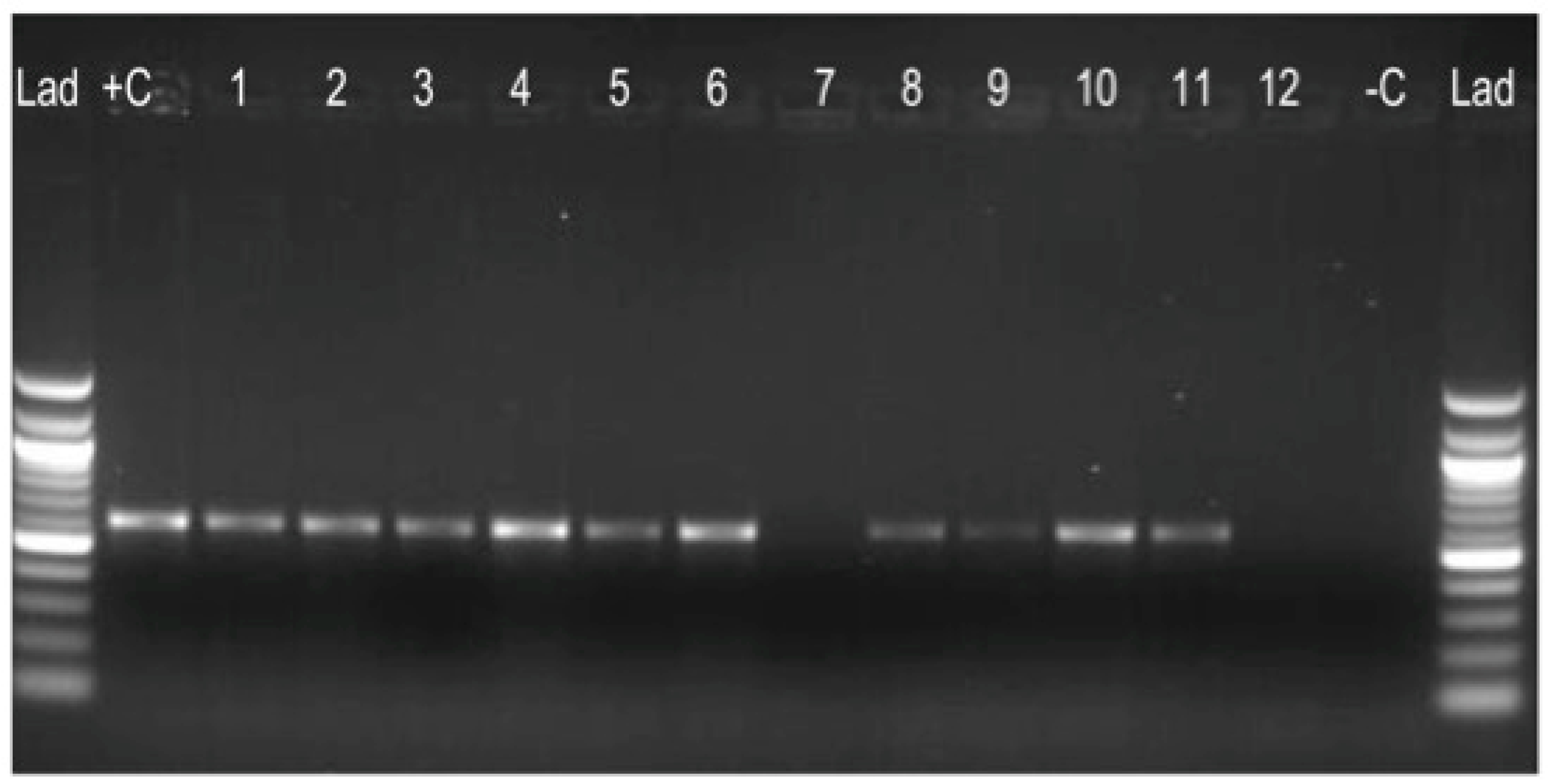
3.2. Regions of Difference (RD) Analysis: RD9, RD4, RD12, and RD1

Of the 133 IS6110 positive samples subjected to RD analyses, 126 were RD9 positive while 7 (5.3%) samples were negative. The RD9 positive results suggested that the isolates were either M. tuberculosis, M. canetti, or M. africanum subtype II. These were not analyzed further. The RD9 negative isolates were further analyzed with RD4 to further delineate the MTC species therein. Of the seven RD9 negative samples analyzed with RD4, five were RD4 positive and two RD4 negative. RD4 is a region deleted from all M. bovis strains (that is, classical M. bovis, M. bovis BCG, and MOTTs) but is present in M. canettii, M. tuberculosis, M. africanum type I and II, and M. microti isolates. To confirm the presence of either classical M. bovis or M. bovis BCG, the RD4 negative samples were analyzed further using both RD12 and RD1 PCR. The two RD4 negative samples were analyzed for the presence of RD12 and RD1 loci. Both isolates were RD12 negative, confirming them as M. bovis. Two isolates were positive for RD1. A summary of the RD analyses is presented in Table 2. These samples were further characterized by genome sequencing (Figure 1).
The prevalence of M. bovis in archived PZA samples at JCRC was determined to be at 1.5% (2/136). The two M. bovis positive samples were Bz_31150 and B2_7505. Sample Bz_31150 was a bronchial wash from a chimpanzee submitted for characterization to the JCRC in May 2007. This sample was sensitive to streptomycin, isoniazid, rifampicin, and ethambutol, first-line anti-tuberculosis drugs. It was resistant to pyrazinamide, which is expected for M. bovis since this organism carries inherent resistance to the drug [13,24,30]. Another sample taken for sequencing was B2_7505, a sputum sample that was a classical M. bovis isolate, from a human patient collected in 2006. This person was part of a multi-resistant TB drug study. This isolate was resistant to all the anti-tuberculosis antibiotics.

3.3. Phenotypic Drug Susceptibility Profiles

All 136 isolates were resistant to at least one anti-TB drug (Figure S4). This was very typical of the trend of drug resistance among the 5676 isolates scanned for analysis in this study. In Figure S4 and Table 3, less than 30% of the isolates were sensitive to the first-line TB drugs and 83 (61%) out of 136 of the isolates were resistant to all first-line TB drugs (e.g., isoniazid, rifampicin, pyrazinamide, and ethambutol) and streptomycin. These patients were characterized as having multi-drug resistant TB or MDR-TB. Twenty-six or 19% of the isolates were sensitive to all first-line TB drugs except for PZA. In addition, 44% or 32% of the isolates were resistant to all the four first-line TB drugs and streptomycin.

3.4. Mycobacterium Bovis from Human Host Among Sequenced Genomes

Eight genomes sequenced by Illumina were uploaded at NCBI [42,43] and the Olive database [36] (Table 4). Two of these isolates were M. bovis and six were M. tuberculosis. Illumina HiSeq2000 whole genome sequencing was performed, with 1.1 M spots yielding 214.9 Mb for M. bovis BZ_31150, and 1.8 M spots yielding 356.7 Mb for M. bovis B2_7505. For M. bovis Bz_31150, Illumina reads were assembled into 141 contigs and 136 scaffolds, resulting in a genome size of ~4.272 Mb, with a GC content of 65.54% at 24x coverage [44]. For M. bovis B2_7505, Illumina reads were assembled into 243 contigs and 206 scaffolds, resulting in a genome size of ~4.17 Mb, with a G + C content of 65.5% at 31.0× coverage [44]. All other genomes sequenced in this study provided at least 30-fold coverage, but the genomes could not be closed due to repetitive region redundancy. Nevertheless, the genome coverage was excellent and provided confidence to extract SNPs from regions that aligned with AF2122/97 at >20-fold coverage.

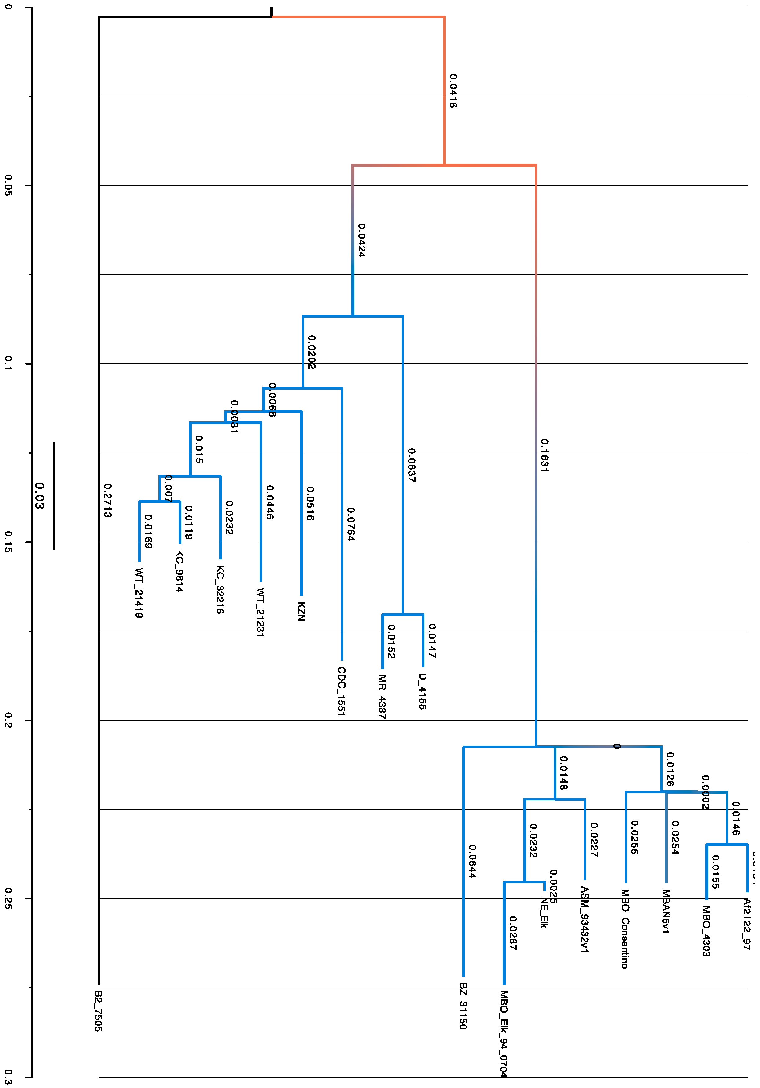
3.5. SNPs Obtained by Phylogenetic Analysis

In total, 7248 SNPs were extracted across the 17 genomes (Table 5) and used to create a phylogenetic tree in MEGA 7.0 using the maximum likelihood method with 1000 bootstrap replicates and the Hasegawa–Kishino–Yano (HYK) model (Figure 2). Phylogenetic analysis of the SNP distribution on genome sequences was carried out to determine the relationships between the isolates and reference genomes as well as likely disease transmission patterns. In our analysis, M. bovis from a human being (B2_7505) was distinct as it neither clustered with any M. bovis nor M. tuberculosis strains (Figure 2).
In addition, we carried out genome annotation of all the eight Ugandan genomes and comparative analysis of the two M. bovis genomes (Bz_31150 and B2_7505) with the reference genome M. bovis Af2122/97, M. bovis BCG, and M. tuberculosis CDC_1551 with the RAST tool [40]

3.6. Genome Analysis of Multi-Drug Resistance Mutations

Mycobacteria lack diversity and this implies that when amino acid polymorphisms are detected, it strongly suggests that the variation has functional importance, like antibiotic resistance [45] (Table 6 and Table 7). All the Ugandan isolates were phenotypically resistant to PZA, but on genome analysis, they had mutations in different locations of the 561 bp pncA gene. The animal isolates (M. bovis Af2122/97, M. bovis Bz_31150, M. bovis B2_7505, M. bovis Elk strain, and M. bovis Corsentino strain) and the human isolate M. bovis B2_7505 had the same pncA single C→G point mutation at nucleotide 169, which resulted in an H57D substitution [46]. In addition, both B2_7505, the human TB isolate, and Bz_31150, the chimpanzee isolate, had an adenine residue at nucleotide 285 in the oxyR gene, further confirming them as M. bovis isolates [47].
Two Ugandan MTb strains (M. tuberculosis D_4155 and M. tuberculosis KC_32216) were phenotypically PZA resistant but exhibited no mutation at the genome level while M. tuberculosis KC_9614 had a mutation four nucleotides upstream of the pncA gene Y(-4)C. This agrees with Sreevatsan et al.’s, [46] work, where 28% of the PZA resistant M. tuberculosis organisms they tested lacked nucleotide sequence changes in pncA. The remaining PZA resistant M. tuberculosis organisms displayed unique single point mutations; WT_21419 had a V163A mutation, MR_4387 had an A134V mutation while WT_21231 had a pncA V131F mutation. AhpC is an alkyl hydroperoxide reductase C, an enzyme that reduces organic peroxides, and it has been shown to be involved in the response to oxidative stress in mycobacterial species. The ahpC gene is located adjacent to the oxyR gene and there were mutations identified in the ahpC gene in two isolates; in MR_4387, there were G(-88)A and T(-76)A nucleotide mutations while in D_4155, there was a G(-88)A mutation upstream of ahpC.
More than 95% of rifampicin (RMP) resistance is associated with mutations in an 81 bp region of the rpoB gene (the RIF resistance determining region (RRDR) from codons 507 to 533 of rpoB), but isoniazid (INH) resistance is more complex and has been associated with multiple genes, most frequently with katG and inhA [48]. Other common mutation patterns are found at codon 315 (55%–90%) of katG and codon 43 (47%–79%) of rpsL for streptomycin resistance [49]. Mutations in the katG and inhA genes together may be responsible for 80% of all organisms resistant to INH [46]. In our work, M. tuberculosis KC_9614 had the S315T mutation in the katG gene (catalase-peroxidase) while M. bovis B2_7505 had both the S315T mutation in katG as well as the S94A mutation in the inhA gene (NADH-dependent enoyl-reductase), defining it as an INH resistant organism [50].
Phenotypically, M. bovis B2_7505 and M. tuberculosis isolates MR_4387, KC_9614, WT_21419, and WT_21231 were all resistant to rifampicin (RMP) yet the mutations they exhibited at the genome level fell outside the RRDR (e.g., S450L, D435Y), which is meant to convey high-level resistance [51]. Gagneux et al. posit that rifampicin resistance-conferring mutations associated with no or low fitness cost in vitro are the most frequent in clinical strains [51]. Several isolates, including the reference strain, M. bovis Af2122/97, had the R463L mutation in katG, but according to Sekiguchi et al., the R463L mutation is not known to be associated with INH resistance [50,52]. This means that the phenotypic resistance to INH observed in some of the isolates (MR_4387, WT_21419, and WT_21231) was probably due to mutation elsewhere. Furthermore, there was an rpoB S531L mutation in WT_21231 and WT_21419 and a D516V mutation for MR_4387, which are classic mutations associated with high-level RMP resistance [53].
Mutations in the gyrA gene related to fluoroquinolone (FQ) resistance were seen in several isolates, with the most common being E21Q, G668D, and T80A; all these mutations were outside the QRDR yet these isolates demonstrated phenotypic resistance [54]. Isolates B2_7505, Bz_31150, D_4155, and MR_4387 had a C206G nucleotide mutation in gyrA. In addition, M. bovis B2_7505 also had a S95T mutation in the QRDR. Fluoroquinolone resistance has been associated with alterations in DNA gyrase, especially mutations in the quinolone resistance determining region (QRDR) of gyrA (codons 74 to 113) and gyrB (codons 500 to 538), respectively [54,55]. Its estimated that 60% to 90% of Mtb clinical isolates with extremely high FQ resistance have mutations in the QRDR of gyrA, particularly codons 88, 90, 91, and 94 [54,56].
For ethambutol resistance, a G884D mutation was identified in embA of MR_4387 and D_4155, Mtb isolates. In addition, there was a T608N mutation in the embA in KC_9614. None of these are documented mutations in the literature for EMB resistance. The mutations for embC were only in the M. bovis isolates except for B2_7505 (see Table 6 and Table 7).

4. Discussion

Although much has been studied on tuberculosis in developed parts of the world and Asia, there are still gaps in the knowledge on tuberculosis—particularly zoonotic tuberculosis—in Africa. In this study, we carried out comparative genomics on archived pyrazinamide resistant MTC isolates from a multicenter study archive in Uganda to determine if there was a non-random distribution of genomotypes of MTC organisms infecting humans and animals in Uganda with the use of genome-wide single nucleotide polymorphisms (SNPs) to differentiate MTC organisms. The guiding hypothesis was whether there was multi-directional transmission of tuberculosis occurring in resource-limited settings, where animal-human contact is intimate and frequent.
Cattle are the main hosts for bTB in Uganda and the region, though there are studies that indicate that goats can be a source of infection as well, but TB in small ruminants is rarely detected at the abattoir as a result of a lower quality of meat inspection than that performed for cattle [8]. In Uganda, milk is usually boiled, but raw and fermented milk are also consumed by some pastoral communities [20,21,22,57]. With the absence of an active bTB surveillance program in Uganda, there is still little known about the scale of the problem in both the animal and the human population. Pasteurized milk is unaffordable for many people, and this coupled with the consumption of raw milk among the pastoral communities, raises important questions about the actual bovine TB status of the country.
Bovine TB generally results in extra-pulmonary lesions. Thus, a bias toward detection on a very small proportion of M. bovis was anticipated as about 80% of the PZA resistant isolates came from sputum. There are limited laboratory facilities for the culture and typing of tubercle bacilli in Uganda especially in rural areas [11,58]. Sputum smear microscopy with acid-fast stains is the primary approach to pulmonary TB detection in Ugandan TB laboratories. It is an easy, fast, and inexpensive way to screen for TB cases but has low sensitivity, especially with persons co-infected with HIV. Furthermore, M. bovis infections are generally missed when microscopy on sputum alone is used for diagnostics and as a triage for culturing for mycobacteria.
The World Health Organization indicates that bovine TB is a neglected disease [16] and given its insidious nature, policy makers need to address it. There is still little published information about the relative proportion of MTC due to M. bovis, the epidemiology as well as the public health aspects of this disease [11,59,60,61]. In fact, Ayele et al. [62] proposed that of all the reports submitted to the Office of International Epizootes (OIE) and WHO by different African member countries, none mention bTB as an important human TB infection. Tanzania, one of the countries neighboring Uganda in East Africa, is one of the few countries in Africa that has quantitative data on the bTB prevalence in humans, with more than 30% of recorded TB cases listed as extra-pulmonary in the Arusha region [59,63]; of these M. bovis was isolated from 7/65 or 10.8% culture positive cases with cervical lymphadenitis. This also highlights the critical need for physicians to be aware of M. bovis and the different routes of transmission and different treatment recommendations.

4.1. Mycobacterium Bovis in PZA Resistant Isolates

We carried out culture, regions of difference (RD) PCR analysis as well as whole genome sequencing on archived pyrazinamide resistant isolates from Uganda and found two M. bovis isolates; M. bovis B2_7505 isolated from a human patient and M. bovis Bz_31150 isolated from a chimpanzee. Out of the over 5000 stored isolates at the JCRC, only 133 or 2.4% were resistant to pyrazinamide, and only two of these turned out to be M. bovis. We expected a higher number of isolates to be positive given that M. bovis is naturally resistant to PZA, a first-line anti-tuberculosis drug [13]. The rest of the PZA resistant isolates were M. tuberculosis, pointing to a high degree of drug resistance to anti-TB drugs amongst the humans from whom the isolates were collected (Supplementary Figure S4). In addition to resistance to PZA, the case patients were resistant to multiple anti-tuberculosis drugs (MDR-TB).
Studies in Ethiopia on bTB have indicated that a higher prevalence of M. tuberculosis in cattle owned by farmers with active TB than in farmers who did not have active tuberculosis; this suggests possible bi-directional transmission of mycobacterial species between cattle and their owners [64,65,66,67,68,69]. In Alemaheyu’s study [64], M. bovis was isolated from the sputum of cattle owners and M. tuberculosis from cow’s milk, which suggests bi-directional transmission of mycobacterial species. Studies in Tanzania also indicate that consumption of raw milk is an important risk factor in bTB transmission [10,13,59,70]. Humans acquire bTB infection through ingestion of infected contaminated milk and milk products and also by inhalation when there is close physical contact between the owner and their cattle, especially at night, since in some cases, they share shelters with their animals [5,64,65,71]. In this study, the person infected with M. bovis (sample B2_7505) was an adult female with MDR TB. Although she received treatment from the JCRC in Kampala, the capital city of Uganda, we cannot rule out the possibility that she had close contact with animals since many Ugandans do have contact with animals.

4.2. Whole Genome Sequencing (WGS) and Analysis

WGS provides unique diagnostic abilities for drug-resistant TB given that it provides information on entire genes and their promoters, expected sensitivity of strains, genetic background, epidemiological data, and an indication of risk of laboratory contamination giving a ‘complete genotypic profile’; features that are critical as incidences of multi-drug resistant and extensively drug resistant TB are increasing [72,73]. In this work, WGS of eight Ugandan isolates and genome analysis with Mauve [37], Mega [39], RAST [40], and the use of the PATRIC database [74] revealed that the two M. bovis isolates from Uganda displayed unique characteristics. The M. bovis reference genome Af2122/97 has a genome of ~4.3 Mb while the Ugandan isolates were ~4.272 Mb (M. bovis Bz_31150) and 4.17 Mb (M. bovis B2_7505) [43]. According to some schools of thought, evolution towards pathogenicity is often accompanied by a reduction in genome size, which is compensated in part by gene duplications and diversification [75]. It has been proposed that genome downsizing of pathogenic bacteria is as a result of adaptation to a pathogenic lifestyle that includes the exploitation of the resources of a host organism, which might make certain gene functions redundant and subject to gene loss [1,75,76]. On the other hand, the reduced genome size could be due to the fact that these were draft genomes, with some regions of the genomes not covered. Another possible reason is that it is difficult to obtain complete MTBC genome sequences with second generation sequencing technologies, like the Illumina Hiseq platform, due to the high GC content and repetitive sequences [77].

4.3. Phylogenetic Analysis

Phylogenetic analysis carried out on the Uganda isolates together with the Af2122/97 reference genome, US M. bovis strains isolated from Elk (M. bovis Elk and Corsentino strains) and isolates from Argentina and Brazil (Figure 2), show that Bz_31150 clustered with the other M. bovis strains. The other Uganda Mtb strains clustered with M. tuberculosis CDC_1551 and in particular, M. tuberculosis WT_21231 clustered very closely with M. tuberculosis KZN, the Kwazulu Natal strain isolated from a patient in South Africa. Mycobacterium bovis B2_7505 did not cluster with any of the two main cluster groups above but formed an intermediate group between the two. This zoonotic Mycobacterium bovis B2_7505 is an extensively drug resistant (XDR) strain that may have been either a mixed infection or a contaminated culture. On the other hand, this may represent a unique isolate that likely underwent multiple genomic changes in-vivo to adapt to the human host to successfully circumvent new host defenses, persisting in the host and developing resistance through various mechanisms, many of which are yet to be fully understood. This would need reconfirmation after re-culturing the bacterium and sequencing multiple individual colonies. Furthermore, long-read sequencing technologies would be very helpful in delineating mixed cultures as well as improving confidence in the genome assemblies.
Analysis of selected isolates (M. bovis B2_7505, M. bovis Bz_31150, M. bovis BCG, M. bovis AF2122/97, and M. tuberculosis CDC_1551) using RAST enabled us to visualize the regions of the genomes that were most similar and others that were not. This is in line with what we obtained from the phylogenetic analysis using single nucleotide polymorphisms (SNPs).
This work demonstrates that although MTCs have a high degree of nucleotide relatedness, there are significant genomic differences between them, which can be harnessed to make critical decisions affecting tuberculosis treatment and management. SNP-based phylogenetic analysis helped to differentiate the different MTC genomes; this provides a population genetic framework to aid in identifying factors responsible for the wide host range and disease phenotypes of M. bovis and its behavior in different host species [78]. Our understanding of TB epidemiology and genomics is constantly being modified and improved with the rapid advances in whole genome sequencing [79,80,81,82].

4.4. Antibiotic Resistance Genes of Uganda Isolates

Resistance to drugs develops when mutations or chromosomal errors occur, in genes that encode drug targets or drug metabolic pathways, which impacts the effectiveness of the drugs. This is problematic when a patient is given a less-than-optimal treatment or there is low adherence to the drugs. In addition, lack of detection and subsequent unsuitable treatment increase the risk transmission of drug resistant tuberculosis and in fact studies have shown that a large proportion of MDR and XDR cases are due to primary transmission [48,83].
The pncA gene encodes the pyrazinamidase (PZase) enzyme, which converts PZA, a pro-drug, into the active pyrazinoic acid [84]. PZA kills semi-dormant tubercle bacilli that are not affected by other anti-tuberculous drugs and its inclusion in the treatment of tuberculosis shortens the period of therapy from 12 to 18 months to 6 months [85,86]. In this study, we sequenced eight genomes from Uganda that were phenotypically PZA resistant but on genome analysis, three of them did not have any mutation in the pncA gene (Mtb D_4155, Mtb KC_32216, and Mtb KC_9614). Further, there was no mutation in the M. bovis Ravenel strain we used yet this mutation (H57D) should be present in this isolate since M. bovis strains are naturally resistant to pyrazinamide [13,85]. This is a laboratory strain and may have mutated over the course of several passages.
Table 6 shows all drug resistance related mutations. Table 7 shows a more detailed study of SNPs related to different TB drug resistance associated mutations. DNA sequencing studies have shown that mutations occur across the entire length of the pncA gene, with some SNPs occurring more frequently than others possibly because these SNPs are rooted in ancestral strains [46,84]. This means that development of a molecular assay for PZA resistance detection would be more challenging compared to other genes, like rpoB, gyrA, and embB, that have well-defined resistance-causing mutations [84]. Studies also show that mutations in the pncA gene can be absent in a small proportion of isolates that are phenotypically resistant to PZA, thus it is said to be indicative of an alternative mechanism of PZA resistance other than mutation in the pncA gene [84].
Isoniazid (INH) targets both active and latent tuberculosis and has a relatively low cost and toxicity, but INH resistance is quite complex and has been associated with multiple genes, usually katG (S315T), which encodes a catalase peroxidase that transforms INH to its active form, and inhA (S94A), which encodes a putative enzyme involved in mycolic acid biosynthesis [48,87,88]. Mycobacterium bovis B2_7505, from a human patient, had both phenotypic resistance to INH as well as mutations in katG (S315T) and inhA (S94A). Two isolates had the S315T mutation and according to Vilcheze et al., this variant is more often found in MDR_TB patients than in INH mono-resistant clinical isolates, hence it may be related to the higher transmission capabilities of these particular strains [50].
Rifampicin (RMP) is another important first-line drug for TB treatment and the most resistance to this drug arises from mutations in the rpoB gene, with the most frequent mutations seen at positions 516, 526, and 531 [89]. The most common mutation seen in our study was S450L and D435Y. The latter mutation (D435Y) is disputed on its association with rifampin resistance. However, its common occurrence in Ugandan isolates may be unique and represent a geographically restricted change because all isolates with the mutation were phenotypically resistant. The isolates screened in this study commonly exhibited the following gyrA mutations (E21Q, D668D), which, according to Lui et al. [83], were also seen in a fluoroquinolone susceptible strain, thus indicating that they may not be the source of fluoroquinolone resistance. Isolates B2_7505, Bz_31150, D_4155, and MR_4387 had a C206G nucleotide mutation in gyrA. Although fluoroqinolones (FQs) are proven to be very effective second-line anti-mycobacterial drugs for the treatment of drug-resistant TB, the increasing rate of drug resistance among them is a concern.
The presence of these mutations (H57D, S315T, S94A) exclusively in the human M. bovis isolate suggests that these occurred after transmission from cattle. Genome analysis in this study identified M. bovis in humans and great apes, suggesting transmission from domesticated ruminants in the area due to a dynamic and changing interface, which has created an exposure opportunity. The acquisition, mutation, or loss of certain sections of genes could provide pathogens with peculiar advantages during infection and transmission and understanding these mechanisms could perhaps shed more light on the secrets these challenging organisms have that makes them so fastidious and successful. Mutations in key genes responsible for drug resistance have ensured that these organisms circumvent MTC therapy and perpetuate themselves in their hosts and in other susceptible hosts. MDR-TB and XDR-TB are a real threat to the achievements made in the fight against TB, one that needs a formidable arsenal of tools to overcome.

4.5. Application of WGS/SNP Chip in the Field

Genome studies suggest that M. bovis transmission occurs between cattle, between cattle and humans, and potentially between humans [33,90,91,92] as well as other species, like wildlife [2,3,79,93]. Genotypic assays, like the Cepheid Xpert MTB/RIF (Cepheid, Sunnyvale, CA, USA) and Hain line-probe (Hain Lifescience, Nehren Germany) assays, provide a short turnaround time (less than a day) in the identification of mycobacteria; but they are not able to detect all resistance conferring mutations [94]. In our work, we used WGS to identify SNP differences in MTC species and determined those that had multi-drug resistance genes [81,94,95,96,97]. A total of 7250 SNPs was used to generate the phylogenetic tree that enabled us to determine the relatedness of the sequenced genomes to reference strains. This data should be regenerated with the addition of global MTC genome collections to help develop an affordable SNP chip to achieve sensitive, specific, and rapid results for MTC detection and typing to enable real-time monitoring of interspecies transmission.

5. Conclusions

Two of the 136 isolates were identified as Mycobacterium bovis. This likely reflects the study population at the JCRC. However, a number of the isolates were MDR-TB. The MTC species are strictly clonal and genetic material is only passed vertically from mother to daughter cells. This clonality facilitates phylogenetic analysis of the MTC complex, where variations occur exclusively by genomic mutations, like single nucleotide polymorphisms (SNPs), deletions, or insertions of small or large pieces of DNA [98,99]. In our work, we demonstrated the presence of M. bovis in a human and wildlife using archived samples from Uganda. This implies that more work in the pastoral communities would present a clearer picture of the TB disease dynamics and possibly the transmission patterns of MTCs between humans and animals. A subset of the SNPs we extracted from this dataset to create an SNP chip for field applications in order to detect MTCS quickly and accurately would be a useful field application. In addition, this work encourages a One Health approach between medical and veterinary departments in order to strengthen the health care and disease surveillance systems, and the need for translating science into policy [15,57,100].

Supplementary Materials

The following are available online at https://www.mdpi.com/2076-2607/7/8/221/s1, Figure S1: Studies from the Joint Clinical Research re (JCRC) studies from which pyrazinamide resistant samples were drawn; Figure S2: Distribution by age and gender of case patients with pyrazinamide (PZA) resistant isolates; Figure S3: Source of PZA samples tested for Mycobacterium tuberculosis complex; Figure S4: Resistance patterns for anti-tuberculosis drugs in PZA resistant isolates.

Author Contributions

S.I.W. conceived ideas, assembled isolates, extracted DNA and helped in genome assembly as well in writing of the manuscript; J.N., P.K., S.O., help in sample sorting, DNA extraction, confirmation of TB by PCR, D.T., W.R.W., T.T., T.J., and S.A.H. helped in genome data analysis and helped writing of the manuscript, T.J., S.I.W. and S.A.H. helped in confirmation of genomic data and SNP calls, S.S. identified the project, provided funding, analyzed data with S.I.W. and T.J., and wrote the paper.

Funding

This project was supported by NIH Research Training Grant # R25 TW009345 funded by the Fogarty International Center, the National Institute of Mental Health, the NIH Office of the Director Office of Research on Women’s Health, and the NIH Office of the Director the Office of AIDS Research.

Acknowledgments

We acknowledge the following organizations for support to make this work possible: The Broad Institute for sequencing of the isolates; University of Minnesota Super Computing Institute for biocomputational resources.

Conflicts of Interest

The authors declare no conflict of interest.

References

  1. Cole, S.T.; Brosch, R.; Parkhill, J.; Garnier, T.; Churcher, C.; Harris, D.; Gordon, S.V.; Eiglmeier, K.; Gas, S.; Barry, C.E., III; et al. Deciphering the biology of Mycobacterium tuberculosis from the complete genome sequence. Nature 1998, 393, 537–544. [Google Scholar] [CrossRef] [PubMed]
  2. Banuls, A.L.; Sanou, A.; Anh, N.T.; Godreuil, S. Mycobacterium tuberculosis: Ecology and evolution of a human bacterium. J. Med. Microbiol. 2015, 64, 1261–1269. [Google Scholar] [CrossRef] [PubMed]
  3. Dos Vultos, T.; Mestre, O.; Rauzier, J.; Golec, M.; Rastogi, N.; Rasolofo, V.; Tonjum, T.; Sola, C.; Matic, I.; Gicquel, B. Evolution and diversity of clonal bacteria: The paradigm of Mycobacterium tuberculosis. PLoS ONE 2008, 3, e1538. [Google Scholar] [CrossRef] [PubMed]
  4. Gutierrez, M.C.; Brisse, S.; Brosch, R.; Fabre, M.; Omais, B.; Marmiesse, M.; Supply, P.; Vincent, V. Ancient origin and gene mosaicism of the progenitor of Mycobacterium tuberculosis. PLoS Pathog. 2005, 1, e5. [Google Scholar] [CrossRef] [PubMed]
  5. O’Reilly, L.M.; Daborn, C.J. The epidemiology of Mycobacterium bovis infections in animals and man: A review. Tuber. Lung Dis. 1995, 76, 1–46. [Google Scholar] [CrossRef]
  6. Michel, A.L.; Muller, B.; Van Helden, P.D. Mycobacterium bovis at the animal-human interface: A problem, or not? Vet. Microbiol. 2010, 140, 371–381. [Google Scholar] [CrossRef] [PubMed]
  7. Nelson, A.M. The cost of disease eradication. Smallpox and bovine tuberculosis. Ann. N. Y. Acad. Sci. 1999, 894, 83–91. [Google Scholar] [CrossRef]
  8. Pesciaroli, M.; Alvarez, J.; Boniotti, M.B.; Cagiola, M.; Di Marco, V.; Marianelli, C.; Pacciarini, M.; Pasquali, P. Tuberculosis in domestic animal species. Res. Vet. Sci. 2014, 97, S78–S85. [Google Scholar] [CrossRef]
  9. El-Sayed, A.; El-Shannat, S.; Kamel, M.; Castaneda-Vazquez, M.A.; Castaneda-Vazquez, H. Molecular Epidemiology of Mycobacterium bovis in Humans and Cattle. Zoonoses. Public Health 2016, 63, 251–264. [Google Scholar] [CrossRef]
  10. Daborn, C.J.; Grange, J.M.; Kazwala, R.R. The bovine tuberculosis cycle—An African perspective. J. Appl. Bacteriol. 1996, 25, 27S–32S. [Google Scholar]
  11. Cosivi, O.; Grange, J.M.; Daborn, C.J.; Raviglione, M.C.; Fujikura, T.; Cousins, D.; Robinson, R.A.; Huchzermeyer, H.F.A.K.; De Kantor, I.; Meslin, F.X. Zoonotic tuberculosis due to Mycobacterium bovis in developing countries. Emerg. Infect. Dis. 1998, 4, 59–70. [Google Scholar] [CrossRef]
  12. Berg, S.; Schelling, E.; Hailu, E.; Firdessa, R.; Gumi, B.; Erenso, G.; Gadisa, E.; Mengistu, A.; Habtamu, M.; Hussein, J.; et al. Investigation of the high rates of extrapulmonary tuberculosis in Ethiopia reveals no single driving factor and minimal evidence for zoonotic transmission of Mycobacterium bovis infection. BMC Infect. Dis. 2015, 15, 112. [Google Scholar] [CrossRef]
  13. Wedlock, D.N.; Skinner, M.A.; De Lisle, G.W.; Buddle, B.M. Control of Mycobacterium bovis infections and the risk to human populations. Microbes. Infect. 2002, 4, 471–480. [Google Scholar] [CrossRef]
  14. Muller, B.; Durr, S.; Alonso, S.; Hattendorf, J.; Laisse, C.J.; Parsons, S.D.; van Helden, P.D.; Zinsstag, J. Zoonotic Mycobacterium bovis-induced tuberculosis in humans. Emerg. Infect. Dis. 2013, 19, 899–908. [Google Scholar] [CrossRef]
  15. Kazwala, R.R.; Daborn, C.J.; Sharp, J.M.; Kambarage, D.M.; Jiwa, S.F.; Mbembati, N.A. Isolation of Mycobacterium bovis from human cases of cervical adenitis in Tanzania: A cause for concern? Int. J. Tuberc. Lung Dis. 2001, 5, 87–91. [Google Scholar]
  16. World Health Organization. Tuberculosis; WHO: Geneva, Switzerland, 2015; Volume 2016. [Google Scholar]
  17. American Thoracic Society; Centers for Disease Control. Mycobacterioses and the acquired immunodeficiency syndrome. Am. Rev. Respir. Dis. 1987, 136, 492–496. [Google Scholar] [CrossRef]
  18. World Health Organization. Uganda TB Profile; WHO: Geneva, Switzerland, 2016; Volume 2016. [Google Scholar]
  19. World Health Organization. TB Diagnostics and Lab Strengthening; WHO: Geneva, Switzerland, 2016; Volume 2016. [Google Scholar]
  20. Oloya, J.; Muma, J.B.; Opuda-Asibo, J.; Djonne, B.; Kazwala, R.; Skjerve, E. Risk factors for herd-level bovine-tuberculosis seropositivity in transhumant cattle in Uganda. Prev. Vet. Med. 2007, 80, 318–329. [Google Scholar] [CrossRef]
  21. Oloya, J.; Opuda-Asibo, J.; Djonne, B.; Muma, J.B.; Matope, G.; Kazwala, R.; Skjerve, E. Responses to tuberculin among Zebu cattle in the transhumance regions of Karamoja and Nakasongola district of Uganda. Trop. Anim. Health Prod. 2006, 38, 275–283. [Google Scholar] [CrossRef]
  22. Oloya, J.; Kazwala, R.; Lund, A.; Opuda-Asibo, J.; Demelash, B.; Skjerve, E.; Johansen, T.B.; Djonne, B. Characterisation of mycobacteria isolated from slaughter cattle in pastoral regions of Uganda. BMC Microbiol. 2007, 7, 95. [Google Scholar] [CrossRef]
  23. Gallivan, M.; Shah, N.; Flood, J. Epidemiology of human Mycobacterium bovis disease, California, USA, 2003–2011. Emerg. Infect. Dis. 2015, 21, 435–443. [Google Scholar] [CrossRef]
  24. Huard, R.C.; Lazzarini, L.C.; Butler, W.R.; Van Soolingen, D.; Ho, J.L. PCR-based method to differentiate the subspecies of the Mycobacterium tuberculosis complex on the basis of genomic deletions. J. Clin. Microbiol. 2003, 41, 1637–1650. [Google Scholar] [CrossRef]
  25. Joint Clinical Research Centre. 2016 Annual Report. Available online: https://www.jcrc.org.ug/resources/reports/annual-report-2016-sep-2017 (accessed on 26 July 2019).
  26. Sambrook, J.; Fritsch, E.F.; Maniatis, T. Molecular Cloning; Cold Spring Harbor Laboratory Press: New York, NY, USA, 1989; Volume 2. [Google Scholar]
  27. Sambrook, J.; Russell, D.W. Storage of bacterial cultures growing on solid medium. CSH Protoc. 2006, 2006, pdb.prot4453. [Google Scholar] [CrossRef]
  28. Huard, R.C.; Fabre, M.; De Haas, P.; Lazzarini, L.C.; Van Soolingen, D.; Cousins, D.; Ho, J.L. Novel genetic polymorphisms that further delineate the phylogeny of the Mycobacterium tuberculosis complex. J. Bacteriol. 2006, 188, 4271–4287. [Google Scholar] [CrossRef]
  29. Pym, A.S.; Brodin, P.; Brosch, R.; Huerre, M.; Cole, S.T. Loss of RD1 contributed to the attenuation of the live tuberculosis vaccines Mycobacterium bovis BCG and Mycobacterium microti. Mol. Microbiol. 2002, 46, 709–717. [Google Scholar] [CrossRef]
  30. Thorne, N.; Borrell, S.; Evans, J.; Magee, J.; Garcia De Viedma, D.; Bishop, C.; Gonzalez-Martin, J.; Gharbia, S.; Arnold, C. IS6110-based global phylogeny of Mycobacterium tuberculosis. Infect. Genet. Evol. 2011, 11, 132–138. [Google Scholar] [CrossRef]
  31. Brosch, R.; Gordon, S.V.; Marmiesse, M.; Brodin, P.; Buchrieser, C.; Eiglmeier, K.; Garnier, T.; Gutierrez, C.; Hewinson, G.; Kremer, K.; et al. A new evolutionary scenario for the Mycobacterium tuberculosis complex. Proc. Natl. Acad. Sci. USA 2002, 99, 3684–3689. [Google Scholar] [CrossRef]
  32. Kamerbeek, J.; Schouls, L.; Kolk, A.; Van Agterveld, M.; Van Soolingen, D.; Kuijper, S.; Bunschoten, A.; Molhuizen, H.; Shaw, R.; Goyal, M.; et al. Simultaneous detection and strain differentiation of Mycobacterium tuberculosis for diagnosis and epidemiology. J. Clin. Microbiol. 1997, 35, 907–914. [Google Scholar] [Green Version]
  33. Van Soolingen, D.; Hermans, P.W.; De Haas, P.E.; Soll, D.R.; Van Embden, J.D. Occurrence and stability of insertion sequences in Mycobacterium tuberculosis complex strains: Evaluation of an insertion sequence-dependent DNA polymorphism as a tool in the epidemiology of tuberculosis. J. Clin. Microbiol. 1991, 29, 2578–2586. [Google Scholar]
  34. Asiimwe, B.B.; Asiimwe, J.; Kallenius, G.; Ashaba, F.K.; Ghebremichael, S.; Joloba, M.; Koivula, T. Molecular characterisation of Mycobacterium bovis isolates from cattle carcases at a city slaughterhouse in Uganda. Vet. Rec. 2009, 164, 655–658. [Google Scholar] [CrossRef]
  35. Tuberculosis. Broad Institute. 2016. Available online: https://www.broadinstitute.org/infectious-disease-and-microbiome/tuberculosis (accessed on 26 July 2019).
  36. Olive, D. TB-ARC M.Bovis.1. 2014, Volume 2016. Available online: https://olive.broadinstitute.org/projects/Mycobacterium%20tuberculosis%20Comparative (accessed on 26 July 2019).
  37. Darling, A.C.; Mau, B.; Blattner, F.R.; Perna, N.T. Mauve: Multiple alignment of conserved genomic sequence with rearrangements. Genome. Res. 2004, 14, 1394–1403. [Google Scholar] [CrossRef]
  38. Darling, A.E.; Mau, B.; Perna, N.T. Progressivemauve: Multiple genome alignment with gene gain, loss and rearrangement. PLoS ONE 2010, 5, e11147. [Google Scholar] [CrossRef]
  39. Tamura, K.; Stecher, G.; Peterson, D.; Filipski, A.; Kumar, S. MEGA6: Molecular Evolutionary Genetics Analysis version 6.0. Mol. Biol. Evol. 2013, 30, 2725–2729. [Google Scholar] [CrossRef]
  40. Aziz, R.K.; Bartels, D.; Best, A.A.; DeJongh, M.; Disz, T.; Edwards, R.A.; Formsma, K.; Gerdes, S.; Glass, E.M.; Kubal, M.; et al. The RAST Server: Rapid annotations using subsystems technology. BMC Genom. 2008, 9, 75. [Google Scholar] [CrossRef]
  41. National Center for Biotechnology Information. Basic Local Alignment Search Tool; National Center for Biotechnology Information: Bethesda, MD, USA, 2016; Volume 2016.
  42. National Center for Biotechnology Information. TB-ARC-M. Bovis; National Center for Biotechnology Information: Bethesda, MD, USA, 2016; Volume 2016.
  43. National Center for Biotechnology Information. Bioproject; National Center for Biotechnology Information: Bethesda, MD, USA, 2016; Volume 2016.
  44. Wanzala, S.I.; Nakavuma, J.; Travis, D.A.; Kia, P.; Ogwang, S.; Sreevatsan, S. Draft Genome Sequences of Mycobacterium bovis BZ 31150 and Mycobacterium bovis B2 7505, Pathogenic Bacteria Isolated from Archived Captive Animal Bronchial Washes and Human Sputum Samples in Uganda. Genome. Announc. 2015, 3, e01102–e01115. [Google Scholar] [CrossRef]
  45. Sreevatsan, S.; Pan, X.; Stockbauer, K.E.; Connell, N.D.; Kreiswirth, B.N.; Whittam, T.S.; Musser, J.M. Restricted structural gene polymorphism in the Mycobacterium tuberculosis complex indicates evolutionarily recent global dissemination. Proc. Natl. Acad. Sci. USA 1997, 94, 9869–9874. [Google Scholar] [CrossRef]
  46. Sreevatsan, S.; Pan, X.; Zhang, Y.; Kreiswirth, B.N.; Musser, J.M. Mutations associated with pyrazinamide resistance in pncA of Mycobacterium tuberculosis complex organisms. Antimicrob. Agents Chemother. 1997, 41, 636–640. [Google Scholar] [CrossRef] [Green Version]
  47. Sreevatsan, S.; Escalante, P.; Pan, X.; Gillies, D.A., II; Siddiqui, S.; Khalaf, C.N.; Kreiswirth, B.N.; Bifani, P.; Adams, L.G.; Ficht, T.; et al. Identification of a polymorphic nucleotide in oxyR specific for Mycobacterium bovis. J. Clin. Microbiol. 1996, 34, 2007–2010. [Google Scholar] [Green Version]
  48. Seifert, M.; Catanzaro, D.; Catanzaro, A.; Rodwell, T.C. Genetic mutations associated with isoniazid resistance in Mycobacterium tuberculosis: A systematic review. PLoS ONE 2015, 10, e0119628. [Google Scholar] [CrossRef]
  49. Brammacharry, U.; Muthaiah, M. Characterization of rpsL Gene Mutations in Streptomycin-Resistant Mycobacterium tuberculosis Isolates. Am. J. Microbiol. Res. 2014, 2, 80–85. [Google Scholar] [CrossRef]
  50. Vilcheze, C.; Jacobs, W.R., Jr. Resistance to Isoniazid and Ethionamide in Mycobacterium tuberculosis: Genes, Mutations, and Causalities. Microbiol. Spectr. 2014, 2, 431–453. [Google Scholar] [CrossRef]
  51. Gagneux, S. Host-pathogen coevolution in human tuberculosis. Philos. Trans. R. Soc. B Biol. Sci. 2012, 367, 850–859. [Google Scholar] [CrossRef]
  52. Sekiguchi, J.; Miyoshi-Akiyama, T.; Augustynowicz-Kopec, E.; Zwolska, Z.; Kirikae, F.; Toyota, E.; Kobayashi, I.; Morita, K.; Kudo, K.; Kato, S.; et al. Detection of multidrug resistance in Mycobacterium tuberculosis. J. Clin. Microbiol. 2007, 45, 179–192. [Google Scholar] [CrossRef]
  53. Dominguez, J.; Boettger, E.C.; Cirillo, D.; Cobelens, F.; Eisenach, K.D.; Gagneux, S.; Hillemann, D.; Horsburgh, R.; Molina-Moya, B.; Niemann, S.; et al. Clinical implications of molecular drug resistance testing for Mycobacterium tuberculosis: A TBNET/RESIST-TB consensus statement. Int. J. Tuberc. Lung Dis. 2016, 20, 24–42. [Google Scholar] [CrossRef]
  54. Avalos, E.; Catanzaro, D.; Catanzaro, A.; Ganiats, T.; Brodine, S.; Alcaraz, J.; Rodwell, T. Frequency and geographic distribution of gyrA and gyrB mutations associated with fluoroquinolone resistance in clinical Mycobacterium tuberculosis isolates: A systematic review. PLoS ONE 2015, 10, e0120470. [Google Scholar] [CrossRef]
  55. Malik, S.; Willby, M.; Sikes, D.; Tsodikov, O.V.; Posey, J.E. New insights into fluoroquinolone resistance in Mycobacterium tuberculosis: Functional genetic analysis of gyrA and gyrB mutations. PLoS ONE 2012, 7, e39754. [Google Scholar] [CrossRef]
  56. Mayer, C.; Takiff, H. The Molecular Genetics of Fluoroquinolone Resistance in Mycobacterium tuberculosis. Microbiol. Spectr. 2014, 2, 455–478. [Google Scholar] [CrossRef]
  57. Kankya, C.; Muwonge, A.; Olet, S.; Munyeme, M.; Biffa, D.; Opuda-Asibo, J.; Skjerve, E.; Oloya, J. Factors associated with pastoral community knowledge and occurrence of mycobacterial infections in human-animal interface areas of Nakasongola and Mubende districts, Uganda. BMC Public Health 2010, 10, 471. [Google Scholar] [CrossRef]
  58. Cousins, D.V. Mycobacterium bovis infection and control in domestic livestock. Rev. Sci. Technol. 2001, 20, 71–85. [Google Scholar] [CrossRef]
  59. Cleaveland, S.; Shaw, D.J.; Mfinanga, S.G.; Shirima, G.; Kazwala, R.R.; Eblate, E.; Sharp, M. Mycobacterium bovis in rural Tanzania: Risk factors for infection in human and cattle populations. Tuberculosis 2007, 87, 30–43. [Google Scholar] [CrossRef]
  60. Cadmus, S.; Palmer, S.; Okker, M.; Dale, J.; Gover, K.; Smith, N.; Jahans, K.; Hewinson, R.G.; Gordon, S.V. Molecular analysis of human and bovine tubercle bacilli from a local setting in Nigeria. J. Clin. Microbiol. 2006, 44, 29–34. [Google Scholar] [CrossRef]
  61. Brudey, K.; Driscoll, J.R.; Rigouts, L.; Prodinger, W.M.; Gori, A.; Al-Hajoj, S.A.; Allix, C.; Aristimuno, L.; Arora, J.; Baumanis, V.; et al. Mycobacterium tuberculosis complex genetic diversity: Mining the fourth international spoligotyping database (SpolDB4) for classification, population genetics and epidemiology. BMC Microbiol. 2006, 6, 23. [Google Scholar] [CrossRef]
  62. Ayele, W.Y.; Neill, S.D.; Zinsstag, J.; Weiss, M.G.; Pavlik, I. Bovine tuberculosis: An old disease but a new threat to Africa. Int. J. Tuberc. Lung Dis. 2004, 8, 924–937. [Google Scholar]
  63. Etter, E.; Donado, P.; Jori, F.; Caron, A.; Goutard, F.; Roger, F. Risk analysis and bovine tuberculosis, a re-emerging zoonosis. Ann. N. Y. Acad. Sci. 2006, 1081, 61–73. [Google Scholar] [CrossRef]
  64. Alemayehu, R.; Girmay, M.; Gobena, A. Bovine tuberculosis is more prevalent in cattle owned by farmers with active tuberculosis in central Ethiopia. Vet. J. 2008, 178, 119–125. [Google Scholar] [CrossRef]
  65. Aylate, A.; Shah, S.N.; Aleme, H.; Gizaw, T.T. Bovine tuberculosis: Prevalence and diagnostic efficacy of routine meat inspection procedure in Woldiya municipality abattoir north Wollo zone, Ethiopia. Trop. Anim. Health Prod. 2013, 45, 855–864. [Google Scholar] [CrossRef]
  66. Ameni, G.; Vordermeier, M.; Firdessa, R.; Aseffa, A.; Hewinson, G.; Gordon, S.V.; Berg, S. Mycobacterium tuberculosis infection in grazing cattle in central Ethiopia. Vet. J. 2011, 188, 359–361. [Google Scholar] [CrossRef] [Green Version]
  67. Ameni, G.; Tadesse, K.; Hailu, E.; Deresse, Y.; Medhin, G.; Aseffa, A.; Hewinson, G.; Vordermeier, M.; Berg, S. Transmission of Mycobacterium tuberculosis between farmers and cattle in central Ethiopia. PLoS ONE 2013, 8, e76891. [Google Scholar] [CrossRef]
  68. Ameni, G.; Erkihun, A. Bovine tuberculosis on small-scale dairy farms in Adama Town, central Ethiopia, and farmer awareness of the disease. Rev. Sci. Technol. 2007, 26, 711–719. [Google Scholar] [CrossRef]
  69. Ameni, G.; Aseffa, A.; Engers, H.; Young, D.; Hewinson, G.; Vordermeier, M. Cattle husbandry in Ethiopia is a predominant factor affecting the pathology of bovine tuberculosis and gamma interferon responses to mycobacterial antigens. Clin. Vaccine Immunol. 2006, 13, 1030–1036. [Google Scholar] [CrossRef]
  70. Roug, A.; Perez, A.; Mazet, J.A.; Clifford, D.L.; VanWormer, E.; Paul, G.; Kazwala, R.R.; Smith, W.A. Comparison of intervention methods for reducing human exposure to Mycobacterium bovis through milk in pastoralist households of Tanzania. Prev. Vet. Med. 2014, 115, 157–165. [Google Scholar] [CrossRef]
  71. Berg, S.; Firdessa, R.; Habtamu, M.; Gadisa, E.; Mengistu, A.; Yamuah, L.; Ameni, G.; Vordermeier, M.; Robertson, B.D.; Smith, N.H.; et al. The burden of mycobacterial disease in ethiopian cattle: Implications for public health. PLoS ONE 2009, 4, e5068. [Google Scholar] [CrossRef]
  72. Ramirez-Busby, S.M.; Valafar, F. Systematic review of mutations in pyrazinamidase associated with pyrazinamide resistance in Mycobacterium tuberculosis clinical isolates. Antimicrob. Agents Chemother. 2015, 59, 5267–5277. [Google Scholar] [CrossRef]
  73. Cirillo, D.M.; Cabibbe, A.M.; De Filippo, M.R.; Trovato, A.; Simonetti, T.; Rossolini, G.M.; Tortoli, E. Use of WGS in Mycobacterium tuberculosis routine diagnosis. Int. J. Mycobacteriol. 2016, 5, S252–S253. [Google Scholar] [CrossRef]
  74. Wattam, A.R.; Abraham, D.; Dalay, O.; Disz, T.L.; Driscoll, T.; Gabbard, J.L.; Gillespie, J.J.; Gough, R.; Hix, D.; Kenyon, R.; et al. PATRIC, the bacterial bioinformatics database and analysis resource. Nucleic Acids Res. 2014, 42, D581–D591. [Google Scholar] [CrossRef]
  75. Le Chevalier, F.; Cascioferro, A.; Majlessi, L.; Herrmann, J.L.; Brosch, R. Mycobacterium tuberculosis evolutionary pathogenesis and its putative impact on drug development. Future Microbiol. 2014, 9, 969–985. [Google Scholar] [CrossRef] [Green Version]
  76. Moran, N.A. Microbial minimalism: Genome reduction in bacterial pathogens. Cell 2002, 108, 583–586. [Google Scholar] [CrossRef]
  77. Votintseva, A.A.; Bradley, P.; Pankhurst, L.; Del Ojo Elias, C.; Loose, M.; Nilgiriwala, K.; Chatterjee, A.; Smith, E.G.; Sanderson, N.; Walker, T.M.; et al. Same-day diagnostic and surveillance data for tuberculosis via whole genome sequencing of direct respiratory samples. J. Clin. Microbiol. 2017, 55, 1285–1298. [Google Scholar] [CrossRef]
  78. Joshi, D.; Harris, N.B.; Waters, R.; Thacker, T.; Mathema, B.; Krieswirth, B.; Sreevatsan, S. Single nucleotide polymorphisms in the Mycobacterium bovis genome resolve phylogenetic relationships. J. Clin. Microbiol. 2012, 50, 3853–3861. [Google Scholar] [CrossRef]
  79. Garcia-Betancur, J.C.; Menendez, M.C.; Del Portillo, P.; Garcia, M.J. Alignment of multiple complete genomes suggests that gene rearrangements may contribute towards the speciation of Mycobacteria. Infect. Genet. Evol. 2012, 12, 819–826. [Google Scholar] [CrossRef]
  80. Salipante, S.J.; SenGupta, D.J.; Cummings, L.A.; Land, T.A.; Hoogestraat, D.R.; Cookson, B.T. Application of whole-genome sequencing for bacterial strain typing in molecular epidemiology. J. Clin. Microbiol. 2015, 53, 1072–1079. [Google Scholar] [CrossRef]
  81. Takiff, H.E.; Feo, O. Clinical value of whole-genome sequencing of Mycobacterium tuberculosis. Lancet Infect. Dis. 2015, 15, 1077–1090. [Google Scholar] [CrossRef]
  82. Jagielski, T.; Minias, A.; Van Ingen, J.; Rastogi, N.; Brzostek, A.; Zaczek, A.; Dziadek, J. Methodological and Clinical Aspects of the Molecular Epidemiology of Mycobacterium tuberculosis and Other Mycobacteria. Clin. Microbiol. Rev. 2016, 29, 239–290. [Google Scholar] [CrossRef] [Green Version]
  83. Liu, F.; Hu, Y.; Wang, Q.; Li, H.M.; Gao, G.F.; Liu, C.H.; Zhu, B. Comparative genomic analysis of Mycobacterium tuberculosis clinical isolates. BMC Genom. 2014, 15, 469. [Google Scholar] [CrossRef]
  84. Whitfield, M.G.; Soeters, H.M.; Warren, R.M.; York, T.; Sampson, S.L.; Streicher, E.M.; Van Helden, P.D.; Van Rie, A. A Global Perspective on Pyrazinamide Resistance: Systematic Review and Meta-Analysis. PLoS ONE 2015, 10, e0133869. [Google Scholar] [CrossRef]
  85. Scorpio, A.; Zhang, Y. Mutations in pncA, a gene encoding pyrazinamidase/nicotinamidase, cause resistance to the antituberculous drug pyrazinamide in tubercle bacillus. Nat. Med. 1996, 2, 662–667. [Google Scholar] [CrossRef]
  86. Maslov, D.A.; Zaichikova, M.V.; Chernousova, L.N.; Shur, K.V.; Bekker, O.B.; Smirnova, T.G.; Larionova, E.E.; Andreevskaya, S.N.; Zhang, Y.; Danilenko, V.N. Resistance to pyrazinamide in Russian Mycobacterium tuberculosis isolates: pncA sequencing versus Bactec MGIT 960. Tuberculosis 2015, 95, 608–612. [Google Scholar] [CrossRef]
  87. Vilcheze, C.; Wang, F.; Arai, M.; Hazbon, M.H.; Colangeli, R.; Kremer, L.; Weisbrod, T.R.; Alland, D.; Sacchettini, J.C.; Jacobs, W.R., Jr. Transfer of a point mutation in Mycobacterium tuberculosis inhA resolves the target of isoniazid. Nat. Med. 2006, 12, 1027–1029. [Google Scholar] [CrossRef]
  88. Aye, K.S.; Nakajima, C.; Yamaguchi, T.; Win, M.M.; Shwe, M.M.; Win, A.A.; Lwin, T.; Nyunt, W.W.; Ti, T.; Suzuki, Y. Genotypic characterization of multi-drug-resistant Mycobacterium tuberculosis isolates in Myanmar. J. Infect. Chemother. 2016, 22, 174–179. [Google Scholar] [CrossRef]
  89. Khosravi, A.D.; Goodarzi, H.; Alavi, S.M. Detection of genomic mutations in katG, inhA and rpoB genes of Mycobacterium tuberculosis isolates using polymerase chain reaction and multiplex allele-specific polymerase chain reaction. Braz. J. Infect. Dis. 2012, 16, 57–62. [Google Scholar] [CrossRef] [Green Version]
  90. Vordermeier, H.M.; Perez De Val, B.; Buddle, B.M.; Villarreal-Ramos, B.; Jones, G.J.; Hewinson, R.G.; Domingo, M. Vaccination of domestic animals against tuberculosis: Review of progress and contributions to the field of the TBSTEP project. Res. Vet. Sci. 2014, 97, S53–S60. [Google Scholar] [CrossRef]
  91. Tschopp, R.; Aseffa, A.; Schelling, E.; Berg, S.; Hailu, E.; Gadisa, E.; Habtamu, M.; Argaw, K.; Zinsstag, J. Bovine tuberculosis at the wildlife-livestock-human interface in Hamer Woreda, South Omo, Southern Ethiopia. PLoS ONE 2010, 5, e12205. [Google Scholar] [CrossRef]
  92. Srivastava, K.; Chauhan, D.S.; Gupta, P.; Singh, H.B.; Sharma, V.D.; Yadav, V.S.; Sreekumaran; Thakral, S.S.; Dharamdheeran, J.S.; Nigam, P.; et al. Isolation of Mycobacterium bovis and M-tuberculosis from cattle of some farms in north India—Possible relevance in human health. Indian J. Med. Res. 2008, 128, 26–31. [Google Scholar]
  93. Ehrt, S.; Rhee, K.; Schnappinger, D. Mycobacterial genes essential for the pathogen’s survival in the host. Immunol. Rev. 2015, 264, 319–326. [Google Scholar] [CrossRef]
  94. Pankhurst, L.J.; Del Ojo Elias, C.; Votintseva, A.A.; Walker, T.M.; Cole, K.; Davies, J.; Fermont, J.M.; Gascoyne-Binzi, D.M.; Kohl, T.A.; Kong, C.; et al. Rapid, comprehensive, and affordable mycobacterial diagnosis with whole-genome sequencing: A prospective study. Lancet Respir. Med. 2016, 4, 49–58. [Google Scholar] [CrossRef]
  95. Sabat, A.J.; Budimir, A.; Nashev, D.; Sa-Leao, R.; Van Dijl, J.; Laurent, F.; Grundmann, H.; Friedrich, A.W.; ESGEM. Overview of molecular typing methods for outbreak detection and epidemiological surveillance. Eurosurveillance 2013, 18, 20380. [Google Scholar] [CrossRef] [Green Version]
  96. Kjeldsen, M.K.; Bek, D.; Rasmussen, E.M.; Prieme, A.; Thomsen, V.O. Line probe assay for differentiation within Mycobacterium tuberculosis complex. Evaluation on clinical specimens and isolates including Mycobacterium pinnipedii. Scand. J. Infect. Dis. 2009, 41, 635–641. [Google Scholar] [CrossRef]
  97. Harris, S.R.; Okoro, C.K. Whole-Genome Sequencing for Rapid and Accurate Identification of Bacterial Transmission Pathways. New Approaches Prokaryotic Syst. 2014, 41, 123–152. [Google Scholar] [CrossRef]
  98. Zakham, F.; Aouane, O.; Ussery, D.; Benjouad, A.; Ennaji, M.M. Computational genomics-proteomics and Phylogeny analysis of twenty one mycobacterial genomes (Tuberculosis and non Tuberculosis strains). Microb. Inf. Exp. 2012, 2, 7. [Google Scholar] [CrossRef]
  99. Schurch, A.C.; van Soolingen, D. DNA fingerprinting of Mycobacterium tuberculosis: From phage typing to whole-genome sequencing. Infect. Genet. Evol. 2012, 12, 602–609. [Google Scholar] [CrossRef]
  100. Hasegawa, M.; Kishino, H.; Yano, T. Dating of the human-ape splitting by a molecular clock of mitochondrial DNA. J. Mol. Evol. 1985, 22, 160–174. [Google Scholar] [CrossRef]
Figure 1. PCR result for RD9 (600 bp) for some of the 133 IS6110 positive samples. Order on gel: 100 bp Lad, +C: Positive control (Mycobacterium tuberculosis strain H37Rv), samples 1–12, -C; negative control (nuclease free PCR water).
Microorganisms 07 00221 g001
Figure 2. Molecular phylogenetic analysis of Mycobacterium tuberculosis complex organisms by the maximum likelihood method. Evolutionary history was inferred by using the maximum likelihood method based on the Hasegawa–Kishino–Yano model (94). The tree with the highest log likelihood (-49,783.9307) is shown. The percentage of trees in which the associated taxa clustered together is shown next to the branches. Initial tree(s) for the heuristic search were obtained automatically by applying Neighbor-Join and Bio neighbor joining algorithms to a matrix of pairwise distances estimated using the maximum composite likelihood (MCL) approach, and then selecting the topology with the superior log likelihood value. The tree is drawn to scale, with branch lengths measured in the number of substitutions per site. The analysis involved 17 nucleotide sequences. Codon positions included were first + second + third + noncoding. All positions containing gaps and missing data were eliminated. There was a total of 7248 positions in the final dataset.
Microorganisms 07 00221 g002
Table 1. Nucleotide sequence of primers used to determine the presence of Mycobacterium bovis [24,28].
Primer Type and Target Locus Forward PrimerReverse PrimerSize (bp)
16SrRNA5′ ACG GTG GGT ACT AGG TGT GGG TTT C 3′5′ TCT GCG ATT ACT AGC GAC TCC GAC TTC A 3′543
IS61105′ TCA GCC GCG TCC ACG CCG CCA 3′5′ CCG ACC GCT CCG ACC GAC GGT 3′786
RD9 (Rv2073c)5′ TCG CCG CTG CCA GAT GAG TC3′5′ TTT GGG AGC CGC CGG TGG TGA TGA 3′600
RD4 (Rv1510)5′ GTG CGC TCC ACC CAA ATA GTT GC3′5′ TGT CGA CCT GGG GCA CAA ATC AGT C 3′1033
RD12 (Rv3120)5′ GTC GGC GAT AGA CCA TGA GTC CGT CTC CAT3′ 5′ GCG AAA AGT GGG CGG ATG CCA GAA TAG T 3′404
RD1 (Rv3877/8)5′ CGA CGG GTC TGA CGG CCA AAC TCA TC3′5′ CTT GCT CGG TGG CCG GTT TTT CAG C 3′999
Table 2. PCR results for 136 Pyranizamide (PZA )resistant Mycobacterium tuberculosis complex.
Regions of Difference (RD)Suspected MTCFrequencyTotal Tested
Positive +Negative −
16S rRNAMycobacteria133 3 136
IS 6110Mycobacterium tuberculosis complex133 3 136
RD9MTC other than M. africanum and M. bovis126 7 133
RD4M. bovis/M. bovis BCG/M. caprae527
RD12M. bovis/M. bovis BCG022
RD1M. bovis202
Table 3. Drug resistance patterns.
Sample TypeDrug Resistance Patterns
NNNNNNNNNRNSSSRRRRRRRRRSRRRSRRRRSSRSRRRRSRRSRSRSSRSSSSRTotal
Blood 2 2
Bronchial wash 11
Gastric Aspirates 22
Isolate 2 1 2 21219
Pleural fluid 1 1 13
Sputum2 44561363210109
Total 22144671403426136
S = sensitive, R = resistant N = not done. Order of first-line drugs and streptomycin: STM—streptomycin, INH—isoniazid, RMP—rifampicin, EMB—ethambutol, and PZA—pyrazinamide. NNNN—not done for all five TB drugs. NNNNR—not done for STM, INH, RMP, EMB, resistant to PZA. NSSSR—not done for STM; sensitive to INH, RMP, EMB, resistant to PZA. RRRRR—resistant to all five TB drugs. RRRSR—resistant to all drugs except EMB. RRSRR—resistant to all TB drugs except RMP, RRSSR—resistant to STM, INH, and PZA, sensitive to RMP, EMB. SRRRR—resistant to all drugs except STM. SRRSR—resistant to INH, RMP, PZA, sensitive to STM and EMB. SRSSR—resistant to RMP, PZA, sensitive to STM, RMP, and EMB. SSSSR—sensitive to all TB drugs except PZA.
Table 4. Mycobacterial genomes with Genbank accession numbers.
Bioproject IDBiosample IDGenome/Genbank AccessionSample NameSample SourceSample TypeInterval
PRJNA233395SAMN02567763JKAK00000000Mycobacterium tuberculosis D 4155Pleural biopsyHumanBaseline
PRJNA233396SAMN02567764JKAJ00000000Mycobacterium tuberculosis Kc 32216IsolateHumanBaseline
PRJNA233392SAMN02567760JKAN00000000Mycobacterium tuberculosis Wt 21231SputumHumanBaseline
PRJNA233393SAMN02567761JKAM00000000Mycobacterium bovis Bz_ 31150Bronchial washChimpanzeeUnknown
PRJNA233394SAMN02567762JKAL00000000Mycobacterium bovis B2_7505SputumHumanUnknown
PRJNA233397SAMN02567765JKAI00000000Mycobacterium tuberculosis Wt 21419SputumHumanBaseline
PRJNA233398SAMN02567766JKAH00000000Mycobacterium bovis Mr 4387SputumHumanUnknown
PRJNA233399SAMN02567767JKAG00000000Mycobacterium tuberculosis Kc 9614SputumHuman3 mo. after start of treatment
Table 5. Global isolates used for phylogenetic analysis.
IsolateHost Country
Af2122_97 M. bovis reference strainCattle United Kingdom
M. bovis B2_7505Human Uganda
M. bovis BZ_31150ChimpanzeeUganda
M. tuberculosis WT 21231Human Uganda
M. tuberculosis WT 21419Human Uganda
M. tuberculosis KC_32216Human Uganda
M. tuberculosis KC 9614Human Uganda
M. tuberculosis D 4155HumanUganda
M. tuberculosis MR 438xzHumanUganda
MBO_4303- Mycobacterium bovis strain 04-303Wild boar living on free-rangeArgentina
MBAN5v1 = Mycobacterium bovis Strain AN5Used for Production of Purified Protein Derivative—BrazilBrazil
ASM_93432v1 = Mycobacterium bovis strain SP38Isolated from the lungs of a bovineSao Paolo, Brazil
NE_ElkElk, Cervus canadensisMinnesota, USA
MBO_Consentino Elk, Cervus canadensisMinnesota, USA
MBO_Elk_94_0704-Mycobacterium bovisElk, Cervus canadensis(USDA/APHIS/VS/NVSL)
M. tuberculosis KZN strain (Kwazulu Natal Isolate)Human South Africa
M. tuberculosis CDC 1551Human USA
Table 6. Drugs, genes, and drug-resistance associated mutations.
Drug Gene Gene Product Mutations
INHKatG
inhA
Catalase/peroxidase NADH-dependent enoyl-(ACP) reductaseS315T, R463L, A243P, V73A, W191G, Q525P S94A
RMPrpoBDNA directed RNA polymerase subunit betaD435Y, D435V, S450L, M390T
PZApncAPyrazinamidase/ nicotinamidase pncAH57D, P54Q, A134V, V163A, V131F
STRrpsL rrsrpsL 30S ribosomal protein S12 16S ribosomal RNANone seenR309C, R338G, R173C
EMBembA
embC
Indolylacetylinositol arabinosyltransferase A
Indolylacetylinositol arabinosyltransferase C
G884D, T608N T270I, V52X, M800V
OFXgyrADNA gyrase subunit AE21Q, S95T, G668D, D639A, R418W, D199G, T80A
Table 7. Single nucleotide olymorphisms (SNPs) identified in antibiotic resistance associated regions in Mycobacterium tuberculosis complex isolates.
Mutations in Target Gene (Corresponding Drugs)
Isolate Antimycobacterial Agent Resistance ProfilePncA- Rv2043c (PZA)KatG—Rv1908c
(INH)
RpoB- Rv0667
(RMP)
EmbA-
Rv3794
EmbC
Rv3793
InhA
-Rv1484
(INH)
GyrA-
Rv0006
16s rRNA-rrs
Rvnr01
M. bovis Af2122/97PZAH57DR463L--T270I-E21Q; S95T
D639A; G668D
-
M. tuberculosis CDC_1551n/a----V52X-E21Q; S95T
G668D
-
M. bovis B2_7505PZA, INH, RMP, EMB, STRH57DS315T
R463L
D435Y--S94AE21Q; S95T
R418W; G668D
R309C
M. bovis Bz_31150PZAH57D R463L--T270I-E21Q; S95T
D199G; G668D
-
MBO Raveneln/a -R463L
A243P
--M800V-D639A
G668D
-
MBO Elkn/a H57DR463L--T270I-E21Q; S95T
D639A; G668D
-
MBO Consentinon/a H57DR463L
V73A
--T270I-E21Q; S95T
D639A; G668D
-
Mtb MR 4387 STM, RMP, INH, EMB, PZAA134VR463LD435VG884D--E21Q; S95T
G668D
R173C
R338G
Mtb D4155PZA-R463L-G884D--E21Q; S95T
G668D
R338G
Mtb Kc 32216PZA_-----E21Q; T80A
S95T; G668D
-
Mtb Kc 9614RMP, INH, EMB, PZAY(-4)CS315TS450LT608N--E21Q; T80A
S95T; G668D
-
WT 21419RMP, INH, EMB, PZAV163AW191GS450LT608N--E21Q; T80A
S95T; G668D
-
WT 21231STM, RMP, INH, EMB, PZAV131FQ525PM390T; S450L---E21Q-

