Article

Genome Mining Coupled with OSMAC-Based Cultivation Reveal Differential Production of Surugamide A by the Marine Sponge Isolate Streptomyces sp. SM17 When Compared to Its Terrestrial Relative S. albidoflavus J1074

by
Eduardo L. Almeida
1,
Navdeep Kaur
2,
Laurence K. Jennings
2,
Andrés Felipe Carrillo Rincón
1,
Stephen A. Jackson
1,3,
Olivier P. Thomas
2 and
Alan D.W. Dobson
1,3,*
1
School of Microbiology, University College Cork, T12 YN60 Cork, Ireland
2
Marine Biodiscovery, School of Chemistry and Ryan Institute, National University of Ireland Galway (NUI Galway), University Road, H91 TK33 Galway, Ireland
3
Environmental Research Institute, University College Cork, T23 XE10 Cork, Ireland
*
Author to whom correspondence should be addressed.
Microorganisms 2019, 7(10), 394; https://doi.org/10.3390/microorganisms7100394
Submission received: 12 August 2019 / Revised: 21 August 2019 / Accepted: 24 September 2019 / Published: 26 September 2019

Abstract

:
Much recent interest has arisen in investigating Streptomyces isolates derived from the marine environment in the search for new bioactive compounds, particularly those found in association with marine invertebrates, such as sponges. Among these new compounds recently identified from marine Streptomyces isolates are the octapeptidic surugamides, which have been shown to possess anticancer and antifungal activities. By employing genome mining followed by an one strain many compounds (OSMAC)-based approach, we have identified the previously unreported capability of a marine sponge-derived isolate, namely Streptomyces sp. SM17, to produce surugamide A. Phylogenomics analyses provided novel insights on the distribution and conservation of the surugamides biosynthetic gene cluster (sur BGC) and suggested a closer relatedness between marine-derived sur BGCs than their terrestrially derived counterparts. Subsequent analysis showed differential production of surugamide A when comparing the closely related marine and terrestrial isolates, namely Streptomyces sp. SM17 and Streptomyces albidoflavus J1074. SM17 produced higher levels of surugamide A than S. albidoflavus J1074 under all conditions tested, and in particular producing >13-fold higher levels when grown in YD and 3-fold higher levels in SYP-NaCl medium. In addition, surugamide A production was repressed in TSB and YD medium, suggesting that carbon catabolite repression (CCR) may influence the production of surugamides in these strains.

1. Introduction

Members of the Streptomyces genus are widely known to be prolific producers of natural products. Many of these compounds have found widespread use in the pharmaceutical industry as antibiotics, immunosuppressant, antifungal, anticancer, and anti-parasitic drugs [1]. However, there continues to be an urgent need to discover new bioactive compounds, and especially antibiotics, primarily due to the emergence of antibiotic resistance in clinically important bacterial pathogens [2,3]. In particular, the increase in multi-resistant ESKAPE pathogens (Enterococcus faecium, Staphylococcus aureus, Klebsiella pneumoniae, Acinetobacter baumannii, Pseudomonas aeruginosa, and Enterobacter species) has focused research efforts to develop new antibiotics to treat these priority antibiotic-resistant bacteria [4].
Up until relatively recently, marine ecosystems had largely been neglected as a potential source for the discovery of novel bioactive compounds, in comparison to terrestrial environments, primarily due to issues of accessibility [5]. Marine sponges are known to host a variety of different bacteria and fungi, which produce a diverse range of natural products, including compounds with antiviral, antifungal, antiprotozoal, antibacterial, and anticancer activities [5,6]. Marine sponge-associated Streptomyces spp. are a particularly important source of bioactive compounds, with examples including Streptomyces sp. HB202, isolated from the sponge Halichondria panicea, which produces mayamycin, a compound with activity against Staphylococcus aureus [7]; and streptophenazines G and K, with activity against Bacillus subtilis [8]; together with Streptomyces sp. MAPS15, which was isolated from Spongia officinalis, which produces 2-pyrrolidine, with activity against Klebsiella pneumoniae [9]. Additionally, our group has reported the production of antimycins from Streptomyces sp. SM8 isolated from the sponge Haliclona simulans, with antifungal and antibacterial activities [10,11]. In further work, we genetically characterised 13 Streptomyces spp. that were isolated from both shallow and deep-sea sponges, which displayed antimicrobial activities against a number of clinically relevant bacterial and yeast species [12,13]. Amongst these strains, the Streptomyces sp. SM17 demonstrated an ability to inhibit the growth of E. coli NCIMB 12210, methicillin-resistant S. aureus (MRSA), and Candida spp., when employing deferred antagonism assays [12,13].
Among other clinically relevant natural products derived from marine Streptomyces isolates are the recently identified surugamides family of molecules. The cyclic octapeptide surugamide A and its derivatives were originally identified in the marine-derived Streptomyces sp. JAMM992 [14], and have been shown to belong to a particularly interesting family of compounds due not only to their relevant bioactivity, but also due to their unusual metabolic pathway involving D-amino acids [14,15,16]. Since their discovery, concerted efforts have been employed in order to chemically characterise these compounds and determine the genetic mechanisms involved in their production [14,15,17,18,19,20]. The surugamides and their derivatives have been shown to possess a number of bioactivities, with the surugamides A–E and the surugamides G–J being shown to possess anticancer activity by inhibiting bovine cathepsin B, a cysteine protease reported to be involved in the invasion of metastatic tumour cells [14,16]; while another derivative, namely acyl-surugamide A, has been shown to possess anti-fungal activity [16]. It has been determined that the non-ribosomal peptide synthase-encoding surABCD genes are the main biosynthetic genes involved in the biosynthesis of surugamides and their derivatives [19], with these genes being involved in the production of at least 20 different compounds [16]. Surugamides A–E have been reported to be produced by the surA and surD genes, while the linear decapeptide surugamide F has been shown to be produced by the surB and surC genes, involving a unique pattern of intercalation of the biosynthetic genes [19]. Further metabolic pathways studies have reported that the expression of the surABCD gene cluster is strongly regulated by the surR transcriptional repressor [16], while the cyclisation of the cyclic surugamides has been shown to involve a penicillin binding protein (PBP)-like thioesterase encoded by the surE gene [17,18,21].
Although apparently widespread in marine-derived Streptomyces isolates [18,19], the production of surugamides has also been reported in the S. albidoflavus strain J1074 [16,22], a derivative of the soil isolate S. albus G [23,24]. The S. albidoflavus strain J1074 is a well-characterised Streptomyces isolate, which is frequently used as a model for the genus and has commonly been successfully employed in the heterologous expression of biosynthetic gene clusters (BGCs) [25,26,27,28,29]. This strain was originally classified as an S. albus isolate, however, due to more recent taxonomy studies, it has been reclassified as a S. albidoflavus species isolate [30,31]. Interestingly, surugamides and their derivatives have been shown to only be produced by S. albidoflavus J1074 under specific conditions, such as when employing chemical stress elicitors [16], and more recently when cultivating the strain in a soytone-based liquid-based medium SG2 [22].
In a previous study [32], we reported that the S. albidoflavus J1074 and Streptomyces sp. SM17 possessed morphological and genetic similarities. Differences were observed, however, when both strains were exposed to high salt concentrations using culture media, such as TSB or ISP2, in which the marine sponge-derived strain SM17 grew and differentiated more rapidly in comparison with the soil strain S. albidoflavus J1074, which appeared to have trouble growing and differentiating when salts were present in the growth medium [32]. Genome mining based on the prediction of secondary metabolites BGCs also showed many similarities between the two strains [32]. Among these predicted BGCs, both the S. albidoflavus J1074 and Streptomyces sp. SM17 isolates appeared to possess the sur BGC, encoding for the production of surugamides A/D. Due to the fact that marine-derived Streptomyces isolates have been shown to produce good levels of surugamides when grown under standard conditions [18,19], and that production of surugamides and derivatives can be induced in the presence of chemical stress elicitors in S. albidoflavus J1074 [16], it appears likely that marine-derived Streptomyces isolates and their sur BGCs could share genetic similarities that might help to optimise production of the compound. To investigate this possibility we 1) employed genome mining approaches together with phylogenomics in order to better characterise the SM17 strain, and to investigate the distribution and differences/similarities between marine- (or aquatic saline-) and terrestrial-derived sur BGCs and sur BGC-harbouring microorganisms; and 2) experimentally compared the metabolic profiles of surugamide A production between a marine (SM17) and a terrestrial (J1074) Streptomyces isolate. With respect to the latter, we employed an "one strain many compounds" (OSMAC)-based approach, which has been shown to be a useful strategy in eliciting production of natural products from silent gene clusters by employing different culture conditions [33,34]; together with analytical chemistry methods such as liquid chromatography–mass spectrometry to monitor production of surugamide A in both S. albidoflavus J1074 and Streptomyces sp. SM17.

2. Materials and Methods

2.1. Bacterial Strains and Nucleotide Sequences

The Streptomyces sp. SM17 strain was isolated from the marine sponge Haliclona simulans, from the Kilkieran Bay, Galway, Ireland, as previously described [13]. The Streptomyces albidoflavus J1074 strain was provided by Dr Andriy Luzhetskyy (Helmholtz Institute for Pharmaceutical Research Saarland, Saarbrücken, Germany). Their complete genome sequences are available from the GenBank database [35] under the accession numbers NZ_CP029338 and NC_020990, for Streptomyces sp. SM17 and S. albidoflavus J1074, respectively. The surugamides biosynthetic gene cluster (sur BGC) sequence used as a reference for this study was the one previously described in Streptomyces albidoflavus LHW3101 (GenBank accession number: MH070261) [18]. Other genomes used in this study’s analyses were obtained from the GenBank RefSeq database [35].

2.2. Phylogenetic Analyses

The NCBI BLASTN tool [36,37] was used to determine the closest 30 Streptomyces strains with complete genome available in the GenBank RefSeq database [35] to the Streptomyces sp. SM17. Then, phylogeny analysis was performed with the concatenated sequences of the 16S rRNA, and the housekeeping genes atpD, gyrB, recA, rpoB, and trpB. The sequences were aligned using the MAFFT program [38], and the phylogeny analysis was performed using the MrBayes program [39]. In MrBayes, the general time reversible (GTR) model of nucleotide substitution was used [40], with gamma-distributed rates across sites with a proportion of invariable sites, with 1 million generations sampled every 100 generations. Final consensus phylogenetic tree generated by MrBayes was processed using MEGA X [41], with a posterior probability cut-off of 95%.
Phylogeny analysis of the surugamides biosynthetic gene cluster (sur BGC) was performed by using the S. albidoflavus LHW3101 sur BGC nucleotide sequence as reference [18] and searching for similar sequences on the GenBank RefSeq database using the NCBI BLASTN tool [35,36,37], only taking into account complete genomes. The genome regions with similarity to the S. albidoflavus LHW3101 sur BGC undergone phylogeny analysis using the same aforementioned tools and parameters.

2.3. Prediction of Secondary Metabolites Biosynthetic Gene Clusters

In order to assess the similarities and differences between the Streptomyces isolates belonging to the albidoflavus phylogroup, in regard to their potential to produce secondary metabolites, BGCs were predicted in their genomes, using the antiSMASH (version 5 available at https://docs.antismash.secondarymetabolites.org/) program [42]. The predicted BGCs were then processed using the BiG-SCAPE program (version 20190604, available at https://git.wageningenur.nl/medema-group/BiG-SCAPE) [43], with the MiBIG database (version 1.4 available at https://mibig.secondarymetabolites.org/) as reference [44], and similarity clustering of gene cluster families (GCFs) was performed. The similarity network was processed using Cytoscape (version 3.7.1, available at https://cytoscape.org/) [45].

2.4. Gene Synteny Analysis

The genome regions previously determined to share similarities with the S. albidoflavus LHW3101 sur BGC were manually annotated, for the known main biosynthetic genes (surABCD), the penicillin binding protein (PBP)-like peptide cyclase and hydrolase surE gene, and the gene with regulatory function surR [15,16,17,18,19,21]. This was performed using the UniPro UGENE toolkit (version 1.32.0, available at http://ugene.net/) [46], the GenBank database, and the NCBI BLASTN tool (available at https://blast.ncbi.nlm.nih.gov/Blast.cgi, accessed on June 2019) [35,36,37]. The gene synteny and reading frame analysis was performed using the UniPro UGENE toolkit [46] and the Artemis genome browser (version 18.0.0, available at https://www.sanger.ac.uk/science/tools/artemis) [47].

2.5. Diagrams and Figures

All the Venn diagrams presented in this study were generated using the Venn package in R [48,49], and RStudio [50]. All the images presented in this study were edited using the Inkscape program (available from https://inkscape.org/).

2.6. Strains Culture, Maintenance, and Secondary Metabolites Production

The same culture media and protocols were employed for both isolates Streptomyces sp. SM17 and Streptomyces albidoflavus J1074. Glycerol stocks were prepared from spores collected from soya-mannitol (SM) medium after 8 days of cultivation at 28 °C and preserved at −20 °C. To verify the secondary metabolites production profile, spores were cultivated for 7 days on SM agar medium at 28 °C, then pre-inoculated in 5 mL TSB medium, and cultivated at 28 °C and 220 rpm for 2 days. Then, 10% (v/v) of the pre-inoculum was transferred to 30 mL of the following media: TSB; SYP-NaCl (1% starch, 0.4% yeast extract, 0.2% peptone, and 0.1% NaCl); YD (0.4% yeast extract, 1% malt extract, and 4% dextrin pH 7.0); P1 (2% glucose, 1% soluble starch, 0.1% meat extract, 0.4% yeast extract, 2.5% soy flour, 0.2% NaCl, and 0.005% K2HPO4 pH 7.3); P2 (1% glucose, 0.6% glycerol, 0.1% yeast extract, 0.2% malt extract, 0.6% MgCl2.6H2O, 0.03% CaCO3, and 10% sea water); P3 (2.5% soy flour, 0.75% starch, 2.25% glucose, 0.35% yeast extract, 0.05% ZnSO4 × 7H2O, and 0.6% CaCO3 pH 6.0); CH-F2 (2% soy flour, 0.5% yeast extract, 0.2% CaCO3, 0.05% citric acid, 5% glucose, and pH 7.0); SY (2.5% soluble starch, 1.5% soy flour, 0.2% yeast extract, and 0.4% CaCO3 pH 7.0); Sporulation medium (2% soluble starch and 0.4 yeast extract); and Oatmeal medium (2% oatmeal). These were cultivated at 28 °C and 220 rpm for 4 days in TSB; and for 8 days in SYP-NaCl, YD, SY, P1, P2, P3, CH-F2, Sporulation, and Oatmeal media. Once the bioprocess was completed, the broth was frozen at −20 °C for further chemical analysis.

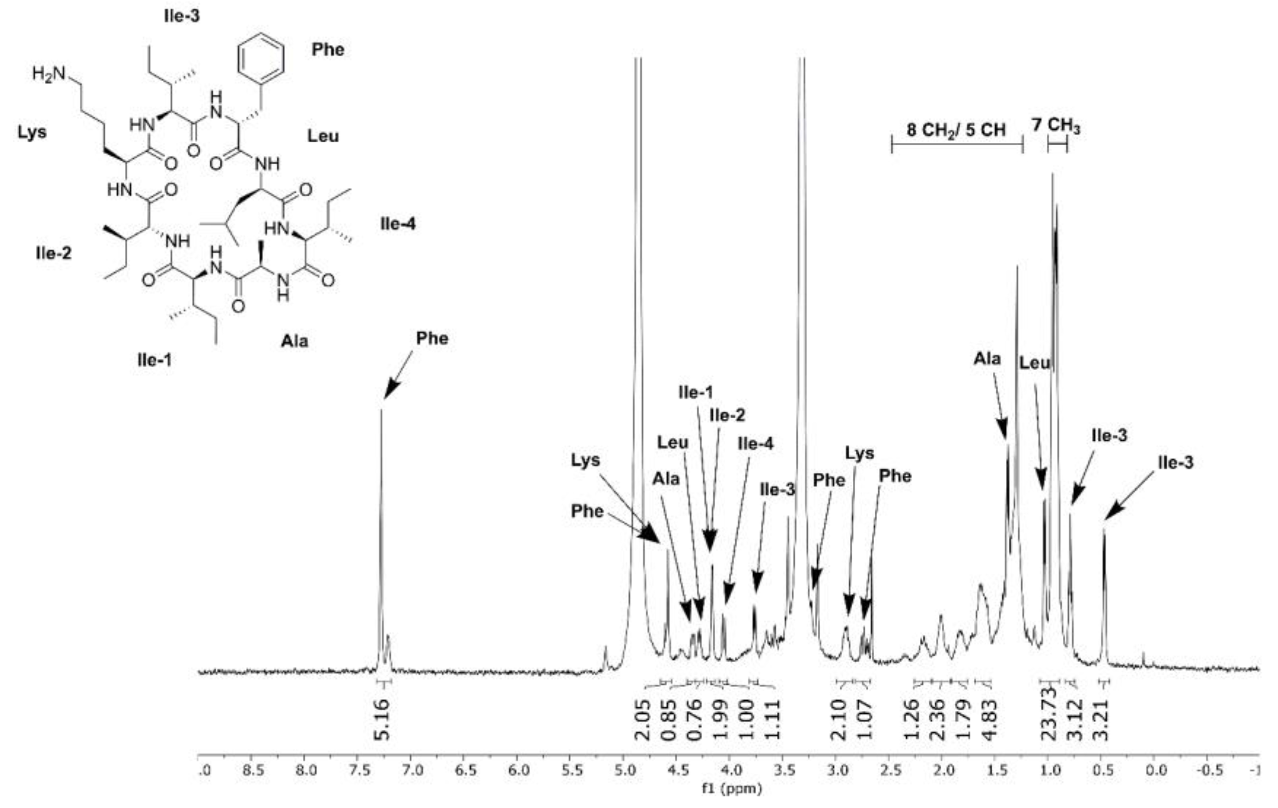
2.7. Metabolic Profiling, Compound Isolation, and Chemical Structure Analysis

The Streptomyces broth of TSB, SYP-NaCl, and YD medium cultures (180 mL) was exhaustively extracted using a solvent mixture of 1:1 MeOH:DCM yielding a crude extract (3.89 g). This crude extract was first separated using SPE on C18 bonded silica gel (Polygoprep C18 (Fisher Scientific, Dublin, Ireland) 12%C, 60 Å, 40–63 µm), eluting with varying solvent mixtures to produce five fractions: H2O (743.62 mg), 1:1 H2O:MeOH (368.6 mg), MeOH (15.4 mg), 1:1 MeOH:DCM (10.9 mg), DCM (8.2 mg). The final three fractions (MeOH, 1:1 MeOH:DCM, DCM, 34.5 mg) were then combined and subject to analytical reverse phase HPLC on a Waters Symmetry (VWR, Dublin, Ireland) C18 5 µm, 4.6 × 250 mm column. The column was eluted with 10% MeCN (0.1% TFA)/90% H2O (0.1% TFA) for 5 min, then a linear gradient to 100% MeCN (0.1% TFA) over 21 min was performed. The column was further eluted with 100% MeCN (0.1% TFA) for 6 min. After the HPLC was complete, a linear gradient back to 10% MeCN (0.1% TFA)/90% H2O (0.1% TFA) over 1 min and then further elution of 10% MeCN (0.1% FA)/90% H2O (0.1% FA) for 4 min was performed. This yielded pure surugamide A (0.8 mg). Surugamide A was characterised using MS and NMR data to confirm the structure for use as an analytical standard.
Surugamide A was quantified in the broth using LC-MS analysis on an Agilent UHR-qTOF 6540 (Agilent Technologies, Cork, Ireland) mass spectrometer. The column used for separation was Waters equity UPLC BEH (Apex Scientific, Kildare, Ireland) C18 1.7 µm 2.1 × 75 mm. The column was eluted with 10% MeCN (0.1% FA)/90% H2O (0.1% FA) for 2 min, then a linear gradient to 100% MeCN (0.1% FA) over 6 min was performed. The column was further eluted with 100% MeCN (0.1% FA) for 4 min. After the UPLC was complete, a linear gradient back to 10% MeCN (0.1% FA)/90% H2O (0.1% FA) over 1 min and then further elution of 10% MeCN (0.1% FA)/90% H2O (0.1% FA) for 3 min was performed before the next run. The MS detection method was positive ion. A calibration curve was produce using the LC-MS method above and injecting the pure surugamide A at seven concentrations (100, 25, 10, 2, 1, 0.2, 0.1 mg/L). Thirty millilitres of each Streptomyces strain in broth were extracted using a solvent mixture of 1:1 MeOH:DCM three times to yield a crude extract. These extracts were resuspended in MeOH and filtered through PTFE 0.2 µm filters (Sigma Aldrich, Arklow, Ireland) before being subject to the above LC-MS method.
The surugamide A calibration standards 1–7 and the six extracts were analysed using the Agilent MassHunter Quantification software package. This allowed the quantification of surugamide A in the extracts based on the intensity of peaks in the chromatogram with matching retention time and exact mass.

3. Results and Discussion

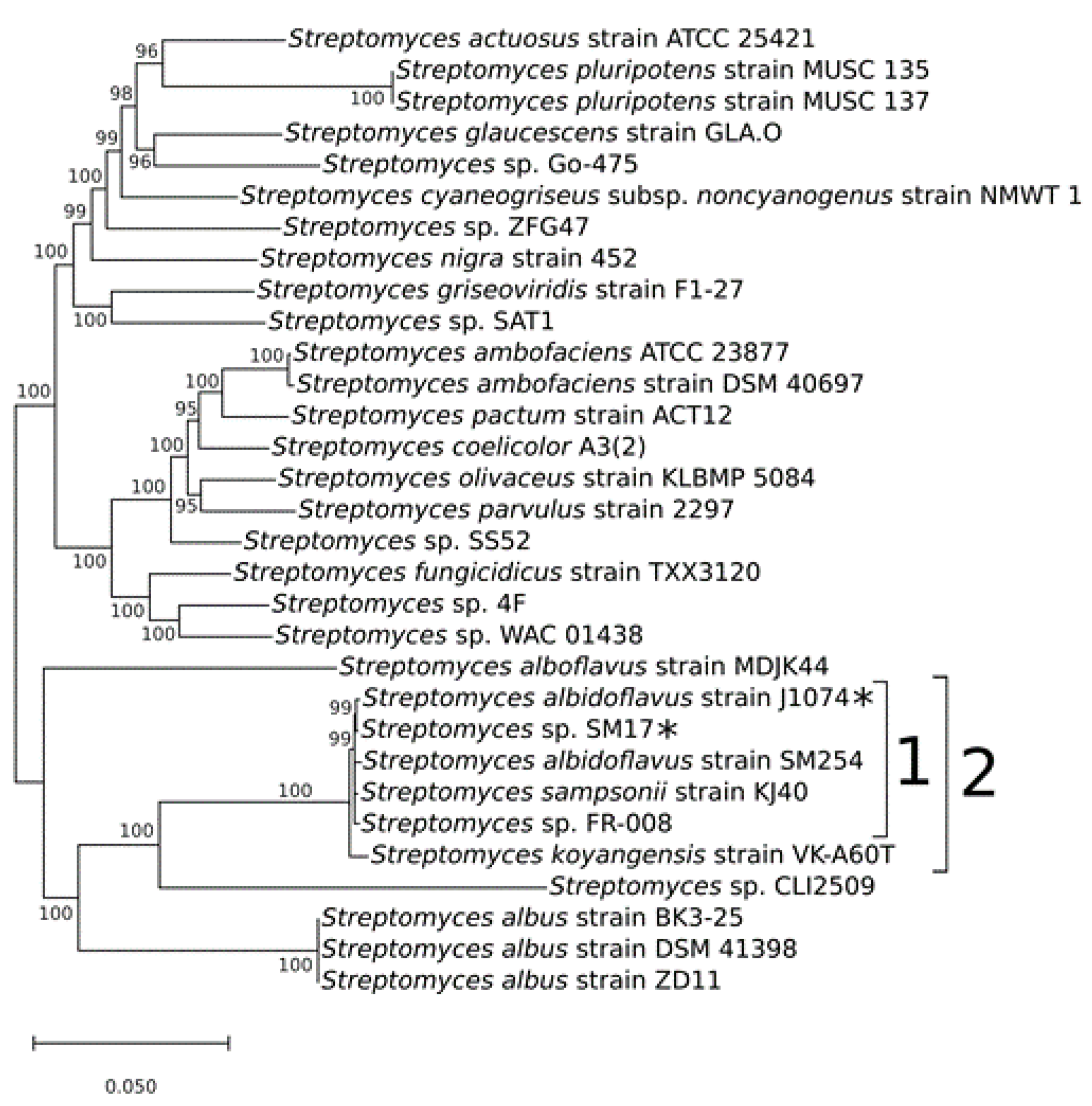
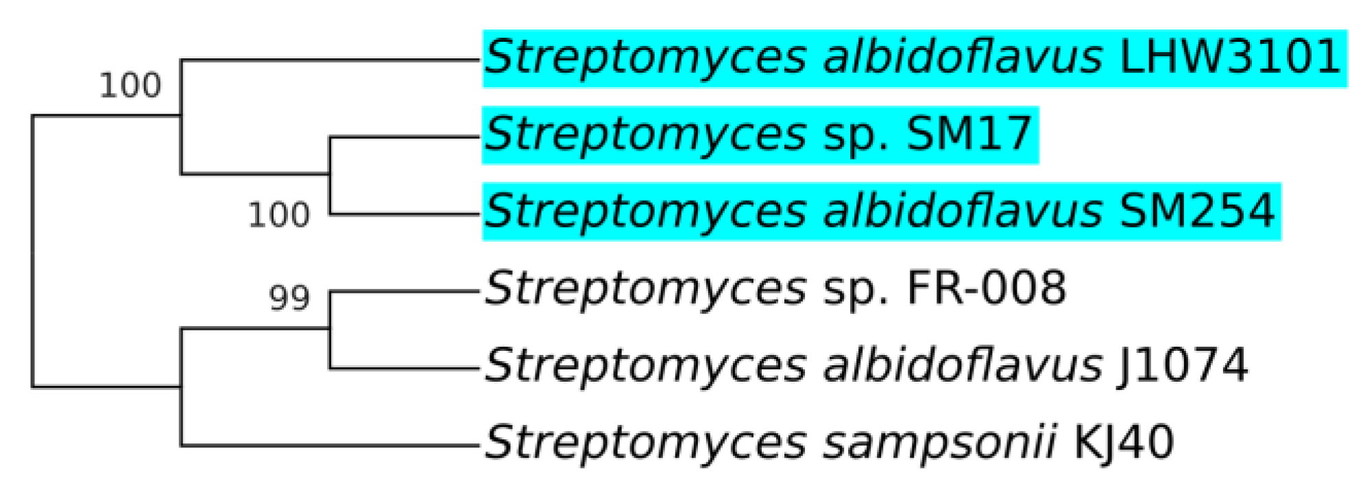
3.1. Multi-locus Sequence Analysis and Taxonomy Assignment of the Streptomyces sp. SM17 Isolate

In order to taxonomically characterise the Streptomyces sp. SM17 isolate based on genetic evidence, multi-locus sequence analysis (MLSA) [51] employing the 16S rRNA sequence, in addition to five housekeeping genes, namely atpD (ATP synthase subunit beta), gyrB (DNA gyrase subunit B), recA (recombinase RecA), rpoB (DNA-directed RNA polymerase subunit beta), and trpB (tryptophan synthase beta chain) was performed, in a similar manner to a previous report [32]. A similarity search was performed in the GenBank database [35], using the NCBI BLASTN tool [36,37], based on the 16S rRNA nucleotide sequence of the SM17 isolate. The top 30 most similar Streptomyces species for which complete genome sequences were available in GenBank were selected for further phylogenetic analysis.
The concatenated nucleotide sequences [51,52] of the 16S rRNA and the aforementioned five housekeeping genes, were first aligned using the MAFFT program [38], and the phylogeny analysis was performed using the MrBayes program [39]. The general time reversible (GTR) model of nucleotide substitution with gamma-distributed rates across sites with a proportion of invariable sites was applied [40], with 1 million generations sampled every 100 generations. The final phylogenetic tree was then processed using MEGA X [41], with a posterior probability cut-off of 95% (Figure 1).
The resulting phylogenetic tree clearly indicates the presence of a clade that includes the isolates Streptomyces albidoflavus strain J1074; Streptomyces sp. SM17; Streptomyces albidoflavus strain SM254; Streptomyces sampsonii strain KJ40; Streptomyces sp. FR-008; and Streptomyces koyangensis strain VK-A60T (clade 2 in Figure 1). In addition, this larger clade contains a sub-clade (clade 1 in Figure 1) that includes Streptomyces isolates similar to the strain Streptomyces albidoflavus J1074. The J1074 strain is a well-studied Streptomyces isolate widely used as a model for the genus and for various biotechnological applications, including the heterologous expression of secondary metabolites biosynthetic gene clusters (BGCs) [25,26,27,28,29]. This isolate was originally classified as “Streptomyces albus J1074”, but due to recent taxonomy data, it has been reclassified as Streptomyces albidoflavus J1074 [30,31]. Hence, in this study, this strain will be referred to as Streptomyces albidoflavus J1074, and this clade will from now on be referred to as the albidoflavus phylogroup (Figure 1).
Interestingly, members of the albidoflavus phylogroup were all isolated from quite different environments. The Streptomyces albidoflavus strain J1074 stems from the soil isolate Streptomyces albus G [23,24]. The Streptomyces sampsonii strain KJ40 was isolated from rhizosphere soil in a poplar plantation [53]. The Streptomyces sp. strain FR-008 is a random protoplast fusion derivative of two Streptomyces hygroscopicus isolates [54]. On the other hand, two of these strains were isolated from aquatic saline environments, with Streptomyces sp. SM17 being isolated from the marine sponge Haliclona simulans [13]; while the Streptomyces albidoflavus strain SM254 strain was isolated from copper-rich subsurface fluids within an iron mine, following growth on artificial sea water (ASW) [55]. The fact that these isolates, although derived from quite distinct environmental niches, simultaneously share significant genetic similarities is interesting, and raises questions about their potential evolutionary relatedness.

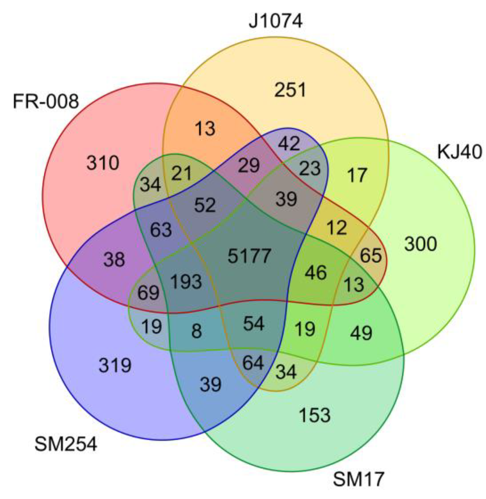
3.2. Analysis of Groups of Orthologous Genes in the Albidoflavus Phylogroup

In an attempt to provide further genetic evidence with respect to the similarities shared among the members of the albidoflavus phylogroup (Figure 1), a pan-genome analysis was performed to determine the number of core genes, accessory genes, and unique genes present in this group of isolates. The Roary program was employed for this objective [56], which allowed the identification of groups of orthologous and paralogous genes (which from now on will be referred to simply as “genes”) present in the set of albidoflavus genomes, with a protein identity cut-off of 95%, which is the identity value recommended by the Roary program manual when analysing organisms belonging to the same species.
A total of 7565 genes were identified in the albidoflavus pan-genome, and among these a total of 5177 were determined to be shared among all the albidoflavus isolates (i.e., the core genome) (Figure 2). This represents a remarkably high proportion of genes that appear to be highly conserved between all the isolates, representing approximately 68.4% of the pan-genome. Additionally, when considering the genomes individually (Table S1), the core genome accounts for approximately 84.5% of the FR-008 genome; 88.5% of J1074; 85.5% of KJ40; 86.7% of SM17; and 83.7% of the SM254 genome. On the other hand, the accessory genome (i.e., genes present in at least two isolates) was determined to consist of 1055 genes (or ~13.9% of the pan-genome); while the unique genome (i.e., genes present in only one isolate) was determined to consist of 1333 genes (or ~17.6% of the pan-genome). This strikingly high conservation of genes present in their genomes together with the previous multi-locus phylogeny analysis are very strong indicators that these microorganisms may belong to the same species.
An additional pan-genome analysis similar to the aforementioned analysis was also performed including the Streptomyces koyangensis strain VK-A60T in the dataset (Figure S1), which was an isolate shown to be a closely related neighbour to the albidoflavus phylogroup (Figure 1, clade 2). When compared to the previous analysis, the pan-genome analysis including the VK-A60T isolate showed significant changes in the values representing the core genome, which changed from 5177 genes (Figure 2) to 3912 genes (Figure S1), with an additional 1273 genes also shared among all of the albidoflavus isolates (Figure S1). The results also showed a much larger number of genes uniquely present in the VK-A60T genome than in the other genomes, with 2059 unique genes identified from a total of 6245 CDSs present in the VK-A60T genome in total, or approximately a third of its total number of genes (Figure S1). This proportion of unique genes present in the VK-A60T genome is considerably higher than the proportions of unique genes observed in the other albidoflavus phylotype genomes (Figure 2), which accounted for approximately only 2.5% of the total number of genes in SM17; 4.2% in J1074; 4.9% in KJ40; 5% in FR-008; and 5.1% in SM254. Taken together, these results further demonstrate the similarities between the isolates belonging to the albidoflavus phylogroup, while the VK-A60T isolate is clearly more distantly related.
Thus, from previous studies [30,31] and in light of the phylogeny analysis and further genomic evidence presented in this study, it is likely that all the isolates belonging to the albidoflavus phylogroup are in fact members of the same species. It is reasonable to infer that, for example, the isolates in the albidoflavus phylogroup that possess no species assignment thus far (i.e., strains SM17 and FR-008) are indeed members of the albidoflavus species. Also, it is possible that the Streptomyces sampsonii KJ40 has been misassigned, and possibly requires reclassification as an albidoflavus isolate.
Misassignment and reclassification of Streptomyces species is a common issue, and an increase in the quantity and the quality of available data from these organisms (e.g., better-quality genomes available in the databases) will provide better support for taxonomy claims, or correction of these when new information becomes available [31,57,58,59].

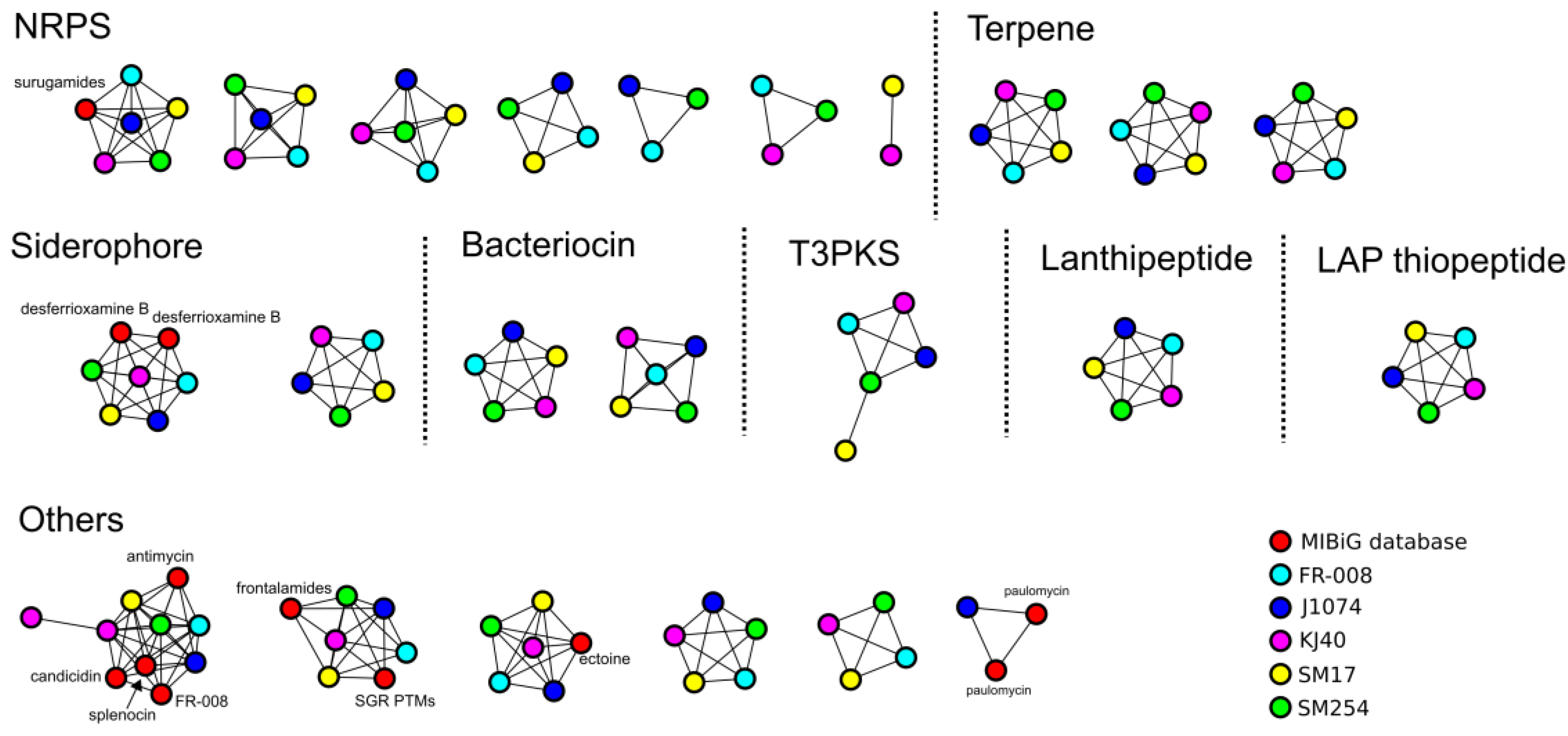
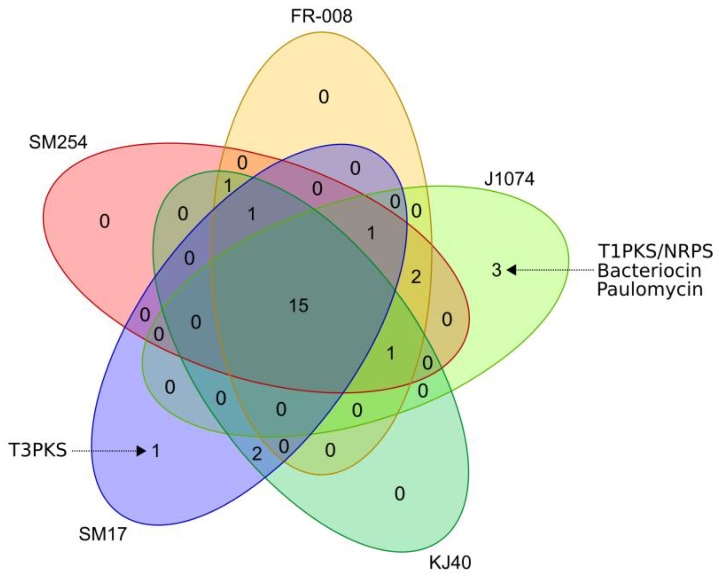
3.3. Prediction of Secondary Metabolites Biosynthetic Gene Clusters in the Albidoflavus Phylogroup

Isolates belonging to the albidoflavus phylogroup have been reported to produce bioactive compounds of pharmacological relevance, such as antibiotics. As mentioned previously, the Streptomyces albidoflavus strain J1074 is the best described member of the albidoflavus phylogroup to date. As such, several of secondary metabolites produced by this isolate have been identified, including acyl-surugamides and surugamides with antifungal and anticancer activities, respectively [16]; together with paulomycin derivatives with antibacterial activity [60]. The Streptomyces sp. FR-008 isolate has been shown to produce the antimicrobial compound FR-008/candicidin [61,62]; while the Streptomyces sampsonii KJ40 isolate has been shown to produce a chitinase that possesses anti-fungal activity against plant pathogens [53]. On the other hand, although no bioactive compounds have been characterised from Streptomyces albidoflavus SM254, this isolate has been shown to possess anti-fungal activity, specifically against the fungal bat pathogen Pseudogymnoascus destructans, which is responsible for the White-nose Syndrome [55,63]. The Streptomyces sp. SM17 isolate has also previously been shown to possess antibacterial and antifungal activities against clinically relevant pathogens, including methicillin-resistant Staphylococcus aureus (MRSA) [13]. However, no natural products derived from this strain have been identified and isolated until now.
In order to further in silico assess the potential of these albidoflavus phylogroup isolates to produce secondary metabolites, and also to determine how potentially similar or diverse they are within this phylogroup, prediction of secondary metabolites biosynthetic gene clusters (BGCs) was performed using the antiSMASH (version 5) program [42]. The antiSMASH prediction was processed using the BiG-SCAPE program [43], in order to cluster the BGCs into gene cluster families (GCFs), based on sequence and Pfam [64] protein families similarity, and also by comparing them to the BGCs available from the minimum information about a biosynthetic gene cluster (MiBIG) repository [44] (Figure 3). When compared to known BGCs from the MiBIG database, a significant number of BGCs predicted to be present in the albidoflavus phylogroup genomes could potentially encode for the production of novel compounds, including those belonging to the non-ribosomal peptide synthetase (NRPS) and bacteriocin families of compounds (Figure 3). The presence/absence of homologous BGCs in the albidoflavus isolates’ genomes was determined using BiG-SCAPE and is represented in Figure 4. Interestingly, the vast majority of the BGCs predicted in the albidoflavus phylogroup are shared among all of its members (15 BGCs); while another large portion (8 BGCs) are present in at least two isolates (Figure 4). Among the five members of the albidoflavus phylogroup, only the J1074 strain and the SM17 strain appeared to possess unique BGCs when compared to the other strains. Three unique BGCs were predicted to be present in the J1074 genome: a predicted type I polyketide synthase (T1PKS)/NRPS without significant similarity to the BGCs from the MiBIG database; a predicted bacteriocin, which also did not show any significant similarity to the BGCs from the MiBIG database; and a BGC predicted to encode for the production of the antibiotic paulomycin, with similarity to the paulomycin-encoding BGCs from Streptomyces paulus and Streptomyces sp. YN86 [65], which has also been experimentally shown to be produced by the J1074 strain [60]. One BGC predicted to encode a type III polyketide synthase (T3PKS)—with no significant similarity to the BGCs from the MiBIG database—was also identified as being unique to the SM17 genome.
Importantly, BGCs with similarity to the surugamide A/D BGC from “Streptomyces albus J1074” (now classified as S. albidoflavus) from the MiBIG database [16] were identified in all the other genomes of the members of the albidoflavus phylogroup. This raises the possibility that this BGC may be commonly present in albidoflavus species isolates. However, as only a few complete genomes of isolates belonging to this phylogroup are currently available, further data will be required to support this hypothesis. Nevertheless, these results further highlight the genetic similarities of the isolates belonging to the albidoflavus phylogroup, even with respect to their potential to produce secondary metabolites.

3.4. Phylogeny and Gene Synteny Analysis of Sur BGC Homologs

In parallel to the previous phylogenomics analysis performed with the albidoflavus phylogroup isolates, sequence similarity and phylogenetic analyses were performed, using the previously described and experimentally characterised Streptomyces albidoflavus LHW3101 surugamides biosynthetic gene cluster (sur BGC, GenBank accession number: MH070261) as a reference [18]. The aim was to assess how widespread in nature the sur BGC might be, and the degree of genetic variation, if any; that might be present in sur BGCs belonging to different microorganisms.
Nucleotides sequence similarity to the sur BGC was performed in the GenBank database [35], using the NCBI BLASTN tool [36,37]. It is important to note that, since the quality of the data is crucial for sequence similarity, homology, and phylogeny inquiries, only complete genome sequences were employed in this analysis. For this reason, for example, the marine Streptomyces isolate in which surugamides and derivatives were originally identified, namely Streptomyces sp. JAMM992 [14], was not included, since its complete genome is not available in the GenBank database.
The sequence similarity analysis identified five microorganisms that possessed homologs to the sur BGC and had their complete genome sequences available in the GenBank database: Streptomyces sp. SM17; Streptomyces albidoflavus SM254; Streptomyces sp. FR-008; Streptomyces albidoflavus J1074; and Streptomyces sampsonii KJ40. Notably, these results overlapped with the isolates belonging to the previously discussed albidoflavus phylogroup (Figure 1), further highlighting the possibility that the sur BGC may be commonly present in and potentially exclusive to the albidoflavus species.
Phylogenetic analysis was performed in the genomic regions determined to be homologs to the Streptomyces albidoflavus LHW3101 sur BGC, using the MrBayes program [39] (Figure 5). Although a larger number of sequences should ideally be employed in this type of analysis, these results suggest the possibility of a clade with aquatic saline environment-derived sur BGCs (Figure 5). Thus, these aquatic saline environment-derived sur BGCs are likely to share more genetic similarities amongst each other, rather than with those derived from terrestrial environments. Since this analysis took into consideration the whole genome regions that contained the sur BGCs of each isolate, it is likely that the similarities and differences present in these regions involve not only coding sequences (CDSs) for biosynthetic genes and/or transcriptional regulators, but also could include promoter regions and other intergenic sequences.
With this in mind, the genomic regions previously determined to share homology with the sur BGC from S. albidoflavus LHW3101 were further analysed, with respect to the genes present in the surrounding region, the organisation of the BGCs, together with the overall gene synteny (Figure 6). Translated CDSs predicted in the region were manually annotated using the NCBI BLASTP tool [36,37], together with GenBank [35] and the CDD [66] databases. These included the main biosynthetic genes, namely surABCD, the transcriptional regulator surR, and the thioesterase surE—all of which had previously been reported to have roles in the biosynthesis of surugamides and their derivatives [15,16,17,18,19] (Figure 6).
Interestingly, this result indicated that the gene synteny of the biosynthetic genes as well as the flanking genes is highly conserved, with the exception to the 3’ flanking region of the BGC from S. sampsonii KJ40. Notably, even the reading frames of the surE gene and the surABCD genes are conserved amongst all the genomes. As indicated by the numbers in Figure 6, the 5’ region in all the genomic regions consisted of: 1) A MbtH-like protein, which have been reported to be involved in the synthesis of non-ribosomal peptides, antibiotics, and siderophores, in Streptomyces species [67,68]; 2) a putative ABC transporter, which is a family of proteins with varied biological functions, including conferring resistance to drugs and other toxic compounds [69,70]; 3) a BcrA family ABC transporter, which is a family commonly involved in peptide antibiotics resistance [71,72]; 4) a hypothetical protein; followed by 5) the transcriptional repressor SurR, which has been experimentally demonstrated to repress the production of surugamides [16]; 6) a hypothetical membrane protein; 7) the thioesterase SurE, which is homologous to the penicillin binding protein, reported to be responsible for the cyclisation of surugamides molecules [21]; and finally 8–11) the main surugamides biosynthetic genes surABCD, all of which encode non-ribosomal peptide synthetase (NRPS) proteins [19]. The 3’ flanking region consisted of: 12) A predicted multi-drug resistance (MDR) transporter belonging to the major facilitator superfamily (MFS) of membrane transport proteins [73,74]; 13) a predicted TetR/AcrR transcriptional regulator, which is a family of regulators reported to be involved in antibiotic resistance [75]; 14) a hypothetical protein; and 15) another predicted MDR transporter belonging to the MFS superfamily. In contrast, the 3’ flanking region of the KJ40 strain sur BGC, consisted of: 16) A group of four hypothetical proteins, which may represent pseudogene versions of the first MDR transporter identified in the other isolates (gene number 12 in Figure 6); 17) a predicted rearrangement hotspot (RHS) repeat protein, which is a family of proteins reported to be involved in mediating intercellular competition in bacteria [76]; 18) a hypothetical protein; and 19) a MDR transporter belonging to the MFS superfamily, which, interestingly, is a homolog of protein number 15, which is present in all the other isolates.
The conserved gene synteny observed in the sur BGC genomic region, particularly those positioned upstream of the main biosynthetic surABCD genes, together with the observation that even the reading frames of the surE and the surABCD genes are conserved among all the genomes analysed, coupled with the previous phylogenetic and pan-genome analyses, suggest the following. Firstly, it is very likely that these strains share a common ancestry and that the sur BGC genes had a common origin. Secondly, there is a strong evolutionary pressure ensuring the maintenance of not only gene synteny, but also of the reading frames of the main biosynthetic genes involved in the production of surugamides. The latter raises the question of which other genes in this region may be involved in the production of these compounds, or potentially conferring mechanisms of self-resistance to surugamides in the isolates, particularly since many of the genes have predicted functions that are compatible with the transport of small molecules and with multi-drug resistance. These observations are particularly interesting considering that these strains are derived from quite varied environments and geographic locations.

3.5. Growth, Morphology, Phenotype, and Metabolism Assessment of Streptomyces sp. SM17 in Complex Media

In order to assess the metabolic potential of the SM17 strain [77], particularly with respect to the production of surugamide A, the isolate was cultivated in a number of different growth media, within an OSMAC-based approach [33,34]. While the SM17 strain was able to grow in SYP-NaCl, YD, SY, P1, P2, P3, and CH-F2 liquid media, the strain was unable to grow in Oatmeal and Sporulation media. The latter indicated an inability to metabolise oat and starch when nutrients other than yeast extract are not present. Morphologically, the SM17 strain formed cell aggregates or pellets in TSB, YD, and SYP-NaCl, while this differentiation was not observed in the other media. Preliminary chemical analyses of these samples, employing liquid chromatography–mass spectrometry (UPLC-DAD-HRMS), indicated that secondary metabolism in SM17 was not very active when the strain was cultivated in SY, P1, P2, P3, and CH-F2 media. In contrast, significant production of surugamides was evidenced in the extracts from TSB, SYP-NaCl, and YD media, with characteristic ions at m/z 934.6106 (surugamide A) and 920.5949 (surugamide B) [M + Na]+, which correlated with the formation of cell pellets and the production of natural products, as previously described in other Streptomyces strains [77,78].

3.6. Differential Production of Surugamide A by Streptomyces sp. SM17 and S. albidoflavus J1074

To confirm the production of surugamide A by the SM17 isolate, extracts from the TSB, SYP-NaCl, and YD media were combined and purified using high-performance liquid chromatography (HPLC). The structures of the major compounds of the extract were subsequently analysed using nuclear magnetic resonance (NMR) spectroscopy, which allowed for the identification of the chemical structure of the surugamide A molecule as major metabolite by comparison with reference NMR data (Figure 7) [14].
The isolates Streptomyces sp. SM17 and S. albidoflavus J1074 were subsequently cultivated in the aforementioned media in which the SM17 strain had been shown to be metabolically active, namely the TSB, SYP-NaCl, and YD media. This was performed in order to assess whether there were any significant differences in the production of surugamide A when different growth media are employed for the production of this compound, and to compare the levels of surugamide A produced by the SM17 and the J1074 isolates. The MeOH/DCM (1:1) extracts from the aforementioned cultures of SM17 and J1074 were subjected to liquid chromatography–mass spectrometry (UPLC-HRMS) to quantify the levels of surugamide A being produced under each condition (Table 1), using a surugamide A standard calibration curve (Figure S2).
The LC-MS quantification analysis (Table 1) indicated that both strains were capable of producing surugamide A in all the conditions tested. However, the SM17 strain appeared to produce considerably higher yields of the compound when compared to J1074, in all the conditions analysed. In addition, the S. albidoflavus J1074 isolate appeared to produce quite low levels of surugamide A when grown in TSB and YD media, accounting for less than 1% (w/w) of the extracts from these media. Interestingly, higher yields of surugamide A were produced in the SYP-NaCl medium in both strains, when compared with the levels of surugamide A produced by these strains when grown in TSB and the YD media (Table 1). In the SM17 culture in SYP-NaCl, surugamide A accounted for 10.60% (w/w) of the extract, compared to 2.44% and 1.13% from TSB and YD, respectively; while in J1074 it accounted for 3.55% (w/w) of the extract from the SYP-NaCl culture, compared to 0.27% and 0.09% from TSB and YD, respectively (Table 1). These results provide further insights into factors that are potentially involved in regulation the biosynthesis of surugamide A, in the albidoflavus phylogroup and in Streptomyces sp. SM17 in particular.
Firstly, it appears likely that surugamide A biosynthesis may be regulated, at least in part, by carbon catabolite repression (CCR). Carbon catabolite repression is a well-described regulatory mechanism in bacteria that controls carbon metabolism [79,80,81,82], and which has also been reported to regulate the biosynthesis of secondary metabolites in a number of different bacterial species, including in Streptomyces isolates [83,84,85,86]. While the TSB and the YD media contain glucose and dextrins as carbon sources, respectively; the complex polysaccharide starch is the carbon source in the SYP-NaCl medium. Therefore, it is reasonable to infer that glucose and dextrin may repress the production of surugamide A in Streptomyces sp. SM17 and in Streptomyces albidoflavus J1074, while starch does not. Further evidence for this can be found when considering the different production media previously employed in the production of surugamides by different Streptomyces isolates. For example, in the original research that led to the discovery of surugamides in Streptomyces sp. JAMM992 [14], the PC-1 medium (1% starch, 1% polypeptone, 1% meat extract, 1% molasses, pH 7.2) was employed for production of these compounds. Similar to the SYP-NaCl medium employed in our study, the PC-1 medium also contains starch as the carbon source, together with another complex carbon source, namely molasses. Likewise, for the production of surugamides in S. albidoflavus strain LHW3101 [18], the TSBY medium (3% tryptone soy broth, 10.3% sucrose, 0.5% yeast extract) was employed, which utilises sucrose as its main carbon source. In contrast, when elicitors were employed to induce the production of surugamides and their derivatives in the J1074 strain [16], by activating the sur BGC, which appeared to be silent in this isolate, the R4 medium (0.5% glucose, 0.1% yeast extract, among other non-carbon related components) was employed, which utilises glucose as its main carbon supply, and, as shown in this study, it potentially represses the production of surugamide A. Thus, from these previous reports and from our observations, it appears likely that CCR plays an important role in regulating the biosynthesis of surugamides.
Secondly, it is important to note the presence of salts in the form of NaCl in the SYP-NaCl medium. As previously mentioned, genetic and phylogenetic analyses of the sur BGC indicated similarities between those BGCs belonging to aquatic saline-derived Streptomyces isolates (Figure 5), together with the likelihood that these sur BGCs might have had a common origin. Thus, it is plausible that this origin may have been marine, and hence the presence of salts in the growth medium may also have an influence on the biosynthesis of surugamide A. Different concentrations of salts in the form of NaCl in the culture medium have also previously been shown to impact on the chemical profile of metabolites produced in the marine-obligate bacteria Salinispora arenicola [87].
Nevertheless, it is interesting to observe that, despite the repression/induction of the biosynthesis of surugamide A observed when different media were employed, the SM17 isolate clearly produces considerably higher amounts of surugamide A when compared to S. albidoflavus J1074—reaching yields up to >13-fold higher in the YD medium, and around 3-fold higher when grown in the SYP-NaCl medium (Table 1).

4. Conclusions

Marine-derived bacteria, particularly those isolated in association with marine invertebrates, such as sponges, have been shown to be reservoirs of bioactive molecules, including those with antibacterial, antifungal, and anticancer activities. Among these newly identified bioactive compounds, the surugamides and their derivatives are of particular interest due to their clinically relevant bioactivities, i.e., anticancer and antifungal, and their original metabolic pathway.
Based on genome mining, this study identified the previously unreported capability of the marine sponge-derived isolate Streptomyces sp. SM17 to produce surugamide A and also sheds new light on factors such as the carbon catabolite repression (CCR) that may be involved in regulating production of this molecule. Phylogenomics analysis indicated that the sur BGC is commonly present in members of the proposed albidoflavus phylogroup, and that the sur BGCs present in different isolates derived from varied environmental niches may possess a common ancestry. Although high quality genomic data from this proposed albidoflavus phylogroup are still lacking, results presented here suggest that the sur BGCs derived from Streptomyces isolated from aquatic saline environment are more similar to each other, when compared to those isolated from terrestrial environments.
Chemical analysis was performed in order to assess differential production of surugamide A when comparing a marine Streptomyces isolate with a terrestrial Streptomyces isolate, namely SM17 and J1074 strains, respectively, following an OSMAC-based approach employing different culture media. This analysis showed that not only the marine-derived isolate SM17 was capable of producing more surugamide A when compared to J1074 under all the conditions tested, but also that the biosynthesis of surugamide A is likely to be influenced by the CCR, and potentially by the presence of salts in the growth medium. These results also highlight the importance of employing an OSMAC-based approach even when analysing the production of known compounds, since there is a clear difference in the yields of surugamide A obtained when employing different culture media. Thus, it is possible to gain further insights into the production of bacterial types of compounds by 1) discovering strains that possess a higher capability to produce these compounds; 2) establishing optimal conditions for the biosynthesis of their production; and 3) providing a better understanding of the genetic and regulatory mechanisms potentially underpinning the production of these compounds.

Supplementary Materials

The following are available online at https://www.mdpi.com/2076-2607/7/10/394/s1, Figure S1: Groups of orthologous genes in the genomes of the albidoflavus phylogroup, Figure S2: Calibration curve for surugamide A, Table S1: Genome statistics of the albidoflavus phylogroup.

Author Contributions

Conceived and designed the experiments: E.L.A., A.F.C.R, O.P.T, A.D.W.D. Performed the experiments: E.L.A., A.F.C.R., N.K., L.K.J. Analysed the data: E.L.A., A.F.C.R., N.K., L.K.J., O.P.T., S.A.J., A.D.W.D. Wrote the paper: E.L.A., O.P.T., A.D.W.D.

Funding

E.L.A. acknowledges the funding support of the Brazilian National Council for Scientific and Technological Development (CNPq). This project was supported by a research grant from the Marine Institute through the NMBLI project (Grant-Aid Agreement PBA/MB/16/01) and by the Marine Biotechnology ERA/NET, NEPTUNA project (contract No. PBA/MB/15/02) and by Science Foundation Ireland (SSPC-2, 12/RC/2275).

Conflicts of Interest

The authors declare no conflict of interest.

References

  1. Hwang, K.-S.; Kim, H.U.; Charusanti, P.; Palsson, B.Ø.; Lee, S.Y. Systems biology and biotechnology of Streptomyces species for the production of secondary metabolites. Biotechnol. Adv. 2014, 32, 255–268. [Google Scholar] [CrossRef] [PubMed]
  2. Thabit, A.K.; Crandon, J.L.; Nicolau, D.P. Antimicrobial resistance: Impact on clinical and economic outcomes and the need for new antimicrobials. Expert Opin. Pharmacother. 2015, 16, 159–177. [Google Scholar] [CrossRef] [PubMed]
  3. Tommasi, R.; Brown, D.G.; Walkup, G.K.; Manchester, J.I.; Miller, A.A. ESKAPEing the labyrinth of antibacterial discovery. Nat. Rev. Drug Discov. 2015, 14, 529–542. [Google Scholar] [CrossRef] [PubMed]
  4. Demers, D.; Knestrick, M.; Fleeman, R.; Tawfik, R.; Azhari, A.; Souza, A.; Vesely, B.; Netherton, M.; Gupta, R.; Colon, B.; et al. Exploitation of Mangrove Endophytic Fungi for Infectious Disease Drug Discovery. Mar. Drugs 2018, 16, 376. [Google Scholar] [CrossRef] [PubMed]
  5. Indraningrat, A.; Smidt, H.; Sipkema, D. Bioprospecting Sponge-Associated Microbes for Antimicrobial Compounds. Mar. Drugs 2016, 14, 87. [Google Scholar] [CrossRef] [PubMed]
  6. Calcabrini, C.; Catanzaro, E.; Bishayee, A.; Turrini, E.; Fimognari, C. Marine Sponge Natural Products with Anticancer Potential: An Updated Review. Mar. Drugs 2017, 15, 310. [Google Scholar] [CrossRef]
  7. Schneemann, I.; Kajahn, I.; Ohlendorf, B.; Zinecker, H.; Erhard, A.; Nagel, K.; Wiese, J.; Imhoff, J.F. Mayamycin, a Cytotoxic Polyketide from a Streptomyces Strain Isolated from the Marine Sponge Halichondria panicea. J. Nat. Prod. 2010, 73, 1309–1312. [Google Scholar] [CrossRef] [PubMed]
  8. Kunz, A.; Labes, A.; Wiese, J.; Bruhn, T.; Bringmann, G.; Imhoff, J. Nature’s Lab for Derivatization: New and Revised Structures of a Variety of Streptophenazines Produced by a Sponge-Derived Streptomyces Strain. Mar. Drugs 2014, 12, 1699–1714. [Google Scholar] [CrossRef]
  9. Sathiyanarayanan, G.; Gandhimathi, R.; Sabarathnam, B.; Seghal Kiran, G.; Selvin, J. Optimization and production of pyrrolidone antimicrobial agent from marine sponge-associated Streptomyces sp. MAPS15. Bioprocess Biosyst. Eng. 2014, 37, 561–573. [Google Scholar] [CrossRef]
  10. Almeida, E.L.; Margassery, L.M.; Kennedy, J.; Dobson, A.D.W. Draft Genome Sequence of the Antimycin-Producing Bacterium Streptomyces sp. Strain SM8, Isolated from the Marine Sponge Haliclona simulans. Genome Announc. 2018, 6, e01535-17. [Google Scholar] [CrossRef]
  11. Viegelmann, C.; Margassery, L.; Kennedy, J.; Zhang, T.; O’Brien, C.; O’Gara, F.; Morrissey, J.; Dobson, A.; Edrada-Ebel, R. Metabolomic Profiling and Genomic Study of a Marine Sponge-Associated Streptomyces sp. Mar. Drugs 2014, 12, 3323–3351. [Google Scholar] [CrossRef] [PubMed]
  12. Jackson, S.; Crossman, L.; Almeida, E.; Margassery, L.; Kennedy, J.; Dobson, A. Diverse and Abundant Secondary Metabolism Biosynthetic Gene Clusters in the Genomes of Marine Sponge Derived Streptomyces spp. Isolates. Mar. Drugs 2018, 16, 67. [Google Scholar] [CrossRef] [PubMed]
  13. Kennedy, J.; Baker, P.; Piper, C.; Cotter, P.D.; Walsh, M.; Mooij, M.J.; Bourke, M.B.; Rea, M.C.; O’Connor, P.M.; Ross, R.P.; et al. Isolation and Analysis of Bacteria with Antimicrobial Activities from the Marine Sponge Haliclona simulans Collected from Irish Waters. Mar. Biotechnol. 2009, 11, 384–396. [Google Scholar] [CrossRef] [PubMed]
  14. Takada, K.; Ninomiya, A.; Naruse, M.; Sun, Y.; Miyazaki, M.; Nogi, Y.; Okada, S.; Matsunaga, S. Surugamides A–E, Cyclic Octapeptides with Four d-Amino Acid Residues, from a Marine Streptomyces sp.: LC–MS-Aided Inspection of Partial Hydrolysates for the Distinction of d- and l-Amino Acid Residues in the Sequence. J. Org. Chem. 2013, 78, 6746–6750. [Google Scholar] [CrossRef] [PubMed]
  15. Matsuda, K.; Kuranaga, T.; Sano, A.; Ninomiya, A.; Takada, K.; Wakimoto, T. The Revised Structure of the Cyclic Octapeptide Surugamide A. Chem. Pharm. Bull. 2019, 67, 476–480. [Google Scholar] [CrossRef] [PubMed]
  16. Xu, F.; Nazari, B.; Moon, K.; Bushin, L.B.; Seyedsayamdost, M.R. Discovery of a Cryptic Antifungal Compound from Streptomyces albus J1074 Using High-Throughput Elicitor Screens. J. Am. Chem. Soc. 2017, 139, 9203–9212. [Google Scholar] [CrossRef] [PubMed]
  17. Thankachan, D.; Fazal, A.; Francis, D.; Song, L.; Webb, M.E.; Seipke, R.F. A trans-Acting Cyclase Offloading Strategy for Nonribosomal Peptide Synthetases. ACS Chem. Biol. 2019, 14, 845–849. [Google Scholar] [CrossRef] [PubMed]
  18. Zhou, Y.; Lin, X.; Xu, C.; Shen, Y.; Wang, S.-P.; Liao, H.; Li, L.; Deng, H.; Lin, H.-W. Investigation of Penicillin Binding Protein (PBP)-like Peptide Cyclase and Hydrolase in Surugamide Non-ribosomal Peptide Biosynthesis. Cell Chem. Biol. 2019, 26, 737–744.e4. [Google Scholar] [CrossRef] [PubMed]
  19. Ninomiya, A.; Katsuyama, Y.; Kuranaga, T.; Miyazaki, M.; Nogi, Y.; Okada, S.; Wakimoto, T.; Ohnishi, Y.; Matsunaga, S.; Takada, K. Biosynthetic Gene Cluster for Surugamide A Encompasses an Unrelated Decapeptide, Surugamide F. ChemBioChem 2016, 17, 1709–1712. [Google Scholar] [CrossRef]
  20. Kuranaga, T.; Matsuda, K.; Sano, A.; Kobayashi, M.; Ninomiya, A.; Takada, K.; Matsunaga, S.; Wakimoto, T. Total Synthesis of the Nonribosomal Peptide Surugamide B and Identification of a New Offloading Cyclase Family. Angew. Chemie Int. Ed. 2018, 57, 9447–9451. [Google Scholar] [CrossRef] [PubMed]
  21. Matsuda, K.; Kobayashi, M.; Kuranaga, T.; Takada, K.; Ikeda, H.; Matsunaga, S.; Wakimoto, T. SurE is a trans-acting thioesterase cyclizing two distinct non-ribosomal peptides. Org. Biomol. Chem. 2019, 17, 1058–1061. [Google Scholar] [CrossRef] [PubMed]
  22. Koshla, O.T.; Rokytskyy, I.V.; Ostash, I.S.; Busche, T.; Kalinowski, J.; Mösker, E.; Süssmuth, R.D.; Fedorenko, V.O.; Ostash, B.O. Secondary Metabolome and Transcriptome of Streptomyces albus J1074 in Liquid Medium SG2. Cytol. Genet. 2019, 53, 1–7. [Google Scholar] [CrossRef]
  23. Chater, K.F.; Wilde, L.C. Restriction of a bacteriophage of Streptomyces albus G involving endonuclease SalI. J. Bacteriol. 1976, 128, 644–650. [Google Scholar] [PubMed]
  24. Chater, K.F.; Wilde, L.C. Streptomyces albus G Mutants Defective in the SalGI Restriction-Modification System. Microbiology 1980, 116, 323–334. [Google Scholar] [CrossRef] [PubMed]
  25. Huang, C.; Yang, C.; Zhang, W.; Zhu, Y.; Ma, L.; Fang, Z.; Zhang, C. Albumycin, a new isoindolequinone from Streptomyces albus J1074 harboring the fluostatin biosynthetic gene cluster. J. Antibiot. (Tokyo) 2019, 72, 311–315. [Google Scholar] [CrossRef] [PubMed]
  26. Zaburannyi, N.; Rabyk, M.; Ostash, B.; Fedorenko, V.; Luzhetskyy, A. Insights into naturally minimised Streptomyces albus J1074 genome. BMC Genomics 2014, 15, 97. [Google Scholar] [CrossRef] [PubMed]
  27. Myronovskyi, M.; Rosenkränzer, B.; Nadmid, S.; Pujic, P.; Normand, P.; Luzhetskyy, A. Generation of a cluster-free Streptomyces albus chassis strains for improved heterologous expression of secondary metabolite clusters. Metab. Eng. 2018, 49, 316–324. [Google Scholar] [CrossRef]
  28. Bilyk, O.; Sekurova, O.N.; Zotchev, S.B.; Luzhetskyy, A. Cloning and Heterologous Expression of the Grecocycline Biosynthetic Gene Cluster. PLoS ONE 2016, 11, e0158682. [Google Scholar] [CrossRef]
  29. Jiang, G.; Zhang, Y.; Powell, M.M.; Zhang, P.; Zuo, R.; Zhang, Y.; Kallifidas, D.; Tieu, A.M.; Luesch, H.; Loria, R.; et al. High-Yield Production of Herbicidal Thaxtomins and Thaxtomin Analogs in a Nonpathogenic Streptomyces Strain. Appl. Environ. Microbiol. 2018, 84, e00164-18. [Google Scholar] [CrossRef]
  30. Labeda, D.P.; Doroghazi, J.R.; Ju, K.-S.; Metcalf, W.W. Taxonomic evaluation of Streptomyces albus and related species using multilocus sequence analysis and proposals to emend the description of Streptomyces albus and describe Streptomyces pathocidini sp. nov. Int. J. Syst. Evol. Microbiol. 2014, 64, 894–900. [Google Scholar] [CrossRef]
  31. Labeda, D.P.; Dunlap, C.A.; Rong, X.; Huang, Y.; Doroghazi, J.R.; Ju, K.-S.; Metcalf, W.W. Phylogenetic relationships in the family Streptomycetaceae using multi-locus sequence analysis. Antonie Van Leeuwenhoek 2017, 110, 563–583. [Google Scholar] [CrossRef] [PubMed]
  32. Almeida, E.L.; Carillo Rincón, A.F.; Jackson, S.A.; Dobson, A.D. Comparative genomics of marine sponge-derived Streptomyces spp. isolates SM17 and SM18 with their closest terrestrial relatives provides novel insights into environmental niche adaptations and secondary metabolite biosynthesis potential. Front. Microbiol. 2019, 10, 1713. [Google Scholar] [CrossRef] [PubMed]
  33. Romano, S.; Jackson, S.; Patry, S.; Dobson, A. Extending the “One Strain Many Compounds” (OSMAC) Principle to Marine Microorganisms. Mar. Drugs 2018, 16, 244. [Google Scholar] [CrossRef] [PubMed]
  34. Pan, R.; Bai, X.; Chen, J.; Zhang, H.; Wang, H. Exploring Structural Diversity of Microbe Secondary Metabolites Using OSMAC Strategy: A Literature Review. Front. Microbiol. 2019, 10, 294. [Google Scholar] [CrossRef] [PubMed] [Green Version]
  35. Benson, D.A.; Cavanaugh, M.; Clark, K.; Karsch-Mizrachi, I.; Ostell, J.; Pruitt, K.D.; Sayers, E.W. GenBank. Nucleic Acids Res. 2018, 46, D41–D47. [Google Scholar] [CrossRef] [PubMed]
  36. Johnson, M.; Zaretskaya, I.; Raytselis, Y.; Merezhuk, Y.; McGinnis, S.; Madden, T.L. NCBI BLAST: A better web interface. Nucleic Acids Res. 2008, 36, W5–W9. [Google Scholar] [CrossRef] [PubMed]
  37. Camacho, C.; Coulouris, G.; Avagyan, V.; Ma, N.; Papadopoulos, J.; Bealer, K.; Madden, T.L. BLAST+: Architecture and applications. BMC Bioinform. 2009, 10, 421. [Google Scholar] [CrossRef] [PubMed]
  38. Katoh, K.; Standley, D.M. MAFFT Multiple Sequence Alignment Software Version 7: Improvements in Performance and Usability. Mol. Biol. Evol. 2013, 30, 772–780. [Google Scholar] [CrossRef] [PubMed] [Green Version]
  39. Ronquist, F.; Teslenko, M.; van der Mark, P.; Ayres, D.L.; Darling, A.; Höhna, S.; Larget, B.; Liu, L.; Suchard, M.A.; Huelsenbeck, J.P. MrBayes 3.2: Efficient Bayesian Phylogenetic Inference and Model Choice Across a Large Model Space. Syst. Biol. 2012, 61, 539–542. [Google Scholar] [CrossRef] [PubMed] [Green Version]
  40. Waddell, P.J.; Steel, M. General Time-Reversible Distances with Unequal Rates across Sites: Mixing Γ and Inverse Gaussian Distributions with Invariant Sites. Mol. Phylogenet. Evol. 1997, 8, 398–414. [Google Scholar] [CrossRef] [PubMed]
  41. Kumar, S.; Stecher, G.; Li, M.; Knyaz, C.; Tamura, K. MEGA X: Molecular Evolutionary Genetics Analysis across Computing Platforms. Mol. Biol. Evol. 2018, 35, 1547–1549. [Google Scholar] [CrossRef] [PubMed]
  42. Blin, K.; Shaw, S.; Steinke, K.; Villebro, R.; Ziemert, N.; Lee, S.Y.; Medema, M.H.; Weber, T. antiSMASH 5.0: Updates to the secondary metabolite genome mining pipeline. Nucleic Acids Res. 2019, 47, 81–87. [Google Scholar] [CrossRef] [PubMed]
  43. Navarro-Muñoz, J.C.; Selem-Mojica, N.; Mullowney, M.W.; Kautsar, S.; Tryon, J.H.; Parkinson, E.I.; Santos, E.L.C.D.L.; Yeong, M.; Cruz-Morales, P.; Abubucker, S.; et al. A computational framework for systematic exploration of biosynthetic diversity from large-scale genomic data. bioRxiv 2018, 10, 445270. [Google Scholar]
  44. Medema, M.H.; Kottmann, R.; Yilmaz, P.; Cummings, M.; Biggins, J.B.; Blin, K.; de Bruijn, I.; Chooi, Y.H.; Claesen, J.; Coates, R.C.; et al. Minimum Information about a Biosynthetic Gene cluster. Nat. Chem. Biol. 2015, 11, 625–631. [Google Scholar] [CrossRef]
  45. Shannon, P.; Markiel, A.; Ozier, O.; Baliga, N.S.; Wang, J.T.; Ramage, D.; Amin, N.; Schwikowski, B.; Ideker, T. Cytoscape: A software environment for integrated models of biomolecular interaction networks. Genome Res. 2003, 13, 2498–2504. [Google Scholar] [CrossRef] [PubMed]
  46. Okonechnikov, K.; Golosova, O.; Fursov, M. Unipro UGENE: A unified bioinformatics toolkit. Bioinformatics 2012, 28, 1166–1167. [Google Scholar] [CrossRef] [PubMed]
  47. Rutherford, K.; Parkhill, J.; Crook, J.; Horsnell, T.; Rice, P.; Rajandream, M.-A.; Barrell, B. Artemis: Sequence visualization and annotation. Bioinformatics 2000, 16, 944–945. [Google Scholar] [CrossRef] [PubMed]
  48. Dusa, A. Venn: Draw Venn Diagrams 2018. Available online: https://cran.r-project.org/web/packages/venn/index.html (accessed on 25 September 2019).
  49. R Core Team R: A Language and Environment for Statistical Computing, version 3.5.3; Vienna, Austria, 2018. Available online: https://www.R-project.org/ (accessed on 25 September 2019).
  50. RStudio Team. RStudio Team RStudio: Integrated Development Environment for RStudio Inc.; RStudio Inc.: Boston, MA, USA, 2015; Volume 14. [Google Scholar]
  51. Glaeser, S.P.; Kämpfer, P. Multilocus sequence analysis (MLSA) in prokaryotic taxonomy. Syst. Appl. Microbiol. 2015, 38, 237–245. [Google Scholar] [CrossRef]
  52. Gadagkar, S.R.; Rosenberg, M.S.; Kumar, S. Inferring species phylogenies from multiple genes: Concatenated sequence tree versus consensus gene tree. J. Exp. Zool. Part B Mol. Dev. Evol. 2005, 304B, 64–74. [Google Scholar] [CrossRef]
  53. Li, S.; Zhang, B.; Zhu, H.; Zhu, T. Cloning and Expression of the Chitinase Encoded by ChiKJ406136 from Streptomyces sampsonii (Millard & Burr) Waksman KJ40 and Its Antifungal Effect. Forests 2018, 9, 699. [Google Scholar]
  54. Liu, Q.; Xiao, L.; Zhou, Y.; Deng, K.; Tan, G.; Han, Y.; Liu, X.; Deng, Z.; Liu, T. Development of Streptomyces sp. FR-008 as an emerging chassis. Synth. Syst. Biotechnol. 2016, 1, 207–214. [Google Scholar] [CrossRef] [PubMed]
  55. Badalamenti, J.P.; Erickson, J.D.; Salomon, C.E. Complete Genome Sequence of Streptomyces albus SM254, a Potent Antagonist of Bat White-Nose Syndrome Pathogen Pseudogymnoascus destructans. Genome Announc. 2016, 4, e00290-16. [Google Scholar] [CrossRef] [PubMed]
  56. Page, A.J.; Cummins, C.A.; Hunt, M.; Wong, V.K.; Reuter, S.; Holden, M.T.G.; Fookes, M.; Falush, D.; Keane, J.A.; Parkhill, J. Roary: Rapid large-scale prokaryote pan genome analysis. Bioinformatics 2015, 31, 3691–3693. [Google Scholar] [CrossRef] [PubMed]
  57. Rong, X.; Doroghazi, J.R.; Cheng, K.; Zhang, L.; Buckley, D.H.; Huang, Y. Classification of Streptomyces phylogroup pratensis (Doroghazi and Buckley, 2010) based on genetic and phenotypic evidence, and proposal of Streptomyces pratensis sp. nov. Syst. Appl. Microbiol. 2013, 36, 401–407. [Google Scholar] [CrossRef] [PubMed]
  58. Ward, A.; Allenby, N. Genome mining for the search and discovery of bioactive compounds: The Streptomyces paradigm. FEMS Microbiol. Lett. 2018, 365, fny240. [Google Scholar] [CrossRef]
  59. Li, Y.; Pinto-Tomás, A.A.; Rong, X.; Cheng, K.; Liu, M.; Huang, Y. Population Genomics Insights into Adaptive Evolution and Ecological Differentiation in Streptomycetes. Appl. Environ. Microbiol. 2019, 85, e02555-18. [Google Scholar] [CrossRef]
  60. Hoz, J.F.-D.L.; Méndez, C.; Salas, J.A.; Olano, C. Novel Bioactive Paulomycin Derivatives Produced by Streptomyces albus J1074. Molecules 2017, 22, 1758. [Google Scholar] [CrossRef]
  61. Chen, S.; Huang, X.; Zhou, X.; Bai, L.; He, J.; Jeong, K.J.; Lee, S.Y.; Deng, Z. Organizational and Mutational Analysis of a Complete FR-008/Candicidin Gene Cluster Encoding a Structurally Related Polyene Complex. Chem. Biol. 2003, 10, 1065–1076. [Google Scholar] [CrossRef] [Green Version]
  62. Zhao, Z.; Deng, Z.; Pang, X.; Chen, X.-L.; Zhang, P.; Bai, L.; Li, H. Production of the antibiotic FR-008/candicidin in Streptomyces sp. FR-008 is co-regulated by two regulators, FscRI and FscRIV, from different transcription factor families. Microbiology 2015, 161, 539–552. [Google Scholar]
  63. Hamm, P.S.; Caimi, N.A.; Northup, D.E.; Valdez, E.W.; Buecher, D.C.; Dunlap, C.A.; Labeda, D.P.; Lueschow, S.; Porras-Alfaro, A. Western Bats as a Reservoir of Novel Streptomyces Species with Antifungal Activity. Appl. Environ. Microbiol. 2017, 83, e03057-16. [Google Scholar] [CrossRef]
  64. El-Gebali, S.; Mistry, J.; Bateman, A.; Eddy, S.R.; Luciani, A.; Potter, S.C.; Qureshi, M.; Richardson, L.J.; Salazar, G.A.; Smart, A.; et al. The Pfam protein families database in 2019. Nucleic Acids Res. 2019, 47, D427–D432. [Google Scholar] [CrossRef] [PubMed]
  65. Li, J.; Xie, Z.; Wang, M.; Ai, G.; Chen, Y. Identification and Analysis of the Paulomycin Biosynthetic Gene Cluster and Titer Improvement of the Paulomycins in Streptomyces paulus NRRL 8115. PLoS ONE 2015, 10, e0120542. [Google Scholar] [CrossRef] [PubMed]
  66. Marchler-Bauer, A.; Derbyshire, M.K.; Gonzales, N.R.; Lu, S.; Chitsaz, F.; Geer, L.Y.; Geer, R.C.; He, J.; Gwadz, M.; Hurwitz, D.I.; et al. CDD: NCBI’s conserved domain database. Nucleic Acids Res. 2015, 43, D222–D226. [Google Scholar] [CrossRef] [PubMed]
  67. Quadri, L.E.; Sello, J.; Keating, T.A.; Weinreb, P.H.; Walsh, C.T. Identification of a Mycobacterium tuberculosis gene cluster encoding the biosynthetic enzymes for assembly of the virulence-conferring siderophore mycobactin. Chem. Biol. 1998, 5, 631–645. [Google Scholar] [CrossRef]
  68. Lautru, S.; Oves-Costales, D.; Pernodet, J.-L.; Challis, G.L. MbtH-like protein-mediated cross-talk between non-ribosomal peptide antibiotic and siderophore biosynthetic pathways in Streptomyces coelicolor M145. Microbiology 2007, 153, 1405–1412. [Google Scholar] [CrossRef]
  69. Glavinas, H.; Krajcsi, P.; Cserepes, J.; Sarkadi, B. The role of ABC transporters in drug resistance, metabolism and toxicity. Curr. Drug Deliv. 2004, 1, 27–42. [Google Scholar] [CrossRef]
  70. Polgar, O.; Bates, S.E. ABC transporters in the balance: Is there a role in multidrug resistance? Biochem. Soc. Trans. 2005, 33, 241–245. [Google Scholar] [CrossRef]
  71. Podlesek, Z.; Comino, A.; Herzog-Velikonja, B.; Zgur-Bertok, D.; Komel, R.; Grabnar, M. Bacillus licheniformis bacitracin-resistance ABC transporter: Relationship to mammalian multidrug resistance. Mol. Microbiol. 1995, 16, 969–976. [Google Scholar] [CrossRef]
  72. Ohki, R.; Tateno, K.; Okada, Y.; Okajima, H.; Asai, K.; Sadaie, Y.; Murata, M.; Aiso, T. A Bacitracin-Resistant Bacillus subtilis Gene Encodes a Homologue of the Membrane-Spanning Subunit of the Bacillus licheniformis ABC Transporter. J. Bacteriol. 2003, 185, 51–59. [Google Scholar] [CrossRef]
  73. Kumar, S.; He, G.; Kakarla, P.; Shrestha, U.; Ranjana, K.C.; Ranaweera, I.; Willmon, T.M.; Barr, S.R.; Hernandez, A.J.; Varela, M.F. Bacterial Multidrug Efflux Pumps of the Major Facilitator Superfamily as Targets for Modulation. Infect. Disord. Drug Targets 2016, 16, 28–43. [Google Scholar] [CrossRef]
  74. Yan, N. Structural Biology of the Major Facilitator Superfamily Transporters. Annu. Rev. Biophys. 2015, 44, 257–283. [Google Scholar] [CrossRef] [PubMed]
  75. Cuthbertson, L.; Nodwell, J.R. The TetR Family of Regulators. Microbiol. Mol. Biol. Rev. 2013, 77, 440–475. [Google Scholar] [CrossRef] [PubMed] [Green Version]
  76. Koskiniemi, S.; Lamoureux, J.G.; Nikolakakis, K.C.; de Roodenbeke, C.T.K.; Kaplan, M.D.; Low, D.A.; Hayes, C.S. Rhs proteins from diverse bacteria mediate intercellular competition. Proc. Natl. Acad. Sci. USA 2013, 110, 7032–7037. [Google Scholar] [CrossRef] [PubMed] [Green Version]
  77. Manteca, Á.; Yagüe, P. Streptomyces as a Source of Antimicrobials: Novel Approaches to Activate Cryptic Secondary Metabolite Pathways. In Antimicrobials, Antibiotic Resistance, Antibiofilm Strategies and Activity Methods; IntechOpen: London, UK, 2019. [Google Scholar] [CrossRef]
  78. Manteca, A.; Alvarez, R.; Salazar, N.; Yague, P.; Sanchez, J. Mycelium Differentiation and Antibiotic Production in Submerged Cultures of Streptomyces coelicolor. Appl. Environ. Microbiol. 2008, 74, 3877–3886. [Google Scholar] [CrossRef] [PubMed]
  79. Stülke, J.; Hillen, W. Carbon catabolite repression in bacteria. Curr. Opin. Microbiol. 1999, 2, 195–201. [Google Scholar] [CrossRef]
  80. Kremling, A.; Geiselmann, J.; Ropers, D.; de Jong, H. Understanding carbon catabolite repression in Escherichia coli using quantitative models. Trends Microbiol. 2015, 23, 99–109. [Google Scholar] [CrossRef]
  81. Deutscher, J. The mechanisms of carbon catabolite repression in bacteria. Curr. Opin. Microbiol. 2008, 11, 87–93. [Google Scholar] [CrossRef] [PubMed]
  82. Brückner, R.; Titgemeyer, F. Carbon catabolite repression in bacteria: Choice of the carbon source and autoregulatory limitation of sugar utilization. FEMS Microbiol. Lett. 2002, 209, 141–148. [Google Scholar] [CrossRef]
  83. Romero-Rodríguez, A.; Ruiz-Villafán, B.; Tierrafría, V.H.; Rodríguez-Sanoja, R.; Sánchez, S. Carbon Catabolite Regulation of Secondary Metabolite Formation and Morphological Differentiation in Streptomyces coelicolor. Appl. Biochem. Biotechnol. 2016, 180, 1152–1166. [Google Scholar] [CrossRef]
  84. Inoue, O.O.; Schmidell Netto, W.; Padilla, G.; Facciotti, M.C.R. Carbon catabolite repression of retamycin production by Streptomyces olindensis ICB20. Braz. J. Microbiol. 2007, 38, 58–61. [Google Scholar] [CrossRef]
  85. Gallo, M.; Katz, E. Regulation of secondary metabolite biosynthesis: Catabolite repression of phenoxazinone synthase and actinomycin formation by glucose. J. Bacteriol. 1972, 109, 659–667. [Google Scholar] [PubMed]
  86. Magnus, N.; Weise, T.; Piechulla, B. Carbon Catabolite Repression Regulates the Production of the Unique Volatile Sodorifen of Serratia plymuthica 4Rx13. Front. Microbiol. 2017, 8, 2522. [Google Scholar] [CrossRef] [PubMed]
  87. Bose, U.; Hewavitharana, A.; Ng, Y.; Shaw, P.; Fuerst, J.; Hodson, M. LC-MS-Based Metabolomics Study of Marine Bacterial Secondary Metabolite and Antibiotic Production in Salinispora arenicola. Mar. Drugs 2015, 13, 249–266. [Google Scholar] [CrossRef] [PubMed]
Figure 1. Phylogenetic tree of the concatenated sequences of the 16S rRNA and the housekeeping genes atpD, gyrB, recA, rpoB, and trpB, from the Streptomyces sp. SM17 together with 30 Streptomyces isolates for which complete genome sequences were available in the GenBank database. Analysis was performed using MrBayes, with a posterior probability cut-off of 95%. 1) albidoflavus phylogroup. 2) Clade including the neighbour isolate Streptomyces koyangensis strain VK-A60T. The strains SM17 and J1074 are indicated with asterisks.
Microorganisms 07 00394 g001
Figure 2. Venn diagram representing the presence/absence of groups of orthologous genes in the organisms belonging to the albidoflavus phylogroup.
Microorganisms 07 00394 g002
Figure 3. Biosynthetic gene clusters (BGCs) similarity clustering using BiG-SCAPE. Singletons, i.e., BGCs without significant similarity with the BGCs from the minimum information about a biosynthetic gene cluster (MiBIG) database or with the BGCs predicted in other genomes, are not represented.
Microorganisms 07 00394 g003
Figure 4. Venn diagram representing BGCs presence/absence in the genomes of the members of the albidoflavus phylogroup, determined using antiSMASH and BiG-SCAPE.
Microorganisms 07 00394 g004
Figure 5. Consensus phylogenetic tree of the sur BGC region of the S. albidoflavus LHW3101 reference sur BGC sequence, plus five Streptomyces isolates determined to have sur BGC homologs, generated using MrBayes and Mega X, with a 95% posterior probability cut-off. Aquatic saline environment-derived isolates are highlighted in cyan.
Microorganisms 07 00394 g005
Figure 6. Gene synteny of the sur BGC region, including the reference sur BGC nucleotide sequence (LHW3101) and each of the albidoflavus phylogroup genomes. Arrows at different positions represent genes transcribed in different reading frames.
Microorganisms 07 00394 g006
Figure 7. Structure of surugamide A isolated from SM17 grown in TSB, SYP-NaCl, and YD medium with annotated 1H NMR spectrum obtained in CD3OD at 500 MHz.
Microorganisms 07 00394 g007
Table 1. Surugamide A production by SM17 and J1074 measured using different media.
StrainMediaPercent (w/w) of ExtractConcentration of Surugamide A (mg/L) Corrected in 5 mg/mL of Extract
SM17TSB2.44%122.01
SM17SYP-NaCl10.60%530.15
SM17YD1.13%56.27
J1074TSB0.27%13.32
J1074SYP-NaCl3.55%176.82
J1074YD0.09%4.26

