Article

In Silico Analysis of a Novel Plasmid from the Coral Pathogen Vibrio coralliilyticus Reveals Two Potential “Ecological Islands”

Department of Biological Sciences, Northern Illinois University, DeKalb, IL 60115, USA
*
Author to whom correspondence should be addressed.
Microorganisms 2016, 4(1), 3; https://doi.org/10.3390/microorganisms4010003
Submission received: 30 October 2015 / Revised: 1 December 2015 / Accepted: 10 December 2015 / Published: 4 January 2016
(This article belongs to the Special Issue Diversity and Dynamics of Marine Microbial Communities)

Abstract

:
As virulence often correlates with the presence of plasmid replicons in several Vibrio spp., this study investigated whether non-chromosomal DNA could be found in the coral pathogen, Vibrio coralliilyticus BAA-450. A circular plasmid, 26,631 bp in size, was identified. DNA sequence analysis indicated that the plasmid contained 30 open reading frames, six tRNA genes, 12 inverted repeats, three direct repeats and presented no continuous sequence identity to other replicons within the database. Consequently, these findings indicate that this is a novel, previously unidentified, plasmid. Two putative “ecological islands” were also identified and are predicted to encode for various factors that would facilitate growth and survival under different ecological conditions. In addition, two open reading frames may encode proteins that contribute to the pathogenicity of the organism. Functional cooperativity is also indicated between several plasmid- and chromosomally-encoded proteins, which, in a single instance, would allow a fully functioning nutrient uptake system to be established.

1. Introduction

Vibrio coralliilyticus has been identified as a major infectious agent causing tissue damage in scleratinian corals located in the Indian Ocean, the Red Sea and throughout the Indo-Pacific; in soft corals from the Mediterranean Sea; in fish and oyster larvae along the British coastline; and, in bivalve larvae from Brazil. Consequently, it is regarded as a global marine pathogen [1,2]. V. coralliilyticus initiates a coral infection by chemotaxically moving towards the mucus secreted by healthy P. damicornis corals and adhering to the coral tissue through the expression of β-galactosidase-containing adhesins [3]. The secretion of two enzymes, superoxide dismutase and catalase, detoxifies the oxygen radicals produced during zooxanthellae photosynthesis and potentiates the survival of the organism [4]. Infection not only causes coral bleaching but can also damage the coral tissue itself causing the condition known as white syndrome pathology [5,6]. White syndrome occurs in various locations within the Indo-Pacific region and only affects scleratinian corals, with advanced tissue necrosis ultimately leading to coral death [6].
Plasmids, as well as filamentous bacteriophage, are common among Vibrios and often provide an organism with virulence or growth factors [7,8,9]. The shrimp pathogen V. nigripulchritudo contains two plasmids which contribute to virulence, with the smaller being almost identical to the V. shilonii plasmid, pAK-1 [8]. Vibrio plasmids also contain genes that aid in the survival of the cell. For example, pJM1 of V. angruillarum encodes an iron sequestering system [7], whereas other plasmids may encode proteins that facilitate organic nutrient degradation, nutrient acquisition, as well as confer heavy metal resistance [10].
As plasmids are known to be instrumental in the emergence of certain pathogenic Vibrio strains, this study focused on identifying and annotating a novel plasmid in the global marine pathogen V. coralliilyticus BAA-450. Currently, five V. coralliilyticus genome sequences are in the NCBI data base with three mega-plasmids (approximately 300 kb or larger) having been identified [11,12,13,14,15]. The plasmid identified in this study is much smaller and would appear to be unique to the V. coralliilyticus BAA-450 strain as BLAST analysis against these five genome sequences did not reveal any matches. Annotation and in silico analysis has revealed several potential proteins predicted to be encoded from the plasmid that might confer properties on the organism that may facilitate survival within unique environments.

2. Experimental Studies

2.1. Bacterial Strains and Growth Conditions

Vibrio coralliilyticus BAA-450 was obtained from ATCC (Manassas, VA, USA). Eschericia coli DH5α (Gibco BRL: Gaithersburg, MD, USA) was used for all cloning experiments. V. coralliilyticus BAA-450 was propagated in Heart Infusion broth (HI) (BD Medical, Downer’s Grove, IL, USA) that included 2% (w/v) NaCl. Stock cultures of V. coralliilyticus BAA-450 were maintained every 2–3 days on HI plates following overnight incubation at 30° C. All liquid growth experiments were performed using 100 mL cultures at 30 °C with shaking at 150 rpm. E. coli was grown using Luria-Bertani (LB) medium at 37 °C. Where appropriate, the LB medium was supplemented with 100 µg/mL carbenicillin/ampicillin (Sigma: St. Louis, MO, USA).

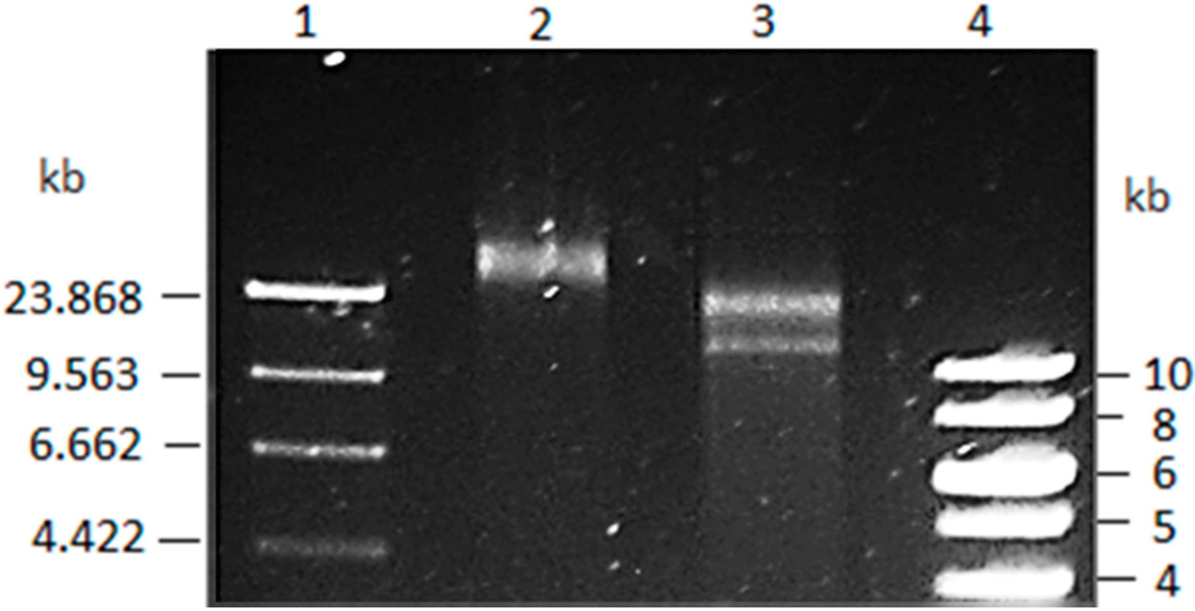
2.2. Vibrio Coralliilyticus Plasmid Isolation

For large-scale plasmid isolation from V. coralliilyticus BAA-450, a 100 mL culture was incubated for 12 h, with plasmid DNA being released by alkaline lysis. The aqueous fraction was further extracted with phenol/chloroform (1:1) prior to plasmid precipitation with 100% ethanol [16]. The resulting plasmid pellets were resuspended in 50 µL of TEN buffer (10 mM Tris-Hcl, 10 mM EDTA, 150 mM NaCl).

2.3. Molecular Biology Protocols

Standard molecular biological protocols were used during the study. Polymerase chain reaction (PCR) experiments were performed using an Applied Biosystems 2720 Thermal Cycler (Singapore). For 1–4.5 kb amplicon fragments, GoTaq® Flexi DNA Polymerase was used (Promega: Madison, WI, USA) according to the manufacturer’s directions; for 4–10 kb fragments, PrimeSTAR HS DNA Polymerase (TaKaRa: Otsu, Japan) was used as per the manufacturer’s directions. Usually, GoTaq® PCR reactions ran for 30 cycles with annealing temperatures set in accordance with the predicted melting temperature of the oligonucleotides; PrimeSTAR PCR reactions typically involved 35 cycles that consisted of a 10 s. denaturing step at 98 °C, an annealing temperature of 63 °C for 5 s, and an elongation time of 1 min per kilobase at 72 °C.

2.4. Vibrio coralliilyticus BAA-450 Plasmid Sequencing

Purified V. coralliilyticus BAA-450 plasmid was digested with various restriction enzymes with the DNA fragments being cloned into pBluescript allowing PCR amplicons to be generated using the M13 universal primers; the amplicons then underwent dideoxy sequencing (Macrogen: Seoul, Korea). DNA sequence was analyzed using Geneious software [17]. Based on the sequence data from 38 individual fragments, custom primers were then designed to amplify novel V. coralliilyticus BAA-450 plasmid sequences (Table S1). A total of 115 DNA fragments were sequenced which provided coverage of the entire plasmid. All DNA sequences were initially assessed at the nucleotide level by BLAST analysis against the V. shilonii pAK-1 plasmid in the NCBI database [18]. Following the alignment of the unique V. coralliilyticus plasmid DNA sequence using the Geneious software, open reading frames (orfs) were identified using the NCBI ORF finder [19]. The predicted orfs were then assessed by BLAST analysis against the NCBI database using the blastp program [18]. Alignments were translated using the Show Translation program; the percentage of adenine and thymine bases (AT) and codon usage patterns were calculated using DNA STATS and Codon Usage available on The Sequence Manipulation Suite [20]. Enzyme restriction sites were identified with the NEBcutter V2.0 tool made available through New England Biolabs [21]. Putative promoters for each orf were identified using BPROM available on Softberry [22]. tRNAs were identified using the tRNAscan-SE program [23]. Inverted repeats and tandem repeats were identified using EMBOSS explorers’ inverted program [24]. Insertion sequence elements were identified using the IS Finder database search tool [25]. Protein alignments were constructed using Clustal Omega available through the European Bioinformatics Institute [26]. The RefSeq ID of the Vibrio coralliilyticus plasmid is NC_020451.1

3. Results and Discussion

3.1. Vibrio Coralliilyticus BAA-450 Plasmid

The purified V. coralliilyticus BAA-450 plasmid was initially assessed by restriction analysis where a linearized molecule in excess of 23 kb was identified (Figure 1). Following DNA sequencing of 115 useable fragments, a circular plasmid, 26,631 bp in length, was assembled. The initial BLAST analysis against V. shilonii pAK-1 plasmid revealed no sequence similarity, with subsequent BLAST analyses being unable to uncover any significant continuous sequence similarity with any DNA sequence within the NCBI database. Consequently, the BAA-450 plasmid appeared to be unique to V. coralliilyticus. The circularity of the plasmid was determined by restriction analysis using the derived DNA sequence as template; BglI digestion (a predicted single cutter) yielded a single band of appropriate size (26,631 bp), whereas SacII (a predicted double cutter) yielded two bands (14,316 and 12,315 bp).
Figure 1. Isolation of the BAA-450 plasmid Lane 1: λ DNA digested with HindIII; Lane 2: V. coralliilyticus Plasmid digested with BglI; Lane 3: V. coralliilyticus Plasmid SacII; Lane 4: Thermo Scientific™ GeneRuler 1 kb Ladder.
Microorganisms 04 00003 g001

3.2. Annotation and Identification of Plasmid-Encoded Orfs

The derived DNA sequence was annotated and the complete linkage map is presented in Figure 2. Most of the putative orfs that were identified began with the ATG codon. However, one putative orf (orf80) was identified that began with a TTG initiation codon. Additionally, as some of the predicted orfs overlapped one another, expression of transcripts arising from these orfs was analyzed with quantitative real time PCR (qRT-PCR) using primers that were outside of the overlapped regions. Overlapping orfs whose transcripts were not detected through qRT-PCR were not included in further analysis (data not shown). However, several overlapping orfs (orf80, orf134, and orf236) could not be analyzed through qRT-PCR due to an erroneously high background. Nonetheless, due to computational probability, these overlapping orfs were included in subsequent analysis although their validity has not been confirmed through experimental data. Transcripts were detected through qRT-PCR analysis from all other overlapping orfs (orf148, orf291, orf287, and orf244) (data not shown). A complete listing of the putative orfs identified by BLASTP analysis, their location on the plasmid and their hypothetical protein functions can be found in Table 1. Predicted promoter elements, as well as the identification of potential operons, are presented in Tables S2 and S3, respectively.
Figure 2. Linkage map of the BAA-450 plasmid. Following DNA sequencing, the identified orfs were assembled and their locations on the plasmid are displayed using [27]. The shading of the orfs corresponds to the putative proteins they encode and are as follows: transporter/permease proteins are shaded pink; transposases are shaded purple; ligase is shaded green; synthase is shaded blue; phosphorylase is shaded a dull green; decarboxylase is shaded a lime green; carboxykinase is shaded a dark blue; mobilization, relaxase and replication orfs are shaded plum purple; reductase is shaded a dusty blue; glycosyltransferase is shaded gold; and the orf encoding a hypothetical protein is white. tRNAs, indirect repeats (IR), and direct repeats (DR) are shown in gray.
Microorganisms 04 00003 g002

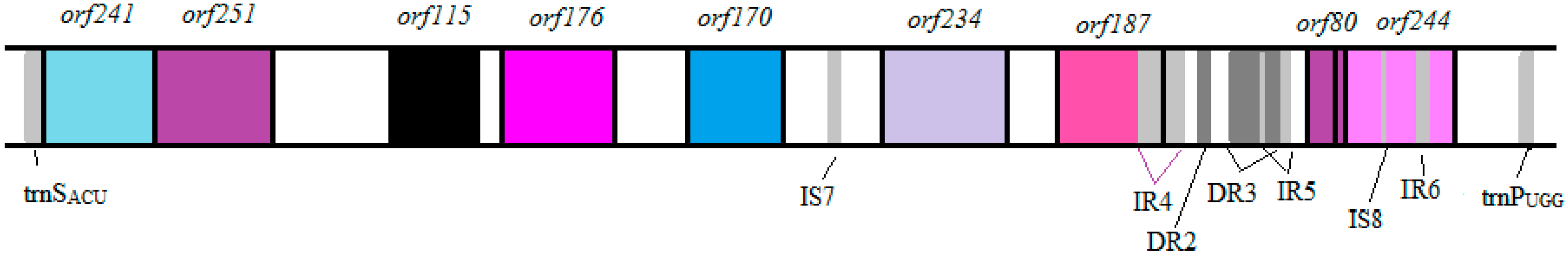
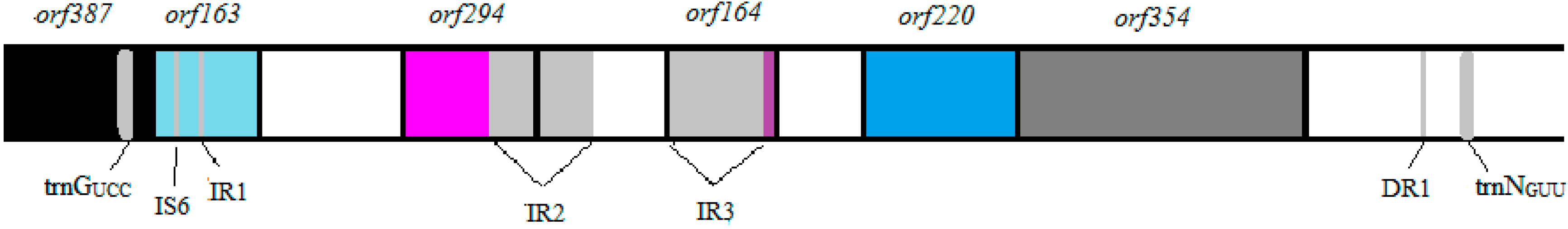
3.3. Identification of Potential “Ecological” Islands within the Plasmid Replicon

Analysis of the BAA-450 plasmid also revealed the presence of several miscellaneous features that included the presence of six tRNA genes (Table 2), eight putative insertion sequences (IS) (Table S4), 12 inverted repeats (IR) (Table S5), and three direct repeats (Table S6). In pathogenic bacteria, tRNA genes often flank pathogenicity islands, with the tRNA loci being the target sites for integration of horizontally acquired foreign DNA [8,9,28,29]. In the BAA-450 plasmid, the region of DNA between trnGUCC and trnNGUU contains five orfs, with the trnGUCC gene being located within orf387. Interestingly, orf387 encodes a putative transposase which may have been instrumental in the insertion of this 6111 bp segment of DNA into the plasmid (Figure 3). Similarly, eight orfs were found between trnPUGG and trnSACU, including orf244, which is again believed to encode a transposase and could explain the presence of this 7,052 bp DNA fragment (Figure 4). Assessment of the guanine and cytosine base (GC) content between the BAA-450 plasmid and the genome indicated a significant difference, which is more noticeably apparent when the DNA encoding the tRNA segments is removed from the calculation (Table 3). Furthermore, a comparison of the plasmid-located tRNA genes to those present on the V. coralliilyticus BAA-450 genome also shows a much lower GC composition indicating that the plasmid tRNA genes were not acquired from the chromosome. Collectively, these observations suggest that these two stretches of DNA located between flanking tRNA genes may constitute “ecological” islands that were horizontally acquired.
Table 1. Putative orfs within the V. coralliilyticus plasmid.
NameStarting and Ending BaseLength (bp)Reading FramePutative Proteine-Value% AT
orf2361–711711+1ABC transporter, ATP-binding protein LivF-like3 × 10-1757
orf387742–19051164+1transposase, IS46 × 10-12343
orf1632058–2549492+3Na+/H+ antiporter2 × 10-6559
orf2943053–3937885+2Biotin-protein ligase2 × 10-4659
orf1644967–4473495−2branched-chain amino acid ABC transporter permease protein, LivM-like2 × 10-2462
orf2205694–6356663+3oligoendopeptidase F8 × 10−8062
orf3546380–74441065+2Trk transporter membrane-spanning protein-K+ transport9 × 10−10362
orf2419244–8519726−3methylglyoxyl synthase9 × 10−6858
orf25110,005–9250756−1purine nucleoside phophorylase7 × 10−7364
orf11510,790–10,443348−2oxaloacetate decarboxylase α-chain3 × 10−3461
orf17611,426–10,896531−2phosphoenolpyruvate carboxykinase4 × 10−4465
orf14211,793–12,218426+3maltose/maltodextrin ABC transporter, permease protein4 × 10−1957
orf23412,438–13,142705+3ykvW heavy metal-(Cd/Co/Hg/Pb/Zn)-translocating P-type ATPase1 × 10−3962
orf18713,290–13,853564+3mobilization protein3 × 10−1859
orf80 *14,423–14,662240+2RepE replication protein2 × 10−1573
orf21315,225–14,587639−1transposase1 × 10−3565
orf38917,190–16,0211170−1glutathione reductase6 × 10−10458
orf20017,234–17,836603+2replication protein6 × 10−2967
orf29717,904–18,797894+3DNA replication protein, putative1 × 10−3662
Clp protease protein5 × 10−32
orf33219,200–20,198999+3maltodextrin-binding protein MdxE3 × 10−12958
orf16720,696–21,199504+2branched-chain amino acid ABC transporter, permease protein, LivH-like4 × 10−5756
orf13621,865–21,455411−3rlx domain protein9 × 10−5657
orf22322,518–21,847672−1mobilization protein1 × 10−10854
orf23723,324–22,611714−2ABC superfamily ATP binding cassette transporter, permease protein1 × 10−3461
orf27624,222–23,392831−1YKVW heavy metal-(Cd/Co/Hg/Pb/Zn)-translocating P-type ATPase7 × 10−5163
orf23824,465–25,181717+3hypothetical protein CAT7 095854 × 10−9160
orf14825,392–25,838447+3PTS system, IIC component5 × 10−2261
orf13425,636–25,232405−3glycosyltransferase6 × 10−2162
orf29126,533–25,658876−3ABC exporter, membrane-spanning/permease subunit2 × 10−14362
* Begins with TTG initiation codon.
Table 2. Putative tRNAs Encoded in the V. Coralliilyticus Plasmid.
NameStarting and Ending BaseLengthAmino AcidCodon
trnDGUC1807–187973Aspartic acidGAC
trnFGAA1896–196873PhenylalanineTTC
trnGUCC1979–204971GlycineGGA
trnNGUU8159–823072AsparagineAAC
trnPUGG15,608–1,568174ProlineCCA
trnSACU8557–848474SerineAGT
Figure 3. The region between trnGUCC and trnNGUU in the BAA-450 plasmid. This 6111 bp fragment contains five orfs, including orf387 which encodes a putative transposase that may have been instrumental in the insertion of this fragment into the plasmid (not drawn to scale).
Microorganisms 04 00003 g003
Figure 4. The region between trnPUGG and trnSACU in the BAA-450 plasmid. This 7052 bp fragment contains eight orfs, including orf244 which encodes a putative transposase enzyme that may have been involved in the incorporation of this fragment into the plasmid (not drawn to scale).
Microorganisms 04 00003 g004
Table 3. GC content of the V. coralliilyticus BAA-450 plasmid and genome.
Vibrio coralliilyticusLength (bp)% GC Content
Entire Plasmid26,63140
Plasmid without tRNA segments13,47242
Segment between trnGUCC and trnNGUU611141
Segment between trnPUGG and trnSACU705237
Genome 5,680,62845
Genome data for V. coralliilyticus BAA-450 genome was obtained from the NCBI database.
Fourteen putative proteins are found within the proposed "ecological" islands. Of these, four have predicted properties that would confer an ecological advantage. Analysis of ORF163 identified a multi-domain NhaC (sodium/hydrogen antiporter) component between amino acid residues 89–143. Significant sequence homology was shared between ORF163 and a Na+/H+ antiporter of Carnobacterium sp. 17-4 (e-value of 2 × 10−65) with the majority of the amino acids (1–144) aligning with the Carnobacterium protein. While the Na+/H+ antiporter of Carnobacterium sp. 17-4 is a more complex 567 amino acid protein, the presence of the NhaC domain within ORF163 infers a role for ORF163 in energy production/conversion utilizing a Na+/H+ antiporter.
Within ORF241 a MGS-like (methylglyoxal synthase) superfamily domain was identified that incorporates an active site, catalytic residues and the appropriate dimer interfaces within amino acids 18–129. This putative protein also contains a P-loop NTPase multi-domain with an ATP binding site between amino acids 154–220. A blastp search identified high sequence homology (e-value of 9 × 10−68) with the Carnobacterium sp. 17-4 MGS with the majority (amino acids 7–139) aligning with ORF241, inferring a methylglyoxal synthase role for orf241. A methylglyoxal synthase (151 amino acids) is also encoded within the V. coralliilyticus genome and shares sequence homology (e-value: 8 × 10⁻³⁵) to ORF241 as well as containing a similar conserved domain. However, no nucleotide homology is shared between the two genes indicating that orf241 did not originate from the genome. Consequently, ORF241 may provide the bacterium with an alternative to triosephosphate isomerase for metabolizing dihydroxyacetone phosphate that is produced during glycolysis.
ORF251 contains a DUF1250 superfamily (unknown function) domain (amino acids 1–69), a PNP UDP 1 phosphorylase domain, and a deoD purine nucleoside phosphorylase domain within amino acids 82–215. Blastp analysis identified a Carnobacterium sp. 17-4 purine nucleoside phosphorylase of similar length (236 amino acids) with significant sequence homology (e-value of 7 × 10−73) to ORF251. Consequently, orf251 may encode a putative purine nucleoside phosphorylase deoD which could provide flexibility with nucleic acid metabolism.
Although the open reading frames are located 10,250 nucleotides apart, two putative proteins, ORF234 (a proposed “ecological” island protein) and ORF276, were found to contain the same conserved domains and highly similar amino acid sequences (amino acids 166–276 in ORF276 and amino acids 1–110 in ORF234). ORF276 contains a HAD-like (haloacid dehalogenase) superfamily from amino acids 156-246 plus a ZntA cation transport ATPase multi-domain encompassing aa 46–271. The HAD-like superfamily of ORF234 is located between amino acids 20–88 with the ZntA multi-domain from amino acids 2–173. Both ORF276 and ORF234 also share significant sequence similarities (e-values: 1 × 10−149 ORF276; 2 × 10−88, ORF234) with a 641 amino acids P-type ATPase cadmium transporter of Enterococcus italicus DSM 15952. A refined blastp search further revealed that ORF276 shared sequence homology (e-value of 7 × 10−51) to a YKVW 325 amino acid heavy metal-(Cd/Co/Hg/Pb/Zn)-translocating P-type ATPase from Bacillus halodurans, and that ORF234 shared sequence homology (e-value 1 × 10−39) with a 225 amino acid ykvW protein of Bacillus cereus ATCC 10987. Based on the presence of conserved domains and sequence homologies, it is hypothesized that both ORF234 and ORF276 function as part of a heavy metal-(Cd/Co/Hg/Pz/Zn)-translocating P-type ATPase complex.

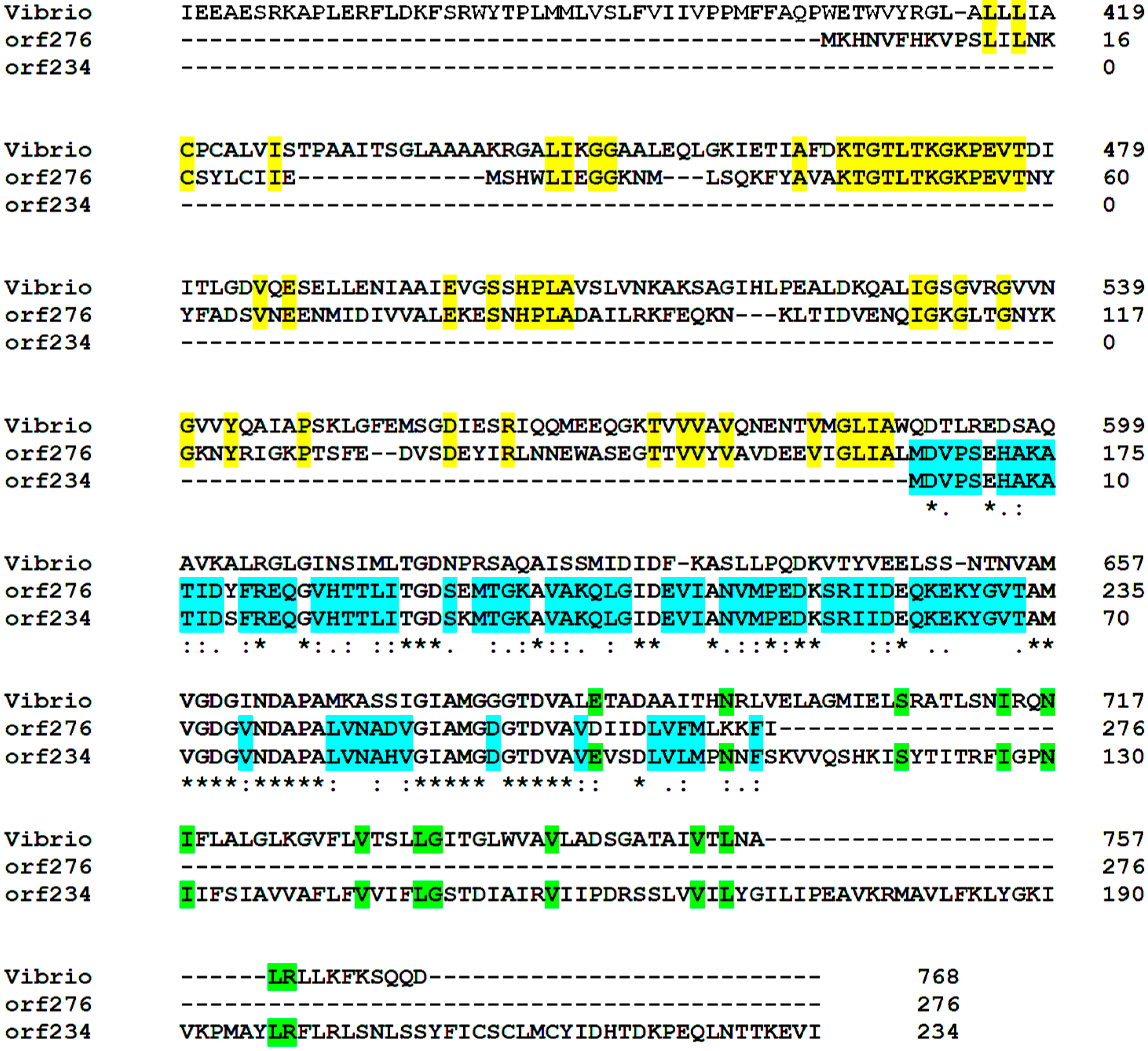
3.4. Identification of Putative Pathogenicity Proteins on the BAA-450 Plasmid

ORF389 contains a PRK06116 glutathione reductase domain within amino acids 10–264 with high sequence homology (e-value of 6 × 10−104) to a glutathione reductase from Salmonella enterica subsp. enterica serovar Typhi str. E98-0664. However, both ORF389 and the glutathione reductase of Salmonella appear to lack a dimerization domain at the carboxy terminus, which could affect the functioning of these proteins as dimerization is essential for catalytic activity. Despite the truncated carboxy-terminal end, the conserved domains, and the extensive protein alignment, supports a role for ORF389 as a glutathione reductase [30]. The chromosomally encoded glutathione reductase (451 amino acids) shares sequence homology (e-value of 5 × 10−90) with ORF389. The chromosomal protein contains a NADB Rossmann superfamily, a PRK06116 multi-domain, plus a Pyr redox dim (Pyridine nucleotide-disulphide oxidoreductase dimerization domain) superfamily, the last domain being absent in ORF389. Partial homology (e-value of 4 × 10⁻³⁵) was also detected between the chromosomal and plasmid nucleotide sequences encoding these proteins suggesting that the chromosomally- and plasmid-encoded proteins may indeed interact.
While ORF134 lacked various conserved domains, sequence homology (e-value of 6 × 10⁻²¹) was found between ORF134 and a putative glycosyltransferase enzyme (207 amino acids) from Melissococcus plutonius ATCC 35311. However, while sequence homology to the putative glycosyltransferase enzyme is present, the specific function of ORF134 is still uncertain since the putative protein lacks the glycosyltransferase domain which would allow for carbohydrate binding in a sugar-nucleoside disphosphate- and manganese-dependent fashion.

3.5. Functional Cooperativity between Putative Plasmid- and Chromosomally-Encoded Proteins

The possibility that plasmid-encoded proteins interact with chromosomally-encoded proteins was further explored following analysis of other protein homologies. The two putative plasmid-encoded heavy metal-(Cd/Co/Hg/Pb/Zn) transporter ATPase proteins (ORF276 and ORF234) were found to share functional similarity to a genomic lead, cadmium, zinc and mercury transporting ATPase protein. While the protein encoded on the chromosome is larger (759 amino acids), a portion of both plasmid putative proteins share sequence similarity to the chromosomal protein (2 × 10⁻²⁰ for ORF234 and 2×10⁻³² for ORF276) (Figure 5). In fact, the chromosomally-encoded protein, ORF234 and ORF276 all share conserved domains that include a zinc/cadmium/mercury/lead-transporting ATPase as well as a haloacid dehalogenase superfamily. However, the DNA sequences of each of these genes share no sequence homology indicating that the plasmid genes did not originate from the chromosome. Indeed, of the 14 putative proteins located within the proposed “ecological” islands, six putative proteins (ORF294, ORF163, ORF115, ORF220, ORF241 and ORF251) shared significant homologies to chromosomally-encoded proteins suggesting functional cooperativity. However, there are some plasmid-encoded proteins which do not share sequence homology to similar chromosomally-encoded proteins. These include the putative Trk protein (ORF354), PEP carboxykinase (ORF176), glycosyltransferase (ORF134), the putative ABC exporter (ORF291), plus the putative Clp protease and replication proteins ORF80, ORF200 and ORF297).
Functional cooperativity is further indicated following analysis of the plasmid-encoded maltose/maltodextrin ABC transporter permease subunit (ORF170) and the maltodextrin-binding protein, MdxE (ORF332). In contrast, the V. coralliilyticus genome encodes a maltose/maltodextrin ABC transporter substrate binding protein (MalE), a maltose/maltodextrin transport ATP-binding protein (MalK), maltose/maltodextrin ABC transporter permease proteins (MalF and MalG), a maltose operon periplasmic protein (MalM), and several glycosidases. In V. cholerae, the homologues of these maltose/maltodextrin genes have been shown to be essential for the expression of virulence factors including cholera toxin and soluble hemaggtluninin-protease [31]. The plasmid and chromosomal maltose/maltodextrin genes may combine to form a functional maltose/maltodextrin transporter system which may play a role in the virulence of this organism.
Figure 5. Clustal alignment of putative chromosomally-encoded and plasmid-encoded lead, cadmium, zinc and mercury transporting ATPase proteins. The alignment begins at amino acid 419 of the chromosomally-encoded protein. The gray shaded amino acids show similarity between all three proteins; the yellow shaded amino acids show similarity between ORF276 and the chromosomal protein; the green shaded amino acids show similarity between ORF234 and the chromosomal protein; and, the blue shaded amino acids show similarity between ORF276 and ORF234.
Microorganisms 04 00003 g005

4. Conclusions

The sequencing and annotation of the BAA-450 plasmid supports a potential role for the plasmid-encoded proteins in the growth and survival of V. coralliilyticus. Several putative orfs may provide factors that would protect the cell from harmful substances that may be encountered in coral ecosystems (e.g., orf389, which codes for a putative glutathione reductase may remove photosynthetically-derived oxygen radicals that accumulate within intracellular environments), with other proteins possibly being implicated in virulence (the putative glycosyltransferase ORF134 possibly being involved in the expression of adhesins), while others may encode various nutrient uptake systems, or, otherwise facilitate growth in high salinity environments. The DNA analysis also indicates the presence of two “ecological islands” that may have been acquired at two distinct intervals which may also facilitate growth within changing marine environments.

Supplementary Materials

Supplementary materials can be found at www.mdpi.com/2076-2607/4/1/3/s1.

Acknowledgements

The work was supported by a graduate student award to J.W. by the Department of Biological Sciences, Northern Illinois University.

Author Contributions

J.W. performed the research, analyzed the data and helped write the manuscript; S.A.H. designed the experiments, analyzed the data and helped write the manuscript.

Conflicts of Interest

The authors declare no conflict of interest.

References

  1. Bally, M.; Garrabou, J. Thermodependent bacterial pathogens and mass mortalities in temperate benthic communities: A new case of emerging disease linded to climate change. Glob. Change Biol. 2007, 13, 2078–2088. [Google Scholar] [CrossRef]
  2. Vizcaino, M.I.; Johnson, W.R.; Kimes, N.E.; Williams, K.; Torralba, M.; Nelson, K.E.; Smith, G.W.; Weil, E.; Moeller, P.D.; Morris, P.J. Antimicrobial Resistance of the Coral Pathogen Vibrio coralliilyticus and Carribbean Sister Phylotypes Isolated from a Diseased Octocoral. Microb. Ecol. 2010, 59, 646–657. [Google Scholar] [CrossRef] [PubMed]
  3. Meron, D.; Efrony, R.; Johnson, W.R.; Schaefer, A.L.; Morris, P.J.; Rosenberg, E.; Greenberg, E.P.; Banin, E. Role of Flagella in Virulence of the Coral Pathogen Vibrio coralliilyticus. Appl. Environ. Microbiol. 2009, 75, 5704–5707. [Google Scholar] [CrossRef] [PubMed]
  4. Rosenberg, E.; Falkovitz, L. The Vibrio shiloi/Oculina patagonica Model System of Coral Bleaching. Ann. Rev. Microbiol. 2004, 58, 143–159. [Google Scholar] [CrossRef] [PubMed]
  5. Ben-Haim, Y.; Thompson, F.L.; Thompson, C.C.; Cnockaert, M.C.; Hoste, B.; Swings, J.; Rosenberg, E. Vibrio coralliilyticus sp. nov., a temperature dependent pathogen of the coral Pocillopora damicornis. Int. J. Sys. Evol. Microbiol. 2003, 53, 309–315. [Google Scholar] [CrossRef] [PubMed]
  6. Sussman, M.; Willis, B.; Victor, S.; Bourne, D. Coral Pathogens Identified for White Syndrome (WS) Epizootics in the Indo-Pacific. PLoS ONE 2008, 3. [Google Scholar] [CrossRef] [PubMed]
  7. Davidson, L.S.; Oliver, J.D. Plasmid Carriage in Vibrio vulnificus and other Lactose-Fermenting Marine Vibrios. Appl. Environ. Microbiol. 1986, 51, 211–213. [Google Scholar]
  8. Reynaud, Y.; Saulnier, D.; Mazel, D.; Goarant, C.; Le Roux, F. Correlation between Detection of a Plasmid and High-Level Virulence of Vibrio nigripulchritudo, a Pathogen of the Shrimp Litopenaeus stylirostris. Appl. Environ. Microbiol. 2008, 74, 3038–3047. [Google Scholar] [CrossRef] [PubMed]
  9. Vivian, A.; Murrillo, J.; Jackson, R.W. The roles of plasmids in phytopathogenic bacteria: Mobile arsenals? Microbiology 2001, 147, 763–780. [Google Scholar] [CrossRef] [PubMed]
  10. Caldwell, B.A.; Ye, C.; Griffiths, R.P.; Moyer, C.L.; Morita, R.Y. Plasmid Expression and Maintenance during Long-Term Starvation-Survival of Bacteria in Well Water. App. Environ. Microbiol. 1989, 55, 1860–1864. [Google Scholar]
  11. Richards, G.P.; Bono, J.L.; Watson, M.A.; Needleman, D.S. Complete genome sequence for the shellfish pathogen Vibrio coralliilyticus RE98 isolated from a shellfish hatchery. Genome Announc. 2014, 2. [Google Scholar] [CrossRef] [PubMed]
  12. De Santos, E.; Alves, N.; Dias, G.M.; Mazotto, A.M.; Vermelho, A.; Vora, G.J.; Wilson, B.; Beltran, V.H.; Bourne, D.G.; Le Roux, F.; et al. Genomic and proteomic analysis of the coral pathogen Vibrio coralliilyticus reveal a diverse viruence repertoire. ISME J. 2011, 5, 1471–1483. [Google Scholar] [CrossRef] [PubMed]
  13. Temperton, B.; Thomas, S.; Tait, K.; Parry, H.; Emery, M.; Allen, M.; Quinn, J.; Macgrath, J.; Gilbert, J. Permanent draft genome sequence of Vibrio tubiashii strain NCIMB 1337 (ATCC19106). Stand. Genomic. Sci. 2011, 4, 183–190. [Google Scholar] [CrossRef] [PubMed]
  14. Ushijima, B.; Videau, P.; Aeby, G.S.; Callahan, S.M. Draft genome sequence of Vibrio coralliilyticus strain OCN008, isolated from Kane’ohe Bay, Hawaii. Genome. Announc. 2013, 1. [Google Scholar] [CrossRef] [PubMed]
  15. Ushijima, B.; Videau, P.; Poscablo, D.; Vine, V.; Salcedo, M.; Aeby, G.S.; Callahan, S.M. Complete genome sequence of Vibrio coralliilyticus strain OCN014, isolated from a diseased coral at Palmyra Atoll. Genome. Announc. 2014, 2. [Google Scholar] [CrossRef] [PubMed]
  16. Birnboim, H.C.; Dohly, J. Plasmid Alkaline Plasmid Preparation. Nucl. Acids Res. 1979, 7, 1513–1522. [Google Scholar] [PubMed]
  17. Kearse, M.; Moir, R.; Wilson, A.; Stones-Havas, S.; Cheung, M.; Sturrock, S.; Buxton, S.; Cooper, A.; Markowitz, S.; Duran, T.; et al. Geneious Basic: An integrated and extendable desktop software platform for the organization and analysis of sequence data. Bioinformatics 2012, 28, 1647–1649. [Google Scholar] [CrossRef] [PubMed]
  18. Altschul, S.F.; Madden, T.L.; Schaffer, A.A.; Zhang, J.; Zhang, Z.; Miller, W.; Lipman, D.J. Gapped BLAST and PSI-BLAST: A new generation of protein database search programs. Nucleic. Acids Res. 1997, 25, 3389–3402. [Google Scholar] [CrossRef] [PubMed]
  19. Wheeler, D.L.; Church, D.M.; Federhen, S.; Lash, A.E.; Madden, T.L.; Pontius, J.U.; Schuler, G.D.; Schriml, L.M.; Sequeira, E.; Tatusova, T.A.; et al. Database resources of the National Center for Biotechnology. Nucleic. Acids Res. 2003, 31, 28–33. [Google Scholar] [CrossRef] [PubMed]
  20. Stothard, P. The Sequence Manipulation Suite. Available online: http://www.bioinformatics.org/sms2/ (accessed on 14 December 2015).
  21. Vincze, T.; Posfai, J.; Roberts, R.J. NEB cutter: A program to cleave DNA with restriction enzymes. Nucleic. Acids Res. 2003, 31, 3688–3691. [Google Scholar] [CrossRef] [PubMed]
  22. Solovyev, V.; Salamov, A. Automatic Annotation of Microbial Genomes and Metagenomics Sequences. In Metagenomics and its Applications in Agriculture, Biomedicine, and Environmental Studies; Li, R.W., Ed.; Nova Science Publishers: New York, NY, USA; pp. 61–78.
  23. Schattner, P.; Brooks, A.N.; Lowe, T.M. The tRNA scan-SE, snoscan and snoGPS web servers for the detection of tRNAs and snoRNAs. Nucleic Acids Res. 2005, 33, w686–w689. [Google Scholar] [CrossRef] [PubMed]
  24. Rice, P.; Longden, I.; Bleasby, A. EMBOSS: The European Molecular Biology Open Software Suite. Trends Genet. 2000, 16, 276–277. [Google Scholar] [CrossRef]
  25. Siguier, P.; Perochon, J.; Lestrade, L.; Mahillon, J.; Chandler, M. ISfinder: The reference center for bacterial insertion sequences. Nucleic. Acids Res. 2006, 34, D32–D36. [Google Scholar] [CrossRef] [PubMed]
  26. Sievers, F.; Wilm, A.; Dineen, D.; Gibson, T.J.; Karplus, K.; Li, W.; Lopez, R.; McWilliam, H.; Remmert, M.; Soding, J.; Thompson, J.D.; Higgins, D.G. Fast, scalable generation of high-quality protein multiple sequence alignments using Clustal Omega. Mol. Syst. Biol. 2011, 7, 539. [Google Scholar] [CrossRef] [PubMed]
  27. Conant, G.C.; Wolfe, K.H. GenomeVx: Simple web-based creation of editable circular chromosome maps. Bioinformatics 2008, 24, 861–862. [Google Scholar] [CrossRef] [PubMed]
  28. Blanc-Potard, A.B.; Groisman, E.A. The Salmonella selC locus contains a pathogenicity island mediating intramacrophage survival. EMBO J. 1997, 16, 5376–5385. [Google Scholar] [CrossRef] [PubMed]
  29. Hacker, J.; Blum-Oehler, G.; Muhldorfer, I.; Tschape, H. Pathogenicity islands of virulent bacteria: Structure, function and impact on microbial evolution. Mol. Microbiol. 1997, 23, 1089–1097. [Google Scholar] [CrossRef] [PubMed]
  30. Bashir, A.; Perham, R.N.; Scrutton, N.S.; Berry, A. Altering kinetic mechanism and enzyme stability by mutagenesis of the dimer interface of glutathione reductase. Biochem. J. 1995, 312, 527–533. [Google Scholar] [CrossRef] [PubMed]
  31. Lang, H.; Jonson, G.; Holmgren, J.; Palva, E.T. The maltose regulon of Vibrio cholera affects production and secretion of virulence factors. Infect. Immun. 1994, 62, 4781–4788. [Google Scholar] [PubMed]

