Article

Curcumin Attenuates the Pathogenicity of Entamoeba histolytica by Regulating the Expression of Virulence Factors in an Ex-Vivo Model Infection

by
Itzia Azucena Rangel-Castañeda
1,
Pilar Carranza-Rosales
2,
Nancy Elena Guzmán-Delgado
3,
José Manuel Hernández-Hernández
4,
Sirenia González-Pozos
5,
Armando Pérez-Rangel
4 and
Araceli Castillo-Romero
6,*
1
Departamento de Fisiología, Centro Universitario de Ciencias de la Salud, Universidad de Guadalajara, Guadalajara 44340, Mexico
2
Centro de Investigación Biomédica del Noreste, Instituto Mexicano del Seguro Social, Monterrey 64720, Mexico
3
Unidad Médica de Alta Especialidad (UMAE), Hospital de Cardiología No. 34, Monterrey 64360, Mexico
4
Departamento de Biología Celular, Centro de Investigación y Estudios Avanzados del Instituto Politécnico Nacional, Ciudad de México 07360, Mexico
5
Unidad de Microscopía Electrónica LaNSE, Centro de Investigación y Estudios Avanzados del Instituto Politécnico Nacional, Ciudad de México 07360, Mexico
6
Departamento de Microbiología y Patología, Centro Universitario de Ciencias de la Salud, Universidad de Guadalajara, Guadalajara 44340, Mexico
*
Author to whom correspondence should be addressed.
Pathogens 2019, 8(3), 127; https://doi.org/10.3390/pathogens8030127
Submission received: 30 June 2019 / Revised: 8 August 2019 / Accepted: 10 August 2019 / Published: 15 August 2019
(This article belongs to the Special Issue Emerging Parasitic Protozoa)

Abstract

:
Infection with the enteric protozoan Entamoeba histolytica is still a serious public health problem, especially in developing countries. Amoebic liver abscess (ALA) is the most common extraintestinal manifestation of the amoebiasis, and it can lead to serious and potentially life-threatening complications in some people. ALA can be cured by metronidazole (MTZ); however, because it has poor activity against luminal trophozoites, 40–60% of treated patients get repeated episodes of invasive disease and require repeated treatments that can induce resistance to MTZ, this may emerge as an important public health problem. Anti-virulence strategies that impair the virulence of pathogens are one of the novel approaches to solving the problem. In this study, we found that low doses of curcumin (10 and 50 μM) attenuate the virulence of E. histolytica without affecting trophozoites growth or triggering liver injury. Curcumin (CUR) decreases the expression of genes associated with E. histolytica virulence (gal/galnac lectin, ehcp1, ehcp5, and amoebapore), and is correlated with significantly lower amoebic invasion. In addition, oxidative stress is critically involved in the etiopathology of amoebic liver abscess; our results show no changes in mRNA expression levels of superoxide dismutase (SOD) and catalase (CAT) after E. histolytica infection, with or without CUR. This study provides clear evidence that curcumin could be an anti-virulence agent against E. histolytica, and makes it an attractive potential starting point for effective treatments that reduce downstream amoebic liver abscess.

1. Introduction

The protozoa Entamoeba histolytica causes several clinical syndromes, ranging from dysentery of acute colitis to amoebic liver abscess (ALA), and other extra-intestinal diseases [1,2,3]. Diverse in vitro and in vivo studies have demonstrated that multiple molecules from E. histolytica are involved directly or indirectly in pathogenesis. Among them: galactose/N-acetyl D-galactosamine lectin (Gal/GalNAc), the Cysteine proteinases (CPs) and the amoebapores are three well-studied virulence factors [4,5,6,7,8,9].
The complete sequence of morphologic events during ALA formation has been studied in vivo and in vitro models. The use of precision-cut hamster liver slices has brought remarkable contributions, from the lodgment of amebas in the hepatic sinusoids to the development of extensive liver necrosis. Importantly, the ex vivo models have contributed to use reduction of experimental animals [10,11,12].
In general, metronidazole (MTZ), tinidazole, ornidazole, and nitazoxanide are active in invaded tissues [13,14]. Due to its excellent response, the tissue agent of choice is MTZ, the cure rate for which is >90%, but because of its rapid absorption after oral administration it is not effective against luminal trophozoites; 40–60% of treated patients get repeated episodes of invasive disease and require repeated treatments that induce resistance to MTZ [15]. For these reasons, there is a significant need for new drugs or approaches to combat amoebic drug resistance. Anti-virulence strategies disarm the pathogens, thereby rendering them harmless and more susceptible to immune clearance [16,17,18]. Curcumin, the major component present in the rhizome of Curcuma longa L., has been extensively studied for its anti-oxidant, anticancer, anti-inflammatory, antiparasitic, and hepatoprotective properties [19,20,21,22,23,24]. Some studies have shown that low concentrations of CUR (5–50 µM) prevent hepatic injury caused by parasites and ethanol [25,26,27]. Recently, we demonstrated that 100–300 μM of CUR has amoebicidal activity, affecting E. histolytica trophozoites, growth, and morphology, in a dose-dependent manner [28]. In this study, we found that CUR at low concentrations (10 and 50 μM) displayed direct activity against E. histolytica, inhibiting the virulence factors expression and pathogenicity of this parasite; histopathological analysis and microscopy images revealed that the presence of CUR during the infection prevents liver amoebic invasion.

2. Results

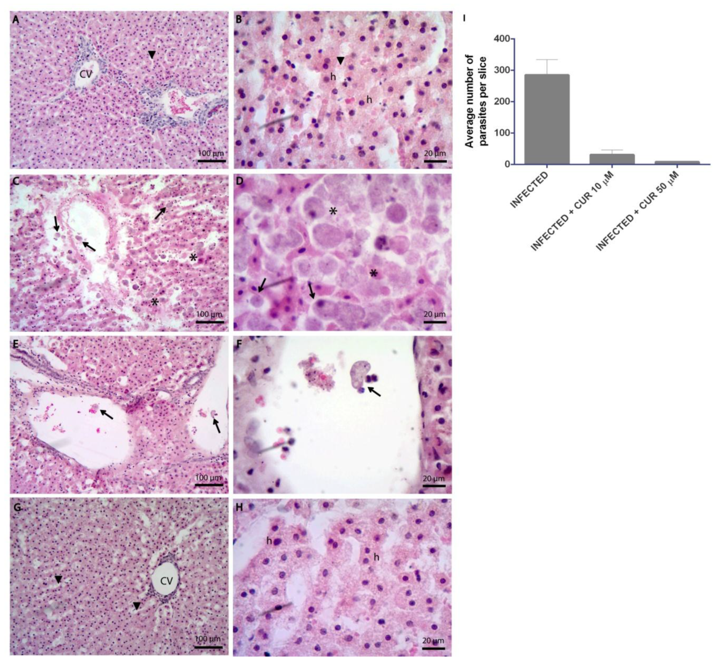
2.1. Curcumin Prevents the Tissue Damage Caused by Entamoeba histolytica

To evaluate the effect of CUR during the infection of precision-cut hamster liver slices (PCHLS) by E. histolytica, hematoxylin and eosin staining and scanning electron microscopy assays were used. Images from H&E showed normal liver architecture in non-infected PCHLS; hepatic parenchyma, sinusoidal spaces, and central veins were observed without change (Figure 1A,B). Infected tissue slices with trophozoites showed architecture alterations, parasites invading the sinusoidal spaces; the amoebas were located close to the hepatocytes (Figure 1C,D). On the other hand, the presence of CUR during infection prevented liver tissue damage; no alterations were seen in the hepatic parenchyma, and the number of trophozoites in the sinusoidal spaces was significantly reduced (Figure 1I). With CUR 10 μM, few parasites with erythrophagocytic activity were observed in the central veins (Figure 1E,F). Moreover, CUR 50 µM-infected slices showed similar histological characteristics to non-infected liver slices (Figure 1G,H).
To further verify effects of CUR during E. histolytica infection, structural tissue details were analyzed by SEM. Comparative SEM images revealed non-infected PCHLS with plates of hepatocytes around the sinusoids, and the portal tract architecture remained unaltered (Figure 2A,B). Infected PCHLS showed trophozoites on the liver parenchyma surface in close contact with hepatocytes and alterations in sinusoids, as well as disorganization of the hepatic cords (Figure 2C,D). Figure 2E–H show that the presence of 10 or 50 µM CUR during infection of PCHLS prevented tissue injury. Tissue with normal characteristics, and intact histological relationship between the sinusoidal spaces and the hepatocyte plates, was observed.

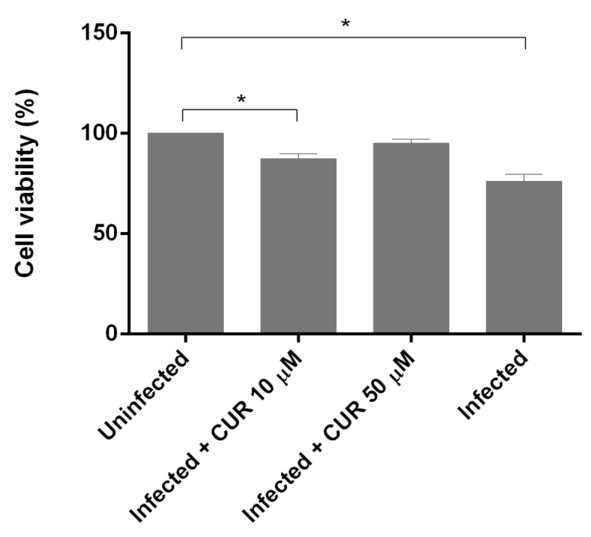
2.2. Effect of Curcumin on PCHLS Viability

The cell viability of infected and CUR-infected PCHLS was determined using the reduction of the metabolic indicator resazurin. After 18 h of infection, the level of reduction of resazurin suggested a significant inhibition of cell viability (23.8%) by E. histolytica, compared with uninfected slices. With CUR 10 µM-infected, the viability of PCHSL was inhibited only 12%. Interestingly, there was no significant differences between CUR 50 µM-infected and uninfected PCHLS (Figure 3).

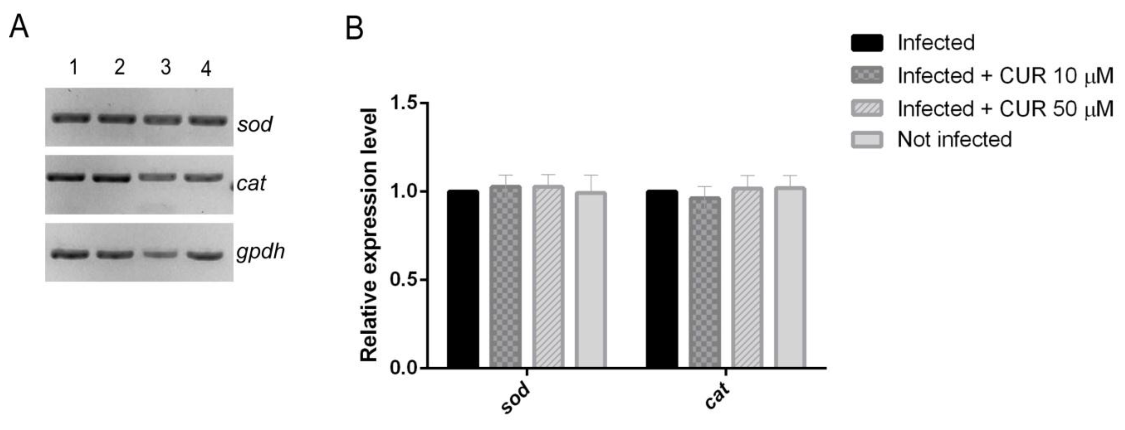
2.3. Effect of Curcumin on Antioxidant Enzymes Expression

Oxidative stress is critically involved in the etiopathology of ALA [29]. There has been evidence that CUR exerts a hepatoprotective effect against oxidative stress via up-regulation of antioxidant enzyme genes [30,31]. The SOD and CAT mRNA levels in the PCHLS were determined to evaluate the impact of CUR during the amoebic infection. Results showed no significant difference in the expression between infected and CUR-infected slices (Figure 4).

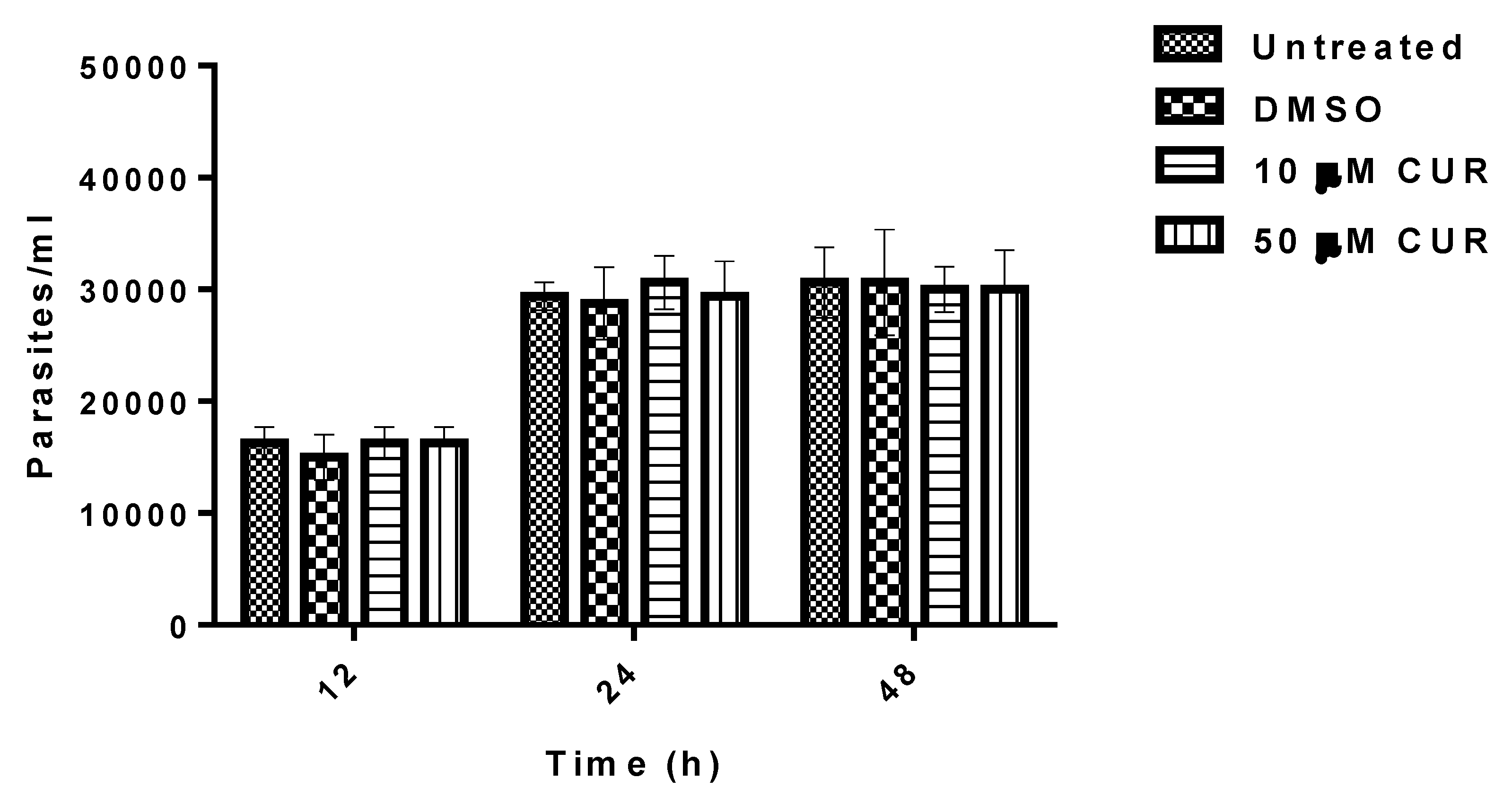
2.4. Effect of Curcumin on the Growth of Entamoeba histolytica

In studies conducted to determine whether CUR can inhibit the growth of E. histolytica in vitro. CUR was found that at 10 or 50 μM has null activity against trophozoites (Figure 5).

2.5. Curcumin Regulates the Expression of Virulence Factors

To determine whether low concentrations of CUR had a direct effect on parasite virulence during PCHLS amoebic infections, we measured the expression levels of gal/galnac lectin, ehcp1, ehcp5, and amoebapore mRNA by semiquantitative RT-PCR in infected PCHLS (Figure 6). After 18 h of amoebic infection, there were no statistically significant differences in the expression level of virulence factors between uninfected and CUR 10 µM-infected slices. However, the presence of CUR 50 µM on infected slices, interestingly, down-regulated mRNA levels of gal/galnac lectin, ehcp1, ehcp5, and amoebapore. The most dramatic impact on gene expression was observed for gal/galnac lectin (90%) and ehcp1 (89.6%). The effect of CUR on expression levels of virulence factors also was evaluated in vitro, and no difference was found with the results obtained in the PCHLS (data no shown).

3. Discussion

E. histolytica has been associated with around 100 million cases of amoebic dysentery, colitis, and ALA that lead to almost 50,000 deaths each year [32,33]. Virulence factors of the pathogen and specific and nonspecific host defense mechanisms are key factors in determining outcome to infection [34]. CUR, the main constituent of turmeric, has demonstrated therapeutic potential to control of parasitic and liver diseases [35]. Recently, we reported the amoebicidal effect of CUR against E. histolytica [28]. However, the mechanism of action has not been elucidated. In this study, we describe the role of CUR on the expression of the major virulence factors of this parasite and on liver damage. Using an ex vivo model to induce the formation of ALA, we demonstrate destruction of liver tissue by the parasite. Similar to Carranza and co-worker results [36,37], our histological and SEM analysis of infected PCHLS showed changes in the tissue architecture with trophozoites in close contact with hepatocytes, especially on the fenestrated surface of the endothelium that covers these cells. On the other hand, some authors have reported the hepatoprotective properties of CUR [26,27,35,38]; in this study, we found that during the amoebic infection the tissue was well preserved by the presence of this phytochemical, and the liver conserved its normal architecture. Furthermore, we observed a portal space with characteristic hepatic artery and portal vein. The presence of parasites in infected PHCLS significantly decreased with CUR 10 µM, and with 50 µM of CUR no parasites were observed, which correlates with the viability assay where, although the slices were infected in the presence of CUR 10 µM, the viability cell was reduced by 12.6 %, while no significant difference was detected between uninfected and CUR 50 µM-infected-PCHLS. Oxidative stress is critically involved in the etiopathology of ALA; SOD and CAT mRNA levels are upregulated during the amoebic liver infection in hamster [29]. Our observations confirmed a high-level expression of both enzymes in amoebic-infected PHCLS. In addition, several studies have shown that CUR exerts remarkable protection against oxidative stress associated liver diseases, through various cellular and molecular mechanisms. Those mechanisms include ameliorating cellular responses to oxidative stress such as the expression of SOD, CAT, and others [26,27,35,38]. In contrast to this evidence, in amoebic liver damage the presence of CUR during infection did not modify the expression levels of these genes (Figure 4), suggesting that CUR prevents ALA by a mechanism that does not involve its antioxidant properties.
Diverse in vitro studies have demonstrated that the principal virulence factors involved directly or indirectly in the pathogenesis of E. histolytica are the GalNAc lectin, the CPs, and the amoebapores [5,7,8,9]. In our infected-PCHLS, the expression of Gal/GalNAc was upregulated, which is consistent with a previous report that shows that Gal/GalNAc expression is upregulated steadily following host–parasite interaction [5]. Therefore, we investigated the effect of CUR in the mRNA expression of the most studied E. histolytica virulence factors, and our evidence demonstrated that all virulence factors tested were down regulated by CUR. With CUR 50 µM, gal/galnac and ehcp1 expression was inhibited more than 89%. The same trend was observed when trophozoites were incubated with 10 or 50 μM of CUR. This explains why the trophozoites in the presence of CUR were not able to invade liver slices. All our findings indicate that CUR has direct activity against E. histolytica, and attenuates the pathogenicity of this parasite during amoebic liver infection. These findings are further supported by our in vitro findings that show that low doses of CUR did not affect the trophozoites growth. Previous studies have demonstrated that the inhibition of cysteine proteinase expression significantly decreased the formation of liver lesions [39,40]. In addition, in bacteria has been described that CUR attenuates its virulence [41,42,43]. In Pseudomonas aeruginosa, treatment with CUR downregulates virulence factors (pyocyanin, elastase, and protease) and inhibits the adherence of the bacteria to polypropylene surfaces [41,43]. This study provides clear evidence that CUR reduces the pathogenicity of Entamoeba histolytica by regulating the expression of virulence required to the processes of tissue invasion, suggests that this polyphenol could be an anti-virulence agent against Entamoeba histolytica, and makes it an attractive potential starting point for effective treatments that reduce downstream ALA.

4. Materials and Methods

4.1. Ethics Statement

The animal management protocols were approved by the institutional committee (C.I. 010/15). All the procedures were carried out following federal regulations for the production, care, and use of laboratory animals (NOM-062-Z00-1999).

4.2. Entamoeba histolytica Trophozoites Culture

Virulent trophozoites of E. histolytica strain HM1-IMSS were grown axenically at 37 °C in PEHPS media [44] and supplemented with 10% bovine serum. Cultures were routinely maintained by amoeba sub-culturing twice a week. The virulence was maintained by routinely direct inoculation of trophozoites into liver hamster. For in vitro experiments, trophozoites were harvested in the log phase growth.

4.3. Precision-Cut Hamster Liver Slices Preparation

Precision-cut hamster liver slices were prepared as described previously [36]. Briefly, 2 months old male Syrian golden hamster (Mesocricetus auratus) were sacrificed with sodium pentobarbital (6 mg/100 g), then the liver was removed and placed into Krebs buffer. The hepatic lobes were cut into cylinders of 10 mm diameter and sliced into 200–250 µm thick tissue slices with a Brendel Vitron tissue slicer (Vitron, Tucson, AZ, USA) in oxygenated KB buffer (95:5% O2:CO2). After, liver slices were placed in 24-well polystyrene microplates with 1 mL of DMEM/F12 media and incubated 1 h at 37 °C to stabilize the tissue before infection.

4.4. Preparation of Curcumin Stocks

The CUR was obtained from Sigma Chemicals Co. (St Louis, MO, USA). The stock of CUR was prepared to a concentration of 40 mg/mL, dissolved in dimethyl sulfoxide (DMSO), and diluted with media to a concentration of 10 and 50 µM.

4.5. Ex Vivo Infection

For infection experiments, an inoculum of 100,000 trophozoites per slice, resuspended in 1 mL of DMEM/F12:PEHPS media (1:1), in a 24-well plate was used. The culture media was supplemented with 2.24 g/L sodium bicarbonate, 50 µg/mL gentamicin, 25 mM glucose, 1% insulin-transferrin-selenium mix (Sigma-Aldrich), and 10% fetal bovine serum. At the same time, PCHLS were exposed to 10 and 50 µM of CUR. Slices without CUR were used as control. First, the plates were incubated for 2 h at 37 °C to allow the interaction and penetration of the trophozoites into the tissue, and then incubated for 18 h at 37 °C with slow agitation (25 rpm) to allow the formation of liver abscess. Finally, the PCHLS were processed for histopathological, scanning electron microcopy (SEM), and molecular analyses.

4.6. Histopathological Image Analysis

To evaluate whether CUR reduced the histopathological damage caused by E. histolytica in PCHLS, uninfected and infected tissue slices were fixed in 10 % neutral formalin for 24 h and embedded in paraffin. For the study, 5 μm tissue sections from each slice were placed in xylene, rehydrated using a graded ethanol (100–75%) (each time for 3 minutes and rinsed with water). Later, the slices were stained with hematoxylin for 10 minutes, rinsed with water, and then stained with eosin. Finally, the tissue sections were dehydrated in a graded ethanol series (75–100%) and examined by light microscopy. All photomicrographs were obtained using a Zeiss Axiostar Plus Brightfield microscope.

4.7. Scanning Electron Microscopy

The morphological changes caused by E. histolytica infection in PCHLS were also analyzed by SEM. The uninfected and infected PCHLS were fixed with 2.5% glutaraldehyde in phosphate buffered saline (PBS). The PCHLS were washed three times with PBS and post-fixed for 2 h in 2% osmium tetroxide. Next, the fixed PCHLS were washed with PBS, dehydrated in a graded ethanol series (50–100%), and subjected to critical-point drying with CO2. Finally, the slices were sputter-coated with gold and examined in a JEOL-JSM6510LV SEM (JEOL, Japan).

4.8. Viability Assay

Resazurin assay was used to determine the cell viability/metabolic activity [45]. Briefly, uninfected and infected PCHLS and exposed to CUR 10 or 50 μM were placed in a 24-well plate, washed with PBS once, and incubated at 37 °C for 4 h with slow agitation (25 rpm) in DMEM medium containing 10% resazurin reagent (0.1 mg/mL) (Sigma). After incubation time, fluorescence intensity was measured in a microplate reader (Synergy HT, BioTek) at 530 nm excitation/590 nm emission wavelengths. Medium without resazurin and completely reduced resazurin were used as controls. The quantitative analysis of viability was determinated using the alamarBlue® Fluorometric Calculator (https://www.bio-rad-antibodies.com/colorimetric-calculator-fluorometric-alamarblue.html#).

4.9. Expression of Virulence Factors and Antioxidant Enzymes in PCHLS

The effect of CUR on the expression of genes coding for antioxidant enzymes superoxide dismutase (SOD) and catalase (CAT) in hamsters and genes associated with the virulence of E. histolytica (gal/galnac lectin, ehcp1, ehcp5, and amoebapore) was evaluated by semi-quantitative RT-PCR. Total RNA from uninfected and infected PCHLS and incubated along with CUR was isolated using total RNA purification kit (NORGEN). The quantity and purity were measured using a biophotometer (Eppendorf D30). The RNA samples (1 μg) were reverse transcribed to cDNA using SuperScript III Reverse Transcriptase (INVITROGEN) and oligo dT primer, according to the manufacturer’s instructions. For RT PCR, the oligonucleotides sequences used in this study are summarized in Table 1. Actin and glyceraldehyde-3-phosphate dehydrogenase (gpdh) were used as endogenous control genes. The RT-PCR included an initial denaturation at 98 °C for 30 s and 40 cycles of 98 °C for 10 s; 54 °C (gal/galnac, sod, cat and gpdh), 60 °C (ehcp1, amoebapore and actin), or 65 °C (ehcp5) for 30 s; and 72 °C for 1 min. A final extension of 7 min at 72 °C was performed. The amplification of each gene was performed by triplicate, and its differential expression calculated through normalization against a reference gene.

4.10. Growth Inhibition Assay

To evaluate the effect of CUR on Entamoeba histolytica growth, an inoculum of 15,000 parasites/mL was grown in the presence of 10 or 50 μM of CUR (Sigma-Aldrich) in TYI-S-33 medium at 37 °C for 12, 24 and 48 h. The diluent of CUR, 0.5% DMSO (Sigma-Aldrich) was used as negative control. After incubation periods, the cells were harvested by cooling and counted using a Neubauer chamber. The percentage of parasite growth inhibition was calculated in relation to negative control, which was defined as 100% parasite growth development. Experiments were performed by triplicate and repeated three different times.

4.11. Statistical Analysis

Densitometric analysis of gene expression levels was performed with Image J analysis software (NIH). All data were presented as mean values with standard deviations and analyzed using two-way ANOVA followed by Dunnett’s multiple comparisons test (GraphPad Prism version 6.01 for Windows, GraphPad Software, La Jolla California USA). p values of ≤ 0.05 were considered significantly different.

Supplementary Materials

The following are available online at https://www.mdpi.com/2076-0817/8/3/127/s1

Author Contributions

Conceived and designed the experiments: A.C.-R., P.C.-R., and I.A.R.-C. Performed the experiments: I.A.R.-C., P.C.-R., and S.G.-P. Analyzed the data: I.A.R.-C., N.E.G.-D., and A.C.-R. Contributed reagents/materials/analysis tools: A.C.-R., J.M.H.-H., and A.P.-R. Wrote the manuscript: A.C.-R. and I.A.R.-C. All authors reviewed the manuscript.

Funding

The current study did not receive any specific funding.

Acknowledgments

The authors are grateful to Q.B.P Consuelo Coronado Martínez for excellent technical assistance. Itzia Azucena Rangel-Castañeda wants to thank CONACYT for the undergraduate scholarship (575535).

Conflicts of Interest

The authors declare that there are no conflict of interest.

References

  1. Espinosa-Cantellano, M.; Martinez-Palomo, A. Pathogenesis of intestinal amebiasis: From molecules to disease. Clin. Microbiol. Rev. 2000, 13, 318–331. [Google Scholar] [CrossRef] [PubMed]
  2. Salles, J.M.; Moraes, L.A.; Salles, M.C. Hepatic amebiasis. Braz. J. Infect. Dis. 2003, 7, 96–110. [Google Scholar] [CrossRef] [PubMed] [Green Version]
  3. Rao, S.; Solaymani-Mohammadi, S.; Parker, S.K. Hahram Hepatic amebiasis: A reminder of the complications. Curr. Opin. Pediatr. 2009, 21, 145–149. [Google Scholar] [CrossRef] [PubMed]
  4. Moncada, D.; Keller, K.; Chadee, K. Entamoeba histolytica cysteine proteinases disrupt the polymeric structure of colonic mucin and alter its protective function. Infect. Immun. 2003, 71, 838–844. [Google Scholar] [CrossRef] [PubMed]
  5. Sanchez, V.; Serrano-Luna, J.; Ramírez-Moreno, E.; Tsutsumi, V.; Shibayama, M. Entamoeba histolytica: Overexpression of the gal/galnac lectin, ehcp2 and ehcp5 genes in an in vivo model of amebiasis. Parasitol. Int. 2016, 65, 665–667. [Google Scholar] [CrossRef] [PubMed]
  6. Lidell, M.E.; Moncada, D.M.; Chadee, K.; Hansson, G. Entamoeba histolytica cysteine proteases cleave the MUC2 mucin in its C-terminal domain and dissolve the protective colonic mucus gel. Proc. Natl. Acad. Sci. USA 2006, 103, 9298–9303. [Google Scholar] [CrossRef] [PubMed]
  7. Cornick, S.; Moreau, F.; Chadee, K. Entamoeba histolytica Cysteine Proteinase 5 Evokes Mucin Exocytosis from Colonic Goblet Cells via alphavbeta3 Integrin. PLoS Pathog. 2016, 12, 1005579. [Google Scholar] [CrossRef]
  8. Zhang, X.; Zhang, Z.; Alexander, D.; Bracha, R.; Mirelman, D.; Stanley, S.L. Expression of amoebapores is required for full expression of Entamoeba histolytica virulence in amebic liver abscess but is not necessary for the induction of inflammation or tissue damage in amebic colitis. Infect. Immun. 2004, 72, 678–683. [Google Scholar] [CrossRef]
  9. Melendez-Lopez, S.G. Use of recombinant Entamoeba histolytica cysteine proteinase 1 to identify a potent inhibitor of amebic invasion in a human colonic model. Eukaryot. Cell 2007, 6, 1130–1136. [Google Scholar] [CrossRef]
  10. Tsutsumi, V.; Shibayama, M. Experimental amebiasis: A selected review of some in vivo models. Arch. Med. Res. 2006, 37, 210–220. [Google Scholar] [CrossRef]
  11. Bansal, D.; Ave, P.; Kerneis, S.; Frileux, P.; Boché, O.; Baglin, A.C.; Dubost, G.; Leguern, A.S. An ex-vivo human intestinal model to study Entamoeba histolytica pathogenesis. PLoS Negl. Trop. Dis. 2009, 3, e551. [Google Scholar] [CrossRef] [PubMed]
  12. Ximenez, C. Differential expression of pathogenic genes of Entamoeba histolytica vs E. dispar in a model of infection using human liver tissue explants. PLoS ONE 2017, 12, e0181962. [Google Scholar]
  13. Lasserre, R. Single-day drug treatment of amebic liver abscess. Am. J. Trop. Med. Hyg. 1983, 32, 723–726. [Google Scholar] [CrossRef] [PubMed]
  14. Rossignol, J.F. Nitazoxanide in the treatment of amoebiasis. Trans. R. Soc. Trop. Med. Hyg. 2007, 101, 1025–1031. [Google Scholar] [CrossRef] [PubMed]
  15. Haque, R. Amebiasis. N. Engl. J. Med. 2003, 348, 1565–1573. [Google Scholar] [CrossRef] [PubMed]
  16. Johnson, B.K.; Abramovitch, R.B. Small Molecules That Sabotage Bacterial Virulence. Trends Pharmacol. Sci. 2017, 38, 339–362. [Google Scholar] [CrossRef] [PubMed] [Green Version]
  17. Dickey, S.W.; Cheung, G.Y.C.; Otto, M. Different drugs for bad bugs: Antivirulence strategies in the age of antibiotic resistance. Nat. Rev. Drug Discov. 2017, 16, 457–471. [Google Scholar] [CrossRef] [PubMed]
  18. Allen, R.C. Targeting virulence: Can we make evolution-proof drugs? Nat. Rev. Microbiol. 2014, 12, 300–308. [Google Scholar] [CrossRef] [PubMed]
  19. Pavan, A.R.; Silva, G.D.; Jornada, D.H.; Chiba, D.E. Unraveling the Anticancer Effect of Curcumin and Resveratrol. Nutrients 2016, 8, 628. [Google Scholar] [CrossRef]
  20. Gupta, S.C. Discovery of curcumin, a component of golden spice, and its miraculous biological activities. Clin. Exp. Pharmacol. Physiol. 2012, 39, 283–299. [Google Scholar] [CrossRef]
  21. Wachter, B. In vitro efficacy of curcumin on Trichomonas vaginalis. Wien. Klin. Wochenschr. 2014, 126, S32–S36. [Google Scholar] [CrossRef] [PubMed]
  22. Aguiar, D. Curcumin Generates Oxidative Stress and Induces Apoptosis in Adult Schistosoma mansoni Worms. PLoS ONE 2016, 11, e0167135. [Google Scholar]
  23. Gutierrez-Gutierrez, F.; Palomo-Ligas, L.; Hernández-Hernández, J.M. Curcumin alters the cytoskeleton and microtubule organization on trophozoites of Giardia lamblia. Acta Trop. 2017, 172, 113–121. [Google Scholar] [CrossRef] [PubMed]
  24. Yin, H.; Qiu, M.; He, D.; Zhang, R.; Tao, P.; Liang, Y.; Yang, Y.; Guo, Y.; Ye, X. Protective effect of curcumin on hepatocytes in rats with sepsis. Zhonghua Wei Zhong Bing Ji Jiu Yi Xue 2017, 29, 162–166. [Google Scholar] [PubMed]
  25. Gressler, L.T. Trypanocidal activity of free and nanoencapsulated curcumin on Trypanosoma evansi. Parasitology 2015, 142, 439–448. [Google Scholar] [CrossRef]
  26. Bao, W. Curcumin alleviates ethanol-induced hepatocytes oxidative damage involving heme oxygenase-1 induction. J. Ethnopharmacol. 2010, 128, 549–553. [Google Scholar] [CrossRef] [PubMed]
  27. Naik, R.S.; Mujumdar, A.M.; Ghaskadbi, S. Protection of liver cells from ethanol cytotoxicity by curcumin in liver slice culture in vitro. J. Ethnopharmacol. 2004, 95, 31–37. [Google Scholar] [CrossRef] [PubMed]
  28. Rangel-Castaneda, I.A.; Hernández-Hernández, J.M.; Pérez-Rangel, A.; González-Pozos, S.; Carranza-Rosales, P.; Charles-Niño, C.L. Amoebicidal activity of curcumin on Entamoeba histolytica trophozoites. J. Pharm. Pharmacol. 2018, 70, 426–433. [Google Scholar] [CrossRef] [PubMed]
  29. Aldaba-Muruato, L.R. Adrenergic regulation during acute hepatic infection with Entamoeba histolytica in the hamster: Involvement of oxidative stress, Nrf2 and NF-KappaB. Parasite 2017, 24, 46. [Google Scholar] [CrossRef]
  30. Han, W. Curcumin alleviated liver oxidative stress injury of rat induced by paraquat. Zhonghua Lao Dong Wei Sheng Zhi Ye Bing Za Zhi 2014, 32, 352–356. [Google Scholar]
  31. El-Bahr, S.M. Effect of curcumin on hepatic antioxidant enzymes activities and gene expressions in rats intoxicated with aflatoxin B1. Phytother. Res. 2015, 29, 134–140. [Google Scholar] [CrossRef] [PubMed]
  32. Fonseca, Z.; Díaz-Godínez, C.; Mora, N.; Alemán, O.R.; Uribe-Querol, E.; Carrero, J.C.; Rosales, C. Entamoeba histolytica Induce Signaling via Raf/MEK/ERK for Neutrophil Extracellular Trap (NET) Formation. Front. Cell Infect. Microbiol. 2018, 8, 226. [Google Scholar] [CrossRef] [PubMed]
  33. Lozano, R. Global and regional mortality from 235 causes of death for 20 age groups in 1990 and 2010: A systematic analysis for the Global Burden of Disease Study 2010. Lancet 2012, 380, 2095–2128. [Google Scholar] [CrossRef]
  34. Nakada-Tsukui, K.; Sekizuka, T.; Sato-Ebine, E.; Cadiz, A. AIG1 affects in vitro and in vivo virulence in clinical isolates of Entamoeba histolytica. PLoS Pathog. 2018, 14, e1006882. [Google Scholar] [CrossRef] [PubMed]
  35. Farzaei, M.H.; Zobeiri, M.; Parvizi, F.; El-Senduny, F.F.; Marmouzi, I.; Coy-Barrera, E. Curcumin in Liver Diseases: A Systematic Review of the Cellular Mechanisms of Oxidative Stress and Clinical Perspective. Nutrients 2018, 10, 855. [Google Scholar] [CrossRef] [PubMed]
  36. Carranza-Rosales, P.; Santiago-Mauricio, M.G.; Guzmán-Delgado, N.E.; Vargas-Villarreal, J.; Lozano-Garza, G. Precision-cut hamster liver slices as an ex vivo model to study amoebic liver abscess. Exp. Parasitol. 2010, 126, 117–125. [Google Scholar] [CrossRef] [PubMed]
  37. Carranza-Rosales, P. Induction of virulence factors, apoptosis, and cytokines in precision-cut hamster liver slices infected with Entamoeba histolytica. Exp. Parasitol. 2012, 132, 424–433. [Google Scholar] [CrossRef] [PubMed]
  38. Girish, C.; Pradhan, S.C. Hepatoprotective activities of picroliv, curcumin, and ellagic acid compared to silymarin on carbon-tetrachloride-induced liver toxicity in mice. J. Pharmacol. Pharmacother. 2012, 3, 149–155. [Google Scholar]
  39. Stanley, S.L. Role of the Entamoeba histolytica cysteine proteinase in amebic liver abscess formation in severe combined immunodeficient mice. Infect. Immun. 1995, 63, 1587–1590. [Google Scholar] [Green Version]
  40. Ankri, S. Antisense inhibition of expression of cysteine proteinases affects Entamoeba histolytica-induced formation of liver abscess in hamsters. Infect. Immun. 1999, 67, 421–422. [Google Scholar]
  41. Rudrappa, T.; Bais, H.P. Curcumin, a known phenolic from Curcuma longa, attenuates the virulence of Pseudomonas aeruginosa PAO1 in whole plant and animal pathogenicity models. J. Agric. Food Chem. 2008, 56, 1955–1962. [Google Scholar] [CrossRef] [PubMed]
  42. Packiavathy, I.A. Prevention of quorum-sensing-mediated biofilm development and virulence factors production in Vibrio spp. by curcumin. Appl. Microbiol. Biotechnol. 2013, 97, 10177–10187. [Google Scholar] [CrossRef] [PubMed]
  43. Li, B.; Li, X.; Lin, H. Curcumin as a Promising Antibacterial Agent: Effects on Metabolism and Biofilm Formation in S. mutans. Biomed. Res. Int. 2018, 2018, 4508709. [Google Scholar] [CrossRef] [PubMed]
  44. Said-Fernandez, S. PEHPS medium: An alternative for axenic cultivation of Entamoeba histolytica and E. invadens. Trans. R. Soc. Trop. Med. Hyg. 1988, 82, 249–253. [Google Scholar] [CrossRef]
  45. Riss, T.L.; Moravec, R.A.; Niles, A.L.; Duellman, S. Cell Viability Assays; National Center for Biotechnology Information: Bethesda, MD, USA, 2004.
Figure 1. Representative histological views of H&E staining of precision-cut hamster liver slices (PCHLS) showed liver injury and Entamoeba histolytica invasion. (A,B) Non-infected. (C,D) Infected. (E,F) CUR 10 µM-infected. (G,H) CUR 50 µM-infected. CV = central vein and h = hepatocyte. Arrows indicate trophozoites, asterisks denote parasite aggregation, and arrowheads indicate the sinusoidal spaces. Images are representative of five independent experiments. (I) Average number of parasites in PCHLS, estimated by counting trophozoites in a series of three independent slices.
Pathogens 08 00127 g001
Figure 2. Comparative scanning electron microscopy micrographs of PCHLS. (A,B) Non-infected PCHLS. (C,D) Infected. (E,F) CUR 10 µM-infected PCHLS. (G,H) CUR 50 µM-infected PCHLS. CV = central vein and h = hepatocyte. Arrow heads denote sinusoidal spaces, arrows indicate trophozoites, and asterisks denote disorganization of the hepatic cord.
Pathogens 08 00127 g002
Figure 3. Effect of curcumin on viability of PCHLS infected with E. histolytica. Data correspond to mean values ± SD. * p < 0.05.
Pathogens 08 00127 g003
Figure 4. Effect of CUR on the mRNA expression levels of sod and cat in PCHLS. (A) Representative gel image. Lane 1, infected; lane 2, infected + CUR 10 µM; lane 3, infected + CUR 50 µM; lane 4, not infected. (B) Graphical representation of the densitometric analysis. Data correspond to mean values ± SD of three independent experiments (Supplementary Figure S1).
Pathogens 08 00127 g004
Figure 5. Effect of curcumin on the growth of E. histolytica. Data correspond to mean values ± SD of three independent experiments.
Pathogens 08 00127 g005
Figure 6. Effect of CUR on relative expression of gal/galnac, ehcp1, ehcp5 and amoebapore. (A) Representative gel image. Lane 1, infected; lane 2, infected + CUR 50 µM; lane 3, infected + CUR 10 µM. (B) Graphical representation of densitometric analysis. Data correspond to mean values ± SD of three independent experiments (Supplementary Figure S2). * p < 0.05 and ** p < 0.001.
Pathogens 08 00127 g006
Table 1. List of oligonucleotides used.
GeneGen Bank Accession Sequence (5ʹ-3ʹ)Amplicon Size (bp)
sodXM_005073823AGGGCACCATCCACTTCGAGCAG224
AGCAGTCACGTTGCCCAGGT
catXM_005064849GCAGCTCAGAAATCCTACACCA332
CCAATGTGCTCAAACACCTTTGC
gpdhU10983CCAACGTGTCCGTCGTGGAT255
ACCCTGTTGCTGTAGCCGAA
ehcp 1XM_645064CATGTAGAAGTGATGTGAAAGC247
TTCTTTCCCATCAACAACA
ehcp 5XM_645845TCCAGCTATTAGAGACCAAGCATC398
TAACTCCAGAAGCATCAATAGC
amoebaporeM83945AAGGAGAAATCCTCTGCAAC216
CAAATAGCATTGGCATCAAC
gal/galnacXM_651089CACTTGTCAAATACACAGCAGGAC484
GGTTTAGCTTTAGGCCATGGAA
actinXM_001914602AATGAAAGATTCAGATGCCC283
ATTGATCCTCCAATCCAGAC

