Article

Red Foxes (Vulpes vulpes) Are Exposed to High Diversity of Borrelia burgdorferi Sensu Lato Species Infecting Fox-Derived Ixodes Ticks in West-Central Poland

1
Department of Genetics and Genomics, Institute of Biology, University of Szczecin, 71-412 Szczecin, Poland
2
Department of Animal Morphology, Faculty of Biology, Adam Mickiewicz University, 61-712 Poznań, Poland
3
Department of Biotechnology, Faculty of Biological Sciences, University of Zielona Góra, 65-516 Zielona Góra, Poland
*
Author to whom correspondence should be addressed.
Pathogens 2022, 11(6), 696; https://doi.org/10.3390/pathogens11060696
Submission received: 17 May 2022 / Revised: 9 June 2022 / Accepted: 14 June 2022 / Published: 16 June 2022

Abstract

:
The role of red fox, Vulpes vulpes, and its associated ticks in maintaining Borrelia burgdorferi sensu lato (s.l.) was studied. A total of 1583 ticks were removed from ears of 120 infested animals and were identified as species using a nested PCR targeting the ITS2 and coxI fragments of Ixodes DNA. Ixodes kaiseri prevailed (76%), followed by I. canisuga, I. ricinus, and I. hexagonus. In total, 32.4% of 943 ticks revealed Borrelia DNA and 10 species of B. burgdorferi s.l. complex were identified. Borrelia garinii and B. afzelii comprised 70% of all infections. The other eight species included B. americana, B. bissettiae, B. burgdorferi sensu stricto (s.s.), B. californiensis, B. carolinensis, B. lanei, B. spielmanii, and B. valaisiana. Analysis of tissues from 243 foxes showed that 23.5% were infected with B. burgdorferi s.l. Borrelia garinii was detected in 91% of the infected animals, including 31% of mixed infections with B. afzelii, the second most prevalent species, followed by B. spielmanii. The predominance of B. garinii in PCR-positive animals and infected larval ticks (38.1%), suggests that this spirochete and B. afzelii are preferentially associated with foxes. Although red foxes are exposed to a high diversity of B. burgdorferi s.l. species found in engorged Ixodes ticks, their reservoir competence for most of them appears to be low.

1. Introduction

The red fox Vulpes vulpes is the most widely distributed terrestrial carnivore species, with its native range including temperate and subarctic regions of the Northern Hemisphere [1]. The implementation of anti-rabies vaccination during the past three decades increased population density of foxes in Central Europe, including Poland [2]. In contrast to cervids or wild boars, the red fox is a game animal hunted predominantly as a pest, mostly in farmland areas without the emphasis on trophy hunting [1]. In Poland, according to the Polish Hunters Association, the red fox is the most abundant carnivore, with an annual harvest estimated at approximately 150,000 animals.
Foxes are hosts for hematophagous ectoparasites, primarily fleas and ixodid ticks, transmitting a wide variety of blood-borne bacterial agents [3,4,5,6]. European eco-epidemiological studies on the identification of potentially zoonotic vector-borne bacteria in foxes, documented the presence of Anaplasma phagocytophilum [7,8], A. platys, Ehrlichia canis [9,10], and Candidatus Neoehrlichia sp. (FU98) [11]. Moreover, Bartonella rochalimae was confirmed in red foxes sampled in Spain [12,13]. However, the role of the red fox in the epidemiology of these vector-borne agents is still poorly understood. This concerns the spirochete species from the complex of Borrelia burgdorferi sensu lato (s.l.), some of which are the causative agents of Lyme disease (LD). To date, there are only a few European reports on the occurrence of B. burgdorferi s.l. in tissues of red foxes sampled in Germany [14,15], Romania [16], and Norway [17]. These carnivores may host all of the developmental stages of Ixodes ricinus, the primary vector of LD spirochetes and, additionally, all instars of three nidicolous Ixodes species: I. canisuga, I. hexagonus, and I. kaiseri [5,18]. The co-occurrence of these three host-specific tick species on the red fox could potentially increase diversity of the Borrelia species, which may be transferred to local populations of the generalist tick I. ricinus acting as a bridge vector to humans. However, so far, the involvement of the burrow-dwelling Ixodes species in the ecology of Borrelia species and other tick-borne pathogens is limited mainly to I. hexagonus that has been demonstrated as a competent vector for LD spirochetes [19]. Moreover, B. burgdorferi s.l. infection was also found in I. canisuga ticks removed from foxes in north-eastern Spain [20]. Recently, based on molecular tick species identification, we have confirmed the presence of the bacterium in the three carnivore-associated ticks: I. canisuga, I. hexagonus, and I. kaiseri collected from the racoon dog Nyctereutes procyonoides and the European badger Meles meles [21]. These sparse reports seem to result from difficulties in the accurate morphological identification of tick species, especially their immature stages, which prevail on carnivorous mammals. In the present study, we report the results of a three-year study on the role of red foxes and their ticks in the ecology of Borrelia spirochetes.

2. Material and Methods

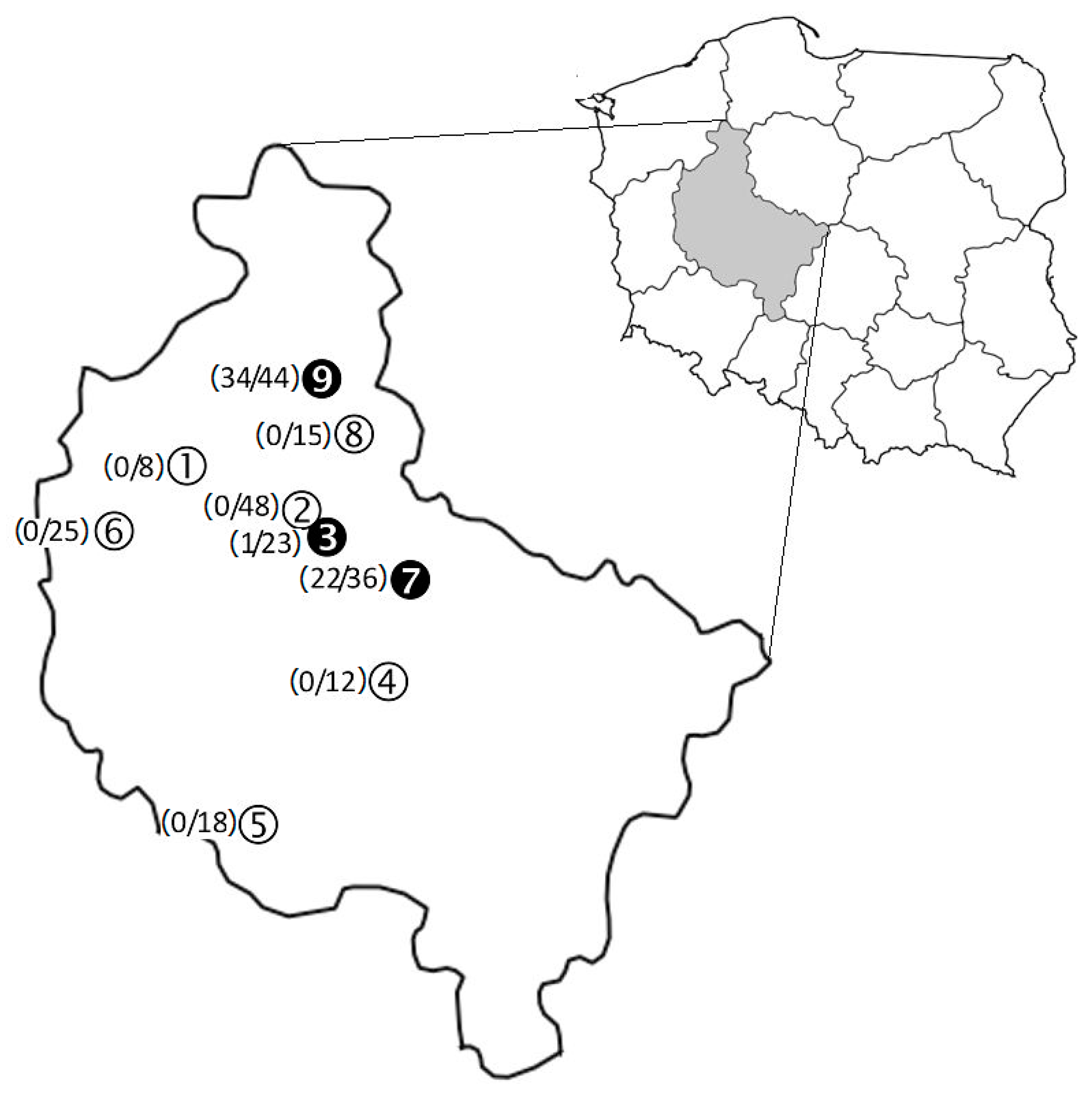
2.1. Study Area and Sample Collection

Foxes were derived from six forest ranges and three forest districts, all situated in Wielkopolska province in west-central Poland. Their localization is shown in Figure 1.
Most of the foxes were harvested in agricultural landscapes neighbouring various forest ecosystems during the fox-hunting seasons (1 June to 31 March) from 2009 to 2011. Autumn and winter are preferred seasons for fox hunting in Poland. Infestation parameters, such as mean prevalence, abundance, and intensity, were estimated for each Ixodes species. Seasons were grouped as (i) spring–summer: June–August, (ii) autumn: September–November, and (iii) winter: December–March. Comparable numbers of foxes were obtained from each of the three consecutive seasons (70, 82, and 77, respectively).
In Poland most of the hunted foxes are left in the hunting ground and buried so acquired material was limited only to blood and biopsies of skin and liver collected by the hunters. In total, 558 tissue samples were obtained from 243 foxes. EDTA-whole blood was collected from the body cavity out of 216 (88.9%) of the animals tested. Furthermore, biopsies of skin (from ear) and liver were obtained from 243 and 99 of the foxes, respectively. These three types of tissue samples were collected within one hour after each animal was culled and stored at −20 °C until extraction.
Both ears of 243 red foxes were cut off basally by hunters and placed into a separate plastic bag, and stored at −20 °C. In the laboratory, after thawing, ears were inspected for ticks under a stereoscopic microscope. Ticks were counted and stored in plastic vials containing 75% ethanol. A total of 1583 Ixodes ticks were removed from 120 infested animals. Since morphological identification of adult female ticks to the species level is easier than immature ticks, females were determined by using taxonomic keys by Siuda [22] and Hornok et al. [18]. Molecular procedures were used for the species determination of larval and nymphal stages.

2.2. DNA Extraction

DNA extraction from animal tissues and engorged ticks was performed with a phenol–chloroform protocol [23]. To avoid any contamination only undamaged ticks rinsed with 75% ethanol before DNA extraction were selected for bacterial DNA detection. DNA samples were stored at −70 °C before PCR analyses.

2.3. Molecular Identification of Ixodes Tick Species

To confirm the accuracy of morphological tick identification according to taxonomic keys, nested PCR assays based on two molecular markers and primer sets targeting (i) the ITS2 (internal transcribed spacer 2) fragment from the nuclear genome of Ixodidae [21], and (ii) the mitochondrial cytochrome c oxidase subunit I (coxI) were used (Table 1).
In each PCR run, DNA isolates of four reference Ixodes spp. female ticks—I. hexagonus from the European hedgehog Erinaceus europaeus, I. canisuga and I. kaiseri from the racoon dog, and I. ricinus from the vegetation—were used. The identities of these ticks had been previously validated with morphological taxonomic keys and molecular studies [18,21,22]. The PCR products were separated on 1.5% agarose gel (Bioshop, Burlington, ON, Canada) and archived, as described elsewhere [21].
In the first stage, including all tick specimens, molecular identification was performed, based on PCR-restriction fragment length polymorphism analysis (PCR-RFLP). PCR-amplified sequences of the ITS2 gene generated with primers SP2-100f and SP2-1274r were digested with enzyme AluI (Thermo Fisher Scientific, Waltham, MA, USA) to obtain RFLP patterns for different Ixodes species according to the protocol previously described [21]. In the second stage, to validate identification done by PCR-RFLP analysis of the ITS2 gene, partial sequencing of Ixodes DNA of ITS2 fragments amplified with inner primer sets SP-100f/SP-1274r was performed for subset of amplicons representing different restriction patterns. Furthermore, partial sequencing of the coxI gene fragments of Ixodes obtained with inner primer sets CO1-375f/CO1-1086r was performed (Table 1). Sequencing was conducted in Macrogen Europe (The Netherlands). Representative partial sequences (n = 78) were deposited in GenBank. The ITS2 sequences (n = 43) are listed as follows: MG962859–MG962867 (I. kaiseri), MG962868–MG962875 (I. hexagonus), MG962876–MG962886 (I. canisuga), and MG962887–MG962901 (I. ricinus). The coxI sequences (n = 35) are listed as follows: MH109183–MH109187 (I. canisuga), MH109188–MH109204 (I. ricinus), MH109205–MH109212 (I. hexagonus), and MH109213-MH109217 (I. kaiseri).

2.4. Detection of Borrelia DNA by Nested PCR

Altogether, 943 undamaged, fully, or partially, engorged ticks (76 females, 166 nymphs, and 701 larvae) and the 558 tissues from foxes were selected. Both groups of samples were tested for the presence of Borrelia DNA using a nested PCR with two primer sets amplifying a fragment of the flaB gene; the protocol has been previously described by Wodecka et al. [24]. All positive samples were rerun using nested PCR assays with two independent sets of primers targeting parts of the p66 gene and intergenic spacer between 3-methyladenine glycosylase (mag) gene and trnI gene encoding tRNA for isoleucine (Table 1). To confirm the specificity and sensitivity of implemented protocols in each PCR run, randomly selected DNA isolated from one of the 11 reference strains of Borrelia spp. was used as a positive control and TE buffer was used as a negative control. In total, 11 reference strains were included: B. burgdorferi sensu stricto (s.s.) IRS, B. garinii 20047, B. afzelii VS461, B. valaisiana VS116, B. bissettiae DN127, B. spielmanii PC-Eq17, B. californiensis CA446, B. carolinensis SCW-22, B. lanei CA28, B. americana CA8, and B. turcica IST7 (German Collection of Microorganisms and Cell Cultures—DSMZ, Germany). The PCR products were separated on 1.5% agarose gel and the results were written, as described elsewhere [21].

2.5. Identification of Borrelia Species by PCR-RFLP and Sequencing

The flaB gene fragments amplified with primers 220f and 823 r were digested with enzymes HpyF3I and Ecl136II (Thermo Fisher Scientific, Waltham, MA, USA) to obtain RFLP patterns of different Borrelia species, according to the protocol previously described [25]. To confirm Borrelia species identification based on PCR-RFLP analysis, partial sequencing of the flaB gene products amplified with primers 220f and 823r or FL120F and FL908R, p66 gene fragments obtained with primers P66-487F and P66-1087R and fragments of intergenic spacer between mag gene and trnI gene generated with primers glz435F and ile65R (Table 1) was performed for positive amplicons, representing different restriction patterns. DNA sequencing was performed in Macrogen Europe (Amsterdam, The Netherlands). Obtained sequences were compared with those available in the GenBank databases using BLAST program (US National Institutes of Health, Bethesda, MD, USA) [www.ncbi.nlm.nih.gov/blast/Blast.cgi (accessed on 23 March 2018)]. A total of 189 selected sequences of the flab gene (n = 112), the p66 gene (n = 33), and the magtrnI intergenic spacers (n = 44) of Borrelia spp. were deposited in GenBank.
The flaB gene sequences are listed as follows: HM802182, HM802184–HM802185, HM802188, KF422758–KF422759, KF422762–KF422767, KF422774, KF422779–KF422782, KF422817, KF422838–KF422846, KF918600–KF918612, MG944996 (B. garinii), HM802193, KF422787, KF422789–KF422791, KF422794–KF422796, KF422858–KF422865, KF918614–KF918616, MG944961–MG944963 (B. afzelii), HM802191, KF422799, KF422802, KF918617, MG944978–MG944983 (B. burgdorferi s.s.), MT118979–MT118980 (B. valaisiana), KF422806–KF422807, KF918618, MG944964 (B. bissettiae), JF732881, MG944976–MG944977, MT118981–MT118982 (B. spielmanii), MG944984–MG944995 (B. californiensis), MG944970–MG944975 (B. carolinensis), MG944965–MG944969 (B. lanei), KF918619–KF918622 (B. americana), and MG944997 (B. turcica).
The p66 gene sequences are listed as follows: MT118983–MT118984 (B. garinii), MT118985–MT118987 (B. afzelii), MT118988–MT118991 (B. burgdorferi s.s.), MT118992–MT118993 (B. valaisiana), MT118998 (B. bissettiae), MT118994–MT118997 (B. spielmanii), MT119004–MT119007 (B. californiensis), MT118999–MT119002 (B. carolinensis), MT119009–MT119013 (B. lanei), and MT119015–MT119018 (B. americana).
The magtrnI intergenic spacer sequences are listed as follows: MT119020–MT119023 (B. garinii), MT119024–MT119029 (B. afzelii), MT119030–MT119034 (B. burgdorferi s.s.), MT119035–MT119036 (B. valaisiana), MT110941–MT119042 (B. bissettiae), MT119037–MT119040 (B. spielmanii), MT119050–MT119054 (B. californiensis), MT119043–MT119048 (B. carolinensis), MT119056–MT119060 (B. lanei), MT119062–MT119065 (B. americana), and MT119067 (B. turcica).

2.6. Contamination Control in DNA Analysis

To minimize contamination, the processes of DNA isolation, the reaction mixture preparation and electrophoresis were carried out in separate rooms.

2.7. DNA Sequence Analysis

Aligned sequences representing 43 nuclear ITS2 gene fragments and 35 mitochondrial coxI gene fragments of Ixodes strains as well as 112 flaB gene, 33 p66 gene, and 44 mag–trnI intergenic spacer fragments of Borrelia strains were examined with MEGAX software (Molecular Evolutionary Genetics Analysis, version X) [26]. Relationships between individuals were assessed by distance estimation between sequences as a measure of the number of allelic substitutions on selected loci, as described earlier [21].

2.8. Statistical Analyses

We analysed Borrelia prevalence in ticks using chi-squared test with Yates’ correction. Differences in mean intensity of tick infestation by Mann–Whitney U test with p < 0.05 were considered statistically significant. All calculations were done using Statistica 8.0 software (StatSoft Inc., Palo Alto, CA, USA).

3. Results

3.1. Tick Species Identification

Of the 243 red foxes, 120 (49.4%) hosted on their ears 1583 Ixodes ticks (Table 2). Using morphological criteria for tick females, four Ixodes species were identified: I. ricinus, I. kaiseri, I. canisuga, and I. hexagonus. Analysis of ITS2 DNA fragments of these females according to the method described by Wodecka et al. [21], confirmed morphological identification.
Analysis of 43 ITS2 sequences obtained from ticks representing different PCR products or restriction patterns, revealed also four distinct groups represented by I. kaiseri (n = 9), I. canisuga (n = 11), I. hexagonus (n = 8), and I. ricinus (n = 15). Comparative analysis of these sequences generated mean distance values within each species as follows: 0.0 for I. canisuga, I. kaiseri, and I. hexagonus, and 0.023 for I. ricinus. The analysis of mean distance values between species showed the highest value for I. ricinus and the three nidicolous tick species: I. canisuga, I. kaiseri, and I. hexagonus (0.262, 0.387, and 0.332, respectively). The distance among these ticks reached: 0.118 for I. kaiseri and I. hexagonus, 0.126 for I. canisuga and I. hexagonus, and 0.185 for I. canisuga and I. kaiseri.
To confirm the ITS2 identification of tick species and its conformity with morphological characterization according to a key by Hornok et al. [18], the analysis of 35 coxI sequences was carried out. This analysis confirmed four distinct groups represented by I. kaiseri (n = 5), I. canisuga (n = 5), I. hexagonus (n = 8), and I. ricinus (n = 17). Comparative analysis of these sequences revealed different mean distance values within each species as represented ITS2 sequences analysis: 0.004 for I. canisuga, 0.002 for I. kaiseri, 0.001 for I. hexagonus, and 0.005 for I. ricinus. The analysis of mean distance values between species confirmed their molecular distinctiveness and the highest value for the exophilic I. ricinus and two nidicolous tick species I. kaiseri and I. hexagonus (0.306 and 0.316, respectively). Surprisingly the distance value for I. ricinus and I. canisuga (0.178) was lower than those for I. canisuga and I. kaiseri (0.212) and I. canisuga and I. hexagonus (0.226). The distance for I. kaiseri and I. hexagonus was the lowest (0.142).

3.2. Occurrence of Tick Species on Ears of Red Foxes

Out of 1583 Ixodes specimens collected from 120 foxes, 1341 were larvae, 166 nymphs, and 76 females. On an average, one infested animal hosted 13.2 ticks (Table 2). Larvae distinctly prevailed over nymphs and females (84.7% vs. 10.5% and 4.8% of total tick numbers, respectively). Larvae infested 34% on animals, nymphs 30%, and females 17%. In total, 55 (45.8%) of the infested animals were parasitized concurrently by at least two of the three different tick developmental stages.
The most abundant tick species was I. kaiseri (76.1%), followed by I. canisuga (11.9%), I. ricinus (10.2%), and I. hexagonus (1.8%, Table 2). Ixodes kaiseri occurred on 33.7% of foxes with a mean intensity of 14.7 ticks (standard deviation, SD—41.01). I. canisuga and I. ricinus reached comparable values of mean intensities (4.2/SD 6.21 and 2.9 ticks/SD 2.83, respectively) and infested 18.5% and 23% of animals, respectively. Only 28 of I. hexagonus ticks were removed from 7.8% of hosts with a mean intensity of 1.5 ticks (SD 0.75). Co-infestations with two, three, or four different tick species (41 double, 16 triple, and 3 quadruple) were noted for 50% of the infested animals. Over half of the double co-infestations (n = 21) was constituted by I. kaiseri and I. ricinus, whereas I. kaiseri, I. canisuga, and I. ricinus accounted for half of the triple infestations (n = 8). Double and triple co-infestations were more prevalent in autumn (39% and 44%, respectively).
Most I. kaiseri (92%, n = 1109) and I. canisuga (81%, n = 152) ticks were larvae which contributed to the larval predominance on the infested foxes. Except for the rare I. hexagonus, loads of nymphs were comparatively low for each of the other three tick species as sparser than larvae intermediate stage (range: 0.1–0.3 ticks per animal). Female ticks were represented mostly by I. ricinus and I. kaiseri, which constituted 84% of the 76 females.
Each of the four Ixodes species occurred on foxes year-round (Table S1). Ixodes ricinus demonstrated clear seasonality, with intensity and prevalence values decreasing steadily from spring–summer to winter. Ixodes canisuga tended to be more prevalent and abundant in winter, whereas the remaining ticks were collected mostly in autumn and winter months. Ixodes kaiseri was more abundant in spring–summer and winter.

3.3. Detection of Borrelia DNA by Nested PCR in Foxes and Their Ticks

Of the 558 tissue samples obtained from the 243 red foxes, 67 (12%) displayed Borrelia spp. specific DNA (Table 3). The bacterium was identified in all three isolate types, but it occurred more frequently in blood samples (23.6%) than in liver (6.1%) or skin (4.1%) biopsies. The infected isolates were derived from 57 (23.5%) of the animals tested. A total of 47 (82.5%) of the infected foxes revealed the bacterium in one isolate type. In this group, blood PCR positive animals (n = 42) prevailed over those carrying the pathogen in liver (n = 3) or skin (n = 2) biopsies. A total of 10 animals (17.5%) harboured spirochetes concurrently in two isolate types: in blood and skin samples (n = 6), in blood and liver samples (n = 3), and in skin and liver biopsies (n = 1, Table S2). The infected foxes were detected in three of the nine sampling sites (Figure 1) with the highest prevalence in Margonin (34/44, 77%) and Czerniejewo (22/36, 61%), and the lowest in Zielonka (1/23, 4.3%).
Borrelia DNA was identified in 306 (32.4%) of the 943 ticks tested and occurred in all parasitic life stages (Table 4). Of the 120 infested animals, 81 (67.5%) hosted at least one infected tick. Of the 81 hosts, 46 (56.8%) carried infected larvae. A total of 12 (14.8%) of the foxes carrying infected ticks proved to harbour concurrently Borrelia spirochetes. Female and nymphal ticks were infected with the bacterium more frequently than larvae (47.4% and 43.9% vs. 28.1%, respectively; p = 0.014). The overall prevalence of infection in I. ricinus ticks was comparable to those recorded in three nidicolous tick species.

3.4. Identification of Borrelia Species

Altogether, PCR positive for flaB gene tissue samples from red foxes and ticks revealed 11 unique RFLP patterns corresponding with different Borrelia species. Of the 11 species, 10 were attributed to the B. burgdorferi s.l. complex i.e., B. garinii, B. afzelii, B. spielmanii, B. burgdorferi s.s., B. valaisiana, B. bissettiae, B. carolinensis, B. californiensis, B. lanei, and B. americana. The first three species were found in the infected foxes, whereas PCR positive ticks revealed all 10 spirochete species (Table 3 and Table 5). Furthermore B. turcica of the reptile-associated borreliae (REP) group was found in a single larva of I. kaiseri.
Borrelia garinii, the most prevalent species infecting foxes, was identified in 52 (21.4%) of the 243 animals tested (36 single and 16 double infections with B. afzelii), followed by B. afzelii (20/8.2%, 4 single and 16 double infections). Moreover B. spielmanii was amplified from one skin sample (Table 3). A total of 10 infected hosts revealed spirochetes concurrently in two different tissue isolate types (Table S2).
Among the 306 Borrelia infected ticks, B. garinii (112/36.6%, including 12 double infections) and B. afzelii (102/33.3%, including 10 double infections) were the most prevalent species (Table 5). The third most frequent species was B. carolinensis (36/11.8%), followed by B. californiensis (24/7.8%), B. burgdorferi s.s. (15/4.9%, including 3 co-infections), B. spielmanii (9/2.9%), B. bissettiae (8/2.6%), B. lanei (5/1.6%), B. americana (4/1.3%, including 1 co-infection), B. valaisiana (3/1%), and B. turcica (1/0.3%). Double infections occurred in 13 (4.2%) of the infected ticks, of which 9 were larvae. Out of the 81 animals carrying at least 1 infected tick, 7 revealed the same spirochete species as their feeding ticks (B. garinii and/or B. afzelii; Table 6).
All 11 identified Borrelia species were detected in I. kaiseri, 10 in I. canisuga (except for B. turcica), 8 in I. ricinus (except for B. bissettiae, B. lanei, and B. turcica), and 4 in I. hexagonus (B. garinii, B. afzelii, B. burgdorferi s.s., and B. bissettiae) (Table 5). The two most prevalent species, namely B. garinii and B. afzelii, were identified in eight out of the nine collection sites. Furthermore, B. burgdorferi s.s. positive ticks were obtained from foxes harvested in five study sites; B. valaisiana, B. spielmanii, B. bissettiae, B. carolinensis, and B. californiensis in three; B. lanei in two, whereas B. americana and B. turcica were detected only in one site (Figure 1).
Subsequent BLAST analysis of 112 selected sequences of the flaB gene fragments from tissue samples and ticks confirmed Borrelia species identification based on PCR-RFLP and revealed also 11 distinct groups represented by all identified species (Table S3). Comparative analysis of these sequences and reference sequences generated mean distance values within each species as follows: 0.0 for B. valaisiana, B. carolinensis, and B. lanei, 0.001 for B. spielmanii, 0.002 for B. californiensis and B. turcica, 0.003 for B. bissettiae, 0.004 for B. burgdorferi s.s., 0.005 for B. americana, 0.013 for B. afzelii, and 0.018 for B. garinii. The analysis of mean distance values between species showed the highest value for B. turcica and 10 Lyme disease species (values from 0.21 to 0.245, Table S4). The distance among LD borreliae species reached values from 0.006 for B. bissettiae and B. carolinensis to 0.075 for B. garinii and B. burgdorferi s.s. (Table S4).
The analysis of 33 sequences of the p66 gene obtained with B. burgdorferi s.l. specific primers confirmed 10 distinct groups of this complex (Table S5). Comparative analysis of obtained and reference sequences revealed different mean distance values within each species, as represented in the case of flaB gene: 0.004 for B. spielmanii, 0.005 for B. lanei, 0.006 for B. valaisiana, 0.01 for B. californiensis, 0.011 for B. burgdorferi s.s., B. afzelii and B. americana, 0.012 for B. carolinensis, and 0.015 for B. garinii and B. bissettiae. The analysis of mean distance values between LD borreliae species confirmed their molecular distinctiveness and revealed the distance values ranged from 0.012 for B. afzelii and B. spielmanii to 0.121 for B. valaisiana and B. carolinensis (Table S6).
The analysis of the 44 sequences of magtrnI intergenic spacer confirmed all 11 identified spirochete species (Table S7). Comparative analysis of these sequences revealed not only different mean distance values within each species, as represented by the flaB and p66 gene sequences analysis, but also differences in length of sequence, depending on Borrelia species (Table S7). Mean distance values within each species are as follows: 0.0 for B. valaisiana, 0.002 for B. spielmanii, 0.003 for B. burgdorferi s.s., B. californiensis, and B. turcica, 0.004 for B. afzelii and B. lanei, 0.007 for B. bissettiae, 0.008 for B. americana, 0.012 for B. carolinensis, and 0.014 for B. garinii. The analysis of mean distance values between species demonstrated the highest values of all examined molecular markers. The highest values were obtained for B. turcica and 10 Lyme disease borreliae species (values from 0.539 to 0.631, Table S8). The distance among LD species reached values from 0.074 for B. lanei and B. americana to 0.31 for B. spielmanii and B. bissettiae (Table S8).

4. Discussion

Based on morphological and molecular methods, we documented the occurrence of four Ixodes tick species on the ears of red foxes. Furthermore, we provide evidence of Borrelia spp. infections in tissue samples of red foxes and in their Ixodes ticks collected from the ears.

4.1. Ixodes Tick Species on Red Foxes

We confirmed the presence of three nidicolous Ixodes species of the subgenus Pholeoixodes, namely I. kaiseri, I. canisuga, I. hexagonus, and the generalist exophilic I. ricinus on red foxes. The first three species are considered nonhuman-biting and are associated mainly with Canidae and Mustelidae and reproduce inside the burrows of their hosts [22,27]. Ixodes hexagonus infests hedgehogs also those inhabiting sub- and urban areas [28]. A taxonomic key published by Hornok et al. [18] allowed us to identify Pholeoixodes females of the three tick species collected in our study. Furthermore, comparison of the coxI sequences obtained from the nidicolous tick species with those published by Hornok et al. [18] confirmed high reliability of this molecular marker in accuracy of species identification of ticks. Therefore, using the coxI gene, we re-examined feeding ticks derived from raccoon dogs and badgers tested in our previous study [21]. This new analysis showed that engorged females of I. kaiseri have been misnamed on the basis of morphological analysis and ITS2 reference sequences deposited in GenBank database as I. canisuga and vice versa. As it turned out, I. kaiseri, and not I. canisuga, dominated both on raccoon dogs and badgers (44.8% vs. 14% and 48.2% vs. 18.5%, respectively). Our present study confirmed the importance of I. kaiseri as the most prevalent tick species associated with wild carnivores in west-central Poland. Moreover, we suggest that in Central Europe, I. kaiseri parasitizes these mammals more frequently than previously thought and its former distribution is not restricted to Romania, the Republic of Moldavia, and Ukraine [22,29]. This confirms the report by Hornok et al. [18] in which the presence of I. kaiseri was documented on carnivores from Germany, Hungary, Romania, and Serbia, expanding its known geographical range in Europe. In our opinion, its hitherto infrequent occurrence on European carnivore species results from misidentification of the three nidicolous Ixodes species using only the morphological approach, therefore, molecular verification is necessary to avoid any misidentification between morphologically similar Ixodes species of the subgenus Pholeoixodes.

4.2. Borrelia Infections in Red Foxes

We found that overall, 23.5% of red foxes revealed DNA of Borrelia spp. Interestingly, over 92% of the 51 blood PCR positive hosts (including 21.3% double infections with B. afzelii) carried B. garinii. Infections caused by B. garinii were also found in skin and liver biopsies. This finding indicates that red foxes develop a disseminated infection with B. garinii more frequently than with B. afzelii, known to exhibit the host specificity for mammals [30]. We also found a predominance of B. garinii over B. afzelii (62.5% vs. 25%) in PCR-positive raccoon dogs [21]. Co-occurrence of both spirochete species has been reported in one European hedgehog Erinaceus europaeus [31] and one Siberian chipmunk Tamias sibiricus barberi [32]. Dumitrache et al. [16] found only B. afzelii (n = 4) and B. burgdorferi s.s. (n = 1) in 1.4% of 353 fox heart isolates tested in Romania. In a German study 24% of skin samples of red foxes had only B. garinii [14]. Furthermore, evidence of B. garinii in blood of a dog from the Czech Republic [33] and in two dogs in Japan was reported [34]. Therefore, we suggest that this avian-adapted spirochete seems to be often associated with canids, including the red fox. However, to clarify its status as a potential reservoir for B. garinii, xenodiagnostic experiments are necessary.

4.3. Borrelia Infections in Ixodes Ticks

The overall infection prevalence found in the four Ixodes tick species collected from ears of red foxes was 32.4% and ranged from 30% to 37%, depending on tick species. The PCR-positive ticks were found year-round. A total of 10 species of the B. burgdorferi s.l. complex were identified, and I. ricinus was infected with eight species, whereas their number in the burrow-dwelling ticks ranged from four (I. hexagonus) to 10 (I. canisuga, and I. kaiseri). So far, there is only one report from Switzerland in I. ricinus ticks infected with eight B. burgdorferi s.l. species [35]. To our knowledge, none of the European mammals has been found to be exposed to such diversity of B. burgdorferi s.l. species as we report here for fox-derived ticks.
Similarly, as in the case of the PCR-positive foxes, B. garinii and B. afzelii comprised most (70%) of the total infections, and reached comparable infection prevalence in each of the four tick species. The distinct predominance of B. garinii and B. afzelii in infected larvae, as well as in foxes, suggests that larvae could have acquired infection while feeding on animals infected with these spirochete species, especially that larval ticks are rarely infected transovarially with B. burgdorferi s.l. Interestingly, the most abundant B. afzelii and B. garinii are associated with rodents and birds, respectively [30], the vertebrates serving as the main food source for red foxes [1]. Therefore, their abundance in our study is not a surprise, especially in the case of I. ricinus that may be shared by the fox and its prey. The presence of the remaining eight species in fox-derived ticks and their absence in hosts, might be explained by co-feeding transmission between spatially clustered infected and uninfected ticks [36]. The observed high aggregation of larval ticks on the ears of the foxes, and the fact that nearly half (46%) of the infested animals were parasitized concurrently by larvae and nymphs or females could greatly enhance the efficiency of this localized non-systemic transmission (Figure 2). Furthermore, the finding that in the case of 11 non-infected foxes, larvae co-feeding with nymphs or females carried the same spirochete species supports our assumption (Table 6). This mode of transmission may facilitate contact and exchange between Borrelia species adapted to different vertebrate host species [37].
Surprisingly, four B. burgdorferi s.l. species previously believed to be restricted only to North America: B. carolinensis, B. californiensis, B. americana, and B. lanei were identified. The first two species were the most prevalent in PCR-positive ticks after B. garinii and B. afzelii and constituted almost 20% of all infections. To date, there are only two European reports documenting the presence of B. carolinensis in ticks: first in one (2.9%) questing I. ricinus from France [38], and the second case together with B. lanei detected for the first time in Europe in three Ixodes tick species associated with European bats from Poland and Romania [39]. In South Carolina and California enzootic cycles of B. carolinensis involve small rodents and a nidicolous I. minor [40,41]. Borrelia californiensis is maintained by Cali–fornia kangaroo rats Dipodomys californicus and two non-human (or rarely) biting I. jellisoni and I. spinipalipis as well as I. pacificus, the key vector of LD species in the western United States [42]. Importantly, in our study B. carolinensis and, for the first time in Europe, B. californiensis were also detected in PCR-positive I. ricinus ticks. Unexpectedly, we detected sequences almost identical to California strains of B. americana in two females of I. ricinus and individual nymphs of I. cansiuga and I. kaiseri. This spirochete was first identified in I. minor from birds in South Carolina as well as from I. pacificus in California [43]. Outside the United States, it was detected only in I. persulcatus ticks in China [44] and, recently, in two I. ricinus specimens in Poland [45]. Thus, our study reaffirms that B. americana occurs in Ixodes ticks in three continents, but its vertebrate host(s) in Europe remains unknown. We identified B. lanei in I. kaiseri and I. canisuga. Considering the study by Michalik et al. [39], it is the second report documenting B. lanei in the group of European Ixodes ticks inhabiting the burrows or caves of their hosts. In the USA this spirochete perpetuates in enzootic cycles involving lagomorphs and rodents and the two tick species, of which I. spinipalpis is nidicolous [46,47].
Borrelia bissettiae like B. burgdorferi s.s., occurs in both the Old and the New World and is considered a human pathogen in Europe and a likely one in the United States [48,49]. In the United States transmission cycles of this species are driven by I. pacificus, and three nidicolous species, I. spinipalpis, I. minor, and I. affinis, and various rodent species acting as reservoirs [42,50,51], and has been detected in the blood of several bird species and I. pacificus feeding on them [52]. Our findings suggest that three burrow-dwelling tick species associated with the red fox could similarly serve as enzootic maintenance vectors of this rarely recorded spirochete in Europe, for which prevalence in I. ricinus is very low [53,54,55]. Furthermore, the finding that B. bissettiae infected ticks were collected only in three collection sites, confirms its highly focal distribution. Although mixed infections of B. bissettiae and B. carolinensis were found in tissues from the European hedgehog, and the Eurasian red squirrel Sciurus vulgaris from the Czech Republic [56], any association of this spirochete with particular vertebrate host(s) in Europe is speculative, at best.
Borrelia spielmanii, with pathogenic potential to humans [57], was detected with a low 1% total prevalence, including six larvae of I. kaiseri, two I. canisuga, and a single I. ricinus female. Together with B. afzelii and B. bavariensis it is considered as maintained by small- and medium-sized mammals [58,59]. Interestingly, it prevailed in bat specific Ixodes ticks [39] and was also reported in bird-derived ticks [60,61].
Enigmatic is evidence of B. turcica in a single larva of I. kaiseri. This spirochete was originally isolated from Hyalomma aegyptium ticks associated with Mediterranean Testudo tortoises in Turkey [62] and is a member of the reptile-associated borreliae (REP), representing the third major group of spirochetes, distinct from LD and relapsing fever (RF) borreliae [63]. Hyalomma aegyptium ticks occur in northern Africa, western Asia, and southern Europe and, apart from their preferential hosts, tortoises, the immature stages may alternatively feed on lizards, birds, and small- or medium-sized mammals [64,65]. Presumably, these vertebrates might influence expansion some pathogens associated with H. aegyptium beyond typical geographical areas. Since the ecology of B. turcica is not sufficiently known [66], the definite cause of detection of this bacterium in a I. kaiseri larva requires further studies.
Of the 14 B. burgdorferi s.l. species so far detected in Europe ([39,61,67],), 10 were detected in red-fox derived ticks, including three identified in tissues of these hosts. Among the 10 species found in ticks, six were discovered for the first time in North America, namely: B. burgdorferi s.s., B. bissettiae, B. americana, B. carolinensis, B. californiensis, and B. lanei. These species together with B. kurtenbachii, detected so far only in North America [68], and B. finlandensis, known solely from Europe [69], are the most closely related in the whole LD stock. To confirm and determine their proper identification we used three molecular markers, i.e., flaB and p66 gene fragments and magtrnI intergenic spacer. Regardless of the marker used, their examination confirmed less genetic distance value between the six species stated in North America and Europe than between the other four LD species detected in this study. A possible explanation why so many closely related species can exist on continents separated by the Atlantic Ocean may be the hypothesis of Borrelia evolution proposed by Estrada-Peña et al. [70]. The authors assume common evolution of the ancestors of all modern ticks and Borrelia species and species level-differentiation at the time of existence of the Pangea supercontinent (between 300 and 180 million years ago). Their contemporary occurrence is the effect of species expansion within Pangea and then separation of individual Borrelia species or populations transmitted by competent tick vectors as the result of the land masses moving apart and then forming the present continents. According to this hypothesis, it is not possible to delineate a “European” or “American” origin of the B. burgdorferi s.l. stock. Their common ancestor might evolve from the relapsing fever group before the Pangea supercontinent splitting and forming of Laurasia and Gondwana [70].
Borrelia garinii identified in red foxes, as well as in their ticks, showed a high level of diversity, which is characteristic of this species. However, unlike birds, carnivore mammals are not recognized as its reservoir. Studies of seabird colonies within the North Atlantic region revealed alongside local genetic variants of B. garinii, also the genotypes similar to those obtained from some human clinical samples in Europe [71]. The North Atlantic region is a particular habitat for B. garinii with the seabird tick Ixodes uriae [72]. Similarly, carnivore mammals, including foxes with their specific tick fauna species, might create conditions favorable to new genetic variant of this bacterium and the remaining LD Borrelia species detected in this study.
Except for the three most prevalent B. burgdorferi s.l. species (B. garinii, B. afzelii, and B. burgdorferi s.s.), the other seven species displayed a highly patchy distribution and were found in three, two or one of our sampling sites. Notably, the highest diversity of the rare spirochetes (i.e., B. bissettiae, B. carolinensis, B. californiensis, and B. lanei) was recorded in Niemierzewo Forest Range. It is located within Sierakowski Landscape Park with diverse ecosystems, including moraine hills, river valleys, dunes, lakes, arable fields, and various forest types. These heterogeneous ecosystems may account for the observed high diversity of Borrelia species, and the park may act as their biodiversity hotspot.

5. Conclusions

Our findings underscore the importance of burrow-dwelling Ixodes species in the ecology of B. burgdorferi s.l. in west-central Poland. The presence of 10 species of this complex in carnivore-associated I. kaiseri and I. canisuga indicates that these non-human biting ticks may serve as maintenance vectors in silent enzootic transmission cycles. Although their vectorial capacity remains unknown, they could potentially increase diversity of Borrelia species which may be transferred to the local population of the generalist tick I. ricinus. Detection in fox-derived samples, only three of the 10 spirochete species infecting engorged ticks, implies that these carnivores appear to be rather incompetent, or exhibit reduced and/or short-lived infectivity for most of them. It cannot be excluded that red foxes in the absence of widespread and long-term infections could serve as so-called “co-feeding transmission hosts” [73], especially that almost in half of the tick-infested animals the co-feeding events were observed. Nevertheless, competent reservoir species for these spirochete species remain unknown and we cannot come to any final conclusions. On the other hand, a distinct predominance of B. garinii in PCR-positive animals, as well as in infected fox-derived larval ticks, suggests that this species, along with B. afzelii, the second most prevalent species, may be preferentially associated with canids. It means that avian and mammalian-adapted spirochete species occupying different ecological niches might be concurrently maintained by red foxes.

Supplementary Materials

The following supporting information can be downloaded at: https://www.mdpi.com/article/10.3390/pathogens11060696/s1, Table S1. Seasonal occurrence of four Ixodes species collected from the ears of 243 red foxes harvested during the fox-hunting seasons (1 June to 31 March) between 2009 and 2011 in west-central Poland. Table S2. Detection of LD Borrelia species concurrently in two isolate types derived from red foxes. BA—B. afzelii, BG—B. garinii. Table S3. Comparison of the selected 112 partial flaB gene sequences of Borrelia species amplified from red foxes (blood, and skin samples: n = 13, and 2, respectively) and their engorged Ixodes ticks (n = 97) sampled in this study. Table S4. MEGA X results of mean distance between Borrelia species obtained on the basis of flaB gene sequence fragment comparison. Table S5. Comparison of the selected 33 partial p66 gene sequences of B. burgdorferi s.l. species amplified from engorged Ixodes ticks removed from red foxes sampled in this study. Table S6. MEGA X results of mean distance between B. burgdorferi s.l. species obtained on the basis of p66 gene sequence fragment comparison. Table S7. Comparison of the selected 44 partial intergenic spacer (IGS) of 3-methyladenine glycosylase (mag) and tRNA-Ile (trnI) genes sequences of Borrelia species amplified from engorged Ixodes ticks removed from red foxes sampled in this study. Table S8. MEGA X results of mean distance between Borrelia species obtained on the basis of intergenic spacer (IGS) of 3-methyladenine glycosylase (mag) and tRNA-Ile genes sequence fragment comparison.

Author Contributions

Conceptualization, B.W. and J.M.; methodology, B.W.; software, B.W. and R.G.; validation, B.W., J.M. and R.G.; formal analysis, B.W.; investigation, B.W. and J.M.; resources, J.M.; data curation, B.W.; writing—original draft preparation, B.W.; writing—review and editing, B.W., J.M. and R.G.; visualization, B.W. and J.M.; supervision, B.W.; project administration, B.W.; funding acquisition, B.W. and J.M. All authors have read and agreed to the published version of the manuscript.

Funding

This research was funded by Grant No. N N303 372136 from the National Science Centre, Kraków, Poland.

Institutional Review Broad Statement

The study was not conducted on live animals and none of the foxes was mortified for the purpose of the study. All foxes used for the study came from hunting conducted as planned wildlife management and were already dead while the tissues and blood were collected. Therefore, the Ethics Committee or Institutional Review Board approval is not required.

Informed Consent Statement

Not applicable.

Data Availability Statement

All data generated or analysed during this study are included in this published article and its supplementary information file. The accession numbers of DNA sequences obtained for ticks and bacteria are mentioned in Material and Methods and are available in the GenBank (https://ww.ncbi.nlm.nih.gov/nuccore, accessed on 23 March 2018).

Acknowledgments

Robert S. Lane, University of California, Berkeley is thanked for his constructive reviews of earlier versions of the manuscript.

Conflicts of Interest

The authors declare no conflict of interest.

References

  1. MacDonald, D.W.; Sillero-Zubiri, C. The Biology and Conservation of Wild Canids, 3rd ed.; Oxford University Press: Oxford, UK, 2004; pp. 207–216. [Google Scholar]
  2. Goszczyński, J.; Misiorowska, M.; Juszko, S. Changes in the density and spatial distribution of red fox dens and cub numbers in central Poland following rabies vaccination. Acta Theriol. 2008, 53, 121–127. [Google Scholar] [CrossRef]
  3. Sréter, T.; Széll, T.; Varga, I. Ectoparasite infestations of red foxes (Vulpes vulpes) in Hungary. Vet. Parasitol. 2003, 115, 349–354. [Google Scholar] [CrossRef]
  4. Kočišová, A.; Lazar, P.; Letková, V.; Čurlík, J.; Goldová, M. Ectoparasitic species from red foxes (Vulpes vulpes) in East Slovakia. Vet. Arch. 2006, 76, 59–63. [Google Scholar]
  5. Meyer-Kayser, E.; Hoffmann, L.; Silaghi, C.; Pfister, K.; Mahling, M.; Passos, L.M.F. Dynamics of tick infestations in foxes in Thuringia, Germany. Ticks Tick-Borne Dis. 2012, 3, 232–239. [Google Scholar] [CrossRef]
  6. Foley, P.; Foley, J.; Sándor, A.D.; Ionica, A.M.; Matei, I.A.; D’Amico, G.; Gherman, C.M.; Dom, A.C.; Mihalca, A.D. Diversity of flea (Siphonaptera) parasites on red foxes (Vulpes vulpes) in Romania. J. Med. Entomol. 2017, 54, 1243–1250. [Google Scholar] [CrossRef]
  7. Karbowiak, G.; Vichova, B.; Majlathova, V.; Hapunik, J.; Petko, B. Anaplasma phagocytophilum infection of red foxes (Vulpes vulpes). Ann. Agric. Environ. Med. 2009, 16, 299–300. [Google Scholar]
  8. Härtwig, V.; von Loewenich, F.D.; Schulze, C.; Straubinger, R.K.; Daugschies, A.; Dyachenko, V. Detection of Anaplasma phagocytophilum in red foxes (Vulpes vulpes) and raccoon dogs (Nyctereutes procyonoides) from Brandenburg, Germany. Ticks Tick-Borne Dis. 2014, 5, 277–280. [Google Scholar] [CrossRef]
  9. Cardoso, L.; Gilad, M.; Cortes, H.C.; Nachum-Biala, Y.; Lopes, A.P.; Vila-Viçosa, M.J.; Simões, M.; Rodrigues, P.A.; Baneth, G. First report of Anaplasma platys infection in red foxes (Vulpes vulpes) and molecular detection of Ehrlichia canis and Leishmania infantum in foxes from Portugal. Parasit. Vectors 2015, 8, 144. [Google Scholar] [CrossRef] [Green Version]
  10. Ebani, V.V.; Rocchigiani, G.; Nardoni, S.; Bertelloni, F.; Vasta, V.; Papini, R.A.; Verin, R.; Poli, A.; Mancianti, F. Molecular detection of tick-borne pathogens in wild red foxes (Vulpes vulpes) from Central Italy. Acta Trop. 2017, 172, 197–200. [Google Scholar] [CrossRef] [PubMed]
  11. Hodžić, A.; Cezanne, R.; Duscher, G.G.; Harl, J.; Glawischnig, W.; Fuehrer, H.P. Candidatus Neoehrlichia sp. in an Austrian fox is distinct from Candidatus Neoehrlichia mikurensis, but closer related to Candidatus Neoehrlichia lotoris. Parasit. Vectors 2015, 8, 539. [Google Scholar] [CrossRef] [Green Version]
  12. Gerrikagoitia, X.; Gil, H.; García-Esteban, C.; Anda, P.; Juste, R.A.; Barral, M. Presence of Bartonella species in wild carnivores of northern Spain. Appl. Environ. Microbiol. 2012, 78, 885–888. [Google Scholar] [CrossRef] [PubMed] [Green Version]
  13. Millán, J.; Proboste, T.; Fernández de Mera, I.G.; Chirife, A.D.; de la Fuente, J.; Altet, L. Molecular detection of vector-borne pathogens in wild and domestic carnivores and their ticks at the human-wildlife interface. Ticks Tick-Borne Dis. 2016, 7, 284–290. [Google Scholar] [CrossRef] [PubMed]
  14. Liebisch, G.; Dimpfl, B.; Finkbeiner-Weber, B.; Liebisch, A.; Frosch, M. The red fox (Vulpes vulpes) a reservoir competent host for Borrelia burgdorferi sensu lato. In Proceedings of the 2nd International Conference on Tick-Borne Pathogens at the Host-Vector Interface: A Global Perspective, Kruger National Park, South Africa, 28 August–1 September 1995; p. 238. [Google Scholar]
  15. Heidrich, J.; Schönberg, A.; Steuber, S.; Nöckler, K.; Schulze, P.; Voigt, W.P.; Schein, E. Investigation of skin samples from red foxes (Vulpes vulpes) in eastern Brandenburg (Germany) for the detection of Borrelia burgdorferi s. l. Zentralbl. Bakteriol. 1999, 289, 666–672. [Google Scholar] [CrossRef]
  16. Dumitrache, M.O.; Matei, I.A.; Ionică, A.M.; Kalmár, Z.; D’Amico, G.; Sikó-Barabási, S.; Ionescu, D.T.; Gherman, C.M.; Mihalca, A.D. Molecular detection of Anaplasma phagocytophilum and Borrelia burgdorferi sensu lato genospecies in red foxes (Vulpes vulpes) from Romania. Parasit. Vectors 2015, 8, 514. [Google Scholar] [CrossRef] [PubMed] [Green Version]
  17. Mysterud, A.; Stigum, V.M.; Jaarsma, R.I.; Sprong, H. Genospecies of Borrelia burgdorferi sensu lato detected in 16 mammal species and questing ticks from northern Europe. Sci. Rep. 2019, 9, 5088. [Google Scholar] [CrossRef] [PubMed]
  18. Hornok, S.; Sándor, A.D.; Beck, R.; Farkas, R.; Beati, L.; Kontschán, J.; Takács, N.; Földvári, G.; Silaghi, C.; Meyer-Kayser, E.; et al. Contributions to the phylogeny of Ixodes (Pholeoixodes) canisuga, I. (Ph.) kaiseri, I. (Ph.) hexagonus and a simple pictorial key for the identification of their females. Parasit. Vectors 2017, 10, 545. [Google Scholar] [CrossRef] [PubMed] [Green Version]
  19. Gern, L.; Rouvinez, E.; Toutoungi, L.N.; Godfroid, E. Transmission cycles of Borrelia burgdorferi sensu lato involving Ixodes ricinus and/or I. hexagonus ticks and the European hedgehog, Erinaceus europaeus, in suburban and urban areas in Switzerland. Folia Parasitol. 1997, 44, 309–314. [Google Scholar]
  20. Estrada-Peña, A.; Oteo, J.A.; Estrada-Peña, R.; Gortázar, C.; Osácar, J.J.; Moreno, J.A.; Castellá, J. Borrelia burgdorferi sensu lato in ticks (Acari: Ixodidae) from two different foci in Spain. Exp. Appl. Acarol. 1995, 19, 173–180. [Google Scholar] [CrossRef] [PubMed]
  21. Wodecka, B.; Michalik, J.; Lane, R.S.; Nowak-Chmura, M.; Wierzbicka, A. Differential associations of Borrelia species with European badgers (Meles meles) and raccoon dogs (Nyctereutes procyonoides) in western Poland. Ticks Tick-Borne Dis. 2016, 7, 1010–1016. [Google Scholar] [CrossRef] [PubMed]
  22. Siuda, K. Ticks (Acari: Ixodida) of Poland. Part II: Taxonomy and Distribution; Polskie Towarzystwo Parazytologiczne: Warsaw, Poland, 1993. (In Polish) [Google Scholar]
  23. Wodecka, B.; Rymaszewska, A.; Skotarczak, B. Host and pathogen DNA identification in blood meals of nymphal Ixodes ricinus ticks from forest parks and rural forests of Poland. Exp. Appl. Acarol. 2014, 62, 543–555. [Google Scholar] [CrossRef] [PubMed] [Green Version]
  24. Wodecka, B.; Leońska, A.; Skotarczak, B. A comparative analysis of molecular markers for the detection and identification of Borrelia spirochetes in Ixodes ricinus. J. Med. Microbiol. 2010, 59, 309–314. [Google Scholar] [CrossRef] [PubMed]
  25. Wodecka, B. FlaB gene as a molecular marker for distinct identification of Borrelia species in environmental samples by the PCR-restriction fragment length polymorphism method. Appl. Environ. Microbiol. 2011, 77, 7088–7092. [Google Scholar] [CrossRef] [PubMed] [Green Version]
  26. Kumar, S.; Stecher, G.; Li, M.; Knyaz, C.; Tamura, K. MEGA X: Molecular Evolutionary Genetics Analysis across computing platforms. Mol. Biol. Evol. 2018, 35, 1547–1549. [Google Scholar] [CrossRef] [PubMed]
  27. Karbowiak, G.; Stanko, M.; Miterpakova, M.; Hurnikova, Z.; Vichova, B. Ticks (Acari: Ixodidae) parasitizing red foxes (Vulpes vulpes) in Slovakia and new data about subgenus Pholeoixodes occurrence. Acta Parasitol. 2020, 65, 636–643. [Google Scholar] [CrossRef] [PubMed]
  28. Dziemian, S.; Michalik, J.; Piłacińska, B.; Bialik, S.; Sikora, B.; Zwolak, R. Infestation of urban populations of the Northern white-breasted hedgehog, Erinaceus roumanicus, by Ixodes spp. ticks in Poland. Med. Vet. Entomol. 2014, 28, 465–469. [Google Scholar] [CrossRef] [PubMed]
  29. Fauna of Ixodid Ticks of the World (Acari, Ixodidae). Available online: http://web.archive.org/web/20150213234618/http:/kolonin.org/13.html (accessed on 15 May 2022).
  30. Kurtenbach, K.; De Michelis, S.; Etti, S.; Schäfer, S.M.; Sewell, H.S.; Brade, V.; Kraiczy, P. Host association of Borrelia burgdorferi sensu lato—The key role of host complement. Trends Microbiol. 2002, 10, 74–79. [Google Scholar] [CrossRef]
  31. Skuballa, J.; Oehme, R.; Hartelt, K.; Petney, T.; Bücher, T.; Kimmig, P.; Taraschewski, H. European hedgehogs as hosts for Borrelia spp., Germany. Emerg. Infect. Dis. 2007, 13, 952–953. [Google Scholar] [CrossRef]
  32. Marsot, M.; Chapuis, J.L.; Gasqui, P.; Dozières, A.; Masséglia, S.; Pisanum, B.; Ferquel, E.; Vourc’h, G. Introduced Siberian chipmunks (Tamias sibiricus barberi) contribute more to Lyme borreliosis risk than native reservoir rodents. PLoS ONE 2013, 8, e55377. [Google Scholar] [CrossRef] [Green Version]
  33. Kybicová, K.; Schánilec, P.; Hulínská, D.; Uherková, L.; Kurzová, Z.; Spejchalová, S. Detection of Anaplasma phagocytophilum and Borrelia burgdorferi sensu lato in dogs in the Czech Republic. Vector Borne Zoonotic Dis. 2009, 9, 655–661. [Google Scholar] [CrossRef] [Green Version]
  34. Inokuma, H.; Maetani, S.; Fujitsuka, J.; Takano, A.; Sato, K.; Fukui, T.; Masuzawa, T.; Kawabata, H. Astasia and pyrexia related to Borrelia garinii infection in two dogs in Hokkaido, Japan. J. Vet. Med. Sci. 2013, 75, 975–978. [Google Scholar] [CrossRef] [Green Version]
  35. Lommano, E.; Bertaiola, L.; Dupasquier, C.; Gern, L. Infections and coinfections of questing Ixodes ricinus ticks by emerging zoonotic pathogens in Western Switzerland. Appl. Environ. Microbiol. 2012, 78, 4606–4612. [Google Scholar] [CrossRef] [Green Version]
  36. Gern, L.; Rais, O. Efficient transmission of Borrelia burgdorferi between cofeeding Ixodes ricinus ticks (Acari: Ixodidae). J. Med. Entomol. 1996, 33, 189–192. [Google Scholar] [CrossRef] [PubMed] [Green Version]
  37. Voodrouw, M.J. Co-feeding transmission in Lyme disease pathogens. Parasitology 2015, 142, 290–302. [Google Scholar]
  38. Cotté, V.; Bonnet, S.; Cote, M.; Vayssier-Taussat, M. Prevalence of five pathogenic agents in questing Ixodes ricinus ticks from western France. Vector Borne Zoonotic Dis. 2010, 10, 723–730. [Google Scholar] [CrossRef] [PubMed]
  39. Michalik, J.; Wodecka, B.; Liberska, J.; Dabert, M.; Postawa, T.; Piksa, K.; Stańczak, J. Diversity of Borrelia burgdorferi sensu lato species in Ixodes ticks (Acari: Ixodidae) associated with cave-dwelling bats from Poland and Romania. Ticks Tick-Borne Dis. 2020, 11, 101300. [Google Scholar] [CrossRef] [PubMed]
  40. Rudenko, N.; Golovchenko, M.; Grubhoffer, L.; Oliver, J.H., Jr. Updates on Borrelia burgdorferi sensu lato complex with respect to public health. Ticks Tick-Borne Dis. 2011, 2, 123–128. [Google Scholar] [CrossRef] [Green Version]
  41. Foley, J.; Ott-Conn, C.; Worth, J.; Poulsen, A.; Clifford, D. An Ixodes minor and Borrelia carolinensis enzootic cycle involving a critically endangered Mojave Desert rodent. Ecol. Evol. 2014, 4, 576–581. [Google Scholar] [CrossRef]
  42. Margos, G.; Lane, R.S.; Fedorova, N.; Koloczek, J.; Piesman, J.; Hojgaard, A.; Sing, A.; Fingerle, V. Borrelia bissettii sp. nov. and Borrelia californiensis sp. nov. prevail in diverse enzootic transmission cycles. Int. J. Syst. Evol. Microbiol. 2016, 66, 1447–1452. [Google Scholar] [CrossRef]
  43. Rudenko, N.; Golovchenko, M.; Grubhoffer, L.; Oliver, J.H., Jr. Borrelia carolinensis sp. nov., a new (14th) member of the Borrelia burgdorferi sensu lato complex from the Southeastern region of the United States. J. Clin. Microbiol. 2009, 47, 134–141. [Google Scholar] [CrossRef] [Green Version]
  44. Yu, P.F.; Niu, Q.L.; Liu, Z.J.; Yang, J.F.; Chen, Z.; Guan, G.Q.; Liu, G.Y.; Luo, J.X.; Yin, H. Molecular epidemiological surveillance to assess emergence and re-emergence of tick-borne infections in tick samples from China evaluated by nested PCRs. Acta Trop. 2016, 158, 181–188. [Google Scholar] [CrossRef]
  45. Dunaj, J.; Drewnowska, J.; Moniuszko-Malinowska, A.; Święcicka, I.; Pancewicz, S. First metagenomic report of Borrelia americana and Borrelia carolinensis in Poland—A preliminary study. Ann. Agric. Environ. Med. 2021, 28, 49–55. [Google Scholar] [CrossRef] [PubMed]
  46. Margos, G.; Fedorova, N.; Kleinjan, J.E.; Hartberger, C.; Schwan, T.G.; Sing, A.; Fingerle, V. Borrelia lanei sp. nov. extends the diversity of Borrelia species in California. Int. J. Syst. Evol. Microbiol. 2017, 67, 3872–3876. [Google Scholar] [CrossRef] [PubMed] [Green Version]
  47. Scott, J.D.; Clark, K.L.; Foley, J.E.; Anderson, J.F.; Durden, L.A.; Manord, J.M.; Smith, M.L. Detection of Borrelia genomospecies 2 in Ixodes spinipalpis ticks collected from a rabbit in Canada. J. Parasitol. 2017, 103, 38–46. [Google Scholar] [CrossRef] [PubMed] [Green Version]
  48. Girard, Y.A.; Fedorova, N.; Lane, R.S. Genetic diversity of Borrelia burgdorferi and detection of B. bissettii-like DNA in serum of north-coastal California residents. J. Clin. Microbiol. 2011, 49, 945–954. [Google Scholar] [CrossRef] [PubMed] [Green Version]
  49. Golovchenko, M.; Vancová, M.; Clark, K.; Oliver, J.; Grubhoffer, L.; Rudenko, N. A divergent spirochete strain isolated from a resident of the southeastern United States was identified by multilocus sequence typing as Borrelia bissettii. Parasit. Vectors 2016, 9, 68. [Google Scholar] [CrossRef] [PubMed] [Green Version]
  50. Eisen, L.; Eisen, R.J.; Mun, J.; Salkeld, D.J.; Lane, R.S. Transmission cycles of Borrelia burgdorferi and B. bissettii in relation to habitat type in northwestern California. J. Vector Ecol. 2009, 34, 81–91. [Google Scholar] [CrossRef] [PubMed]
  51. Fedorova, N.; Kleinjan, J.E.; James, D.; Hui, L.T.; Peeters, H.; Lane, R.S. Remarkable diversity of tick or mammalian-associated Borreliae in the metropolitan San Francisco Bay Area, California. Ticks Tick-Borne Dis. 2014, 5, 951–961. [Google Scholar] [CrossRef]
  52. Newman, E.A.; Eisen, L.; Eisen, R.J.; Fedorova, N.; Hasty, J.M.; Vaughn, C.; Lane, R.S. Borrelia burgdorferi sensu lato spirochetes in wild birds in Northwestern California: Associations with ecological factors, bird behavior and tick infestation. PLoS ONE 2015, 10, e0118146. [Google Scholar] [CrossRef]
  53. Hulínská, D.; Votýpka, J.; Kríz, B.; Holínková, N.; Nováková, J.; Hulínský, V. Phenotypic and genotypic analysis of Borrelia spp. isolated from Ixodes ricinus ticks by using electrophoretic chips and real-time polymerase chain reaction. Folia Microbiol. 2007, 52, 315–324. [Google Scholar] [CrossRef]
  54. Raileanu, C.; Moutailler, S.; Pavel, I.; Porea, D.; Mihalca, A.D.; Savuta, G.; Vayssier-Taussat, M. Borrelia diversity and co-infection with other tick borne pathogens in ticks. Fron. Cell Infect. Microbiol. 2017, 7, 36. [Google Scholar] [CrossRef] [Green Version]
  55. Blazejak, K.; Raulf, M.K.; Janecek, E.; Jordan, D.; Fingerle, V.; Strube, C. Shifts in Borrelia burgdorferi (s.l.) genospecies infections in Ixodes ricinus over a 10-year surveillance period in the city of Hanover (Germany) and Borrelia miyamotoi-specific Reverse Line Blot detection. Parasit. Vectors 2018, 11, 304. [Google Scholar] [CrossRef] [PubMed] [Green Version]
  56. Majerová, K.; Hönig, V.; Houda, M.; Papežík, P.; Fonville, M.; Sprong, H.; Rudenko, N.; Golovchenko, M.; Černá Bolfíková, B.; Hulva, P.; et al. Hedgehogs, squirrels, and blackbirds as sentinel hosts for active surveillance of Borrelia miyamotoi and Borrelia burgdorferi complex in urban and rural environments. Microorganisms 2020, 8, 1908. [Google Scholar] [CrossRef] [PubMed]
  57. Maraspin, V.; Ruzic-Sabljic, E.; Strle, F. Lyme borreliosis and Borrelia spielmanii. Emerg. Infect. Dis. 2006, 12, 1177. [Google Scholar] [CrossRef] [PubMed]
  58. Richter, D.; Schlee, D.B.; Matuschka, F.R. Reservoir competence of various rodents for the Lyme disease spirochete Borrelia spielmanii. Appl. Environ. Microbiol. 2011, 77, 3565–3570. [Google Scholar] [CrossRef] [Green Version]
  59. Jahfari, S.; Ruyts, S.C.; Frazer-Mendelewska, E.; Jaarsma, R.; Verheyen, K.; Sprong, H. Melting pot of tick-borne zoonoses: The European hedgehog contributes to the maintenance of various tick-borne diseases in natural cycles urban and suburban areas. Parasit. Vectors 2017, 10, 134. [Google Scholar] [CrossRef] [Green Version]
  60. Tomassone, L.; Grego, E.; Auricchio, D.; Iori, A.; Giannini, F.; Rambozzi, L. Lyme borreliosis spirochetes and spotted fever group rickettsiae in ixodid ticks from Pianosa island, Tuscany Archipelago, Italy. Vector Borne Zoonotic Dis. 2013, 13, 84–91. [Google Scholar] [CrossRef] [Green Version]
  61. Gryczyńska, A.; Welc-Falęciak, R. Long-term study of the prevalence of Borrelia burgdorferi s.l. infection in ticks (Ixodes ricinus) feeding on blackbirds (Turdus merula) in NE Poland. Exp. Appl. Acarol. 2016, 70, 381–394. [Google Scholar] [CrossRef] [Green Version]
  62. Guner, E.S.; Watanabe, M.; Hashimoto, N.; Kadosaka, T.; Kawamura, Y.; Ezaki, T.; Kawabata, H.; Imai, Y.; Kaneda, K.; Masuzawa, T. Borrelia turcica sp. nov., isolated from the hard tick Hyalomma aegyptium in Turkey. Int. J. Syst. Evol. Microbiol. 2004, 54, 1649–1652. [Google Scholar] [CrossRef]
  63. Margos, G.; Gofton, A.; Wibberg, D.; Dangel, A.; Marosevic, D.; Loh, S.M.; Oskam, C.; Fingerle, V. The genus Borrelia reloaded. PLoS ONE 2018, 13, e0208432. [Google Scholar] [CrossRef]
  64. Paștiu, A.I.; Matei, I.A.; Mihalca, A.D.; D’Amico, G.; Dumitrache, M.O.; Kalmár, Z.; Sándor, A.D.; Lefkaditis, M.; Gherman, C.M.; Cozma, V. Zoonotic pathogens associated with Hyalomma aegyptium in endangered tortoises: Evidence for host-switching behaviour in ticks? Parasit Vectors 2012, 5, 301. [Google Scholar] [CrossRef] [Green Version]
  65. Trevisan, G.; Cinco, M.; Trevisini, S.; di Meo, N.; Chersi, K.; Ruscio, M.; Forgione, P.; Bonin, S. Borreliae Part 1: Borrelia Lyme Group and Echidna-Reptile Group. Biology 2021, 10, 1036. [Google Scholar] [CrossRef] [PubMed]
  66. Kalmar, Z.; Cozma, V.; Sprong, H.; Jahfari, S.; D’Amico, G.; Marcutan, D.I.; Ionica, A.M.; Magdas, C.; Modry, D.; Mihalca, A.D. Transstadial transmission of Borrelia turcica in Hyalomma aegyptium ticks. PLoS ONE 2015, 10, e0115520. [Google Scholar] [CrossRef] [PubMed]
  67. Strnad, M.; Hönig, V.; Růžek, D.; Grubhoffer, L.; Rego, R.O.M. Europe-wide meta-analysis of Borrelia burgdorferi sensu lato prevalence in questing Ixodes ricinus ticks. Appl. Environ. Microbiol. 2017, 83, e00609-17. [Google Scholar] [CrossRef] [PubMed] [Green Version]
  68. Margos, G.; Hojgaard, A.; Lane, R.S.; Cornet, M.; Fingerle, V.; Rudenko, N.; Ogden, N.; Aanensen, D.M.; Fish, D.; Piesman, J. Multilocus sequence analysis of Borrelia bissettii strains from North America reveals a new Borrelia species, Borrelia kurtenbachii. Ticks Tick-Borne Dis. 2010, 1, 151–158. [Google Scholar] [CrossRef] [Green Version]
  69. Qiu, W.G.; Bruno, J.F.; McCaig, W.D.; Xu, Y.; Livey, I.; Schriefer, M.E.; Luft, B.J. Wide distribution of a high-virulence Borrelia burgdorferi clone in Europe and North America. Emerg. Infect. Dis. 2008, 14, 1097. [Google Scholar] [CrossRef] [Green Version]
  70. Estrada-Pena, A.; Alvarez-Jarreta, J.; Cabezas-Cruz, A. Reservoir and vector evolutionary pressures shaped the adaptation of Borrelia. Infect. Genet. Evol. 2018, 66, 308–318. [Google Scholar] [CrossRef] [Green Version]
  71. Munro, H.J.; Ogden, N.H.; Mechai, S.; Lindsay, L.R.; Robertson, G.J.; Whitney, H.; Lang, A.S. Genetic diversity of Borrelia garinii from Ixodes uriae collected in seabird colonies of the northwestern Atlantic Ocean. Ticks Tick-Borne Dis. 2019, 10, 101255. [Google Scholar] [CrossRef]
  72. Olsen, B.; Jaenson, T.G.; Noppa, L.; Bunikis, J.; Bergström, S. A Lyme borreliosis cycle in seabirds and Ixodes uriae ticks. Nature 1993, 362, 340–342. [Google Scholar] [CrossRef]
  73. Kahl, O.; Gern, L.; Eisen, L.; Lane, R.S. Ecological research on Borrelia burgdorferi sensu lato: Terminology and some methodological pitfalls. In Lyme Borreliosis: Biology, Epidemiology and Control; Gray, J.S., Kahl, O., Lane, R.S., Stanek, G., Eds.; CABI Publishing: New York, NY, USA, 2002; pp. 29–46. [Google Scholar]
Figure 1. Location of nine collection sites where red foxes were harvested in Wielkopolska province, west-central Poland: six Forest Ranges: (1) Annogóra, Lubasz Commune: 52°51′ N 16°31′ E; (2) Kąty, Murowana Goślina Commune: 52°37′22″ N 17°00′22″ E; (3) Zielonka, Murowana Goślina Commune: 52°33′00.0″ N 17°07′00.0″ E; (4) Murzynówko, Krzykosy Commune: 52°09′26″ N 17°23′18″ E; (5) Poniec Commune: 51°45′50″ N 16°48′30″ E; (6) Niemierzewo, Kwilcz Commune: 52°33′50″ N 16°10′12″ E), and three Forest Districts: (7) Czerniejewo Commune: 52°26′ N 17°29′ E; (8) Durowo, Wągrowiec Commune 52°48′25″ N 17°12′25″ E; (9) Margonin Commune: 52°58′23′89″ N 17°05′41′27″ E. In brackets: number of PCR positive hosts vs. tested. Black circles denote collection sites with infected foxes. Animals were harvested during fox-hunting seasons (1 June to 31 March) from 2009 to 2011. Borrelia garinii was found in fox-derived ticks in sites no. 1–7 and 9, B. afzelii in sites 1–4 and 6–9, B. burgdorferi s.s. in sites no. 1, 3, 4, 6, and 7, B. valaisiana in sites no. 6, 7, and 9, B. spielmanii in sites no. 1, 6, 7, and 9, B. bissettiae in sites no. 1, 6, and 7; B. carolinensis and B. californiensis in sites no. 6, 7, and 9; B. lanei in sites no. 6, and 9; B. americana in site no. 7, and B. turcica in site no. 6.
Pathogens 11 00696 g001
Figure 2. Larvae co-feeding with a nymph of Ixodes kaiseri on an ear tissue of the red fox (Michalik J.).
Pathogens 11 00696 g002
Table 1. Primers used for the amplification DNA of Ixodes ticks and Borrelia spirochetes.
SpecificityGenetic MarkerSequence
of Primers
(5′->3′)
Anneling Temp.
(°C)
Length of Amplicons (bp)UsageReference
Ixodes *ITS2SP2-26F: CTTCCCGTGGCTTCGTCT48453–694PCR-RFLP, sequencing[21]
SP2-1299R: CTATGCTTAAATTCAGGG
Nested PCR
SP2-100F: TCGTTTTGACTGTGTCGG48
SP2-1274: CCTGATGTGAGGTCGACA
coxICO1-45F: ACTAACCATAAAGACACATTGG44706sequencingThis study
CO1-1100R: GAATTGGCTAAAATAATTCC
Nested PCR
CO1-375F: GGCAGGAACTGGATGAAC47
CO1-1086R: AATTCCTGTTAATCCYCC
Borrelia **flaB132f: TGGTATGGGAGTTTCTGG56604PCR-RFLP, sequencing[24]
905r: TCTGTCATTGTAGCATCTTT
Nested PCR
220f: CAGACAACAGAGGGAAAT54
823r: TCAAGTCTATTTTGGAAAGCACC
FL84F: AGAAGCTTTCTAGTGGGTACAGA57789sequencingThis study
FL976R: GATTGGCCTGTGCAATCAT
Nested PCR
FL120F: TGATGATGCTGCTGGGATGG56
FL908R: TCATCTGTCATTGTAGCATCTT
Borrelia burgdorferi sensu latop66p66-226ldf—GGTACTACATATGCTTCTGT54596–599sequencingThis study
p66-1320htldr—AGGTACACTTCAATTTGGATACA
Nested PCR
p66-487ldf—CCTTTTTTGTTGTCTTCATAGC53
p66-1087ldr—AATTCATCAATAACATACTCT
Borrelia **mag—trnIglz199f—GTAAGTTTGCCAGGACCATT56309–1183sequencingThis study
ile20r—TGAACATCCGACCTCAGG
Nested PCR
glz435f—TAAGCTTCCGTTTCAAC58
ile65r—CAGACCTGCGCTCTAACC
* primers specific to the whole Ixodes genus. ** primers specific to the whole Borrelia genus, including Lyme disease borreliae (B. burgdorferi s.l.), RF borreliae, and REP borreliae.
Table 2. Occurrence of four Ixodes species collected from ears of 243 red foxes harvested in west-central Poland.
Tick
Species
No. (%) Infested Hosts/No. Ticks/No. Ticks per Host/No. Ticks per Infested Host
TotalFemalesNymphsLarvae
I. ricinus56 (23)/162/0.7/2.928 (11.5)/43/0.2/1.531 (12.8)/58/0.2/1.923 (9.5)/61/0.3/2.7
I. kaiseri82 (33.7)/1205/5/14.713 (5.3)/21/0.1/1.639 (16)/75/0.3/1.951 (21)/1109/4.6/21.7
I. canisuga45 (18.5)/188/0.8/4.28 (3.3)/11/0.05/1.419 (7.8)/25/0.1/1.332 (13.2)/152/0.6/4.8
I. hexagonus19 (7.8)/28/0.1/1.51 (0.4)/1/0.004/1.07 (2.9)/8/0.03/1.113 (5.3)/19/0.08/1.5
Total120 (49.4)/1583/6.5/13.241 (16.9)/76/0.3/1.973 (30)/166/0.7/2.383 (34.2)/1341/5.5/16.2
Table 3. Borrelia burgdorferi s.l. (Bb s.l.) species identified by PCR-RFLP procedure in three different types of tissue samples (n = 558) obtained from 243 red foxes harvested in west-central Poland. Cumulated infection prevalence in the hosts is presented in the last line of the table.
Tissue SamplesNo. Tested/ Bb s.l. Species a in Positive Tissue Samples (Animals) (% Prevalence)
Positive (%)BGBABSPBG/BA
Blood216/51 (23.6)37 (72.5)4 (7.8) 10 (19.6)
Skin (ear)243/10 (4.1)4 (40)4 (40)1 (10)1 (10)
Liver99/6 (6.1)2 (33.3)4 (66.7)
Total *558/67 (12)43 (64.2)12 (17.9)1 (1.5)11 (16.4) b
Red foxes(243/57 (23.5))[36 (63.2)][4 (7)][1 (1.7)][16 (28.1)] c
a BG—B. garinii, BA—B. afzelii, and BSP—B. spielmanii; b BG/BA co-infection detected in a single tissue isolate type; c BG/BA co-infection found in one or two different tissue isolate types of the same host; * The number of infected tissue samples is higher than the number of infected foxes (67 vs. 57) because in 10 animals spirochetes were present concurrently in two tissue isolate types.
Table 4. Prevalence of Borrelia DNA in 943 feeding Ixodes ticks obtained from the ears of 243 red foxes harvested in west-central Poland.
Tick
Species
No. (%) Hosts with at Least One Infected TickNo. Tested/Infected (%)
FemalesNymphsLarvaeTotal
I. ricinus26 (10.7) 43/19 (44.2)58/21 (36.2)61/17 (27.9)162/57 (35.2)
I. kaiseri50 (20.6)21/9 (42.9)75/33 (44)479/131 (27.3)575/173 (30.1)
I. canisuga27 (11.1)11/7 (63.6)25/14 (56)143/45 (31.5)179/66 (36.9)
I. hexagonus9 (3.7)1/1 (100)8/5 (62.5)18/4 (22.2)27/10 (37)
Total81 (33.3)76/36 (47.4)166/73 (43.9)701/197 (28.1)943/306 (32.4)
Table 5. Prevalence of Borrelia species identified by PCR-RFLP procedure in 306 infected Ixodes ticks (Females/Nymphs/Larvae) collected from the ears of red foxes * in west-central Poland.
Borrelia spp.No. (F/N/L) (%) of Ticks Infected
I. ricinusI. kaiseriI. canisugaI. hexagonusTotal
B. garinii13 (5/4/4) (22.8)66 (4/13/49) (38.2)17 (1/3/13) (25.8)4 (1/2/1) (40.0)100 (11/22/67) (32.7)
B. afzelii20 (5/10/5) (35.1)49 (0/12/37) (28.3)20 (3/2/15) (30.3)3 (0/2/1) (30.0)92 (8/26/58) (30.1)
B. burgdorferi s.s.2 (1/0/1) (3.5)8 (0/1/7) (4.6) 2 (0/1/1) (20.0)12 (1/2/9) (3.9)
B. valaisiana1 (1/0/0) (1.8)1 (0/0/1) (0.6)1 (0/1/0) (1.5) 3 (1/1/1) (1.0)
B. bissettiae 4 (1/0/3) (2.3)3 (1/2/0) (4.5)1 (0/0/1) (10.0)8 (2/2/4) (2.6)
B. spielmanii1 (1/0/0) (1.8)6 (0/0/6) (3.5)2 (0/0/2) (3.0) 9 (1/0/8) (2.9)
B. carolinensis8 (3/1/4) (14.0)22 (2/3/17) (12.7)6 (1/2/3) (9.1) 36 (6/6/24) (11.8)
B. californiensis9 (1/6/2) (15.8)6 (2/1/3) (3.5)9 (0/2/7) (13.6) 24 (3/9/12) (7.8)
B. lanei 2 (0/0/2) (1.2)3 (0/1/2) (4.5) 5 (0/1/4) (1.6)
B. americana2 (2/0/0) (3.5) 1 (0/1/0) (1.5) 3 (2/1/0) (1.0)
B. garinii/B. afzelii1 (0/0/1) (1.8)5 (0/2/3) (2.9)3 (0/0/3) (4.5) 9 (0/2/7) (2.9)
B. garinii/B. burgdorferi s.s. 1 (0/0/1) (0.6)1 (1/0/0) (1.5) 2 (1/0/1) (0.7)
B. garinii/B. americana 1 (0/1/0) (0.6) 1 (0/1/0) (0.3)
B. afzelii/B. burgdorferi s.s. 1 (0/0/1) (0.6) 1 (0/0/1) (0.3)
B. turcica ** (REP) 1 (0/0/1) (0.6) 1 (0/0/1) (0.3)
Total57 (19/21/17) 173 (9/33/131)66 (7/14/45)10 (1/5/4)306 (36/73/197)
* A total of 18 (33.3%) of the 243 tested animals carried at least one infected tick. ** The member of the reptile-associated (REP) borreliae.
Table 6. Red foxes with at least one infected larva (F—female, N—nymph, L—larva).
Fox No.Fox
Infection
Borrelia Species in Ticks (F/N/L)
BGBABBBVBSPBBIBCLBCRBAMBLN
1-+/+/−+/+/−+/−/− −/−/+ *−/+/−
2BG a/BA a −/−/+ **−/−/+ *
3BG a/BA a,b−/−/+ **−/+/−
4BG a/BA a,b+/−/+ *** +/−/−
5- −/−/+ *
6- −/−/+ *
7-−/+/+ ***−/+/−
8-−/−/+ *
9-−/−/+ *−/−/+ *
10-−/+/+ ***
11-−/−/+ *
12-−/−/+ *−/+/+ *** −/−/+ * −/−/+ *
13- +/+/−−/−/+ *
14BA a,b−/−/+ *−/−/+ * −/+/+ ***−/+/+ ***
15- +/−/+ ***
16BG a/BA a−/−/+ **−/−/+ ** −/+/+ ***+/−/+ *** +/−/−
17BG a −/−/+ *
18BG a−/+/− −/−/+ *
19BG a −/−/+ *
20BG a −/−/+ * −/−/+ *−/−/+ *
21BG a−/−/+ ** −/+/−
22BG a/BA a −/−/+ *−/+/−
23BG a −/−/+ * −/−/+ * −/−/+ * −/−/+ *
24-+/−/−+/+/−−/−/+ *
25-−/−/+ *
26-−/−/+ *
27-−/−/+ *−/+/+ ***
28-−/−/+ *
29-−/−/+ * −/+/−
30-−/−/+ *
31-−/+/+ ***−/−/+ * −/−/+ *
32-+/+/+ ***+/+/+ ***−/+/+ ***
33-−/−/+ * −/−/+ *
34-−/−/+ *−/−/+ *−/−/+ *
35-−/−/+ *
36-−/+/+ ***−/−/+ *−/−/+ *−/−/+ *−/−/+ *−/−/+ * −/−/+ *
37- +/−/+ *** −/−/+ *+/+/+ ***
38- −/−/+ *−/−/+ *−/−/+ *−/−/+ *
39-−/−/+ *−/−/+ *−/−/+ *
40-−/−/+ *−/−/+ *−/−/+ * −/−/+ * −/−/+ * −/−/+ *
41-−/−/+ *−/−/+ *
42-−/−/+ *−/−/+ *
43-−/−/+ *−/−/+ *−/−/+ *
44-−/−/+ *
45-−/−/+ *
46-−/−/+ *−/−/+ *
* Larva infected in the case of non-systemic fox infection. ** Possible larval infection by infected fox. *** Possible larval infection during co-feeding. a—blood infection, b—skin infection.
Publisher’s Note: MDPI stays neutral with regard to jurisdictional claims in published maps and institutional affiliations.

