Article

Polarized Light-Induced Molecular Orientation Control of Rigid Schiff Base Ni(II), Cu(II), and Zn(II) Binuclear Complexes as Polymer Composites

by
Hiroyuki Nakatori
,
Tomoyuki Haraguchi
and
Takashiro Akitsu
*
Department of Chemistry, Faculty of Science, Tokyo University of Science, 1-3 Kagurazaka, Tokyo 162-8601, Japan
*
Author to whom correspondence should be addressed.
Symmetry 2018, 10(5), 147; https://doi.org/10.3390/sym10050147
Submission received: 15 March 2018 / Revised: 13 April 2018 / Accepted: 23 April 2018 / Published: 7 May 2018
(This article belongs to the Special Issue Symmetry in Coordination Chemistry)

Abstract

:
We have investigated linearly polarized UV light-induced molecular orientation due to Weigert effect of composite materials of new six binuclear nickel(II), copper(II), and zinc(II) complexes of two rigid Schiff base ring ligands (L1 and L2) composite materials with methyl orange (MO), an azo-dye, in polyvinylalchol (PVA) cast films. To compare the degree of molecular orientation, two ligands, namely flexible aliphatic cyclohexane (ML1: NiL1, CuL1, ZnL1) and rigid aromatic (ML2: NiL2, CuL2, ZnL2), were synthesized using amine moiety. We have also characterized these complexes by means of elemental analysis, IR, and UV-vis spectra, single crystal or powder X-ray diffraction (XRD) analysis, and so on. Composite materials of ML1 or ML2+MO+PVA were also prepared to separately disperse the solutes in a polymer matrix. For any metal complexes, optical anisotropy (represented as the R parameters) of ML2+MO+PVA was larger than ML1+MO+PVA because of the rigidness of the ligands.

1. Introduction

Nowadays, specific trans-cis photoisomerization of photochromic azobenzene (AZ) and its related compounds are academically and commercially interesting and have been widely studied to date. Many industrial applications of photoisomerization have been reported in the fields such as molecular devices, molecular switches, polymers, and biochemistry, etc. [1,2,3]. In particular, trans-cis photoisomerization of AZ may also lead to the appearance of nonlinear optics, anisotropy (dichroism), and birefringence [4,5], which must be important features in the field of photofunctional materials. Additionally, it is also well-known that azo-compounds show molecular reorientation in polymers due to Weigert effect [6,7] (namely polarized light induces optical anisotropy of azo-compounds directly). When linearly polarized (UV, and vis under a certain condition), light continues to be irradiated to azo-compounds under a restricted environment to some extent; azo-compounds repeat photoisomerization and finally arrange in one direction that does not absorb linearly polarized light. By such mechanism and reason, molecular angular mobility, as well as birefringence and dichroism, also emerged due to azo-compound exhibiting trans-cis photoisomerization in a polymer matrix [8]. Many studies on orientation control based on the reorientation of azo-compounds have been reported so far.
The motivation of this work is to introduce such metal complexes into photosensitive polymer films, as follows. External (mechanical, electric, or magnetic) fields-induced molecular alignment of solutes in a matrix is a promising method for preparing switching or other functional materials beyond single crystalline materials in solid states [9,10]. Classically, planar metal complexes (lowered symmetry from Oh to D4h) may be an important example with which to elucidate their electronic states (assignment of d-d and π-π* transitions and other established associated electronic structures) by means of optical experiments such as polarized crystal (electronic) spectra [11]. However, spectral measurements for single crystals from a certain face (or along one or two axis) are not in agreement with plane-of-coordination-sphere molecular coordinates about d-orbitals symmetry due to orientation in the crystal packing of molecules. Thus, to overcome this experimental difficulty, alignment-tunable (or at least alignment-oriented) molecules of planer metal complexes may be preferable samples with which to study polarized spectral measurements. Moreover, such oriented metal complexes (e.g., [CuCl4]2−) in single crystals may also exhibit important other physical behavior such as thermochromism, as well as optical properties [12]. In this context, organic/inorganic hybrid materials of AZ and (planar) metal complexes deserve to be investigated by means of optical or other physical experiments. Organic/inorganic hybrid materials have the advantage of exhibiting additional electronic properties due to inorganic metal complexes, which is difficult for organic photofunctional polymers solely. However, light or laser (at the same time electric field) controlling of molecular alignment and its evaluation are not simple generally [13,14]. Based on the above-mentioned factors, we aimed to develop new functions and physical properties through the anisotropic arrangement of metal complexes that have azo-components in polymers. We have prepared organic/inorganic composite materials of methyl methacrylate (PMMA) polymer films separately that contain Schiff bases Ni(II), Cu(II), Zn(II) complexes, and AZ with various substituents [15,16,17,18,19]. In recent years, much effort has been devoted to controlling optical anisotropy using not AZ and PMMA but water-soluble methyl orange (MO) and polyvinyl alcohol (PVA) [20], by which application in protein systems may be also expected potentially. In this research, herein, as molecular design comparison from the viewpoint of receiving intermolecular interaction, we discuss the degree of induced optical anisotropy for binuclear complexes that have flexible cyclohexane moieties (ML1) (Scheme 1) and more rigid aromatic phenyl moieties (ML2) (Scheme 2) at the amine site.

2. Materials and Methods

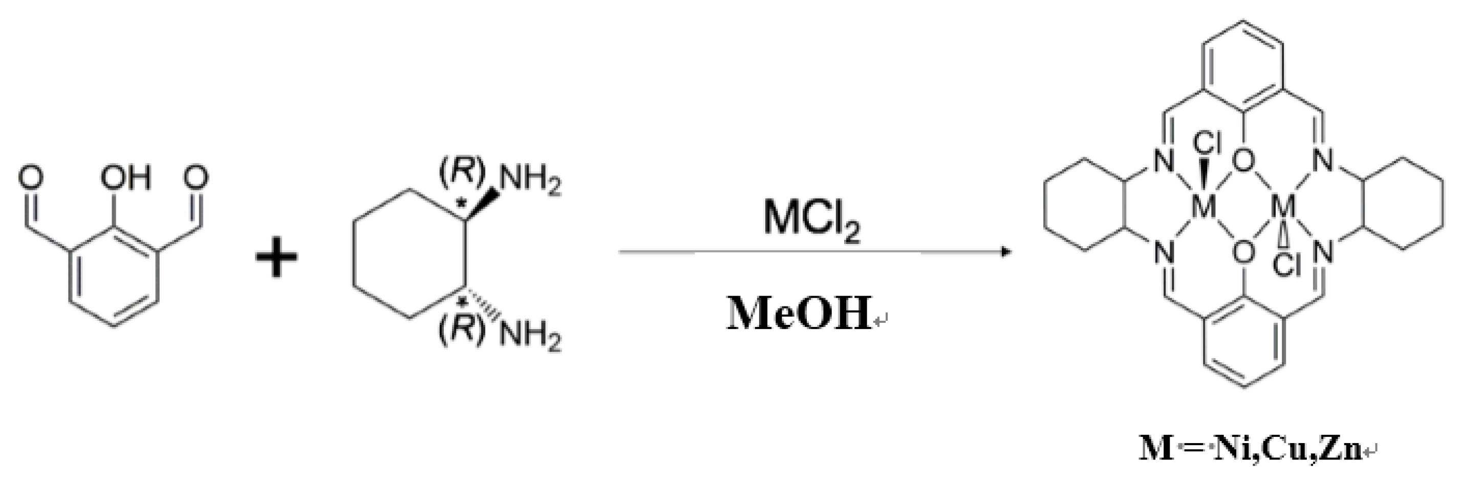
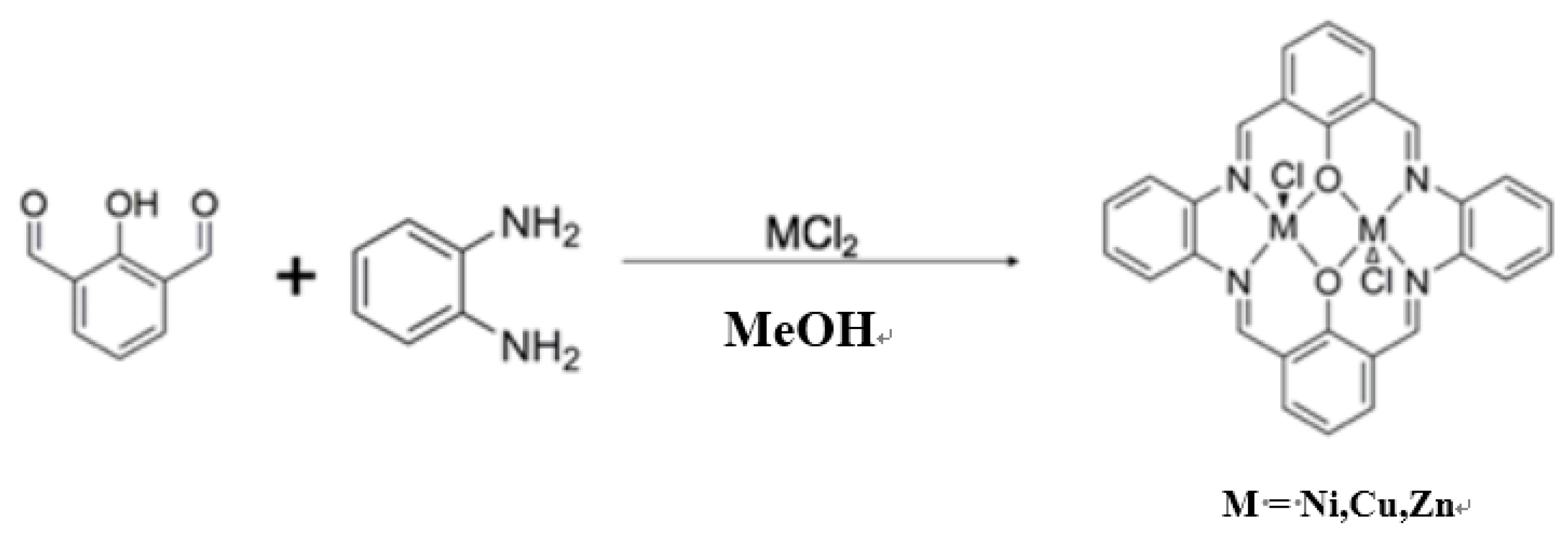
2.1. Preparations of Complexes

All the reagents and solvents were of the highest commercial grade available (Aldrich, Wako, or TCI) were used as received without further purification. Those dissolved in methanol (20 mL), metal sources of chlorides (0.67 mmol), amines, and (1R,2R)-(−)-1,2-cyclohexanediamine for L1 or o-phenylenediamine for L2 (0.067 mmol) were refluxed for 1 day at room temperature to give rise to crude products. The solution was left overnight at room temperature. The resulting crude precipitates were filtered, thoroughly washed with methanol, and dried in vacuo. Only for CuL1, the resulting precipitate was recrystallized in methanol and ethanol (1:1, v/v), taken in sample tube, and left alone for a few days to yield deep green single crystals suitable for single-crystal XRD analysis.
  • NiL1: Yield: 21.8%. Anal. Found: C, 51.31%; H, 4.70%; N, 8.72%. Calcd. for C28H30Cl2N4Ni2O2·H2O: C, 50.89%; H, 4.88%; N, 8.48%. IR(KBr); 1637 (s) (C=N), 1561 (s), 1481, 1446, 1334, 1236, 1055, 568(w), 3420(w) cm−1.
  • CuL1: Yield: 13.9%. Anal. Found: C, 50.54%; H, 4.63%; N, 8.59%. Calcd. for C28H30Cl2Cu2N4O2·H2O: C, 50.15%; H, 4.81%; N, 8.36%. IR(KBr): 1626 (s) (C=N), 2364 (s), 2345(s), 1553, 1438, 1380, 1344, 1237, 1090, 1032, 889, 2930, 3434(w) cm−1.
  • ZnL1: Yield: 20.5%. Anal. Found: C, 51.25%; H, 4.61%; N, 8.54%. Calcd. for C28H30Cl2N4O2Zn2: C, 51.25%; H, 4.61%; N, 8.54%. IR(KBr): 1623 (s) (C=N), 1544, 1430, 1378, 1347, 1230, 1086, 1028, 884, 761, 2922, 3424(w) cm−1.
  • NiL2: Yield: 36.5%. Anal. Found: C, 61.04%; H, 2.88%; N, 9.87%. Calc. for C28H18Cl2N4Ni2O2: C, 53.32%; H, 2.88%; N, 8.88%. IR(KBr): 1625 (s) (C=N), 1536 (s), 1490, 1457, 1431, 1385, 1339, 1273, 1235, 1208, 1060, 909, 748, 3424 (w) cm−1.
  • CuL2: Yield: 32.7%. Anal. Found; C, 52.51%; H, 2.83%; N, 8.75%. Calcd. for C28H18Cl2Cu2N4O2: C, 52.51%; H, 2.83%; N, 8.75%. IR(KBr): 1620 (s) (C=N), 1530 (s), 1548, 1385, 1334, 1248, 1204, 1045, 755, 3459 (w) cm−1.
  • ZnL2: Yield: 32.7%. Anal. Found: C, 52.73%; H, 2.73%; N, 8.50%. Calcd. for C28H18Cl2N4O2Zn2: C, 52.21%; H, 2.82%; N, 8.70%. IR(KBr): 1623 (s) (C=N), 1552 (s), 1489, 1395, 1371, 1334, 1267, 1204, 1038, 760, 3451 (w) cm−1.

2.2. Preparations of Composite Materials

Aqueous solutions of each complex (0.1 mM), MO (0.1 mM), and PVA (10 wt%) were mixed and cast onto a slide glass on the hot plate (333 K) for about 60 min to obtains cast films of the composite materials (MLn+MO+PVA).

2.3. Physical Measurements

Elemental analyses were carried out with a Perkin-Elmer 2400II CHNS/O analyzer Perkin-Elmer, Waltham, MA, USA) at Tokyo University of Science. Infrared (IR) spectra were recorded on a JASCO FT-IR 4200 spectrophotometer (JASCO, Tokyo, Japan) in the range of 4000–400 cm−1 at 298 K. (Polarized) electronic (UV-vis) spectra were measured on a JASCO V-650 spectrophotometer (JASCO, Tokyo, Japan) equipped with polarizer in the range of 800–220 nm at 298 K. Circular dichroism (CD) spectra were measured on a JASCO J-725 spectropolarimeter (JASCO, Tokyo, Japan) in the range of 800–200 nm at 298 K. Photo-illumination was carried out using a lamp (1.0 mW/cm2) with optical filters (UV λ = 200–400 nm), producing a sample using optical fibers and polarizer through optical filters.

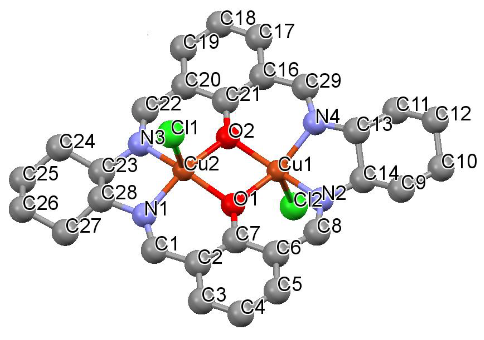
2.4. X-ray Crystallography

Deep greenish prismatic single crystals of CuL1 were glued on top of a glass fiber and coated with a thin layer of epoxy resin to measure the diffraction data. Intensity data were collected on a Bruker APEX2 CCD diffractometer (Bruker, Billerica, MA, USA) with graphite-monochromated Mo Kα radiation (λ = 0.7107 Å). Data analysis was carried out with a SAINT program package. The structures were solved by direct methods with a SHELXS-97, expanded by Fourier techniques, and refined by full-matrix least-squares methods based on F2 using the program SHELXL-97 [21]. An empirical absorption correction was applied by a program SADABS. All non-hydrogen atoms were readily located and refined anisotropic thermal parameters. All hydrogen atoms were located at geometrically calculated positions and refined using riding models. Unfortunately, single crystals of the other complexes could not be obtained.
The powder X-ray diffraction patterns for CuL2 and ZnL2 were collected at 298 K on Rigaku Smart lab with CuKα radiation (λ = 1.5418 Å) at the University of Tokyo. Powder crystal structures were determined by means of Rietveld refinement using a Rigaku PDXL2 program package (Rigaku, Tokyo, Japan).
Crystallographic data for CuL1 (CCDC 1826446): C28H30Cl2Cu2N4O2, Orthorhombic, P21212 (#18), Z = 4, a = 13.841(15) Å, b = 24.705(3) Å, c = 8.198(9) Å, V = 2803.2(6) Å3, ρcalc = 1.660 g/cm3, μ = 1.775 mm−1, F(000) = 1432.0, S = 0.765, R1[I>2σ(I)] = 0.0335, wR2 = 0.1096. Flack parameter = 0.014(10).
Crystallographic data for CuL2 (CCDC 1826434): C28H18Cl2Cu2N4O2, Triclinic, P-1 (#2), Z = 2, a = 7.750(4) Å, b = 8.460(4) Å, c = 15.256(7) Å, α = 85.59(12)°, β = 110.77(9)°, γ = 112.89(7)°, V = 2510(6) Å3, Rwp = 5.28%.
Crystallographic data for ZnL2 (CCDC 1826435): C28H18Cl2N4O2Zn2 Triclinic, P-1 (#2), Z = 2, a = 13.87(4) Å, b = 14.56(4) Å, c = 13.18(4) Å, α = 85.19(15)°, β = 110.60(17)°, γ = 112.84(14)°, V = 2293(11) Å3, Rwp = 4.25%. (See Supplementary Materials)

3. Results

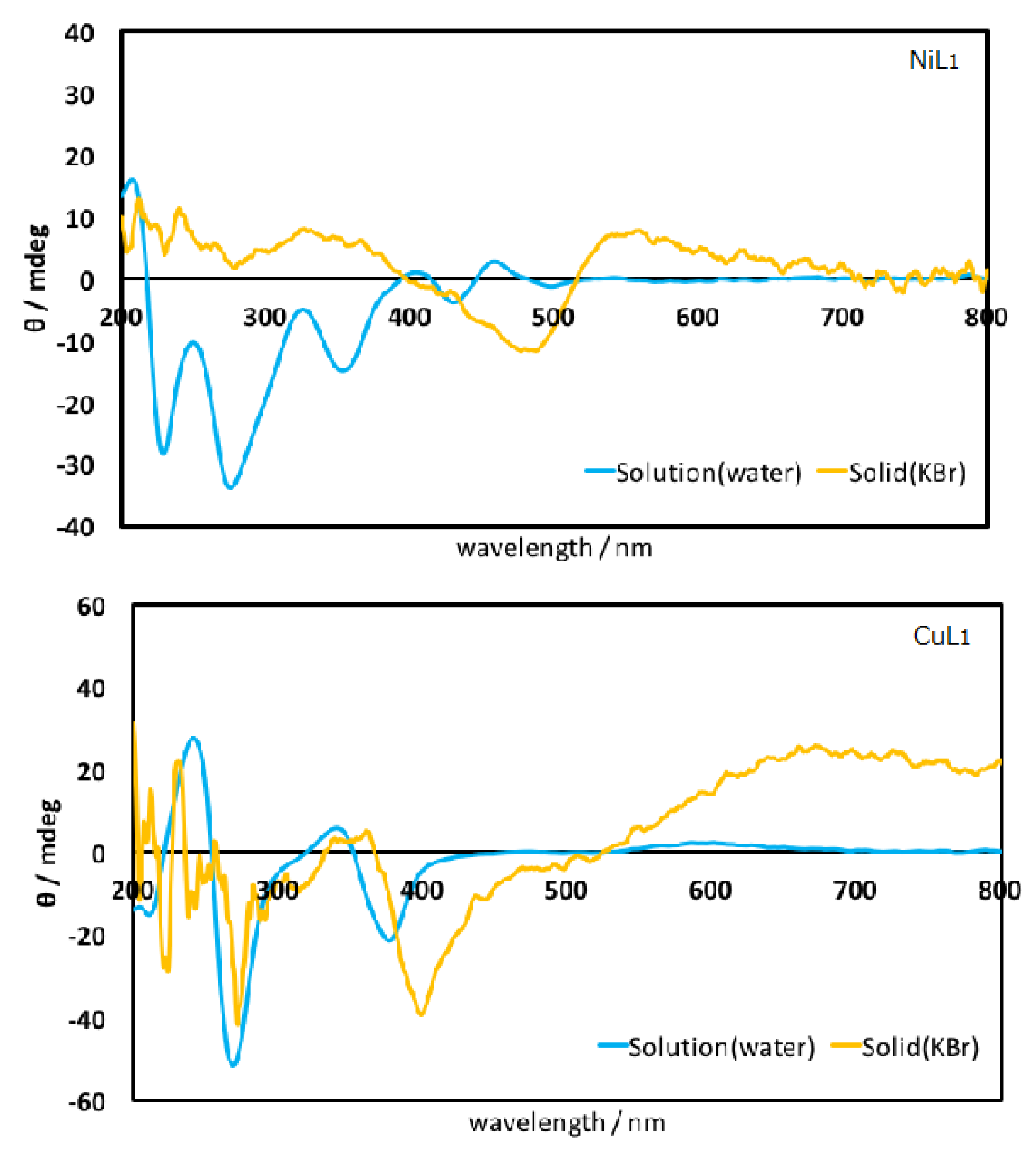
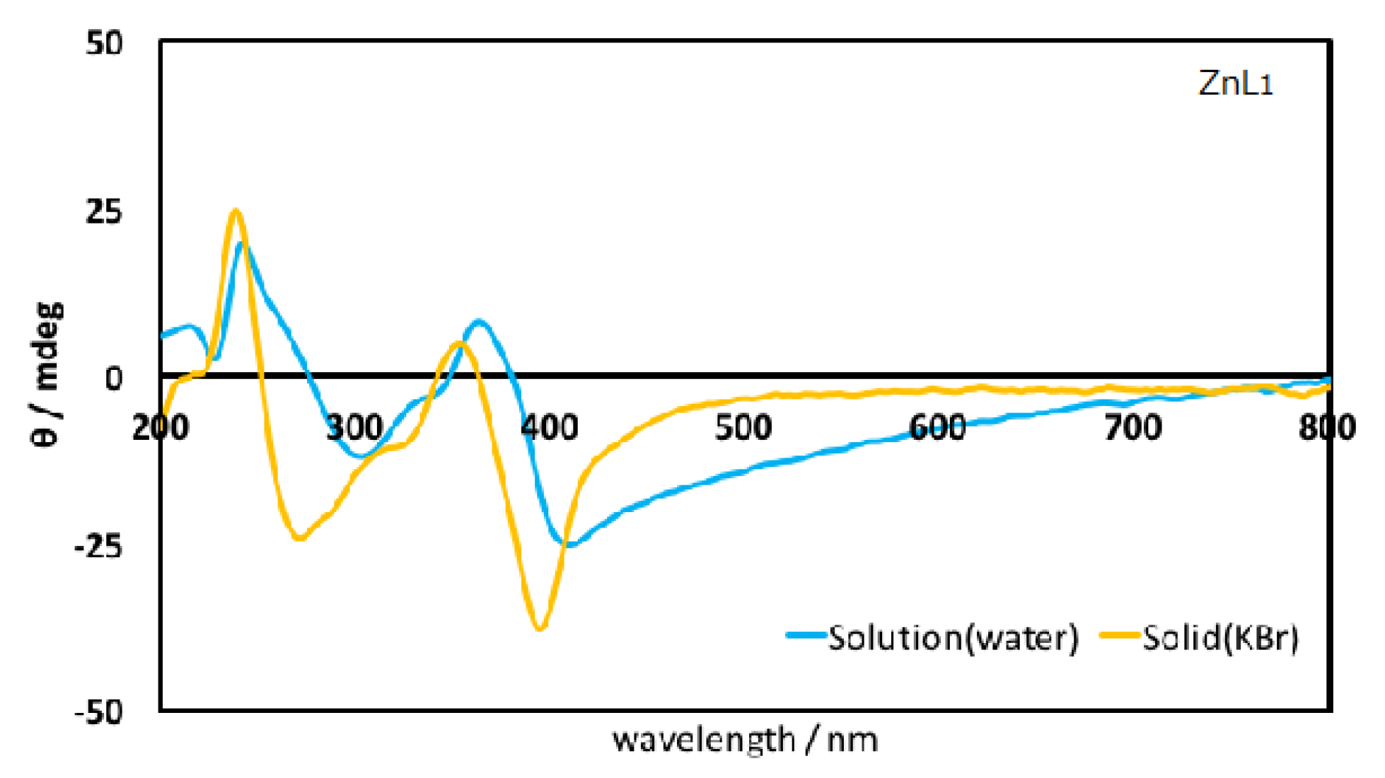
3.1. Spectral Characterization

Figure 1 depicts CD spectra of chiral complexes (ML1). As prepared using chiral amine, it could be confirmed that they are optically active products. Since these complexes afford identical structures except for metal ions, the spectral difference is attributed to transition associated with metal ions (d-d) or between metal and coordination atoms (CT) transitions mainly. The reason why the spectra in solutions are slightly different from that in solid state (KBr) may be artifact peaks due to LD components besides CD components [22].

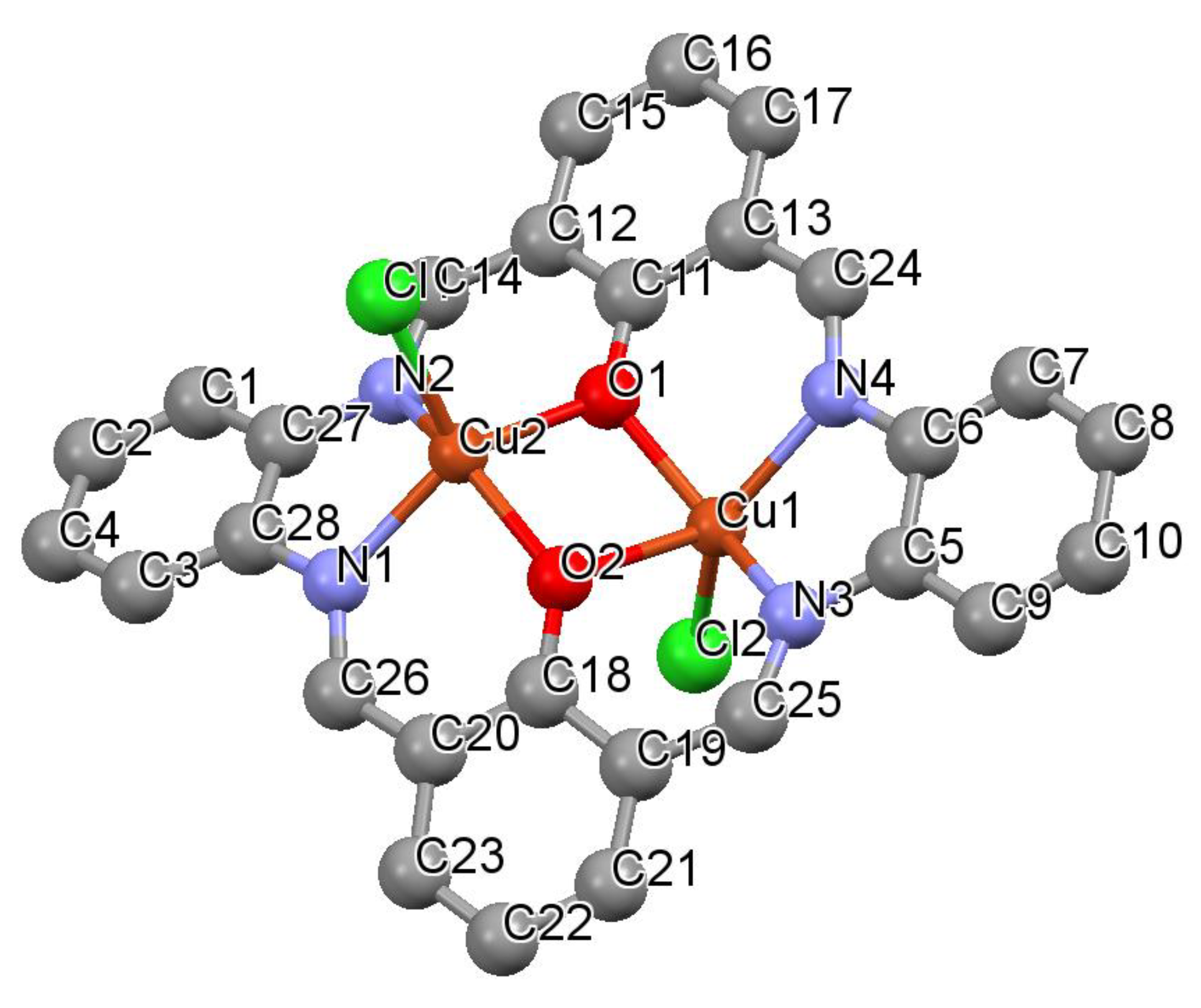
3.2. Structural Characterization

Figure 2, Figure 3 and Figure 4 exhibits crystal structures of CuL1, CuL2, and ZnL2, respectively. Each metal ion of binuclear chromophores affords a five-coordinated square pyramidal coordination geometry, in which metal ions are slightly lifted up towards the apical Cl atom from the [MN2O2] basal planes. Both apical Cl atoms are located on the opposite sides of a ligand plane in a symmetric manner. Rigidity of six-membered C ring derived from amines for CuL1 are CuL2 resulted in conformational difference of whole ligands. Besides them, most of bond lengths and angles are within common ranges of the analogous compounds [20].

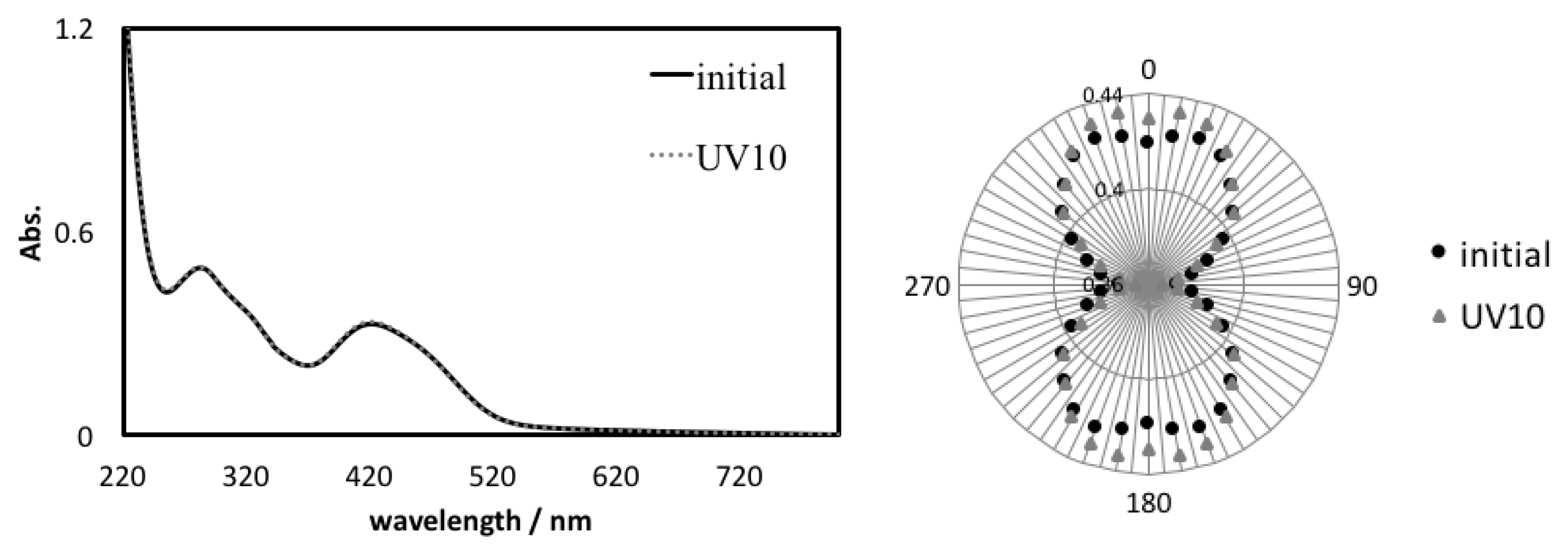
3.3. Linearly Polarized UV Light Induced Optical Anisotropy
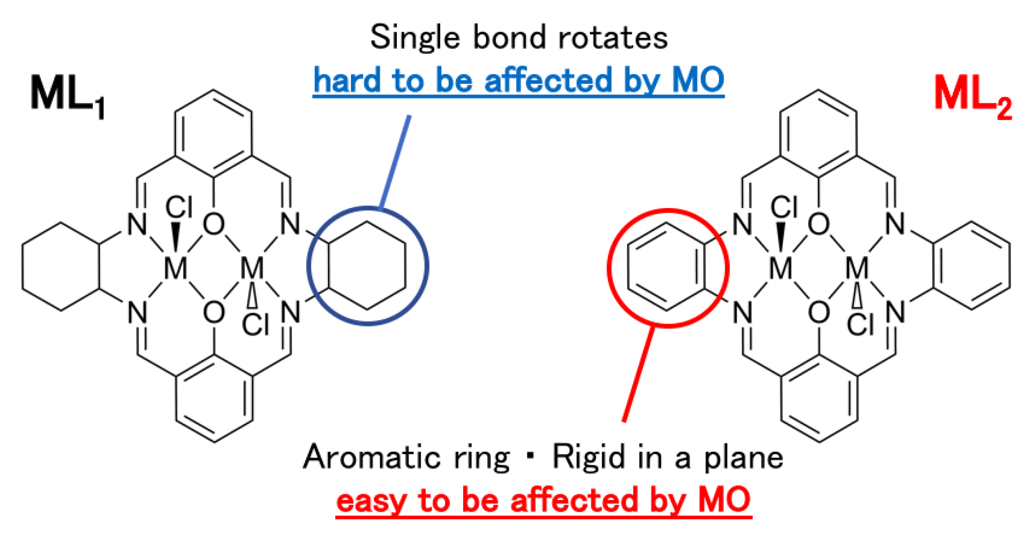
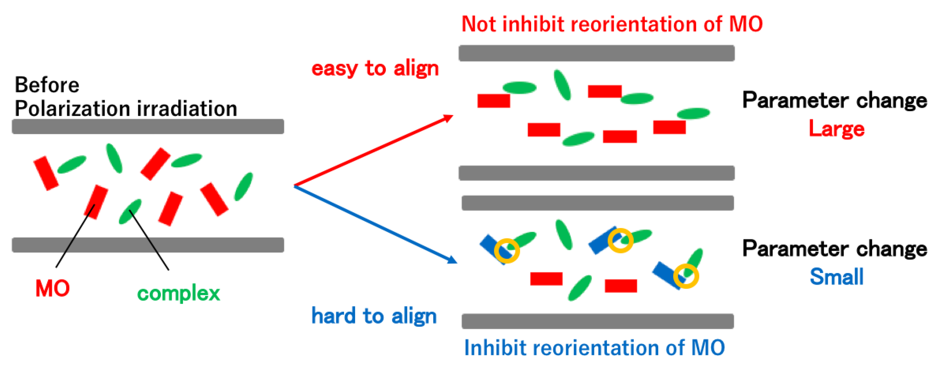
At first, induced optical anisotropy was examined with polarized UV-vis spectra of MO+PVA and MO+ML1+PVA (Table 1). Regarding the relative values (to 0 min) of an anisotropy parameter R (=Abs0deg/Abs90deg) [23], the parameters R increased with linearly polarized UV light irradiation time based on polarized UV-vis spectra (Figure 5). The anisotropy parameter before irradiation is set to 1 for simplicity. From the results, the parameters R increased with increasing irradiation time, which implies that molecules were aligned in a direction to avoid absorbing polarized light. Thus, it is suggested that molecules are aligned in one direction, leading to a situation in which they cannot absorb irradiated polarized light furthermore [6]. Next, the corresponding data for MO+ML2+PVA (Table 2) indicated that the maximum values of the anisotropy parameter for MO+ML2+PVA (1.0279, 1.0222, and 1.0452 for NiL2, CuL2, and ZnL2, respectively) are larger than those of MO+ML1+PVA (0.9850, 1.0141, and 1.0230 for NiL1, CuL1, and ZnL1, respectively), which expressed that MO+ML2+PVA (incorporating more rigid aromatic ligands) are easier to align due to intermolecular interaction between MO and ML2 in PVA in which only MO is affected on Weigert effect directly [18,19].

4. Discussion

Similar to the related study with theoretical analysis [24], explanation of the origin of the observed spectral changes in composite films can be also probed using Weigert effect for MO (as direct driving force) and intermolecular interaction to complexes in a polymer matrix. According to these results, the maximum change in the anisotropic parameter R was larger for ML2+MO+PVA than ML1+MO+PVA for all metals. From the results, it was found that ZnL2 in ZnL2+MO+PVA is the easiest to align. As a consideration, assumption summarized in Scheme 3 can be deduced. Flexibility is more important than chirality of ligand in this case [25]. In previous study, indeed, we exhibited that diastereomers of mononuclear (chiral) complexes indicated similar results (namely slight differences) about anisotropic alignment resulting from Weigert effect. In addition, coordination environment of flexible mononuclear Cu(II) complexes was not as suitable for transferring intermolecular interaction from azo-dyes as that of rigid mononuclear Zn(II) complexes. Since cyclohexane possessed by ML1 is composed of a single bond, it is hardly affected by intermolecular effect [26,27] due to molecular reorientation of MO. Because cyclohexane moiety of ML1 is composed of a single bond, it would be hardly affected by molecular reorientation of MO. On the other hand, phenylenediamine of ML2 is rigid and flat aromatic ring, resulting in its susceptible nature to molecular reorientation of MO. Thus, the change of anisotropic parameter R would be affected by the difference in the susceptibility of ML1 and ML2 to the molecular reorientation of MO. In other words, since phenylenediamine possessed by ML2 is an aromatic ring and a planar shape, it is susceptible to intermolecular effect due to molecular reorientation of MO. As a result, ML2 is oriented without inhibiting the molecular reorientation of MO. However, ML1 inhibits the molecular reorientation of MO [28]. This is related to the magnitude of the change in the anisotropic parameter R depicted in Scheme 4. The quality of data (statistical dispersion) and magnitude of optical anisotropy induced are common ranges for that of the recent examples [29]. By using these mechanisms, potential applications can be expected such as chiroptical probes of spectroscopy [30,31] or sensors [32], and chiral materials of nanostructure in oriented ones [32] or surface [33,34].

5. Conclusions

In summary, we have synthesized two series of new binuclear Schiff base Ni(II), Cu(II), and Zn(II) complexes. One series is the complexes that have flexible cyclohexane moiety on the amine site (ML1: NiL1, CuL1, and ZnL1) and the other series is the complexes that have rigid aromatic six-membered ring moiety on the amine site (ML2: NiL2, CuL2, and ZnL2) instead of the cyclohexane moiety. We have also prepared their hybrid materials (ML1+MO+PVA and ML2+MO+PVA) as PVA cast films. The polarized UV-vis spectra of the hybrid materials after linearly polarized UV light irradiation were measured. From the changes of the anisotropy parameter R, it was found that ZnL2+MO+PVA exhibited the highest anisotropic orientation induced among the six materials. In addition, the magnitude of the anisotropic parameter R was compared among ML1+MO+PVA and ML2+MO+PVA. As a result, it was found that the orientation of ML2, of which the ligand had many aromatic groups, was induced the most effectively of all the metals. Therefore, the susceptibility of the molecular reorientation by MO to the complex through intermolecular interactions leads to enhancement in the anisotropic molecular orientation of the complex. In this way, the change of anisotropic parameter R would be affected by the difference in the susceptibility of ML1, which has flexible cyclohexane moiety, and ML2, which has a rigid phenyl group with regard to the molecular reorientation of MO. These results also support a mechanism based on intermolecular interaction and emphasize the significance of the rigidity of the ligands even for binuclear complexes for effective receiving such as intermolecular interaction from MO (azo-dyes influenced by Weigert effect directly).

Supplementary Materials

The following are available online at https://www.mdpi.com/2073-8994/10/5/147/s1, CCDC 1826446, 1826434, and 1826435 contain the supplementary crystallographic data for CuL1, CuL2, and ZnL2, respectively. These data can be obtained free of charge via www.ccdc.cam.ac.uk, or from the Cambridge Crystallographic Data Centre, 12 Union Road, CB2 1EZ Cambridge, England, UK; Fax: (+44) 01223 336033; or E-Mail: [email protected].

Author Contributions

H.N. performed the experiments, T.H. analyzed the data, and T.A. wrote the paper.

Acknowledgments

This XRD work was conducted at Advanced Characterization Nanotechnology Platform of the University of Tokyo (Kazuhiro Fukawa), supported by “Nanotechnology Platform” of the Ministry of Education, Culture, Sports, Science, and Technology (MEXT), Japan.

Conflicts of Interest

The authors declare no conflict of interest.

References

  1. Velema, W.A.; Szymanski, W.; Feringa, B.L. Photopharmacology: Beyond Proof of Principle. J. Am. Chem. Soc. 2014, 136, 2178–2191. [Google Scholar] [CrossRef] [PubMed]
  2. Xu, Z.; Shi, L.; Jiang, D.; Cheng, J.; Shao, X.; Li, Z. Azobenzene Modified Imidacloprid Derivatives as Photoswitchable Insecticides: Steering Molecular Activity in a Controllable Manner. Sci. Rep. 2015, 5, 13962. [Google Scholar] [CrossRef] [PubMed]
  3. Wang, L.; Li, Q. Photochromism into nanosystems: Towards lighting up the future nanoworld. Chem. Soc. Rev. 2018, 47, 1044–1097. [Google Scholar] [CrossRef] [PubMed]
  4. Delaire, J.A.; Nakatani, K. Linear and Nonlinear Optical Properties of Photochromic Molecules and Materials. Chem. Rev. 2000, 100, 1817–1845. [Google Scholar] [CrossRef] [PubMed]
  5. Grebenkin, S.; Meshalkin, A.B. Wavelength Dependence of the Reorientation Efficiency of Azo Dyes in polymer Matrixes. J. Phys. Chem. B 2017, 121, 8377–8384. [Google Scholar] [CrossRef] [PubMed]
  6. Natansohn, A.; Rochon, P. Photoinduced Motions in Azo-Containing Polymers. Chem. Rev. 2002, 102, 4139–4175. [Google Scholar] [CrossRef] [PubMed]
  7. Ichimura, K. Photoalignment of Liquid-Crystal Systems. Chem. Rev. 2000, 100, 1847–1874. [Google Scholar] [CrossRef] [PubMed]
  8. Sekkat, Z.; Dumont, M. Photoinduced orientation of azo dyes in polymeric films. Characterization of molecular angular mobility. Synth. Met. 1993, 54, 373–381. [Google Scholar] [CrossRef]
  9. Beaudoin, E.; Abecassis, B.; Constantin, D.; Degrouard, J.; Davidson, P. Strain-controlled fluorescence polarization in a CdSe nanoplatelet–block copolymer composite. Chem. Commun. 2015, 51, 4051–4054. [Google Scholar] [CrossRef] [PubMed]
  10. Ma, H.; Zhu, M.; Luo, W.; Li, W.; Fang, K.; Mou, F.; Guan, J. Free-standing, flexible thermochromic films based on one-dimensional magnetic photonic crystals. J. Mater. Chem. C 2015, 3, 2848–2855. [Google Scholar] [CrossRef]
  11. Akitsu, T.; Komorita, S.; Urushiyama, A. Assignment of d-d Transitions of Square Planar [CuIIN4] Complexes Containing Imidate and Amine Ligands by Polarized Crystal Spectra and Angular Overlap Model. Bull. Chem. Soc. Jpn. 2001, 74, 851–860. [Google Scholar] [CrossRef]
  12. Mande, H.M.; Ghalsasi, P.S.; Navamoney, A. Synthesis, structural spectroscopic characterization of the thermochromic compounds A2CuCl4 [(Naphthylethylammonium)2CuCl4]. Polyhedron 2015, 91, 141–149. [Google Scholar] [CrossRef]
  13. Sakai, H.; Safvan, C.P.; Larsen, J.J.; Hilligsoe, K.M.; Hald, K.; Stapelfeldt, H. Controlling the alignment of neutral molecules by a strong laser field. J. Chem. Phys. 1999, 110, 10235–10238. [Google Scholar] [CrossRef]
  14. Ohshima, Y.; Hasegawa, H. Coherent rotational excitation by intense nonresonant laser fields. Int. Rev. Phys. Chem. 2010, 29, 619–663. [Google Scholar] [CrossRef]
  15. Akitsu, T.; Itoh, T. Polarized spectroscopy of hybrid materials of chiral Schiff base cobalt(II), nickel(II), copper(II), and zinc(II) complexes and photochromic azobenzenes in PMMA films. Polyhedron 2010, 29, 477–487. [Google Scholar] [CrossRef]
  16. Aritake, Y.; Takanashi, T.; Yamazaki, A.; Akitsu, T. Polarized spectroscopy and hybrid materials of chiral Schiff base Ni(II), Cu(II), Zn(II) complexes with included or separated azo-groups. Polyhedron 2011, 30, 886–894. [Google Scholar] [CrossRef]
  17. Aritake, Y.; Akitsu, T. The role of chiral dopants in organic/inorganic hybrid materials containing chiral Schiff base Ni(II), Cu(II), and Zn(II) complexes. Polyhedron 2012, 31, 278–284. [Google Scholar] [CrossRef]
  18. Yamazaki, A.; Akitsu, T. Polarized spectroscopy and polarized UV light-induced molecular orientation of chiral diphenyl Schiff base Ni(II) and Cu(II) complexes and azobenzene in a PMMA film. RSC Adv. 2012, 2, 2975–2980. [Google Scholar] [CrossRef]
  19. Ito, M.; Akitsu, T.; Palafox, M.A. Theoretical interpretation of polarized light-induced supramolecular orientation on the basis of normal mode analysis of azobenzene as hybrid materials in PMMA with chiral Schiff base Ni(II), Cu(II), and Zn(II) complexes. J. Appl. Solut. Chem. Model. 2016, 5, 30–47. [Google Scholar]
  20. Takase, M.; Akitsu, T. Linearly polarized light-induced anisotropic orientation of binuclear Ni(II), Cu(II), and Zn(II) Schiff base complexes including or without methyl orange in PVA. In Polymer Science Book Series—Volume #1: “Polymer Science: Research Advances, Practical Applications and Educational Aspects”; Formatex Research Centre: Badajoz, Spain, 2016; pp. 301–308. [Google Scholar]
  21. Sheldrick, G.M. A short history of shelx. Acta Crystallogr. Sect. A 2008, 64, 112–122. [Google Scholar] [CrossRef] [PubMed]
  22. Schellman, J.; Jensen, H.P. Optical spectroscopy of oriented molecules. Chem. Rev. 1987, 87, 1359–1399. [Google Scholar] [CrossRef]
  23. Norden, B.; Todger, A.; Dafforn, T. Linear Dichroism and Circular Dichroism A Textbook on Polarized-Light Spectroscopy; RSC Publishing: Cambridge, UK, 2010. [Google Scholar]
  24. Yamazaki, A.; Kominato, C.; Matsuoka, S.; Watanabe, Y.; Akitsu, T. Polarized Electronic Spectra of 3 Components Organic/ Organic/ Inorganic Hybrid Materials of Chiral Schiff Base Cu(II) Complex, Azobenzene, and Viologens. In Integrating Approach to Photofunctional Hybrid Materials for Energy and the Environment; Nova Science Publishers, Inc.: Hauppauge, NY, USA, 2013; Chapter 6; pp. 125–138. [Google Scholar]
  25. Yamazaki, A.; Akitsu, T. Polarized Spectra of Diastereomers of Schiff Base Ni(II) and Cu(II) Complexes and Azobenzene for Chiral Molecular Recognition in Organic/Inorganic Hybrid Materials. In Magnets: Types, Uses and Safety; Nova Science Publishers, Inc.: Hauppauge, NY, USA, 2012; Chapter 2; pp. 51–67. [Google Scholar]
  26. Takano, H.; Takase, M.; Sunaga, N.; Ito, M.; Akitsu, T. Viscosity and intermolecular interaction of organic/inorganic hybrid systems composed of chiral Schiff base Ni(II), Cu(II), Zn(II) complexes having long ligands, azobenzene and PMMA. Inorganics 2016, 4, 20–29. [Google Scholar] [CrossRef]
  27. Van der Asdonk, P.; Kouwer, P.H.J. Liquid crystal templating as an approach to spatially and temporally organise soft matter. Chem. Soc. Rev. 2017, 46, 5935–5949. [Google Scholar] [CrossRef] [PubMed]
  28. Statman, D.; Janossy, I. Study of photoisomerization of azo dyes in liquid crystals. J. Chem. Phys. 2003, 118, 3222. [Google Scholar] [CrossRef]
  29. Akitsu, T.; Yamazaki, A.; Kobayashi, K.; Haraguchi, T.; Endo, K. Computational Treatments of Hybrid Dye Materials of Azobenzene and Chiral Schiff Base Metal Complexes. Inorganics 2018, 6, 37. [Google Scholar] [CrossRef]
  30. Moletti, A.; Coluccini, C.; Pasini, D.; Taglietti, A. A chiral probe for the detection of Cu(II) by UV, CD and emission spectroscopies. Dalton Trans. 2007, 1588–1592. [Google Scholar] [CrossRef] [PubMed]
  31. Agnes, M.; Nitti, A.; Griend, D.A.V.; Dondi, D.; Merli, D.; Pasini, D. A chiroptical molecular sensor for ferrocene. Chem. Commun. 2016, 52, 11492–11495. [Google Scholar] [CrossRef] [PubMed]
  32. Caricato, M.; Leza, N.J.; Roy, K.; Dondi, D.; Gattuso, G.; Shimizu, L.S.; Griend, D.A.V.; Pasini, D. A Chiroptical Probe for Sensing Metal Ions in Water. Eur. J. Org. Chem. 2013, 6078–6083. [Google Scholar] [CrossRef]
  33. Caricato, M.; Sharma, A.K.; Couluccini, C.; Pasini, D. Nanostructuring with chirality: Binaphthyl-based synthons for the production of functional oriented nanomaterials. Nanoscale 2014, 6, 7165–7174. [Google Scholar] [CrossRef] [PubMed]
  34. Caricato, M.; Delforge, A.; Bonifazi, D.; Dondi, D.; Mazzanti, A.; Pasini, D. Chiral nanostructuring of multivalent macrocycles in solution and on surfaces. Org. Biomol. Chem. 2015, 13, 3593–3601. [Google Scholar] [CrossRef] [PubMed] [Green Version]
Scheme 1. Preparation of ML1.
Symmetry 10 00147 sch001
Scheme 2. Preparation of ML2.
Symmetry 10 00147 sch002
Figure 1. CD spectra in aqueous solutions (blue) and KBr pellets (orange) for chiral complexes (NiL1, CuL1, and ZnL1).
Symmetry 10 00147 g001aSymmetry 10 00147 g001b
Figure 2. Crystal structures of CuL1 showing atom labeling schemes. Hydrogen atoms and crystalline water were omitted for clarity. Selected bond lengths [Å] and angles [°]: Cu1-N2 = 1.900(3), Cu1-O2 = 1.917(3), Cu1-N4 = 1.919(4), Cu1-O1 = 1.928(3), Cu1-Cl2 = 2.5297(13), Cu1-Cu2 = 2.9823(7), Cu2-N3 = 1.912(4), Cu2-N1 = 1.917(4), Cu2-O2 = 1.917(3), Cu2-O1 = 1.926(3), Cu2-Cl = 2.5290(13), N2-Cu1-O2 = 160.33(15), N2-Cu1-N4 = 89.07(15), O2-Cu1-N4 = 92.47(14), N2-Cu1-O1 = 92.97(14), O2-Cu1-O1 = 78.19(12), N4-Cu1-O1 = 157.03(15), N3-Cu2-N1 = 88.85(15), N3-Cu2-O2 = 92.52(14), N1-Cu2-O2 = 162.59(16), N3-Cu2-O1 = 155.27(15), N1-Cu2-O1 = 93.46(14), O2-Cu2-O1 = 78.23(12), Cu1-O2-Cu2 = 102.12(14), Cu2-O1-Cu1 = 101.42(13).
Symmetry 10 00147 g002
Figure 3. Crystal structures of CuL2 showing atom labeling schemes. Hydrogen atoms were omitted for clarity. Selected bond lengths [Å] and angles [°]: Cu1-Cu2 = 3.142(4), Cu1-Cl2 = 2.315(4), Cu2-Cl1 = 2.326(3), Cu1-O1 = 1.989(2), Cu1-O2 = 2.306(4), Cu2-O1 = 2.041(3), Cu2-O2 = 2.018(3), Cu1-N3 = 2.061(3), Cu1-N4 = 2.110(3), Cu2-N1 = 2.077(3), Cu2-N2 = 2.254(3), O1-Cu1-O2 = 78.53(9), O1-Cu2-O2 = 84.47(10), O1-Cu1-N3 = 140.88(9), O1-Cu1-N4 = 84.37(11), O2-Cu1-N3 = 77.09(12), O2-Cu1-N4 = 124.09(9), O2-Cu2-N2 = 140.00(9), O1-Cu2-N1 = 136.46(8), O1-Cu2-N2 = 78.62(14), O2-Cu2-N1 = 87.36(11), Cu1-O1-Cu2 = 102.45(9), Cu1-O2-Cu2 = 92.97(10).
Symmetry 10 00147 g003
Figure 4. Crystal structures of ZnL2 showing atom labeling schemes. Hydrogen atoms were omitted for clarity. Selected bond lengths [Å] and angles [°]: Zn1-Zn2 = 3.244(10), Zn1-Cl2 = 2.250(5), Zn2-Cl1 = 2.250(5), Zn1-O1 = 1.998(7), Zn1-O2 = 2.120(5), Zn2-O1 = 2.120(5), Zn2-O2 = 1.998(7), Zn1-N1 = 2.151(6), Zn1-N4 = 2.231(6), Zn2-N2 = 2.231(6), Zn2-N3 = 2.151(6), O1-Zn1-O2 = 76.1(3), O1-Zn2-O2 = 76.1(3), O1-Zn1-N1 = 127.87(15), O1-Zn1-N4 = 82.8(2), O2-Zn1-N1 = 81.61(18), O2-Zn1-N4 = 130.50(15), O2-Zn2-N2 = 82.8(2), O1-Zn2-N2 = 130.51(15), O1-Zn2-N3 = 81.61(18), O2-Zn2-N3 = 127.88(15), Zn1-O1-Zn2 = 103.9(3), Zn1-O2-Zn2 = 103.9(3).
Symmetry 10 00147 g004
Figure 5. (Left) Polarized UV-vis spectra of ZnL2+MO+PVA before and after 10 min polarized UV light irradiation. (Right) Polarizer angle dependence of absorbance of polarized UV-vis spectra (at 306 nm, π-π* band) of ZnL2+MO+PVA before and after 10 min polarized UV light irradiation.
Symmetry 10 00147 g005
Scheme 3. Flexibility (or rigidity) of ligands and intermolecular effect from MO.
Symmetry 10 00147 sch003
Scheme 4. Molecular orientation and parameter change (proposed mechanism).
Symmetry 10 00147 sch004
Table 1. The relative R parameters for MO+PVA and MO+ML1+PVA after linearly polarized UV light irradiation with detected wavelengths.
Time/minMO+PVA
n-π* 436 nm
MO+NiL1+PVA
π-π* 245 nm
MO+CuL1+PVA
π-π* 253 nm
MO+ZnL1+PVA
π-π* 242 nm
01111
11.02080.98501.01411.0119
31.03110.99391.00331.0140
51.03820.99271.00551.0230
101.04590.99831.00491.0005
Table 2. The relative R parameters for MO+ML2+PVA after linearly polarized UV light irradiation with detected wavelengths.
Time/minMO+NiL2+PVA
π-π* 294 nm
MO+CuL2+PVA
π-π* 306 nm
MO+ZnL2+PVA
π-π* 306 nm
0111
11.00821.01261.0223
31.02151.01091.0258
51.01301.02221.0382
101.02791.01921.0452

