Article

Characterization Analysis of Human Anti-Ferritin Autoantibodies

Laboratory of Veterinary Biochemistry, School of Veterinary Medicine, Kitasato University, Aomori 034-8628, Japan
*
Author to whom correspondence should be addressed.
Antibodies 2014, 3(1), 169-181; https://doi.org/10.3390/antib3010169
Submission received: 6 February 2014 / Revised: 11 March 2014 / Accepted: 12 March 2014 / Published: 21 March 2014

Abstract

:
Anti-ferritin autoantibodies are found in many animals. Human ferritin-binding proteins (FBPs) were partially purified from human serum by ion-exchange chromatography and immobilized metal affinity chromatography with Zn2+. Crude FBPs were immunocoprecipitated with canine liver ferritin followed by the addition of anti-ferritin antibodies. Immunoglobulins in the immunocoprecipitate were detected with antibodies specific for human IgG, IgM or IgA heavy chains, and immunoglobulins IgG, IgM and IgA to bind to expressed recombinant human H and L chain homopolymers were also found. A portion of human serum proteins bound to zinc ions immobilized on beads were released upon the addition of canine liver ferritin, and the released protein was identified as IgM antibody. Additionally, the released proteins recognized peptide sequence (DPHLCDF) commonly found in amino acid sequences of mammalian ferritin H and L subunits. These results suggest that human serum contains anti-ferritin autoantibodies (IgG, IgM and IgA) which bind zinc ions and preferentially bind ferritin over both the H and L subunits, and that a portion of, but not all, the IgM antibodies bound to ferritin with higher affinity than to zinc ions and may recognize the common sequence found in mammalian ferritin H and L subunits.

1. Introduction

Ferritin, a ubiquitous iron storage protein, is a 24-mer protein composed of varying proportions of two subunits termed H (heavy or heart type) and L (light or liver type) [1,2,3,4,5]. Mammalian intracellular ferritin functions by storing and sequestering iron in a non-toxic form [3,4]. H and L subunits have distinct immunological and physiological properties [1,3,4,5,6]. The H subunit possesses ferroxidase, which is essential for the incorporation of iron, while the L subunit lacks ferroxidase but promotes iron core formation by mineral core-catalyzed iron oxidation at iron nucleation sites within the ferritin shell [1,3,4].
Serum ferritin is present at relatively low concentrations (<1 µg/mL) in various animals including humans and is positively correlated with body iron stores [7,8,9,10,11,12,13]. A variety of ferritin-binding proteins (FBPs) in mammalian circulation systems have been identified: H-kininogen [14], alpha-2-macroglobulin [15,16], autoantibodies [17,18,19,20,21], apolipoprotein B [22] and fibrinogen [23]. These proteins are likely involved in the clearance of circulating ferritin through indirect receptor-mediated uptake by forming complexes with it. Some FBPs bind directly (e.g., ferritin binding with H-kininogen or anti-ferritin autoantibody) [19,24] and others bind indirectly (e.g., heme-mediated binding between ferritin and apolipoprotein B or fibrinogen) [22,25].
Anti-ferritin autoantibodies as well as alpha-2-macroglobulin have been identified as common mammalian FBPs [4,15]. Bellotti et al. [26] suggested the presence of many human FBPs (the complement proteins C3 and C4, alpha-2-macroglobulin and immunoglobulins), but these were not examined in detail. Although putative human serum FBPs interfere ferritin immunoassay [27] as in horse [23,28], bovine [18] and feline [20], complex formation between serum ferritin and FBPs has not fully been elucidated. However, Covell et al. [29] reported that human IgM and IgG were unlikely to bind ferritin. Albumin, alpha-2-macroglobulin, ceruloplasmin, transferrin, immunoglobulins, complement C4 and C-reactive protein have been identified as human serum zinc-binding proteins [30,31], and some proteins can be candidate for FBPs. However, iron level is tightly regulated due to its generation of free radicals [3,4,32].
This study showed characterization of partially purified FBPs from human serum with ion-exchange chromatography and immobilized metal affinity chromatography with Zn2+, and demonstrates that human serum contains zinc-binding anti-ferritin autoantibodies (IgM, IgG and IgA). This study also shows the binding analysis of IgM antibody binding with ferritin.

2. Results and Discussion

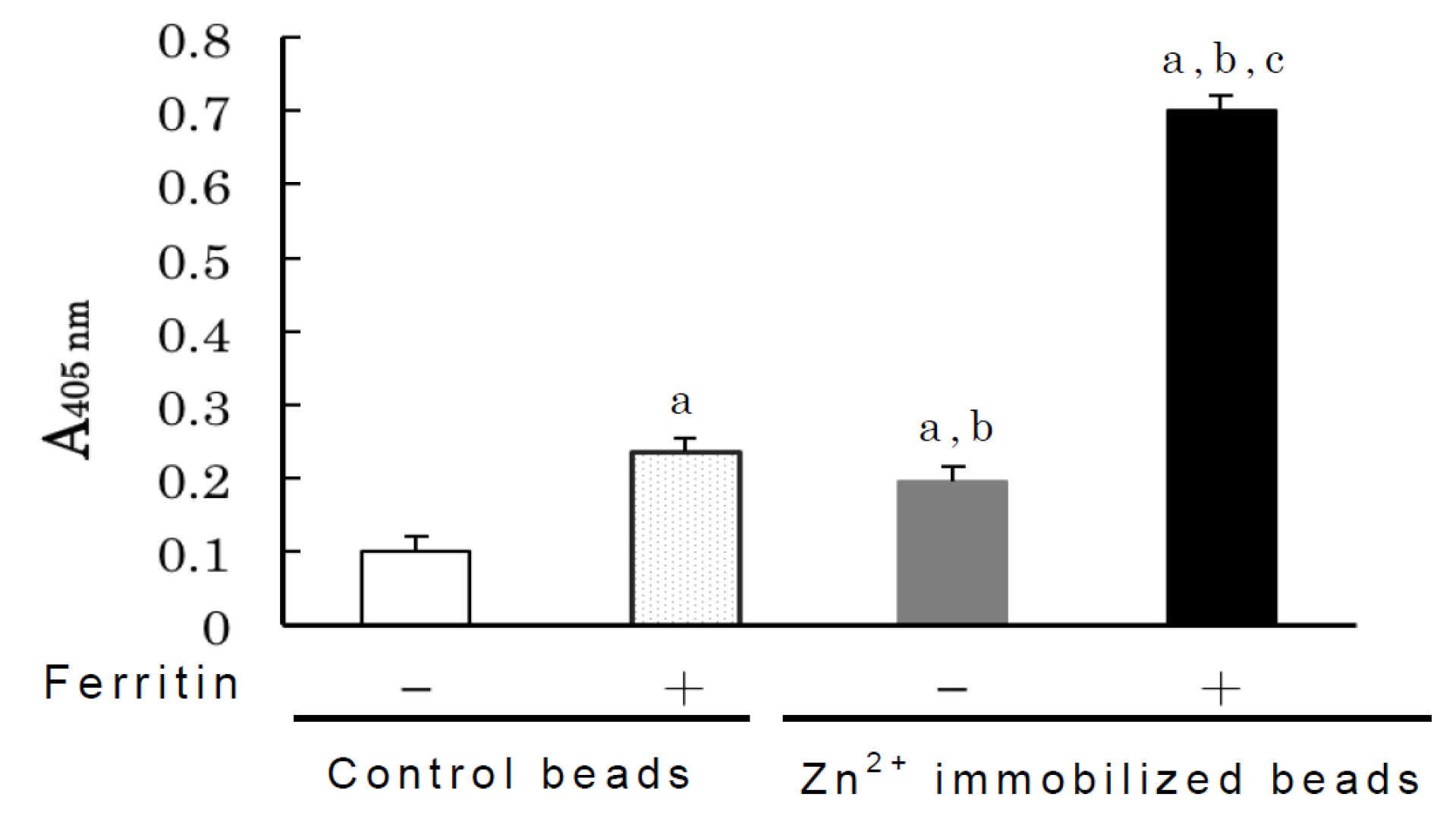
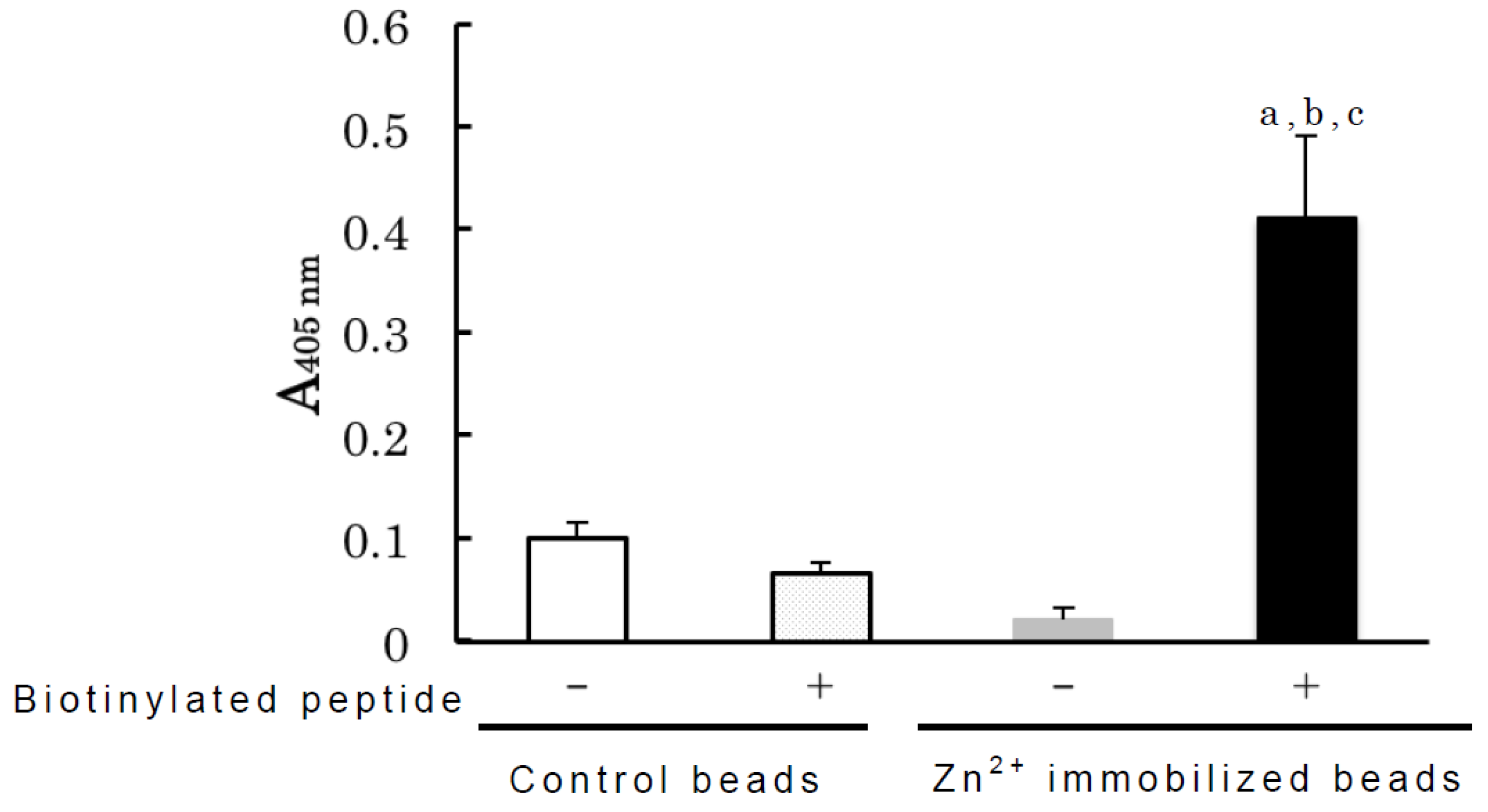
2.1. Identification of Partially Purified Human FBPs as Anti-Ferritin Autoantibodies

Human serum FBPs were partially purified using ion exchange chromatography and immobilized metal affinity chromatography with Zn2+. Pooled fractions containing human FBPs were immunocoprecitated by antibodies to feline liver ferritin after complex formation with canine liver ferritin following a previously described method [20]. All antibody classes (IgM, IgG and IgA) tested were detected in the immunocoprecipitate with ferritin-binding activities as control data in the absence of fractions were subtracted from absorbance value of each antibody class (Figure 1) although an antibody to human IgG Fc fragments strongly cross-reacted with rabbit IgG (data not shown). As shown in Figure 2, all antibodies (IgM, IgG and IgA) tested were detected in the immunocoprecipitate using ferritin H and L subunit homopolymers. Ferritin-binding activity of IgM antibody was detected more strongly than that of other immunoglobulin classes (IgG and IgA), and the antibody to human IgM heavy chain did not cross-react with rabbit IgG than to the antibody to human IgG (Fc) fragment. In this study, further experiments were performed to detect IgM antibody binding ferritin.
Figure 1. Detection of immunoglobulins in immunocoprecitated partially purified human serum ferritin-binding proteins (FBPs). Immunocoprecitation was performed after complex formation with partially purified FBPs and canine liver ferritin followed by the addition of anti-feline liver ferritin antiserum. Immunoglobulin (IgM: solid bar; IgG: gray bar; IgA: open bar) was detected with goat antibodies for human IgM, IgG or IgA heavy chains followed by the addition of alkaline phosphatase (ALP)-labeled anti-goat IgG as coated the immunocoprecipitate on the plate well as described in “Experimental Section”. Control reactions without human FBPs were carried out for each antibody class and subtracted from the reaction values. Each bar and error bar represents the mean ± SD of four determinations.
Antibodies 03 00169 g001
Figure 2. Binding of human anti-ferritin autoantibodies (IgM, IgG and IgA) to the expressed recombinant human H and L subunit homopolymers. After complex formation between partially purified FBPs with the recombinant human ferritin H (solid bar) or L (open bar) subunit homopolymers, specific antibodies to human ferritin H and L subunits were added to precipitate H and L subunit homopolymeres, respectively. Detection of immunoglobulins (IgM, IgG and IgA) was carried out as described in Figure 1. Control reactions without human FBPs were carried out for each antibody class and subtracted from the reaction values. Each bar and error bar represents the mean ± SD of four determinations.
Antibodies 03 00169 g002

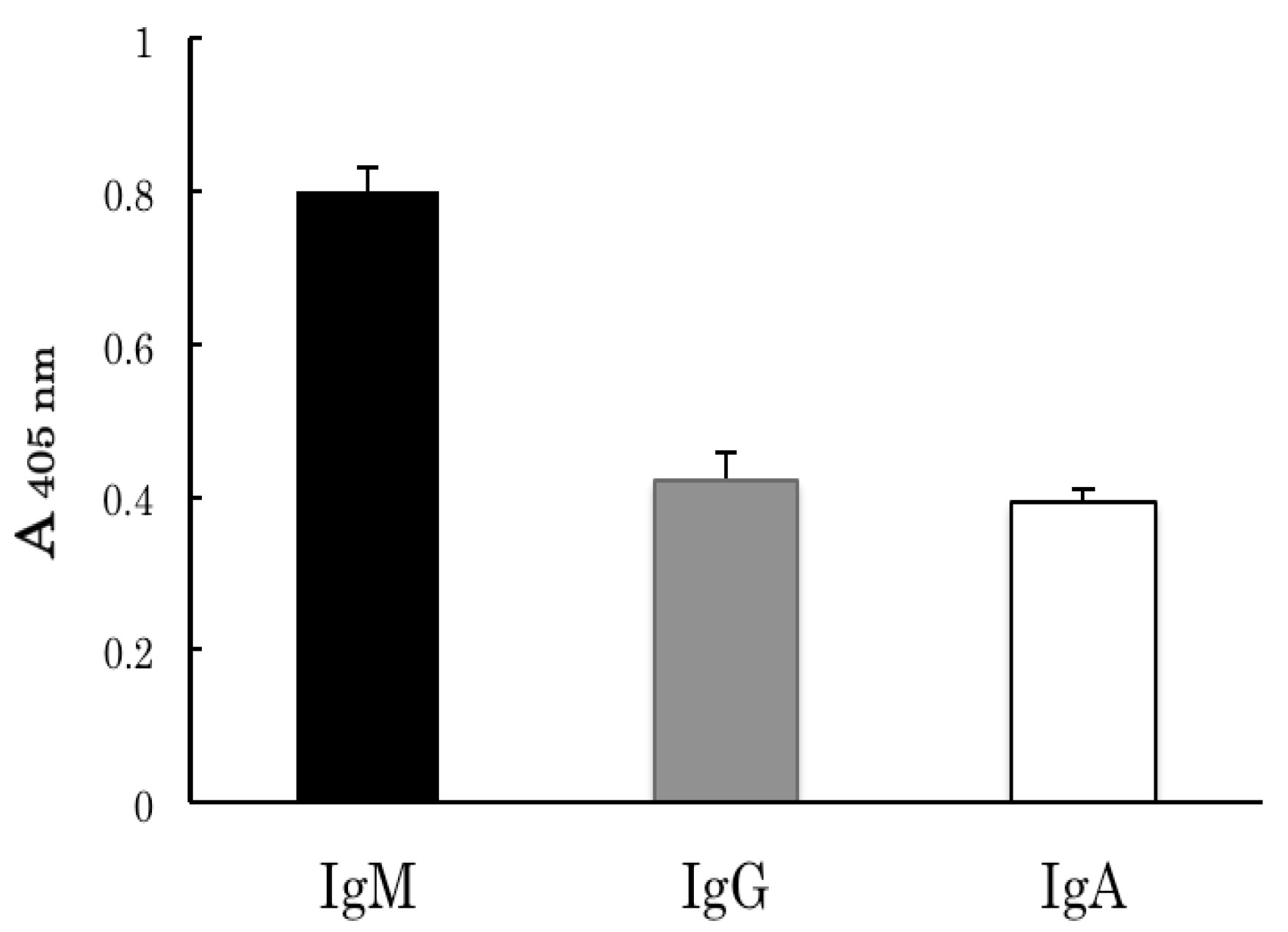
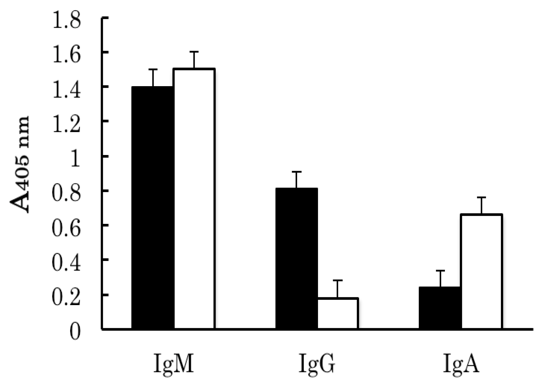
2.2. Binding Analyses of IgM Antibody Binding Ferritin

Human anti-ferritin autoantibodies (IgM, IgG and IgA) recognized different species of ferritin and both human subunits: H and L. Peptide fragment (DPHLCDF) was synthesized based on the combination of ferritin epitope analyses [33] and sequence information found commonly in amino acid sequences of H and L subunits [4]. This peptide fragment inhibited the binding between canine liver ferritin and IgM antibody binding ferritin (Supplementary Figure S1). A much smaller proportion of human serum zinc-binding proteins which bound to Zn2+ immobilized beads recognized biotinylated peptide fragment (Supplementary Figure S2). Although NHS-biotin was prepared by the same method in the absence of peptide, no reaction was detected with NHS-biotin (data not shown). Additionally, canine liver ferritin was incubated with Zn2+ immobilized beads which bound human serum zinc-binding proteins, and thereafter immunoprecitated. As shown in Figure 3, IgM antibody was detected in the immunoprecitate coated on the plate. The IgM antibodies were not released in the absence of ferritin and control beads did not show strong reaction in the presence or absence of ferritin. This result suggests that canine liver ferritin released zinc-binding protein, IgM antibody, from Zn2+ immobilized beads and bound the released protein. The biotinylated peptide (DPHLCDF) bound the released proteins as coated the immunoprecipitate on the plate well (Figure 4). This reaction was not detected in the absence of the ferritin (data not shown), and control beads did not show strong reaction in the presence or absence of ferritin.
Figure 3. Competitive binding of IgM antibody in the binding of zinc ions to ferritin. After removing serum components from non-specifically binding control beads (Sepharose 4B) from human serum as described in “Experimental Section”, zinc ion immobilized on beads (20 µL) was added to the human serum samples, and the mixture was incubated with rotation. After rotation, the mixture was incubated in the presence (+) and the absence (−) of canine liver ferritin (5 µg), and the mixture was centrifuged at 16,000 × g for 7 min. Rabbit anti-feline liver ferritin antiserum was added to resultant supernatant. The detection of IgM antibody was performed as described in Figure 1 with the modification to use 1 mL resuspension volume in place of 2.8 mL. Control values represent the absorbance in the presence of ferritin without any beads, and the control value was subtracted from the measured ferritin-binding activity values. Each bar and error bar represents the mean ± SD of four determinations. a: p < 0.01 compared to the binding activity in the absence of ferritin in the control beads; b: p < 0.01 compared to the binding activity in the presence of ferritin in the control beads; c: p < 0.01 compared to the binding activity in the absence of ferritin in zinc ion immobilized beads.
Antibodies 03 00169 g003
Figure 4. Binding of the zinc-binding protein released from Zn2+ immobilized beads by canine liver ferritin with biotinylated peptide (DPHLCDF). After removing serum components from human serum with non-specifically binding control beads (Sepharose 4B) as described in “Experimental Section”, zinc ion immobilized on beads (20 µL) was added to the human serum samples followed by the addition of canine liver ferritin (5 µg). After incubation, the mixture was centrifuged at 16,000 × g for 7 min, and rabbit anti-feline liver ferritin antiserum was added to the resultant supernatant. Immunocoprecipitate was coated on plate wells as described in Figure 3. After coating and washing, 100 µL of biotinylated peptide (DPHLCDF) was added to the wells (1.5 µg/well), and the plate was incubated at 37 °C for 1.5 h. After the incubation, 100 µL of ALP-labeled avidin was added to each well (0.1 µg/well). The control value was the absorbance in the presence of ferritin without any beads, and the control value was subtracted from the measured values for peptide-binding activity. Each bar and error bar represents the mean ± SD of four determinations. a: p < 0.01 compared to the binding activity in the absence of biotinylated peptide in the control beads; b: p < 0.01 compared to the binding activity in the presence of biotinylated peptide in the control beads; c: p < 0.01 compared to the binding activity in the absence of biotinylated peptide in zinc ion immobilized beads.
Antibodies 03 00169 g004
We proposed that anti-ferritin autoantibody is a common FBP in mammals [4]. In this study, anti-ferritin autoantibodies in human serum were identified as an FBP as suggested by Bellotti et al. [26]. On the other hand, Covell et al. [29] did not show the binding of immunoglobulins and ferritin, and the reasons for not finding the complex formation between immunoglobulins and ferritin remain to be determined, although anti-human IgG and IgM antibodies used for immunocoprecipitation may compete at the binding site in the binding between ferritin and anti-ferritin autoantibodies. Immunocoprecipation method was used to detect anti-ferritin autoantibody in feline and bovine due to lower affinity to immunoprecipitate ferritin [18,20] provided that anti-ferritin antibody for immunocoprecipitation is unlikely to compete the binding site with autoantibody to ferritin. Ferritin is a conservative protein [1,4]. The heterlogus ELISA system has been developed for measurement of canine ferritin with antibody to rat liver ferritin [21], and the combination of canine liver ferritin and anti-bovine spleen antiserum were used to immunocoprecitate feline anti-ferritin antibody [20]. In this study, human anti-ferritin autoantibodies did not precipitate additional ferritin due to a lower affinity to ferritin (data not shown) consisting with findings in other mammals such as feline [20] and horse [18]. Human anti-ferritin antibodies recognized canine liver ferritin, and these complexes were immunocoprecipitaed with anti-feline liver ferritin antibody. Additionally, this study suggests that human anti-ferritin antibodies are present in larger amount than circulating ferritin because of its low concentration [7]. Many proteins including albumin, alpha-2-macroglobulin, ceruloplasmin, transferrin, immunoglobulin, complement C4 and C-reactive protein have been identified as human serum zinc-binding proteins [30,31]. This study demonstrates human anti-ferritin autoantibodies (IgM, IgG and IgA) were also detected in the fractions eluted from immobilized metal affinity columns with Zn2+ on which human serum had been applied. Human γ-globulin (probably IgG) showed zinc binding and these binding sites were occupied with the increase in bound metal content [34]. Although the amounts of zinc-binding immunoglobulins were not elucidated, this study demonstrates that all classes of immunoglobulins (IgM, IgG and IgA) tested have ferritin-binding activities as well as zinc-binding activities. After forming complexes between FBPs and canine liver ferritin, the complexes were immunocuprecipitated by the addition of anti-ferritin antibody. Human anti-ferritin autoantibodies did not precipitate additional ferritin due to a lower affinity to ferritin (data not shown) consistent with findings in other mammals such as feline [20] and horse [17]. Human H and L subunits were found to be immunologically different [6]. To characterize the immunological properties of human anti-ferritin autoantibodies, human ferritin H and L subunit homopolymers were expressed in E. coli. Human anti-ferritin autoantibodies (IgM, IgG and IgA) recognized both of human ferritin H and L subunit homopolymers. IgM class antibody was detected with the highest binding activity for H and L subunit homopolymers compared to other IgG and IgA classes. All classes of antibodies tested showed differences in the specific binding activity in the cross-reaction of H and L subunits although rabbit IgG cross-reacted with antibody to the human IgG (Fc) fragment. Although the differences in ferritin-binding activities among immunoglobulin classes remain to be clarified, experiments were performed in this study to detect IgM antibody binding ferritin.
Human anti-ferritin autoantibodies (IgM, IgG and IgA) showed binding with canine liver ferritin (L/H subunit ratio: 2.3 [35]) and both human H and L subunits. Anti-human liver ferritin antibody and anti-human ferritin H subunit antibody were shown to form complex formation with L and H subunit homopolymers, respectively. Human liver ferritin is composed of 90% of the L subunit and 10% of the H subunit [2], and rabbit antibody to human liver ferritin appears to be specific for the L subunit. Peptide fragment (130–182, excluding N-terminal Met) synthesized from human H subunit sequence is used for antigen in the production of antibody specific for the H subunit. These antibodies are unlikely to inhibit immunocoprecipitation, and human anti-ferritin autoantibodies seem to bind ferritin over other species. In this study, a peptide fragment synthesized was common amino acid sequence (DPHLCDF: 126–132, excluding N-terminal Met) from sequence data of mammalian ferritin subunits, and expected to be the antigenic site [19,33]. This fragment significantly inhibited binding between canine liver ferritin and IgM antibody binding ferritin (Supplementary Figure S1).
To elucidate the binding mechanism of IgM antibody with ferritin, canine liver ferritin was added to a solution containing Zn2+-immobilized beads that bound serum Zn2+-binding proteins beforehand followed by immunoprecipitation with anti-ferritin antibody. IgM antibody was detected as coating the immunoprecipitate on the plate well, suggesting that ferritin releases IgM antibody that binds Zn2+-immobilized beads, and that the IgM antibody has higher affinity to ferritin than Zn2+. After binding human serum zinc-binding proteins to Zn2+-immobilized beads, a portion of, but not all, Zn2+-binding proteins recognized biotinylated peptide fragment (DPHLCDF) (Supplementary Figure S2). Additionally, the released human serum zinc-binding proteins including IgM antibody by ferritin also bound biotinylated peptide fragment (DPHLCDF) (Figure 4). Canine anti-ferritin antibody (IgM) more strongly recognized the H subunit than the L subunit and did not bind this sequence peptide [19]. Human anti-ferritin autoantibodies, such as IgM class antibody, may recognize ferritin from different species than canine anti-ferritin antibody. Although whether the binding site of IgM binding ferritin is the same binding site as for zinc ions remains to be clarified, a common sequence in mammalian ferritin subunits is likely to be involved in binding ferritin and zinc. On the other hand, even after binding ferritin, pentameric IgM antibody may still have zinc-binding ability [35]. Further study needs to elucidate other FBPs, for example alpha-2-macroglobulin, as zinc-binding proteins.
Zinc is an essential element for multiple beneficial functions, including immunity, oxidative stress and chronic inflammation [36]. In mammals, ferritin is removed from circulation via receptor-mediated uptake by the reticuloendothelial system (RES) after complex formation between circulating ferritin and anti-ferritin autoantibody [15,21]. Ferritin iron causes oxidative stress in RES cells; however, zinc ions coupled with antibody may relieve oxidative stress.
Naturally occurring autoantibodies are no longer considered to be an immunological curiosity [37]. In fact, naturally occurring autoantibodies appear to play a beneficial role for functional potency in homeostasis and regulation despite low concentrations and affinity [37]. Most of ferritin in human blood seems to bind with anti-ferritin autoantibodies as already described. Glycosylated ferritin is found in human blood, and it immunologically is similar with L subunit [38] and more slowly cleared than tissue ferritin [39]. Further study needs to elucidate physiological role in the complex formation of circulating ferritin with FBPs including anti-ferritin autoantibody. This study provides preliminary data on autoantibodies (IgG, IgM and IgA) binding to zinc ion as well as ferritin, and provides new insights into iron and zinc metabolism in human circulation.

3. Experimental

3.1. Chemicals

Human serum was purchased from Uniglobe Research Corp. (Reseda, CA, USA). Chelating Sepharose Fast Flow, Sepharose 4B and Vivaspin 6 centrifugal concentrator (MWCO: 100,000) were purchased from GE Healthcare (Milwaukee, WI, USA). Alkaline phosphatase (ALP)-labeled rabbit antibodies for goat IgG were purchased from Millipore Corp. (Temecula, CA, USA). Antibodies specific for the IgG Fc fragment and the heavy chain of IgM or IgA of human immunoglobulins, ALP-conjugated NeutrAvidin, Coomassie Plus-The Better Bradford Assay kit and EZ-linkTM sulfo-NHS-biotin were purchased from Thermo Fisher Scientific (Waltham, MA, USA). Rabbit anti-human liver ferritin and ferritin H chain antibodies were purchased from Dako (Carpinteria, CA, USA) and Santa Cruz Biotechnology, Inc. (Santa Cruz, CA, USA), respectively. A dialysis tube (Spectra/Pore MWCO: 100,000, 10 mm diameter) was purchased from Funakoshi Co., Ltd. (Tokyo, Japan). Competent Escherichia coli BL21(DE3) cells were purchased from BioDynamics Laboratory Inc. (Tokyo, Japan). Isopropyl-β-d-galctopyronoside (IPTG) was purchased from Biolynks Co., Ltd. (Yokohama, Kanagawa Prefecture, Japan). Immuno Plate Maxisorp F96 and assay microtiter plates were purchased from Nunc (Roskilde, Denmark) and Iwaki Brand Div., Asahi Techno Glass (Funabashi, Chiba Prefecture, Japan), respectively. Other reagents were of the highest grade available.

3.2. Preparation of Ferritin and Antiserum to Ferritin

Canine and feline liver ferritin monomers were purified from portions of frozen canine and feline livers, respectively, as described previously [20]. Rabbits were immunized with feline liver ferritin according to a previously described immunization protocol [28] to obtain antisera to feline liver ferritin.

3.3. Preparation of Recombinant Human H and L Subunit Homopolymers

Human ferritin H and L subunit homopolymers were expressed in the host E. coli strain BL21 (DE3) transfected with pET21C vector with human ferritin H or L subunit coding region inserts [40], The expressed ferritin was purified following incubation with 1 mM IPTG. Bacteria cells were precipitated, resuspended with phosphate buffered saline (PBS: 20 mM sodium phosphate, 150 mM NaCl, pH 7.2), and frozen at −20 °C. After freezing, the bacteria suspension was thawed, and subjected to repeated freezing and thawing. After several freezing and thawing cycles, the thawed bacterial suspension was centrifuged at 27,000 × g for 20 min at 4 °C, and the resultant supernatant was dialyzed against PBS with a dialysis tube (MWCO: 100,000). After dialysis, the solution in the dialysis tube was centrifuged at 27,000 × g for 20 min at 4 °C, and resultant supernatant was concentrated with a Vivaspin centrifugal concentrator (MWCO: 100,000). The concentrated solution was applied to Sepharose CL-6B column chromatography (2 × 100 cm), which was equilibrated with PBS at a flow rate of 12.5 mL/h. The fractions (3 mL) were collected, and fractions containing ferritin monomer were pooled and concentrated with the above concentrator. The purity of ferritin homopolymers was examined by sodium dodecyl-polyacrylamide gel electrophoresis as described previously [41].

3.4. Protein Determination

Protein concentrations were determined by Coomassie Plus-The Better Bradford Assay kit following the microplate protocol and using BSA as the standard.

3.5. Biotin Labeling of Peptide Fragment

Synthetized peptide fragment (DPHLCDF) was obtained from Thermo Fisher Scientific GmbH. (Einsteinstrasse, Ulm, Germany). Biotin-labeling of peptide fragment was performed by EZ-linkTM sulfo-NHS-biotin by reacting a 1:1 mole ratio of peptide fragment to sulfo-NHS-biotin according to manufacturer instructions.

3.6. Partial Purification of Human Serum FBPs

Commercial human serum (5 mL) was diluted 5-fold with 80 mM Tris/HCl (pH 7.5) and dialyzed with the same buffer in a dialysis tube (MWCO: 100,000). The dialyzed solution was applied to a DE-52 column (1 × 5 cm), equilibrated with the same buffer and washed with the same buffer until the absorbance of the efluent at 280 nm was below 0.01. The protein fractions were eluted from the column in 3-mL fractions with 300 mM Tris/HCl (pH 7.5), and the eluted protein fractions were dialyzed against 20 mM sodium phosphate buffer containing 0.5 M NaCl (PBHS), and thereafter applied to a Zn2+ immobilized Sepharose column (1 × 5 cm) prepared using Chelating Sepharose Fast Flow according to manufacturer instructions. The protein fractions were eluted with 50 mM citrate buffer (pH 4.2), dialyzed against PBS and used as partially purified FBPs.

3.7. Identification of Human FBPs Using Immunocoprecipitation

To immunocoprecipitate and identify human serum FBPs, canine liver ferritin and the expressed recombinant human ferritin H subunit and L subunit homopolymers were used. After complex formation between FBPs and ferritin, immunocoprecipitations of canine liver ferritin, the expressed recombinant human ferritin L subunit and H subunit homopolemers, respectively (Supplementary Figure S3), were conducted with rabbit anti-serum to feline liver ferritin, rabbit antibody to human liver ferritin and human ferritin H subunit, respectively. Briefly, 1 mL of PBS containing partially purified human FBPs and ferritin (5 µg each) was incubated overnight at 4 °C with rotation was followed by the addition of the corresponding antibody (10 or 15 µL) to the ferritin sample. After overnight incubation, the mixture was centrifuged at 16,000 × g for 7 min. The resulting pellet was suspended in 1 mL of PBHS and washed three times in 1 mL of PBHS by centrifugation with the same conditions as described above. Finally, the resultant pellet was resuspended in 2.8 mL of PBS and the suspension was aliquoted (100 µL per well) to a microtiter plate. The plate was incubated overnight at 4 °C and the wells were washed with PBS containing 0.05% Tween 20 (PBST). The plate wells were washed three times with PBST after every step. After washing, 300 µL of PBS containing 0.1% gelatin and 0.1% Tween 20 (ELISA buffer) was added to each well and blocked with gelatin for 1 h at room temperature to prevent non-specific binding. After washing, 100 µL of goat antibody specific for human IgG (Fc) fragment, IgM heavy chain or IgA heavy chain, was diluted 1,000-fold with ELISA buffer, aliquoted to the wells of a plate and incubated for 1.5 h at 37 °C. After incubation, the plate was washed as described above, and 100 µL of ALP-labeled rabbit anti-goat IgG antibody diluted 1,000-fold with ELISA buffer was aliquoted to each well of the plate. The plate was incubated for 1.5 h at 37 °C, and the plate thereafter was washed as described above followed by the ALP reaction using disodium p-nitrophenyl phosphate as described previously [28].

3.8. Competitive Inhibition of Zinc Binding by Human Serum Zinc-Binding Proteins by Ferritin

To remove serum proteins that bind nonspecifically agarose, 40 µL of 50% (v/v) Sepharose 4B in PBS was added to 1 mL of PBS containing 100 µL of human serum (net volume of beads per sample: 20 µL), and was rotated overnight at 4 °C. After incubation overnight, the mixture was centrifuged at 16,000 × g for 7 min, and 20 µL of Sepharose 4B beads was again added to the resulting supernatant. After overnight incubation, the mixture was centrifuged at 16,000 × g for 15 min, and 20 µL of Zn2+ immobilized Sepharose beads were added to the resulting supernatant. After overnight incubation, the mixture was centrifuged at 16,000 × g for 7 min, and the resultant pellet was washed three times with 1 mL of PBHS as described above. The resulting pellet was resuspended in 1 mL of PBS, and canine liver ferritin (5 µg) was added to the suspension. The mixture was rotated overnight at 4 °C, and thereafter the mixture was centrifuged at 16,000 × g for 7 min. Rabbit antiserum to feline liver ferritin (10 µL) was added to the resultant supernatant to immunoprecipitate the ferritin added. After incubation overnight, the mixture was centrifuged at 16,000 × g for 7 min, and the resulting pellet was washed three times with 1 mL of PBHS by centrifugation as described above. The resulting pellet was resuspended in 1 mL of PBS, and 100 µL of the suspension was aliquoted to each well of a microtiter plate. The plate was incubated overnight at 4 °C and the wells were washed with PBST. After washing, detecting of IgM bound to the wells were performed with goat antibody specific for human IgM heavy chain followed by the incubation with ALP-labeled rabbit anti-goat IgG antibody as described above. The reaction with ALP was also carried out as described above.
To elucidate the ferritin-binding site, 100 µL of 15 µg/mL of biotinylated peptide fragment (DPHLCDF) in ELISA buffer was aliquoted to each well of the plate after coating the immunoprecipitated sample as described above. After incubation for 1.5 h at 37 °C, the plate was washed as described above, and 100 µL of 1 µg/mL ALP-conjugated NeutrAvidin was added to the wells, and the plate was incubated for 1.5 h at 37 °C. After incubation, the plate was washed, and the ALP reaction was performed as described above. Reactions were conducted in parallel with Sepharose 4B beads as a control for Zn2+ immobilized Sepharose beads.

3.9. Statistical Analysis

All data are expressed as the mean ± SD of four measurements. Student’s t-test was used to compare the means of the two groups. Multiple comparisons were analyzed by one-way analysis of variance followed by Tukey’s test. p < 0.01 was considered statistically significant.

4. Conclusions

In conclusions, human FBPs were partially purified from human serum by ion-exchange chromatography and immobilized metal affinity chromatography with Zn2+. Crude FBPs were found to contain anti-ferritin autoantibodies (IgG, IgM abd IgA) which bound to canine liver ferritin and expressed recombinant human H and L chain homopolymers. A portion of human serum proteins bound to zinc ions immobilized on beads were released by canine liver ferritin, and the released protein was identified as IgM antibody. The released proteins recognized peptide sequence (DPHLCDF) commonly found in amino acid sequences of mammalian ferritin subunits. These results suggest that human anti-ferritin autoantibodies (IgG, IgM and IgA) binds ferritin over species, and that a portion of, but not all, the IgM antibodies bind more strongly to ferritin than to zinc ions and recognize the common sequence found in mammalian ferritin subunits.

Supplemental Files

Supplementary materials can be accessed at https://www.mdpi.com/2073-4468/3/1/169/s1.

Supplementary Files

Supplementary File 1

Acknowledgements

The authors are grateful to Torti (Department of Medicine, University of Connecticut Health Center, Farmington, CT 06030) for providing plasmid vector pET21C with human ferritin H or L subunit coding region inserts.

Author Contributions

Conceived ad designed the experiments: S.H., K.N., Y.Y., K.W., K.O. Performed experiments: S.H., K.N., K.O. Analyzed data: S.H., K.N., K.W., K.O. Contributed reagents/materials/analysis tools: Y.Y., K.W., K.O. Wrote the paper: S.H. All authors read and approved the final manuscript.

Conflicts of Interest

The authors declare no conflict of interest.

References

  1. Andrews, S.C.; Arosio, P.; Bottke, W.; Briat, J.F.; von Darl, M.; Harrison, P.M.; Laulhere, J.P.; Levi, S.; Lobreaux, S.; Yewdall, S.J. Structure, function, and evolution of ferritins. J. Inorg. Biochem. 1992, 47, 161–174. [Google Scholar] [CrossRef]
  2. Arosio, P.; Adelman, T.G.; Drysdale, J.W. On ferritin heterogeneity. Further evidence for heteropolymers. J. Biol. Chem. 1978, 253, 4451–4458. [Google Scholar]
  3. Harrison, P.M.; Arosio, P. The ferritins: molecular properties, iron storage function and cellular regulation. Biochim. Biophys. Acta 1996, 1275, 161–203. [Google Scholar] [CrossRef]
  4. Orino, K.; Watanabe, K. Molecular, physiological and clinical aspects of the iron storage protein ferritin. Vet. J. 2008, 178, 191–201. [Google Scholar] [CrossRef]
  5. Theil, E.C. Ferritin: structure, gene regulation, and cellular function in animals, plants, and microorganisms. Annu. Rev. Biochem. 1987, 56, 289–315. [Google Scholar] [CrossRef]
  6. Luzzago, A.; Arosio, P.; Iacobello, C.; Ruggeri, G.; Capucci, L.; Brocchi, E.; de Simone, F.; Gamba, D.; Gabri, E.; Levi, S.; Albertini, A. Immunochemical characterization of human liver and heart ferritins with monoclonal antibodies. Biochim. Biophys. Acta 1986, 872, 61–71. [Google Scholar] [CrossRef]
  7. Addison, G.M.; Beamish, M.R.; Hales, C.N.; Hodgkins, M.; Jacobs, A.; Llewellin, P. An immunoradiometric assay for ferritin in the serum of normal subjects and patients with iron deficiency and iron overload. J. Clin. Pathol. 1972, 25, 326–329. [Google Scholar] [CrossRef]
  8. Andrews, G.A.; Chavey, P.S.; Smith, J.E. Enzyme-linked immunosorbent assay to measure serum ferritin and the relationship between serum ferritin and nonheme iron stores in cats. Vet. Pathol. 1994, 31, 674–678. [Google Scholar] [CrossRef]
  9. Andrews, G.A.; Smith, J.E.; Gray, M.; Chavey, P.S.; Weeks, B.R. An improved ferritin assay for canine sera. Vet. Clin. Pathol. 1992, 21, 57–60. [Google Scholar] [CrossRef]
  10. Miyata, Y.; Furugouri, K. The relationship between serum ferritin concentrations and tissue non-heme iron or tissue ferritin in dairy cattle. Jpn. J. Vet. Sci. 1987, 49, 1157–1159. [Google Scholar] [CrossRef]
  11. Smith, J.E.; Moore, K.; Boyington, D.; Pollmann, D.S.; Schoneweis, D. Serum ferritin and total iron-binding capacity to estate iron storage in pigs. Vet. Pathol. 1984, 21, 597–600. [Google Scholar]
  12. Smith, J.E.; Moore, K.; Cipriano, J.E.; Morris, P.G. Serum ferritin as a measure of stored iron in horses. J. Nutr. 1984, 114, 677–681. [Google Scholar]
  13. Walters, G.O.; Miller, F.M.; Worwood, M. Serum ferritin concentration and iron stores in normal subjects. J. Clin. Pathol. 1973, 26, 770–772. [Google Scholar] [CrossRef]
  14. Torti, S.V.; Torti, F.M. Human H-kininogen is a ferritin-binding protein. J. Biol. Chem. 1998, 273, 13630–13635. [Google Scholar] [CrossRef]
  15. Massover, W.H. α2-Macroglobulin: a ferritin-binding protein. Ann. N. Y. Acad. Sci. 1994, 737, 468–471. [Google Scholar] [CrossRef]
  16. Santambrogio, P.; Massover, W.H. Rabbit serum alpha-2-macroglobulin binds to liver ferritin: association causes a heterogeneity of ferritin molecules. Br. J. Haematol. 1989, 71, 281–290. [Google Scholar] [CrossRef]
  17. Orino, K.; Hamada, S.; Hashimoto, M.; Nambo, Y.; Kondo, T.; Watanabe, K. Identification of horse anti-ferritin autoantibody. J. Equine Sci. 2006, 17, 113–119. [Google Scholar] [CrossRef]
  18. Orino, K.; Ishiji, T.; Yamamoto, S.; Watanabe, K. Characterization of bovine serum ferritin-binding proteins. Comp. Biochem. Physiol. 2004, 137A, 375–381. [Google Scholar]
  19. Orino, K.; Uehara, M; Okano, S.; Watanabe, K. Purification and characterization of canine serum ferritin-binding proteins. Biometals 2006, 19, 315–322. [Google Scholar] [CrossRef]
  20. Sakamoto, H.; Kuboi, T.; Nagakura, T.; Hayashi, S.; Hoshi, F.; Mutoh, K.; Watanabe, K.; Orino, K. Characterization of feline serum ferritin-binding proteins: the presence of a novel ferritin-binding protein as an inhibitory factor in feline ferritin immunoassay. Biometals 2009, 22, 793–802. [Google Scholar] [CrossRef]
  21. Watanabe, K.; Hayashi, K.; Miyamaoto, T.; Tanaka, M.; Okano, S.; Yamamoto, S. Characterization of ferritin and ferritin-binding proteins in canine serum. Biometals 2000, 13, 57–63. [Google Scholar] [CrossRef]
  22. Seki, T.; Kunichika, T.; Watanabe, K.; Orino, K. Apolipoprotein B binds ferritin by hemin-mediated binding: evidence of direct binding of apolipoprotein B and ferritin to hemin. Biometals 2008, 21, 61–69. [Google Scholar] [CrossRef]
  23. Orino, K.; Yamamoto, S.; Watanabe, K. Fibrinogen as a ferritin-binding protein in horse plasma. J. Vet. Med. Sci. 1993, 55, 785–787. [Google Scholar] [CrossRef]
  24. Parthasarathy, N.; Torti, S.V.; Torti, F.M. Ferritin binds to light chain of human H-kininogen and inhibits kallikrein-mediated bradykinin release. Biochem. J. 2002, 365, 279–286. [Google Scholar] [CrossRef]
  25. Takahashi, K.; Kondo, T.; Yoshikawa, Y.; Watanabe, K.; Orino, K. The presence of heat-labile factors interfering with binding analysis of fibrinogen with ferritin in horse plasma. Acta Vet. Scand. 2013, 55, 70–74. [Google Scholar] [CrossRef]
  26. Bellotti, V.; Arosio, P.; Cazzola, M.; Cozzi, A.; Levi, S.; Meloni, F.; Zappone, E. Characterization of a ferritin-binding protein present in human serum. Br. J. Haematol. 1987, 65, 489–493. [Google Scholar] [CrossRef]
  27. Niitsu, Y.; Goto, Y.; Arisato, N.; Sasaki, K.; Kohgo, Y. Ferritin-binding proteins in serum. Br. J. Haematol. 1988, 68, 496–497. [Google Scholar] [CrossRef]
  28. Orino, K.; Saji, M.; Ozaki, M.; Ohya, T.; Yamamoto, S.; Watanabe, K. Inhibitory effects of horse serum on immunoassay of horse ferritin. J. Vet. Med. Sci. 1993, 55, 45–49. [Google Scholar] [CrossRef]
  29. Covell, A.M.; Jacobs, A.; Worwood, M. Interaction of ferritin with serum: implications for ferritin turnover. Clin. Chim. Acta 1984, 139, 75–84. [Google Scholar] [CrossRef]
  30. Chesters, J.K.; Will, M. Zinc transport proteins in plasma. Br. J. Nutr. 1981, 46, 111–118. [Google Scholar] [CrossRef]
  31. Scot, B.J.; Bradwell, A.R. Identification of the serum binding proteins for iron, zinc, cadmium, nickel and calcium. Clin. Chem. 1983, 29, 629–633. [Google Scholar]
  32. Arredondo, A.; Nunez, M.T. Iron and copper metabolism. Mol. Aspects Med. 2005, 26, 313–327. [Google Scholar] [CrossRef]
  33. Addison, J.M.; Treffry, A.; Harrison, P.M. The location of antigenic sites on ferritin molecules. FEBS Lett. 1984, 175, 333–336. [Google Scholar] [CrossRef]
  34. Babaeva, E.E.; Vorobyova, U.A.; Denisova, E.A.; Medvedeva, D.A.; Cheknev, S.B. Binding of zinc cations to human serum γ-globulin. Bull. Exp. Biol. Med. 2006, 141, 602–605. [Google Scholar] [CrossRef]
  35. Watanabe, K.; Muranishi, N.; Murata, Y.; Orino, K.; Okano, S.; Yamamoto, S. Biochemical properties of canine serum ferritin: iron content and nonbinding to concanavalin A. Biometals 2000, 13, 319–324. [Google Scholar] [CrossRef]
  36. Prasad, A.S. Zinc: role in immunity, oxidative stress and chronic inflammation. Cur. Opin. Clin. Nutr. Metab. Care 2009, 12, 646–652. [Google Scholar] [CrossRef]
  37. Lutz, H.U.; Binder, C.J.; Kaveri, S. Naturally occurring auto-antibodies in homeostasis and disease. Trends Immunol. 2008, 30, 43–51. [Google Scholar] [CrossRef]
  38. Santambrogio, P.; Cozzi, A.; Levi, S.; Arosio, P. Human serum ferritin G-subunit is recognized by anti-L ferritin subunit antibodies and concanavalin-A. Br. J. Haematol. 1987, 65, 235–237. [Google Scholar] [CrossRef]
  39. Worwood, M.; Cragg, S.J.; Williams, A.M.; Wagstaff, M.W.; Jacobs, A. The clearance of 131I-human plasma ferritin in man. Blood 1982, 60, 827–833. [Google Scholar]
  40. Rucker, P.; Torti, F.M.; Torti, S.V. Role of H and L subunits in mouse ferritin. J. Biol. Chem. 1996, 271, 33352–33357. [Google Scholar] [CrossRef]
  41. Schägger, H.; von Jagow, G. Tricine-sodium dodecyl sulfate-polyacrylamide gel electrophoresis for the separation of protein in the rage from 1 to 100 kDa. Anal. Biochem. 1987, 166, 368–379. [Google Scholar] [CrossRef]

