Article

Development of an Antibody for Detection of Rhamnolipids Characterized as a Major Bacterial Virulence Factor

by
Dimitrios C. Giagkas
,
Theodora Choli-Papadopoulou
and
Anastasia A. Pantazaki
*
Laboratory of Biochemistry, Department of Chemistry, Aristotle University of Thessaloniki, 54124 Thessaloniki, Greece
*
Author to whom correspondence should be addressed.
Antibodies 2013, 2(3), 501-516; https://doi.org/10.3390/antib2030501
Submission received: 20 June 2013 / Revised: 4 August 2013 / Accepted: 16 August 2013 / Published: 28 August 2013

Abstract

:
Rhamnolipids (RLs), the glycolipidic biosurfactants found initially as exoproducts of the opportunistic pathogen Pseudomonas aeruginosa, are characterized as virulence factors contributing to its pathogenesis infections. However, RLs are also produced by various bacterial species. They consist of a gluconic part, usually containing one or two rhamnoses, and a lipid part, containing one or two hydroxy-fatty acids. In this study, we present both the isolation of RLs from bacterial cultures of the non-pathogenic bacterium Thermus thermophilus as well as the development of the rabbit antibody directed against them. The antibody was titrated and evaluated, in respect of its recognition selectivity. Between both RLs constituents, it specifically recognized only the hydroxydecanoic acid between the fatty acids tested, contrary to rhamnose. The potential of the antibody to recognize both purified RLs and RLs present in crude extracellular media produced by T. thermophilus and Escherichia coli cultures, is evidenced by Dot Blot immuno-reaction. The development of this antibody is addressed in detail, as the sensitive analytical technique, and its potential use would facilitate the implementation of rhamnolipids’ detection, or may be a useful and promising tool for determining these microbial secondary metabolites and virulence factors secreted in extracellular culture media or in biological fluids during infections.

1. Introduction

Rhamnolipids (RLs) are glycolipids that are produced by some bacterial species but mainly and widely spread by Pseudomonas aeruginosa [1,2]. Many microorganisms ensure their carbon and energy requirements by using hydrophobic substrates as a carbon source, which are immiscible in aqueous media. The uptake and growth on these substrates are often associated with the production of biosurfactants, which are amphipathic molecules. This provides these molecules with tensio-active properties among them to cause pseudosolibilization of insoluble substrates facilitating the penetration of these substrates to the periplasmic space. RLs production is regulated by the quorum sensing system (QS) dependent from the bacterial cell density [3,4]. RLs are implicated in bacterial virulence [5]. RLs consist of a gluconic part usually containing one or two rhamnoses and a lipidic part containing one or two hydroxy-fatty acids [5]. P. aeruginosa invades respiratory epithelia reconstituted with primary human respiratory cells due to the RLs production. The mechanism, which involves the incorporation of RLs within the host cell membrane, is followed by tight-junction alterations and the junction-dependent barrier of the respiratory epithelium is selectively altered by RLs [6]. A correlation was reported between increased levels of RLs in the bronchial epithelium of patients with cystic fibrosis with established infection of the bacterium P. aeruginosa and the worsening of the clinical condition of the patients [7]. RLs inhibit the function of the epithelial mucociliary and cause damage to the bronchus, varying the ion transport by reducing the absorption of sodium and single direction chlorine through bronchial epithelium [8].
However, many other bacteria in addition to P. aeruginosa are found to produce RLs [4,5]. Escherichia coli produces RLs in a very small amount, which is greatly increased when the bacterium is recombined with the operon rhlAB from bacterium P. aeruginosa [9]. Yet one more example is the thermophilic bacterium Thermus thermophilus HB8, which also produces RLs (TthRLs) in large amounts using sunflower seed oil, sodium gluconate or glucose as carbon source [10,11]. Bacteria Thermus sp., Thermus aquaticus and Meiothermus ruber produce RLs varying in chain lengths up to unusually long chains with 24 carbon atoms and unsaturation [12].
There are some reports in the literature concerning virulence of RLs. Heat-stable hemolysin produced by bacterium P. aeruginosa was identified as a glycolipid containing rhamnose and beta-hydroxydecanoic acid [13]. The RL Rha-Rha-C10-C10 in P. aeruginosa shows hemolytic activity in red blood cells [14,15]. RLs secreted from the saprophytic bacterium Burkholderia pseudomallei (causative agent of melioidosis, an infectious disease of humans and animals), induced cytopathic changes, which were due to a progressive reorganization of the F-actin network resulting in impaired cell cycle progression and reduced phagocytic function of macrophages [16]. This bacterium produces Rha-Rha-C14-C14 exhibiting a hemolytic activity and found that the albumin inhibits hemolytic activity of the RL [17].
Di-RLs cause hemolysis of human erythrocytes through a lytic mechanism as shown by leakage of hemoglobin and by the absence of osmotic protectants resulting in the change of the shape of erythrocytes from disc form into that of spheroechinocytes [18]. RLs induce lysis of polymorphonuclear leukocytes (PMNs) by a mechanism of action, which consists of intercalation into the biological membrane and destruction by their permeabilizing effect [19,20,21,22,23]. Saponin white (C27H42O3), a relative compound of RLs, isolated from plants has been found to have hemolytic activity in red blood cells [24]. In addition, di-RLs mode of action could involve a direct cell-receptor binding, creating complexes with serum components, binding to the membrane proteins, passing through the phospholipids bi-layer of the cell membrane and interacting with DNA transcriptional and translational machinery [25]. In human epidermal cells, RLs cause inhibition of DNA synthesis without causing chromosomal aberrations [26], and an overall loss in cellular fatty acid content due to release of LPS from the outer membrane, and that is the probable mechanism of enhanced cell surface hydrophobicity [27]. RLs apart from virulence factors act as immune modulators; they have antimicrobial activities, involving in surface motility and in bacterial biofilm development [4]. In particular, a plethora of biological activities ascribed to their detergent-like properties are referred for P. aeruginosa biosurfactants, which exhibit low irritancy and even anti-irritating effects, as well as compatibility with human skin [1]. In vivo data relate specifically to the treatment of autoimmune diseases and include the effects of di-RL on cellular immuno-suppression, oxazolone induced delayed-type hypersensitivity, immuno-modulation and immuno-restoration [15]. Lastly, the clinical trials with di-RLs conducted on the treatment of psoriasis, lichen ruber planus, neurodermatitis and human wound healing have confirmed excellent ameliorative effects of di-RL when compared with conventional therapy using corticosteroids [26].
This article reports the isolation of RLs using cultures of bacteria, a non-pathogenic T. thermophilus and a pathogenic Escherichia coli, that further were used for the production of specific antibody against RLs. The detection limit of this antibody was demonstrated by Dot Blot analysis with purified RLs from both bacteria to be as little as 1 μg of RLs. Furthermore, the recognition selectivity of this antibody against RLs components, the gluconic or the lipidic part, was evaluated as well. The antibody was also validated using as samples extracellular media from T. thermophilus and E. coli cultures containing RLs, as already previously confirmed by the quantitative colorimetric orcinol method.

2. Results and Discussion

2.1. TthRLs Production by T. thermophilus

The ability of T. thermophilus to produce RLs in MSM containing sodium gluconate (1.5% w/v) as sole carbon source was shown previously [11]. Thus, T. thermophilus were grown under conditions permissive for TthRLs production, which were then extracted from the extracellular medium and identified as previously described. These TthRLs were used as antigen for immunization.

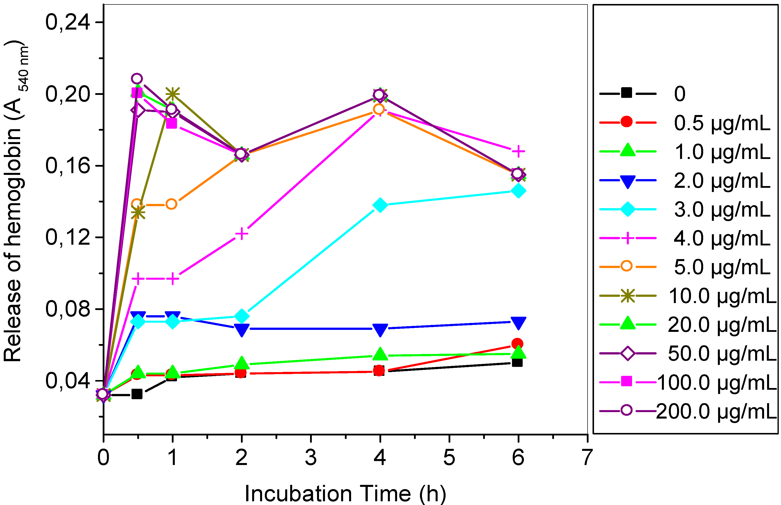
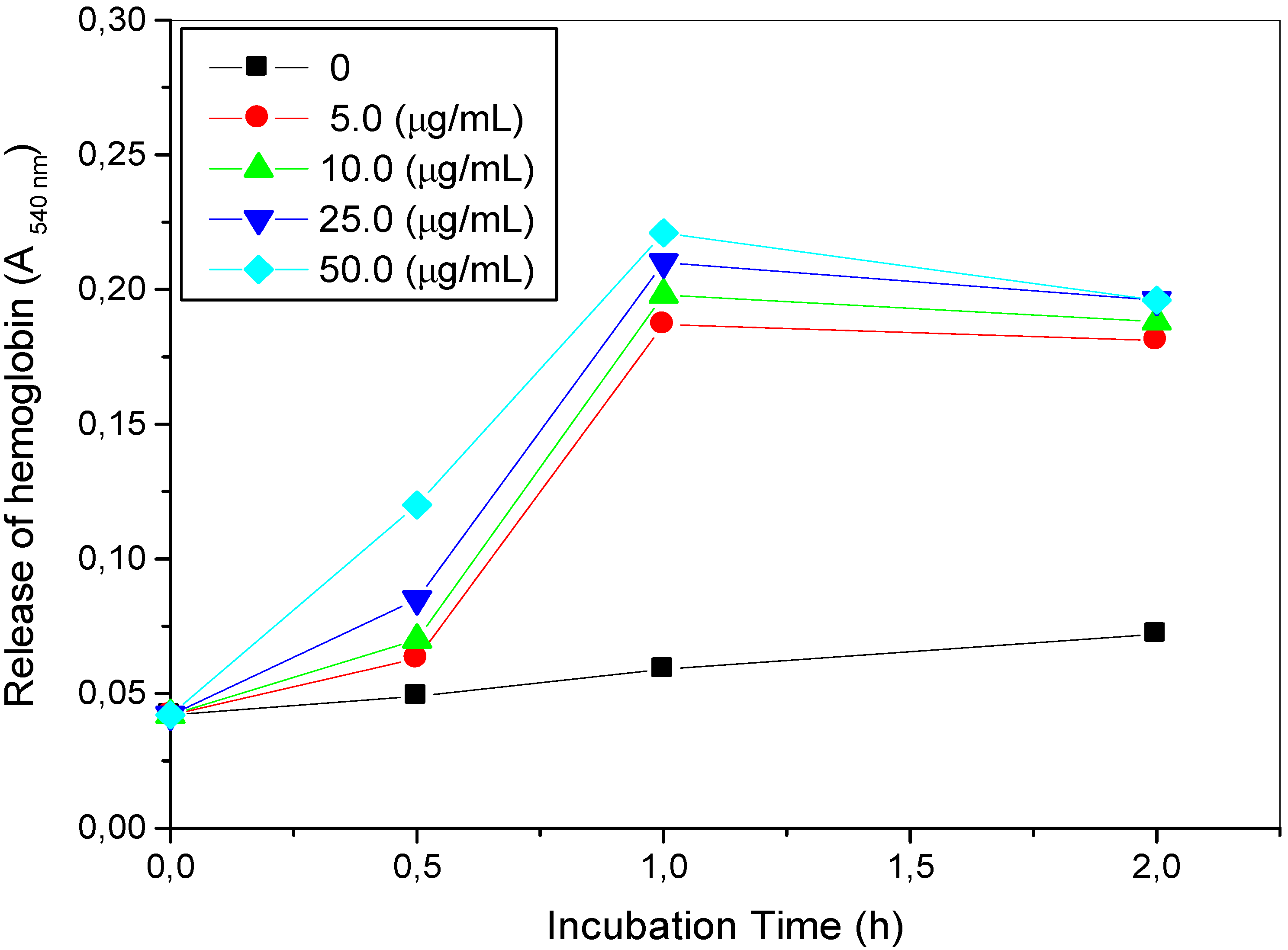
2.2. Hemolytic Reaction Caused by TthRLs or Saponin to Rabbit Erythrocytes

To prevent hemolytic damage caused by purified TthRLs to rabbit, the hemolytic activity of rabbit erythrocytes was investigated in vitro using a wide range of TthRLs concentration ranging from 0 to 200 μg/mL. The measurement of the hemoglobin released showed that a full hemolytic activity of erythrocytes occurred, since the first hour of incubation in the presence of 10 μg/mL TthRLs and more. Analytically, erythrocytes were also subjected to hemolysis, in the presence of 6 μg/mL of TthRLs in 4 h, while in the presence of 4 μg/mL of TthRLs in six hours of incubation at 37 °C. Consequently, whether the concentration of TthRLs present in the suspension of erythrocytes is exceeded the 3 μg/mL a hemolytic activity occurred, reflecting in the hemoglobin release attributed to be caused by TthRLs. This hemolytic activity is due to the property of RLs to act as biosurfactants. Whereas erythrocytes incubated with concentration of TthRLs lower than 2 μg/mL exhibited a negligible hemolysis until 6 h of incubation comparable with this observed with the negative control, without TthRLs (Figure 1).
Figure 1. Hemolysis of erythrocytes induced by purified Thermus thermophilus rhamnolipids (TthRLs) at concentration ranged from 0 to 200 μg/mL of TthRLs, which was examined during incubation time for 0 to 6 h. Hemolysis is expressed by measuring the amount of hemoglobin released as the absorbance at 540 nm of hemoglobin present in the supernatants after centrifugation.
Antibodies 02 00501 g001
The hemolytic activity of rabbit erythrocytes caused by saponin white, used as a standard, was also investigated in vitro at saponin concentrations ranging from 0 to 50 μg/mL. The measurement of the hemoglobin released showed that a full hemolytic activity of erythrocytes occurred, even from the first hour of incubation in the presence of 50 μg/mL or 25 μg/mL of saponin. This was deduced from the sharp and obvious increase in free hemoglobin, which was observed in all the saponin concentrations used, whereas then it reached a plateau (after 2 h of incubation). In conclusion, saponin white showed a rapid hemolytic activity with minimum critical concentration of 5 μg/mL in 1 h (Figure 2).
These results are consistent with the literature where RLs are bio-surfactants that destroy the cell membrane, changing the shape of erythrocytes and eventually causing hemolysis [28]. However, in vivo albumin, the main protein of the serum, might inhibit the hemolytic activity of the RL [16]. The hemolytic activity of saponins, which are mixtures of plant glycosides, depends on the structure of glycoside molecules in commercial products [29]. The concentration of saponin, which hemolyzed 50% of red blood cells estimated to be 9.3 ± 0.6 mg/L [30]. The half maximal concentration that we found with the Merck product was in the same range (11.0 ± 1 mg/L). Either the use of red blood cells obtained from buffy coats or the incubation at 0 °C in our experiments instead of 37 °C [30] may explain the slightly higher resistance.
Figure 2. Hemolysis of erythrocytes induced by saponin white at concentration ranged from 0 to 50 μg/mL of saponin, which was examined during incubation time for 0 to 2 h. Hemolysis is expressed as in Figure 1.
Antibodies 02 00501 g002

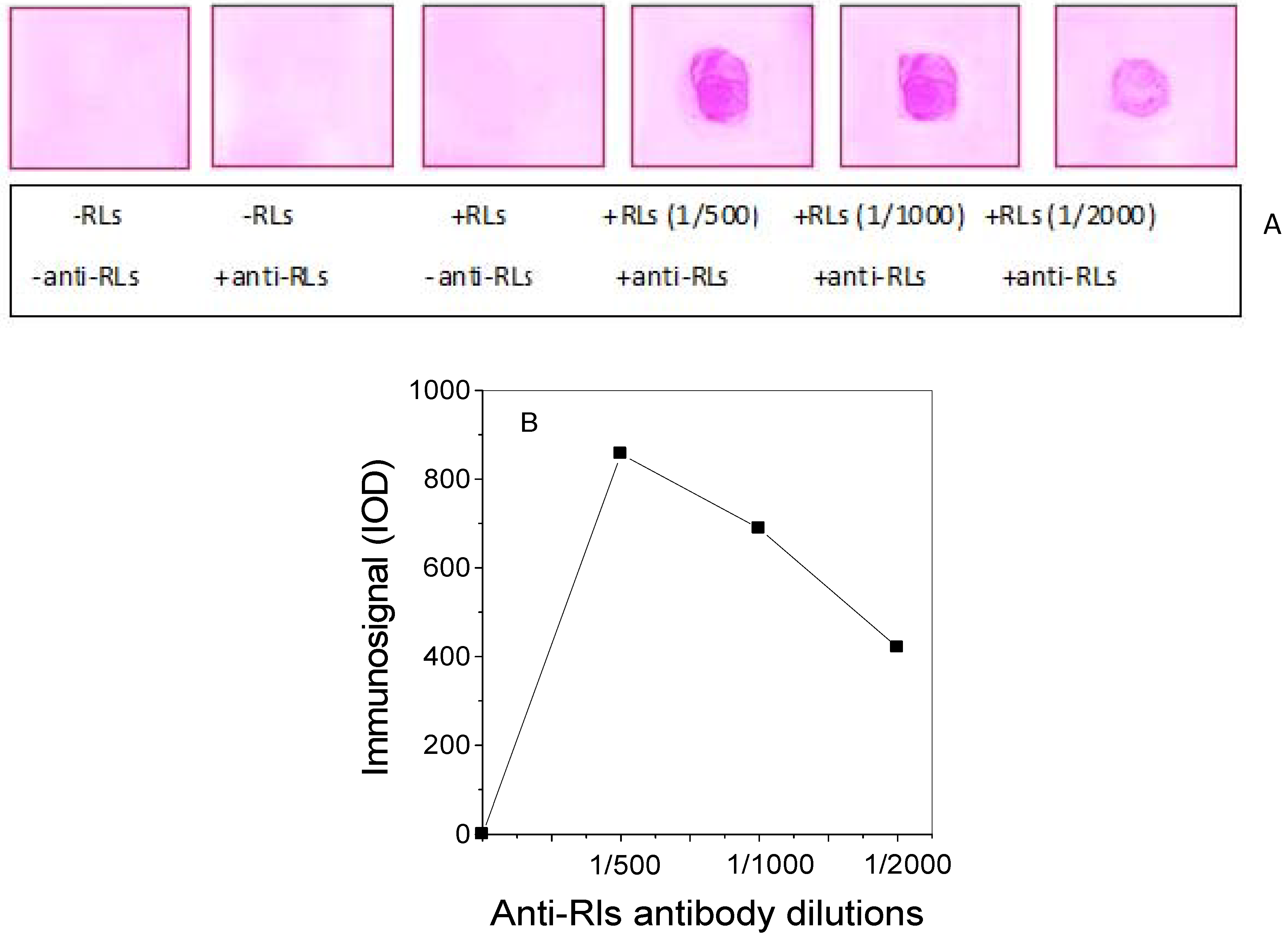
2.3. Production of TthRLs-Specific Antibody and Titration by Dot Blot Analysis

The purified TthRLs, as described previously, were used as an antigen. An amount of 50 μg of these purified TthRLs (as it was quantified by a standard curve performed with the colorimetric orcinol method) was raised by intramuscular injection into a rabbit that was used for each immunization. Dot blots were performed by spotting amounts of TthRLs (50 μg) onto nitrocellulose membrane, and immuno-blotted using three dilutions (1/500, 1/1,000 and 1/2,000) of the polyclonal antiserum which was recovered from rabbit blood, against TthRLs. Control experiments were performed in a sample containing the same amount of TthRLs without polyclonal antiserum and a sample without TthRLs and without polyclonal antiserum. Additionally, a negative control was performed with the pre-immune serum removed from the same rabbit prior to the immunization rounds. The use of this control provides assurance that the immune response is specific, otherwise it is not possible to know if this animal had a prior immune response due to the cross-reaction with RLs. Dot blot analysis showed that the signal of the RLs’s immuno-staining decreased gradually with the successive dilutions but remained still strong even at 1/2,000, suggesting that the immune-signal is dilution-dependent, as it was quantified by the image analysis by Gelpro Analyzer V.3 computer program (Figure 3B). Additionally, to address the concern about the presence of any possible trace of protein in the purified TthRLs preparation used for immunization, this TthRLs sample was monitored on SDS-PAGE and the silver nitrate-stained gel showed the complete absence of protein.
Figure 3. Immuno-dot blot assay performed by spotting TthRLs (50 μg), purified from the extracellular medium of a T. thermophilus culture in the presence of sodium gluconate as sole carbon source, and then the membranes were probed with different dilutions (1/500, 1/1,000 and 1/2,000) of rabbit polyclonal antiserum against TthRLs (A). Quantification of the intensity of immune-staining by the Gelpro Analyzer V.3 computer program was performed, and a curve of the immunosignal as function of anti-RLs antibody dilutions was designed.
Antibodies 02 00501 g003

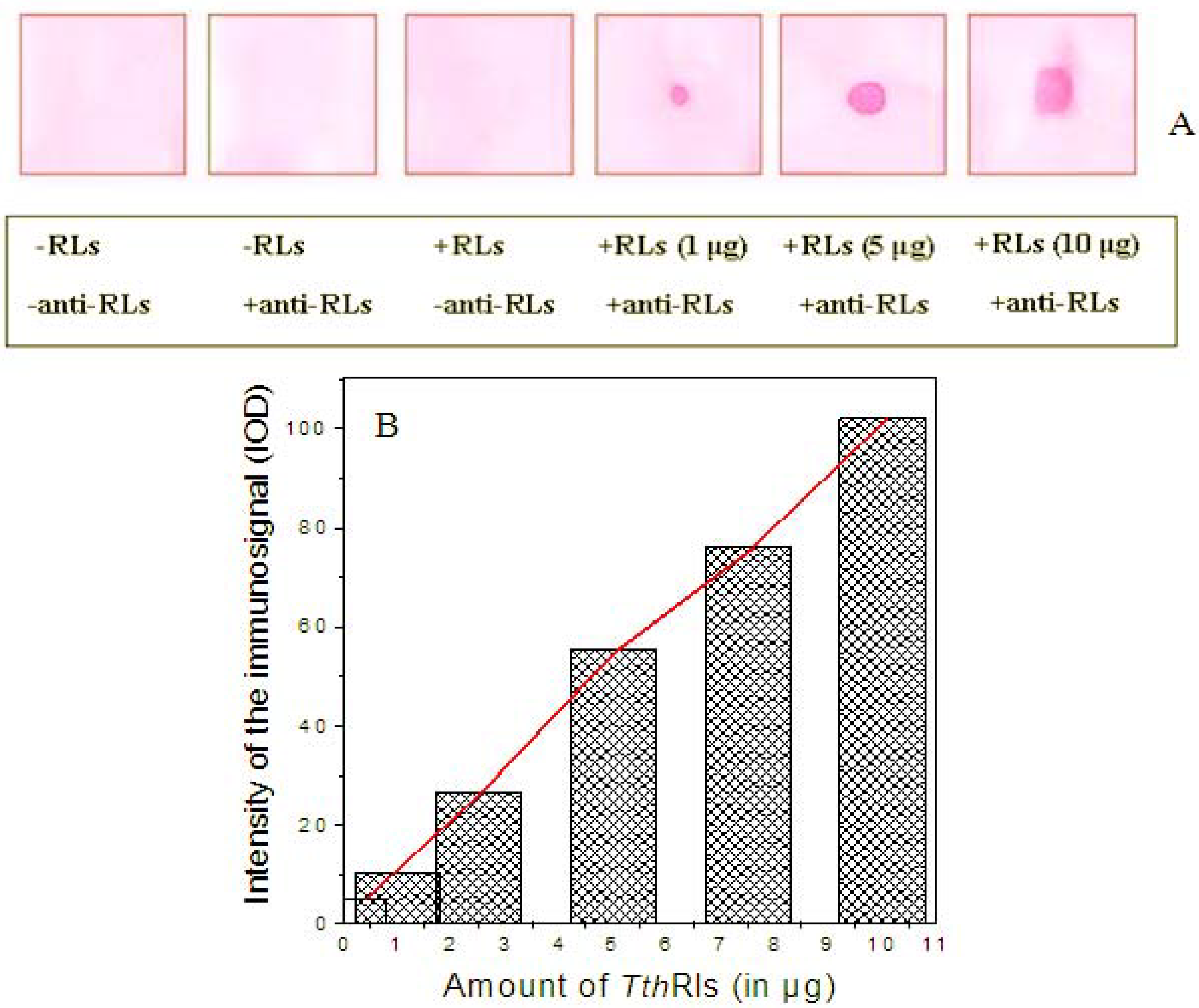
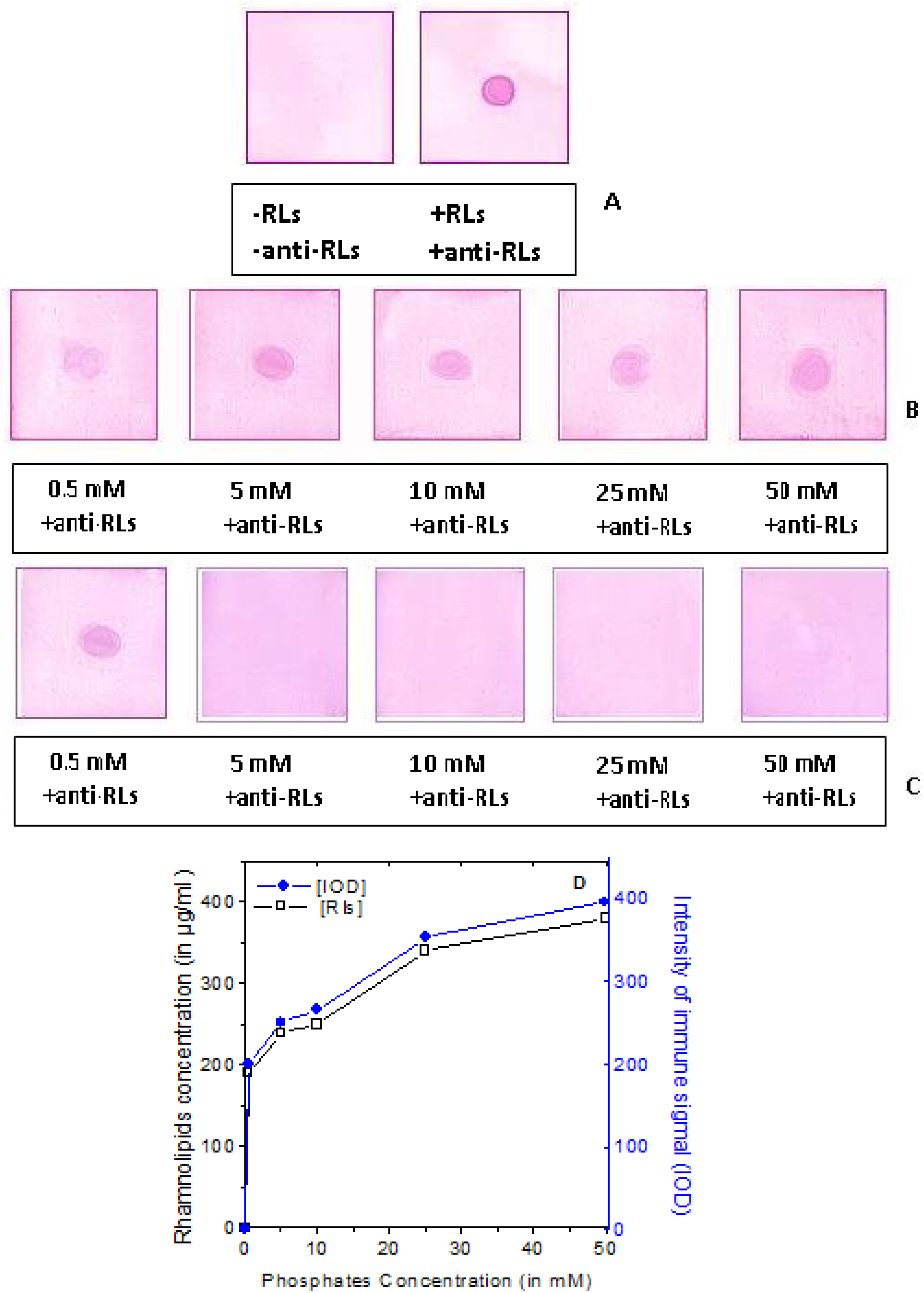
2.4. Detection Limit of the Anti-TthRLs Antibody

An immuno-dot blot experiment, for the detection limit of the anti-TthRLs antibody, was performed by spotting various amounts of purified TthRLs ranging from 1, 5 and 10 μg of TthRLs, as it was quantified previously by the colorimetric orcinol method, using a standard curve of rhamnose [38]. The result showed that the anti-TthRLs antibody recognized TthRLs and the immune-signal intensity was proportional to the TthRLs amount (Figure 4). Additionally, a range of controls, as in the presence and in the absence of TthRLs, and/or antiserum, were performed to assure the experiment. In this context, albumin the most abundant protein in the serum was tested as a non specific protein marker but it did not immuno-cross-reacted.
Figure 4. Detection limits of the anti-TthRLs antibodies. (A) Immuno-dot blot assay performed by spotting various amounts of TthRLs (1, 5 and 10 μg), purified as described in Figure 3, and as it was determined by the colorimetric orcinol method using a standard curve of rhamnose. Then the membranes were probed with 1/500 dilution of rabbit polyclonal antiserum against TthRLs. (B) Quantification of the intensity of immune-staining by the Gelpro Analyzer V.3 computer program (Histograms). Plot of the correlation between the immune-blot quantified by image analysis and the orcinol colorimetric method (in red line).
Antibodies 02 00501 g004
Evaluation of the intensity of Dot Blots immune-signals was performed after quantitative analysis of the membranes by the Gelpro Analyzer V.3 computer program, and the corresponding histograms were obtained. A plot of the correlation between the immune-blot (quantified by image analysis) and the orcinol colorimetric method was established (Figure 4B). It is notable the sensitivity of the anti-TthRLs antibody, which exhibited the potential to detect strongly even a small amount of TthRLs as 1 μg in the immuno-staining (Figure 4A).

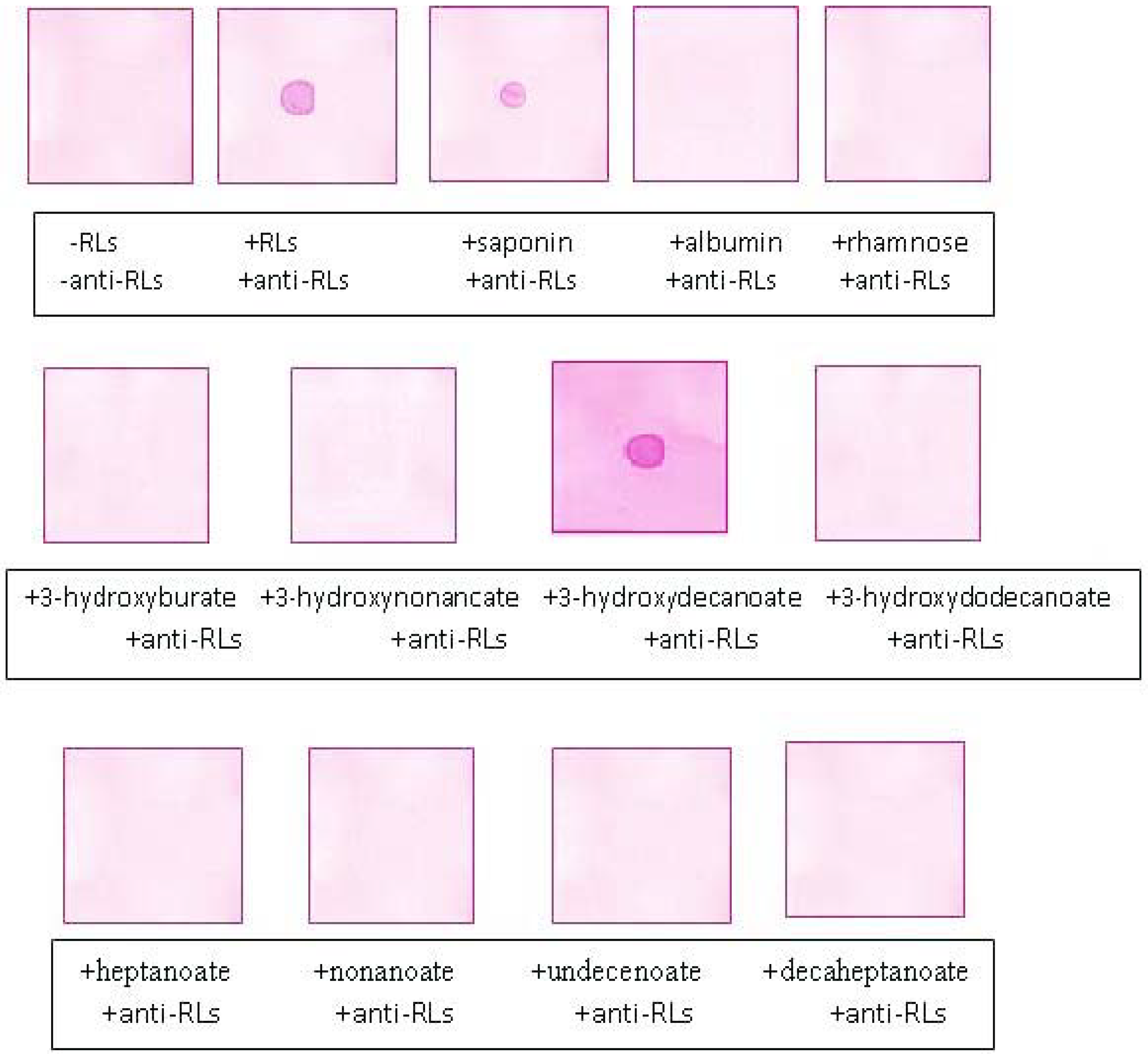
2.5. Recognition Specificity of the Anti-TthRLs Antibodies

To perform the assessment of the antibody preparation, and to delineate the immune-dominant region(s) among the RLs components, the glycoside or the lipidic chain, to be recognized by the anti-TthRLs antibody, these constituents were tested in immuno-dot blot assay. In this concept, a variety of hydroxyalkanoic, and alkanoic acids, methyl-3-hydroxy-alkanoates including 3-hydroxyburate, 3-hydroxynonanoate, 3-hydroxydecanoate, 3-hydroxydodecanoate, etc. with different length of lipidic chain, as well as from the glycoside chain rhamnose, all were spotted on nitrocellulose membranes and then tested in a Dot blot assay with anti-TthRLs antibody.
Concerning the composition of TthRLs it should be noted that the extracted mixture of RLs produced in the presence of sodium gluconate was subjected to identification and characterization using LC-(ESI)-MS. LC-MS analyses revealed that both mono- and di-RL moieties as well as di-rhamno-mono-lipidic congeners were present in the RLs mixture. Additionally, a plethora of diversity of mono- and di-RLs as well, was identified differing in the length of the lipidic chain, which additionally found to be saturated or unsaturated in some cases [11].
Surprisingly, among all the lipidic chains tested the anti-TthRLs antibody recognized only the methyl-3-hydroxy-decanoate (Sigma 24-1003-8), and exhibited a positive immunocross-reactivity. In contrast rhamnose gave no positive immunological signal. Hence from the aforesaid observation, it can be assumed that the antiserum could recognize the lipidic chain, constituent of TthRLs (Figure 5). Furthermore, saponin white, a mixture of plant glycosides (5 μg; C27H42O3), used as a positive control, gave also a strong immuno-reactivity (Figure 5).
Figure 5. Recognition specificity of the anti-TthRLs antibody against 3-hydroxyalkanoates and rhamnose. Immuno-dot blot assay performed by spotting TthRLs, saponin, albumin, rhamnose, and various fatty acids such as 3-hydroxyburate, 3-hydroxynonanoate, 3-hydroxydecanoate, 3-hydroxydodecanoate, heptanoate, nonanoate, undecenoate, decaheptanoate, and then the membranes were probed with 1/500 dilution of rabbit polyclonal antiserum against TthRLs.
Antibodies 02 00501 g005
There is evidence that attributes the implication of the lipidic chain of RLs (but not rhamnose) to triggering some biological effects such as cytotoxicity or heamolytic activity among the TthRLs constituents [31]. RLs possessing the same glycoside chain can also have a different influence, when the aglycone is only slightly modified. The hydrophobic aglycone backbone could probably intercalate into the hydrophobic membrane bi-layer, while the glycoside chain could interact with the polar group [31,32]. The enzyme RhlA (rhamnosyltransferase subunit) supplies the acyl moieties for RL biosynthesis by competing with the enzymes of the type II fatty acid synthase (FASII) cycle for the β-hydroxyacyl-acyl carrier protein (ACP) pathway intermediates. Purified RhlA forms one molecule of β-hydroxydecanoyl-β-hydroxydecanoate from two molecules of β-hydroxydecanoyl-ACP and is the only enzyme required to generate the lipid component of RL, the HAA. The acyl groups in RL are primarily β-hydroxydecanoyl, and in vitro, the enzyme RhlA has a greater affinity for 10-carbon substrates, illustrating that RhlA functions as a molecular ruler that selectively extracts 10-carbon intermediates from type II fatty acid synthase (FASII) [33]. RhlA is highly selective for 10-carbon acyl-ACP intermediates and thus functions as the molecular ruler that controls the acyl chain composition of RLs. This might explain the abundance of the β-hydroxydecanoyl-β-hydroxydecanoate as most frequent constituent of the lipidic chain as well as possibly the recognition selectivity of the antibody.

2.6. Applications of Anti-TthRLs Antibody

As a useful application to valorize the preparation of anti-TthRLs antibody, it was selected to test real samples, which contained various amounts of RLs, like extracellular media from cultures of T. thermophilus and E. coli grown under conditions permissive for RLs production. These samples were previously monitored and assured for the presence and the amount of RLs that were estimated using the quantitative spectrophotometric orcinol method.
For this purpose, an immuno-dot blot assay was performed to detect the presence of RLs by spotting first the purified TthRLs (Figure 6A), and second the extracellular spent supernatants (50 μL) of T. thermophilus (Figure 6B), and E. coli cultures (Figure 6C), containing various concentrations of phosphates (from left to right 0.5, 5, 10, 25, and 50 mM of phosphates) in the presence of sodium gluconate, and grown for 70 h and 24 h at 70 °C and 37 °C respectively. Dot Blot analysis showed that the antibody recognized and immunodetected indiscriminately and quantitatively the presence of RLs in all supernatants derived from T. thermophilus cultures or from the E. coli culture containing 0.5 mM of phosphates spotted in a nitrocellulose membrane (Figure 6B). It is remarkable that the results obtained were consistent with these obtained with the orcinol method. Comparative curves were obtained from the determination of RLs in various cultures using the colorimetric orcinol method, as well as from the quantitative image analysis of the Dot Blots membranes immune-stained with the anti-RLs antibody, by the Gelpro Analyzer V.3 computer program (Figure 6D).
Figure 6. Immuno-dot blot assay performed as application for RLs detection in various samples using the anti-TthRLs antibodies. (A) As a positive control purified TthRLs were used as previously described (right panel, A), and as a negative control a sample without TthRLs was used (left panel, A), which were spotted in nitrocellulose membranes. (B) Extracellular spent supernatants from T. thermophilus cultures grown for 70 h at 75 °C, and E. coli cultures grown for 24 h at 37 °C (C), in the presence of sodium gluconate as sole carbon source containing various concentrations of phosphates affecting TthRLs production (from left to right 0.5, 5, 10, 25, and 50 mM of phosphates) were spotted. Then membranes were probed with 1/500 dilution of rabbit polyclonal antiserum against TthRLs. (D) Comparative curves were obtained from the determination of RLs in various cultures using the colorimetric orcinol method, as well as from the quantitative image analysis of the Dot Blots membranes immune-stained with the anti-RLs antibody by the Gelpro Analyzer V.3 computer program.
Antibodies 02 00501 g006
These results show that the preparation of anti-TthRLs antibodies is a powerful tool for detection of RLs in the extracellular medium able to replace the time-consuming RLs extraction method by a rapid one. The presence of RLs recorded as virulence factors that are implicated in the pathogenicity of a microorganism [4] has rarely been certified in some bacterial infections. To substantiate this view the following experimental data should be presented. It was found that sputum samples obtained from P. aeruginosa-colonized Cystic fibrosis (CF) patients contained RLs (up to 8 μg/mL) and reported a correlation between elevated levels of RLs and worsened patient's clinical status [7]. These RL concentrations in sputum samples, however, might actually underestimate the concentrations present in the lower respiratory tract, as levels of 65 μg/mL were found in secretions of a lung removed from a CF patient [34]. Recently it was found that RL produced by different motile P. aeruginosa strains and clinical isolates from different sources including blood (PA strain 227 63 ± 10 μg/mL RL), skin (ranging approximately from 56, 538 and 702 μg/mL dependent from the strain PA-O, PA 264 and PA 1450 respectively), and Cystic fibrosis (CF) patients (795 ± 1 μg/mL of Rl from strain PA 391) [35]. According to this biochemical approach, tested P. aeruginosa strains produced RLs at concentrations above the critical micelle concentration (CMC) of 20 mg/mL, determined for Rha-C10-C10, Rha-Rha-C10-C10 [36]. The obtained values vary between 20 mg/mL and 795 mg/mL. Since the CMC is dependent on composition of different RLs [37].

3. Experimental Section

3.1. Chemicals and Immunochemicals

Nitroblue tetrazolium (NBT)/5-bromo-4-chloro-3-indolylphosphate (BCIP) (MBI Fermentas, Canada). Goat anti-rabbit IgG conjugated to alkaline phosphatase (AP), Freund’s complete and incomplete adjuvants were purchased from Sigma, Steinheim, Germany). All other chemicals were standard commercial products of analytical-reagent grade.

3.2. Bacterial Strain and Growth for RLs Production

T. thermophilus HB8 (DSM Acc No 579) grown initially in a basal rich medium (BRM) overnight at 70 °C. For RLs production, cultivation was carried out in 2 L Erlenmeyer flasks with 700 mL minimal mineral salt medium (MSM) containing per liter: 1 mM KH2PO4, 25 mM Na2HPO4.∙12 H2O, 0.05 g NH4Cl, 10 g NaCl, 15 mg CaCl2, 123 mg MgSO4.∙7H2O, 6 mL of a mineral solution and sodium gluconate (1.5% w/v) used as sole carbon source. Sodium gluconate was selected because is derived from glucose, its metabolic precursor, in which glucose is transformed via Entner-Doudoroff pathway by glucose dehydrogenase with the NAD(P) ++ reduction. The medium was inoculated with a 5% (v/v) inoculum of exponentially growing in BRM pre-culture and incubated on a rotary shaker at 70 °C for 70 h [10]. Additionally, various T. thermophilus or E. coli cultures were prepared containing different initial concentration of phosphates and sodium gluconate (1.5% w/v) as sole carbon source to assure different productivity of RLs in the extracellular medium [10,11].

3.3. Extraction and Analysis of TthRLs

Produced RLs were extracted from the supernatant of T. thermophilus culture grown in the presence of sodium gluconate and identified as previously described [11]. Purified RLs and extracellular media from the various cultures containing different initial concentration of phosphates, and thus different amount of RLs, were quantified by the colorimetric orcinol method using a standard curve of rhamnose [38]. RLs concentration was calculated by a coefficient of 3.4, obtained from the correlation of pure RLs/rhamnose (1.0 mg of rhamnose corresponds approximately to 3.4 mg of RLs) [39].

3.4. Hemolytic Reaction Caused by TthRLs or Saponin to Rabbit Erythrocytes

Based on the RLs incrimination in hemolytic reaction and since it is destined for immunization of a rabbit, a hemolysis study was performed with purified TthRLs and saponin as well as standard to avoid to cause hemolysis in erythrocytes of the rabbit by the use of high RLs concentration during immunization.
Preparation of erythrocytes. Blood (2 mL) was removed from a rabbit in a tube containing heparin and then was centrifuged at 3,000 rpm for 10 min. The supernatant (plasma) and the medium layer (white blood cells) are removed. Red blood cells were washed 4 times with 0.9% w/v NaCl and finally with 3 ml phosphate buffered saline (PBS), and centrifuged at 3,000 rpm for 10 min. White cells and platelets were carefully aspirated. Red blood cells were diluted with PBS to a final dilution of 1:40.
Hemolytic reaction. A equal volume of erythrocyte solution (0.1 mL) was first put in a series of five tubes, in which then various amounts of saponin or RLs was added, completed with PBS to a final volume of 1 ml. The tubes were then incubated at 37 °C; membranes and intact cells were removed by centrifugation at 10,000 g for 30 min, at 4 °C. Hemolysis recorded as absorbance at 540 nm at different time intervals and is expressed as percentage of free hemoglobin released in the supernatant derived after centrifugation for precipitation of red blood cells. Hemolysis of red blood cells by saponin white was used as positive control to compare the degree of hemolytic activity of RLs.

3.5. Production of Specific Antibody against TthRLs (Anti-TthRLs)

Purified TthRLs with successive extraction of extracellular medium, derived from T. thermophilus HB8 culture grown in the presence of sodium gluconate, were then used as an antigen for the preparation of polyclonal rabbit antisera. The immunization schedules and procedures were performed in the Experimental Animal Handling Facility in the Department of Pharmacy at Aristotle University, Thessaloniki, Greece, in compliance with ethical regulations. The immunogen was emulsified with Freund's incomplete adjuvant using an emulsifier to immunize two white female rabbits. The first immunization was carried out by subcutaneous injection at a dose of 80 μg immunogen per rabbit, using Freund’s incomplete adjuvant for emulsification in advance. The booster inoculations were implemented in four weeks (once every week) with the immunogen at a concentration of 10, 20, 50 and 80 μg per rabbit in 500 μL PBS respectively emulsified with 500 μL Freund’s incomplete adjuvant to meet the requirements of serum titer. The sprint immunization was administered by intraperitoneal injection and sera were recovered from blood collected from the rabbit three days. Pre-immune sera as well as immune sera were collected from the rabbit one week after each immunization. Generation of immune response was followed by immunoblotting at each step of immunization.
To elucidate whether the sample of purified RLs used as antigen for immunization contained the presence of any trace protein, the sample was subjected to SDS-PAGE [40] and stained with silver nitrate.

3.6. Dot Blot Assay of RLs

Purified RLs produced in T. thermophilus TthRLs (or in E. coli) were dissolved in water and aliquots (as described in each experiment) were dotted onto a nitrocellulose membrane (NC). All following steps were done at room temperature. After air drying for 1 h the membranes were blocked with 5% nonfat dry milk in PBS for 2 h to reduce non specific adsorption. The NC membrane was immersed in polyclonal antisera against RLs (anti-RLs) and shaken for 3 h. Diluted polyclonal rabbit anti-RLs sera were used as primary antibody (1:1,000, v/v or as indicated). After washing the membranes with PBS containing 0.03% v/v Tween 20 (T-PBS), a diluted (1:2,000, v/v) secondary antibody goat anti-rabbit IgG-conjugated to alkaline phosphatase (AP) was added and shaken for 2 h. The membranes were washed twice with T-PBS and once with PBS and were developed by conventional method with nitroblue tetrazolium (NBT)/5-bromo-4-chloro-3-indolylphosphate (BCIP) in 100 mM Tris, 100 mM NaCl, and 5 mM MgCl2, pH 9.5. The reaction was stopped by washing with PBS and the immunostained membranes were allowed to dry.

4. Conclusions

Several methods have been investigated for qualitative and quantitative analysis of RLs, such as the spectrophotometric orcinol method or thin layer chromatography (TLC), due to industrial interest of RLs as bacterial biosurfactants as well as their wide applications for disease treatment. Immunoassay systems using antibody against bioactive compounds as RLs will become important tools for this kind of analysis in industry and medicine. The significance of establishing such an antibody is to detect the presence of RLs in extracellular media of cultures easier without successive extractions with a rapid method. It is hoped that this improvement of RLs detection will eventually lead to an extension of the use of this antibody in biological fluids (blood, urine) suspected of bacterial infections containing RLs as bacterial secreted metabolites.

Conflicts of Interest

The authors declare no conflict of interest.

References

  1. Maier, R.M.; Soberón-Chávez, G. Pseudomonas aeruginosa rhamnolipids: Biosynthesis and potential applications. Appl. Microbiol. Biotechnol. 2000, 54, 625–633. [Google Scholar] [CrossRef]
  2. Vatsa, P.; Sanchez, L.; Clement, C.; Baillieul, F.; Dorey, S. Rhamnolipid biosurfactants as new players in animal and plant defense against microbes. Int. J. Mol. Sci. 2010, 11, 5095–5108. [Google Scholar] [CrossRef]
  3. Ochsner, U.A.; Reiser, J. Autoinducer-mediated regulation of rhamnolipid biosurfactant synthesis in Pseudomonas aeruginosa. Proc. Natl. Acad. Sci. USA 1995, 92, 6424–6428. [Google Scholar] [CrossRef]
  4. Abdel-Mawgoud, A.M.; Lépine, F.; Déziel, E. Rhamnolipids: Diversity of structures, microbial origins and roles. Appl. Microbiol. Biotechnol. 2010, 86, 1323–1336. [Google Scholar] [CrossRef]
  5. Abdel-Mawgoud, A.M.; Hausmann, R.; Lépine, F.; Muller, M.M.; Déziel, E. Rhamnolipids: Detection, analysis, biosynthesis, genetic regulation and bioengineering of production. Biosurf. Microbiol. Monog. 2011, 20, 13–55. [Google Scholar] [CrossRef]
  6. Zulianello, L.; Canard, C.; Köhler, T.; Caille, D.; Lacroix, J.S.; Meda, P. Rhamnolipids are virulence factors that promote early infiltration of primary human airway epithelia by Pseudomonas aeruginosa. Infect. Immun. 2006, 74, 3134–3147. [Google Scholar] [CrossRef]
  7. Kownatzki, R.; Tümmler, B.; Döring, G. Rhamnolipid of Pseudomonas aeruginosa in sputum of cystic fibrosis patients. Lancet 1987, 1, 1026–1027. [Google Scholar] [CrossRef]
  8. Stutts, M.J.; Schwab, J.H.; Chen, M.G.; Knowles, M.R.; Boucher, R.C. Effects of Pseudomonas aeruginosa on bronchial epithelial ion transport. Am. Rev. Respiratory Dis. 1986, 134, 17–21. [Google Scholar]
  9. Cabrera-Valladares, N.; Richardson, A.P.; Olvera, C.; Treviño, L.G.; Déziel, E.; Lépine, F.; Soberón-Chávez, G. Monorhamnolipids and 3-(3-hydroxyalkanoyloxy) alkanoic acids (HAAs) production using Escherichia coli as a heterologous host. Appl. Microbiol. Biotechnol. 2006, 73, 187–194. [Google Scholar] [CrossRef]
  10. Pantazaki, A.; Dimopoulou, M.; Simou, O.; Pritsa, A. Sunflower seed oil and oleic acid utilization for the production of rhamnolipids by Thermus thermophilus HB8. Appl. Microbiol. Biotechnol. 2010, 88, 939–951. [Google Scholar] [CrossRef]
  11. Pantazaki, A.A.; Papaneophytou, C.P.; Lambropoulou, D.A. Simultaneous polyhydroxyalkanoates and rhamnolipids production by Thermus thermophilus HB8. AMB Express 2011, 1, 17–30. [Google Scholar] [CrossRef]
  12. Rezanka, T.; Siristova, L.; Sigler, K. Rhamnolipid-producing thermophilic bacteria of species Thermus and Meiothermus. Extremophiles 2011, 15, 697–709. [Google Scholar] [CrossRef]
  13. Fujita, K.; Akino, T.; Yoshioka, H. Characteristics of heat-stable extracellular hemolysin from Pseudomonas aeruginosa. Infect. Immun. 1988, 56, 1385–1387. [Google Scholar]
  14. Johnson, M.K.; Boese-Marrazzo, D. Production and properties of heat-stable extracellular hemolysin from Pseudomonas aeruginosa. Infect. Immun. 1980, 29, 1028–1033. [Google Scholar]
  15. Piljac, G.; Piljac, V. Immunological activity of rhamnolipids. United States—Patent Application Publication 5466675, 1995. [Google Scholar]
  16. Häussler, S.; Rohde, M.; von Neuhoff, N.; Nimtz, M.; Steinmetz, I. Structural and functional cellular changes induced by Burkholderia pseudomallei rhamnolipid. Infect. Immun. 2003, 71, 2970–2975. [Google Scholar]
  17. Häussler, S.; Nimtz, M.; Domke, T.; Wray, V.; Steinmetz, I. Purification and characterization of a cytotoxic exolipid of Burkholderia pseudomallei. Infect. Immun. 1998, 66, 1588–1593. [Google Scholar]
  18. Sánchez, M.; Aranda, F.J.; Teruel, J.A.; Espuny, M.J.; Marqués, A.; Manresa, A.; Ortiz, A. Permeabilization of biological and artificial membranes by a bacterial dirhamnolipid produced by Pseudomonas aeruginosa. J. Colloid Interf. Sci. 2010, 341, 240–247. [Google Scholar]
  19. McClure, C.D.; Schiller, N.L. Effects of Pseudomonas aeruginosa rhamnolipids on human monocyte derived macrophages. J. Leukoc. Biol. 1992, 51, 97–102. [Google Scholar]
  20. Jensen, P.O.; Bjarnsholt, T.; Phipps, R.; Rasmussen, T.B.; Calum, H.; Christoffersen, L. Rapid necrotic killing of polymorphonuclear leukocytes is caused by quorum-sensing-controlled production of rhamnolipid by Pseudomonas aeruginosa. Microbiology 2007, 153, 1329–1338. [Google Scholar]
  21. Van Gennip, M.; Christensen, L.D.; Alhede, M.; Phipps, R.; Jensen, P.Ø.; Christophersen, L.; Pamp, S.J.; Moser, C.; Mikkelsen, P.J.; Koh, A.Y.; et al. Inactivation of the rhlA gene in Pseudomonas aeruginosa prevents rhamnolipid production, disabling the protection against polymorphonuclear leukocytes. APMIS 2009, 117, 537–546. [Google Scholar]
  22. Sotirova, A.; Spasova, D.; Vasileva-Tonkova, E.; Galabova, D. Effects of rhamnolipid-biosurfactant on cell surface of Pseudomonas aeruginosa. Microbiol. Res. 2009, 164, 297–303. [Google Scholar]
  23. Sotirova, A.V.; Spasova, D.I.; Galabova, D.N; Karpenko, E.; Shulga, A. Rhamnolipid-biosurfactant permeabilizing effects on gram-positive and gram-negative bacterial strains. Curr. Microbiol. 2008, 56, 639–644. [Google Scholar]
  24. Voutquenne, L. Saponins and hemolytic activity. Saponins and glycosides from five species of Sapindaceae. Ann. Pharm. Fr. 2001, 59, 407–414. [Google Scholar]
  25. Stipcevic, T.; Piljac, T.; Isseroff, R.R. Di-rhamnolipid from Pseudomonas aeruginosa displays differential effects on human keratinocyte and fibroblast cultures. J. Dermatol. Sci. 2005, 40, 141–143. [Google Scholar]
  26. Piljac, G.; Piljac, V. Pharmaceutical preparation based on rhamnolipid. US Patent 1995. [Google Scholar]
  27. Al-Tahhan, R.A.; Sandrin, T.R.; Bodour, A.A.; Maier, R.M. Rhamnolipid-induced removal of lipopolysaccharide from Pseudomonas aeruginosa: Effect on cell surface properties and interaction with hydrophobic substrates. Appl. Environ. Microbiol. 2000, 66, 3262–3268. [Google Scholar] [CrossRef]
  28. Irie, Y.; O'Toole, G.A.; Yuk, M.H. Pseudomonas aeruginosa rhamnolipids disperse Bordetella bronchiseptica biofilms. FEMS Microbiol. Lett. 2005, 250, 237–243. [Google Scholar] [CrossRef]
  29. Haruma, M.; Tanaka, M.; Sugimoto, T.; Kojima, R.; Suzuki, Y.; Konoshima, T.; Kozuka, M.; Ito, K. Alteration of Na+ permeability in human erythrocytes as studied by 23Na-NMR and inhibition of the kidney Na+, K+-ATPase activities with saponins: Interaction of gleditsia saponins with human erythrocyte membranes. Bioorg. Med. Chem. Lett. 1995, 5, 827–830. [Google Scholar] [CrossRef]
  30. Peuchant, E.; Salles, C.; Vallot, C.; Wone, C.; Jensen, R. Increase of erythrocyte resistance to hemolysis and modification of membrane lipids induced by hemodialysis. Clin. Chim. Acta 1988, 178, 271–282. [Google Scholar] [CrossRef]
  31. Pantazaki, A.A.; Choli-Papadopoulou, T. On the Thermus thermophilus HB8 potential pathogenicity triggered from rhamnolipids secretion: morphological alterations and cytotoxicity induced on fibroblastic cell line. Amino Acids 2012, 42, 1913–1926. [Google Scholar] [CrossRef]
  32. Chwalek, M.; Lalun, N.; Bobichon, H.; Plé, K.; Voutquenne-Nazabadioko, L. Structure-activity relationships of some hederagenin diglycosides: Haemolysis, cytotoxicity and apoptosis induction. Biochim. Biophys. Acta 2006, 1760, 1418–1427. [Google Scholar] [CrossRef]
  33. Zhu, K.; Rock, C.O. RhlA converts b-hydroxyacyl-acyl carrier protein intermediates in fatty acid synthesis to the b-hydroxydecanoyl-b-hydroxydecanoate component of rhamnolipids in Pseudomonas aeruginosa. J. Bacteriol. 2008, 190, 3147–3154. [Google Scholar] [CrossRef]
  34. Read, R.C.; Roberts, P.; Munro, N.; Rutman, A.; Hastie, A.; Shryock, T.; Hall, R.; McDonald-Gibson, W.; Lund, V.; Taylor, G. Effect of Pseudomonas aeruginosa rhamnolipids on mucociliary transport and ciliary beating. J. Appl. Physiol. 1992, 72, 2271–2277. [Google Scholar]
  35. Meyer-Hoffert, U.; Zimmermann, A.; Czapp, M.; Bartels, J.; Koblyakova, Y.; Gläser, R.; Schröder, J.M.; Gerstel, U. Flagellin delivery by Pseudomonas aeruginosa rhamnolipids induces the antimicrobial protein psoriasin in human skin. PLoS One 2011, 6, e16433. [Google Scholar] [CrossRef]
  36. Syldatk, C.; Lang, S.; Matulovic, V.; Wagner, F. Production of four interfacial active rhamnolipids from n-alkanes or glycerol by resting cells of Pseudomonas species DSM 2847. Z Naturforsch 1985, 40c, 61–67. [Google Scholar]
  37. Déziel, E.; Lépine, F.; Dennie, D.; Boismenu, D.; Mamer, O.A.; Villemur, R. Liquid chromatography/mass spectrometry analysis of mixtures of rhamnolipids produced by Pseudomonas aeruginosa strain 57RP grown on mannitol or naphthalene. Biochim. Biophys. Acta Mol. Cell Biol. Lipids 1999, 1440, 244–252. [Google Scholar] [CrossRef]
  38. Koch, A.K.; Kappeli, O.; Ficher, A.; Reiser, J. Hydrocarbon assimilation and biosurfactant production in Pseudomonas aeruginosa mutants. J. Bacteriol. 1991, 173, 4212–4219. [Google Scholar]
  39. Pearson, J.P.; Pesci, E.C.; Iglewski, B.H. Roles of Pseudomonas aeruginosa las and rhl quorum-sensing systems in control of elastase and rhamnolipid biosynthesis genes. J. Bacteriol. 1997, 179, 5756–5767. [Google Scholar]
  40. Laemmli, U.K. Cleavage of structural proteins during the assembly of the head of bacteriophage T4. Nature 1970, 227, 680–685. [Google Scholar] [CrossRef]

