Article

Pleiotrophin and the Expression of Its Receptors during Development of the Human Cerebellar Cortex

by
Margarita Belem Santana-Bejarano
1,2,
Paula Romina Grosso-Martínez
1,3,
Ana Graciela Puebla-Mora
1,
María Guadalupe Martínez-Silva
3,
Mario Nava-Villalba
4,
Ana Laura Márquez-Aguirre
5,
Daniel Ortuño-Sahagún
6,* and
Marisol Godínez-Rubí
1,7,*
1
Laboratorio de Patología Diagnóstica e Inmunohistoquímica, Centro de Investigación y Diagnóstico en Patología, Departamento de Microbiología y Patología, CUCS, Universidad de Guadalajara, Guadalajara 44340, Mexico
2
Doctorado en Ciencias en Biología Molecular en Medicina, Centro Universitario de Ciencias de la Salud, Universidad de Guadalajara, Guadalajara 44340, Mexico
3
Departamento de Anatomía Patológica, Centro Médico Nacional de Occidente, Instituto Mexicano del Seguro Social (IMSS), Guadalajara 44340, Mexico
4
Centro de Investigación y Diagnóstico en Patología, Departamento de Microbiología y Patología, CUCS, Universidad de Guadalajara, Guadalajara 44340, Mexico
5
Unidad de Biotecnología Médica y Farmacéutica, Centro de Investigación y Asistencia en Tecnología y Diseño del Estado de Jalisco A.C. (CIATEJ), Guadalajara 44270, Mexico
6
Laboratorio de Neuroinmunobiología Molecular, Instituto de Investigación en Ciencias Biomédicas (IICB), CUCS, Universidad de Guadalajara, Guadalajara 44340, Mexico
7
Departamento de Morfología, CUCS, Universidad de Guadalajara, Guadalajara 44340, Mexico
*
Authors to whom correspondence should be addressed.
Cells 2023, 12(13), 1733; https://doi.org/10.3390/cells12131733
Submission received: 5 May 2023 / Revised: 19 June 2023 / Accepted: 24 June 2023 / Published: 27 June 2023
(This article belongs to the Special Issue Cerebellar Development: From Cell Imaging to Clinical Applications)

Abstract

:
During embryonic and fetal development, the cerebellum undergoes several histological changes that require a specific microenvironment. Pleiotrophin (PTN) has been related to cerebral and cerebellar cortex ontogenesis in different species. PTN signaling includes PTPRZ1, ALK, and NRP-1 receptors, which are implicated in cell differentiation, migration, and proliferation. However, its involvement in human cerebellar development has not been described so far. Therefore, we investigated whether PTN and its receptors were expressed in the human cerebellar cortex during fetal and early neonatal development. The expression profile of PTN and its receptors was analyzed using an immunohistochemical method. PTN, PTPRZ1, and NRP-1 were expressed from week 17 to the postnatal stage, with variable expression among granule cell precursors, glial cells, and Purkinje cells. ALK was only expressed during week 31. These results suggest that, in the fetal and neonatal human cerebellum, PTN is involved in cell communication through granule cell precursors, Bergmann glia, and Purkinje cells via PTPRZ1, NRP-1, and ALK signaling. This communication could be involved in cell proliferation and cellular migration. Overall, the present study represents the first characterization of PTN, PTPRZ1, ALK, and NRP-1 expression in human tissues, suggesting their involvement in cerebellar cortex development.

Graphical Abstract

1. Introduction

In humans, cerebellar development starts during the fourth gestational week (gw), and it continuously changes until it matures [1]. This process includes cell proliferation, migration, differentiation, and apoptosis events. Specifically, the cerebellum arises from the rhombencephalon, and it undergoes a complex maturation process that finishes at 2 years old [2,3]. In the latest human fetal stage, the cerebellar cortex is structured by the external granule cell layer (EGL), molecular layer (ML), Purkinje cell layer (PCL), and internal granule cell layer (IGL). When a human is approximately two years old, granule cells culminate their migration from the EGL to the IGL via Bergmann glia [4,5,6]. The Bergmann glia have an astroglial phenotype, which expresses glial fibrillary acidic protein (GFAP) during human fetal development [7,8,9,10]. Any disruption in these processes can lead to cerebellar pathology in the form of medulloblastoma, the molecular subgroup and prognosis of which depend on the specific developmental abnormalities [11]. Additionally, it has been demonstrated that extracellular growth factors, such as pleiotrophin (PTN) and midkine (MK), play an important role in rodent cerebral and cerebellar cortex ontogenesis, with special emphasis on the postnatal stage [12,13,14,15,16,17]. Nevertheless, its expression has not been demonstrated in the developing human cerebellum. On the other hand, alterations in PTN are related to tumor cell proliferation and prognosis of neoplasms, such as glioblastoma, testicular germ cell neoplasms, and hepatocellular carcinoma [18,19,20,21].
PTN is a protein composed of 168 amino acids (a.a.) that is secreted into the extracellular matrix [22,23], where it has a signaling function for neural survival, differentiation, and migration molecules during mouse and rat embryonic induction [12,24], and is beginning to be recognized as a neuromodulatory cytokine [17]. This signaling function is mediated by different receptors, such as receptor-type protein-tyrosine kinase Z receptor (PTPRZ1), anaplastic lymphoma kinase receptor (ALK), and neuropilin-1 receptor (NRP-1) [25,26,27,28]. PTPRZ1 is a chondroitin sulfate proteoglycan that has intracellular phosphatase activity. It has been found in Purkinje cells (PCs) and Bergmann glia in postnatal rat cerebellar cortices and neural axon fibers during growth [16,29]. Additionally, the PTN–PTPRZ complex induces PTPRZ1 tyrosine phosphatase inactivation in U373-MG cells, and this signaling pathway is involved in neural cell migration in the brain cortex [30,31]. On the other hand, ALK is a receptor tyrosine kinase that is structurally similar to the insulin receptor subfamily [32]. The mRNA and protein expression of ALK has been described in the neural cells of the diencephalon, neuroepithelium, thalamus, midbrain, and spinal cord in mouse development [33]. Moreover, the interaction between PTN and ALK depends on PTPRZ1 inhibition by the PTN–PTPRZ1 complex [34]. NRP-1 is a 130 KDa non-tyrosine kinase receptor, and it has a transmembrane single-pass structure [35]. NRP-1 has been described as a chemo-repulsive molecule involved in the neuronal growth cone in the peripheral nerves [36,37]. Furthermore, NRP-1 was found expressed in vitro in Schwann cells and hippocampal neurons [37].
Even though these molecules are reported in nervous system development, most knowledge is based on animal models, and their cerebellar cortex localization is still debated. For this reason, this study aims to describe the expression of PTN and its receptors in the human fetal and neonatal cerebellar cortex.

2. Materials and Methods

2.1. Human Paraffin-Embedded Tissues

The present study was approved by the Ethics Committee of Centro Médico Nacional de Occidente (México) and Centro Universitario de Ciencias de la Salud, Universidad de Guadalajara, Jalisco, México (approval numbers: R-2021-1301-123 and CI-02021, respectively). Five fetal samples and one neonatal sample were collected from pathology autopsy studies between 2013 and 2015. For human fetal samples, there were three females at 31, 37.4, and 38 gw; two males at 17 and 36.6 gw. For human neonatal samples, there was one female at 38 gw. Any infectious process was discarded in the clinical history.

2.2. Tissue Preparation and Histological Staining

Fetal ages were calculated from the first day of the mother’s last menstrual period and fetal somatometry study. The human fetal cerebellum was fixed in 4% paraformaldehyde with a pH of 7.4. They were dehydrated with ethanol and paraffin embedded. Then, 3 µm thick sections were made, and hematoxylin and eosin staining (H&E) was performed for histological examination.

2.3. Immunohistochemistry (IHC)

The analyses were performed on paraffin-embedded tissues from the fetal cerebellar cortex. The paraffin block was cut into 3 µm thick sections, deparaffinized, and rehydrated. For all IHC analyses, a NovolinkTM Polymer Detection System was used (RE7290-K, Leica BIOSYSTEMS). Antigen retrieval was performed using citrate buffer solution (pH, 6.0) to analyze PTN, PTPRZ1, and ALK; EDTA buffer solution (pH, 8.0) was used for NRP-1 antigen retrieval. Endogenous peroxidase was blocked with 3% (v/v) H2O2. NovocastraTM protein block was used for 30 min to reduce the non-specific binding of primary antibodies and polymers. Anti-PTN (1:100, sc-74443, Santa Cruz) and anti-PTPRZ1 (1:100, sc-33664, Santa Cruz) primary antibodies were incubated for 60 min; anti-ALK (1:10, ACI 3041 A, Biocare) was hatched for 90 min; anti-NRP-1 (1:250, Ab81321, Abcam) and anti-GFAP (1:250, 258R, Cell Marque) were incubated for 30 min. The tissues were then treated with post-primary (rabbit anti-mouse IgG) and Novolink TM polymer solutions (anti-rabbit poly-HRP-IgG). The chromogen was 5% 3,3′-diaminobenzidine in a buffer solution containing 0.1% H2O2. The sections were counterstained with hematoxylin, dehydrated, and cover slipped. The specificity of antibody labeling was previously verified in several control tissues (Supplementary Figure S1). Seminoma tissue was utilized as a PTN-positive control tissue; cecal appendix and cerebral cortex as PTPRZ1-positive control tissues; cecal appendix as an NRP-1-positive control tissue; cerebellar cortex as an ALK-positive control tissue. The same tissues without primary antibodies were used as the negative control. Immunohistochemical staining was evaluated by one pathologist.

2.4. Histological Analysis

The tissue was digitalized using Aperio LV1 (Leica Biosystem Imaging Inc., Deer Park, IL, USA). The analysis was conducted on a total of 4 different locations in each sample using Qupath (open-source software for digital pathology image analysis) [38]. EGL cell density was counted at 5000 µm2 per section using hematoxylin detection as a measure of total cells. The thickness of the EGL and ML was defined with the spatial analysis function. Purkinje cell area was automatically determined by the software using an ellipse selection form. The staining intensity measurement in immunohistochemistry was conducted with a set cell intensity classification function according to DAB subcellular localization. All staining intensity measurements were evaluated as a percentage of four different fields analyzed and reported using three categories: 1+ (weak), 2+ (moderate), and 3+ (strong) (Supplementary Figure S2).

3. Results

The cerebellar cortex was analyzed for all human samples. The majority of them were female (67%), with a median age of 37 gw (range: 17–38 gw). The cause of death of the unique neonatal sample was respiratory failure. The clinical characteristics of the human cases are summarized in Table 1.

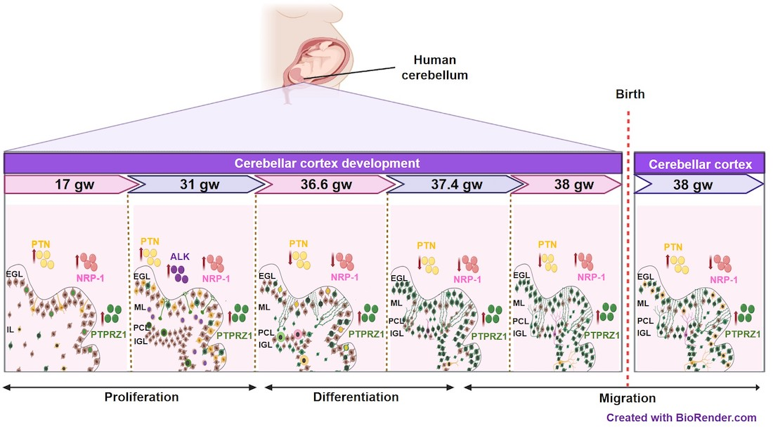
3.1. The Topological Architecture of the Developing Human Cerebellar Cortex

Cerebellar cortex cellularity was observed by using hematoxylin and eosin staining. The EGL was constantly detected in all gestational ages analyzed. The cell thickness and density showed variations at different gestational ages. The age with the highest EGL cell thickness and density of EGL was 36.6 gw, and the lowest density was at 17 gw (Table 2, Figure 1A,B,F). Additionally, at 31 gw, the PC bodies could be easily detected in comparison to those at 17 gw. PCs increase the surface area of their soma as the gestational age increases, and this tendency is maintained in the postnatal stage, wherein the largest cell size for this type of cell is recorded. The molecular layer was not visible at 17 gw and only became evident at 31 gw; its maximum thickness could be seen at 36.6 gw, with a drastic reduction in cases of higher gestational age (Figure 1A–F, Table 2). From 36.6 gw, the IGL was noticeable (Figure 1C).
Interestingly, the fetal and neonatal samples at 38 gw showed histological differences. The EGL density was higher in the fetal cerebellar cortex than in the neonatal cerebellar cortex. The Purkinje axonal projections were more prominent in the neonatal cerebellar cortex than in the fetal cerebellum (Figure 1E,F, Table 2). A summary of histological changes in the cerebellar cortex during embryogenesis is depicted in Figure 1G and Table 2.

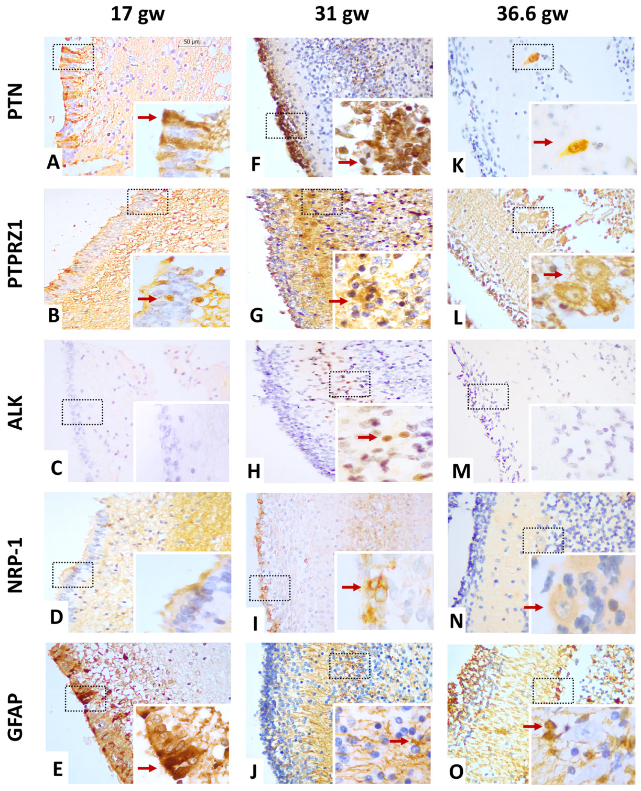
3.2. Pleiotrophin Cell-Type Expression Changes during Cerebellar Cortex Development in Humans

The anti-PTN antibody recognizes a 168 a.a. protein, the expression and localization of which were variable through the different stages of the human fetal cerebellar cortex development evaluated (Table 3). As shown in Figure 2A,E and Table 3, PTN protein expression was lower at 17 gw, where it was immunoreactive in the nuclei and somas of cells that were vertically extended throughout the EGL. These patterns were compared to GFAP-positive stains in the EGL. At 31 gw, PTN expression was increased; it was located with high intensity in all nuclei of the EGL where GFAP staining was reduced to some glial projections (Figure 2F,J). PTN expression depicted a new decrease at 36.6 gw in the EGL, but GFAP staining showed an increase in intensity and expression (Figure 2O); PTN localization was limited to a few nuclei of the EGL and in some of the cytoplasm and nuclei of PC neurons (Figure 2K). At 37.4 gw, the intensity of PTN decreased and was only expressed in the cytoplasm of PCs, while GFAP stains showed diffuse distribution across all cerebellar cortex layers (Figure 3A). Thus, PTN intensity became barely detectable in the cytoplasm of PCs at 38 gw (Figure 3F). Interestingly, PTN expression reappeared in the cell cytoplasm of the EGL in the neonatal sample, but it disappeared in PCs (38 gw) (Figure 3K).

3.3. PTN Receptor Expression Spatiotemporally Changes in Cerebellar Cortex Development

As PTN receptors, the expression of PTPRZ1, ALK, and NRP-1 proteins was evaluated using immunohistochemistry. All characterizations are shown in Figure 2, Figure 3 and Figure 4.

3.3.1. Gestational Week 17

At this stage, the EGL was clearly detected in cerebellar cortex tissue (Figure 1A). The immunoreactivity to PTPRZ1 was low in some cell nuclei of this layer. The PTPRZ1 protein also showed diffuse distribution and high intensity in IGL cell soma (Figure 2B, Table 4). In contrast, ALK protein staining was negative in all cerebellar cortex layers (Figure 2C, Table 5). NRP-1 expression was diffusely distributed in the neuropil zone where it had moderate intensity. The EGL was completely negative to NRP-1 expression (Figure 2D, Table 6).

3.3.2. Gestational Week 31

At this stage, cell density began to increase in the EGL, and PC bodies were easily seen (Figure 1B). The intensity of PTPRZ1 expression was higher in all soma of external granular cells. PTPRZ1 was also distributed abundantly in the cytoplasm of PCs. In the IGL, PTPRZ1 was positive in the soma and nuclei of ganglion neurons (Figure 2G, Table 4). PTPRZ1-positive patterns were similar to GFAP-positive cells, showing the morphology and location of Bergmann glia (Figure 2J). In addition, ALK protein expression was only observed in the cell nuclei of the ML at this age, with high-intensity staining in most cell nuclei (Figure 2H, Table 5). The NRP-1 protein was localized in the soma of the external granule cells, too. NRP-1 expression was also shown in the neuropil zone. Its staining intensity was higher in the EGL than in the neuropil zone (Figure 2I, Table 6).

3.3.3. Gestational Week 36.6

At this stage, PC body size was increased (Figure 1C). The expression of PTPRZ1 was diffusely distributed through all cerebellar cortex layers, as was a GFAP staining pattern (Figure 2L,O). In granule cells and Bergmann glia, PTPRZ1 showed nuclear and soma staining with high intensity. The PCL was positive for PTPRZ1 in the cell cytoplasm, where it retained moderate intensity. PTPRZ1 was also located in the neuropil zone (Figure 2L, Table 4). On the other hand, the ALK protein was again negative in all cerebellar cortex layers (Figure 2M, Table 5). NRP-1 expression was reduced in this gestational age, it was only positive in the cytoplasm of PC, and it showed low-intensity staining (Figure 2N, Table 6).

3.3.4. Gestational Week 37.4

At this stage, PTPRZ1 and GFAP staining patterns were consistently similar (Figure 3B,E). PTPRZ1 expression was seen in the soma of external granule cells and glial projections. In the PCL, PTPRZ1 was positive in the cytoplasm of PCs, which began to demonstrate a long axon. Additionally, PTPRZ1 was diffusely distributed in the ML and neuropil zone of all cerebellar cortex layers (Figure 3B, Table 4). The ALK protein was not detected in the cerebellar cortex layers (Figure 3C, Table 5), but NRP-1 expression was increased in the cytoplasm of PCs compared to the cerebellar cortex at 36.6 wg (Figure 3D, Table 6).

3.3.5. Gestational Week 38

At this stage, PTPRZ1 expression showed high-intensity staining along the axon projection of PCs, which was extended to the EGL. In the ML, PTPRZ1 continued showing diffuse distribution, as the GFAP pattern did, and it was positive at the neuropil zone of all cerebellar cortex layers. In the IGL, PTPRZ1 was observed in the soma of PCs (Figure 3G, Table 4). The ALK protein was negative in the cerebellar cortex (Figure 3H, Table 5), and NRP-1 showed enhanced staining intensity in the cytoplasm of PCs (Figure 3I, Table 6).

3.3.6. Neonatal Sample

At this stage, the thickness and cell density of the EGL were gradually reduced (Table 2). The staining intensity of PTPRZ1 in the EGL was lower in neonatal tissue than in fetal tissue at 38 gw (Table 4). PTPRZ1 expression was maintained along the axon projection of PCs. It showed diffuse distribution in the ML with moderate intensity, where cell nuclei were negative, and had the same staining pattern as the GFAP protein. PTPRZ1 was also positive in the neuropil zone of all cerebellar cortices (Figure 3L,O). ALK expression was not seen in the cerebellar cortex (Figure 3M, Table 5). NRP-1 expression showed lower-intensity staining in neonatal tissue than in fetal tissue at 38 gw and was limited to the cytoplasm of PCs (Figure 3N, Table 6).

4. Discussion

This study described, for the first time, the expression pattern of PTN and its receptors—PTPRZ1, NRP-1, and ALK—throughout the development of the human cerebellar cortex, from 17 gw to the early neonatal stage, using an immunohistochemical technique. The results indicate that, in the cases analyzed, the localization and intensity of PTN, PTPRZ1, and NRP-1 staining changed according to the gestational weeks, but the ALK protein was only expressed in the cerebellar cortex at 31 gw. We also contrasted the cell type that expresses them using the GFAP marker as an astroglial phenotype in human fetal stages.
This work shows that PTN is predominantly expressed in the EGL at 17 gw and increases at 31 gw. This immunoreactivity retains a pattern similar to Bergmann glia only at 17 gw. This shows that, in humans, PTN could be produced by Bergmann glia at 17 gw, but granular cell precursors may be the major sources of PTN at 31 gw. Even though PTN protein expression has been described in Bergmann glia, it was only reported for the postnatal stage in rats [16]. However, a similar pattern between PTN and radial glia has been reported in the cerebral cortex of rat embryos (E13), where it seems to be related to the perpendicularly running processes of neurons [12]. The PTPRZ1 protein was observed in some granule cell precursors at 17 gw, but it has a similar expression pattern to GFAP in the ML at 31 gw. This suggests that there is continuous communication between granule cell precursors and Bergmann glial cells that could implicate the PTN–PTPRZ1 pathway, which increases at 31 gw. Additionally, Bergmann glial cells may be stimulated by other molecules to lower PTN expression in humans between 17 and 31 gw. This may be related to the expression of the PTPRZ1 protein by Bergmann glia in the latest stage of rat and mouse development [29,39].
Additionally, the ALK protein was absent at 17 gw, where PTN and PTPRZ1 were weakly expressed, too, but they were highly expressed in the ML at 31 gw. This could suggest another cell type in which ALK and PTPRZ1 are co-expressed in the ML at 31 gw. It has been demonstrated that the PTN–PTPRZ1 complex promotes ALK activation in humans [40] and ALK can stimulate its expression by promoting MYCN transcription [41]; therefore, PTN–PTPRZ1 signaling can promote ALK expression in the ML. An important role of ALK in synaptic plasticity has been described in mouse AlkKO [42], but the activation of interconnecting pathways (RAS/ERK; JAK/STAT; and PI3K/AKT) is also known, which may activate cell proliferation [43,44,45]. The latter finding is consistent with the increase in ML thickness observed in the present study after 31 gw (peak ALK expression). In parallel, NRP-1 was only localized in the neuropil zone at 17 gw, but it was highly expressed in the soma of granule cell precursors at 31 gw. These results could represent a co-localization of PTN and NRP-1 in the EGL. Although NRP-1 is a receptor that should be observed in membrane cells, it was also described that the PTN–NRP-1 interaction could stimulate NRP-1 internalization into the cell cytoplasm [27]. These reports suggest autocrine signaling in granular cell precursors. Remarkably, the increase in protein expression at 31 gw is consistent with the peak of proliferation in the human fetal cerebellar cortex [46]. Thus, autocrine signaling activated by PTN/NRP-1 axe may be involved in granule cell growth and proliferation in the cerebellar cortex.
From 36.6 gw to the postnatal stage, the ALK protein was negative in all cerebellar cortex layers. Consistent with this result, ALK protein expression has been described in the central and peripheral nervous systems in mouse embryos [33,47], but negative expression has been shown in the cerebellum of 14-day embryos to adult mouse stages [32]. Therefore, it is noteworthy to describe the negative expression of ALK in the human cerebellum, too, as a sign of other similar embryonic processes between both species. In the same period, we described PTN and PTPRZ1 expression in the human cerebellar cortex where they were co-localized in PCs. This result could involve autocrine signaling (PTN–PTPRZ1) in PCs that do not produce ALK activation. Some mutations in ALK cause its aberrant activation, which is related to the development of neuroblastoma, a tumor originating from multipotent cells of the neural crest [48,49,50]. It is hypothesized that ALK overexpression generates a defect in the differentiation of neural crest cells in neuroblastoma [51]. Therefore, in the development of the central nervous system, the inactivation of ALK is important for the correct process during embryogenesis. Interestingly, PTPRZ1 had the same staining pattern as GFAP, which suggests that Bergmann glia express PTPRZ1, too, and have intense communication with PCs. Disturbance to PTN–PTPRZ1 signaling causes some effects in the glutamate/aspartate transporter (GLAST) on Bergmann glia and, at the same time, induces PC abnormalities in organotypic slice culture [52]. These findings support possible human intercellular communication between PC and Bergmann glia that involves PTN–PTPRZ1 signaling and regulates the morphogenesis of PC dendrites.
It is also important to consider the change in the localization of PTN expression in the postnatal stage [14]. We found negative expression of PTN in PCs but moderate intensity in precursor granular cells in the EGL. This finding was also noted by Matsumoto and collaborators in 1994, who described the extracellular expression of PTN in the EGL of the postnatal mouse cerebellum [12]. Based on Matsumoto’s results and the peak migration process required to mature the cerebellum in human and mouse development [13,53,54], PTN expression could be implicated in granular cell migration from the EGL to the IGL, which may also be related to PTPRZ1 expression in Bergmann glia. Moreover, NRP-1 expression decreased at 36.6 gw and was only localized in PCs. Even though NRP-1 has been described in the peripheral nervous system [55,56], its expression in the human cerebellar cortex has not been demonstrated before. The localization of NRP-1 described in this study suggests its implication in the PC maturation process, too. NRP-1 has been described as a neuronal axon guide, which is important for synapsis connections [57]; therefore, this function can explain why we found a gradual increase in NRP-1 intensity in PCs from 37.4 gw to the neonatal stage. During these gestational weeks, PCs need dendritic arborization and axon projection to the deep cerebellar nuclei in the adult brain [58,59]. This finding implies a more complex process in PCs during cerebellar cortex development, which could implicate the auto-stimulation of PTN in complexes with PTPRZ1 and NRP-1 receptors. The knowledge of anatomical and physiological changes in the cerebellum without these proteins is limited, but an assay using PRPTZ1 −/− mice showed a decrease in normal motor activity and balance function in comparison with miceWT [60].
We are aware that this is an exploratory study based on a small number of cases, and we hypothesize rather than provide conclusive results about the role of each protein studied in human cerebellar maturation. Nevertheless, we believe that the information presented here is valuable because it provides insights into the biology of cerebellar development. In addition, since it is known that there are neoplasms originating from immature cerebella, such as medulloblastomas, these results raise the question of whether these molecules—the involvement of which has been demonstrated in other neoplasms of the nervous system—may also play a role in medulloblastomas. Further studies with larger samples are needed to deepen the investigation of signaling pathways that confirm the degree to which PTN and its receptors are involved in human cerebellar development, both prenatally and postnatally.

5. Conclusions

The present study represents the first spatial–temporal characterization of PTN and the expression of its receptors in the human fetal and neonatal cerebellum. PTN, PTPRZ1, and NRP-1 change their expression and localization, indicating that PTN could activate different signaling pathways and may be involved in generating intercellular communication in the fetal cerebellum and neonatal cells during their development. Thus, PTN could be involved in the morphogenesis of PCs as well as granular cell proliferation and migration in the human cerebellar cortex. Furthermore, PTPRZ1 expression in all cerebellar cortex layers suggests that these proteoglycans play an important role in the maturation of the human cerebellum. Given the exploratory nature of this work, the extent to which these molecules are expressed in immature human cerebellar tissue needs to be confirmed by using other methodological approaches and larger samples.

Supplementary Materials

The following supporting information can be downloaded at: https://www.mdpi.com/article/10.3390/cells12131733/s1, Figure S1: PTPRZ1 control tissue. (A) Lymphoid follicles of cecal appendix tissue with positive stain reaction in germinal center cell cytoplasm. PTPRZ1 expression has been demonstrated in human mature B cells, with important participation in the regulation of cell survival mechanisms [61]. (B) Negative control in appendix cecal tissue (without primary antibody). (C) Immature brain cortex tissue with cytoplasmic PTPRZ1 diffuse stain reaction, with moderate to intense staining in neurons, cytoplasm, and neuropil. PTPRZ1 expression has been demonstrated in neurons and astrocytes of the developing cerebral cortex and hippocampus in mammals, as well as in human neuroblastoma cells [62,63]. (D) Desmoplastic/nodular medulloblastoma tissue shows a positive cytoplasmic stain reaction into the nodules. (E) Classic medulloblastoma shows positive stain reaction in neoplastic cell cytoplasm. (F) Classic medulloblastoma shows negative reaction to PTPRZ1. Although RPTPZ1 expression has been demonstrated in neuroblastoma cells, there are no records of its expression in medulloblastoma cells. In our group, we have identified differential expression in subgroups of this type of neoplasm (unpublished data); Figure S2: Histological and immunohistochemistry analysis. (A) The red circles indicate the limited cytoplasm of granule cells in EGL; the green line shows the thickness of EGL; the blue line specifies the thickness of ML. The pink structure marks the PC somas. (B–D) The red circle indicates cells with strong intensity (3+); the yellow circle shows cells with moderate intensity (2+); the orange circle limits the cell with weak intensity (1+); and the blue circle shows the negative cell. The big rectangle separates each cerebellar cortex layer [38].

Author Contributions

Conceptualization, M.B.S.-B., D.O.-S. and M.G.-R.; formal analysis, D.O.-S. and M.G.-R.; funding acquisition, D.O.-S. and M.G.-R.; investigation, M.B.S.-B., A.G.P.-M., M.G.M.-S. and M.G.-R.; methodology, M.B.S.-B., P.R.G.-M., M.N.-V., A.L.M.-A., D.O.-S. and M.G.-R.; project administration, D.O.-S. and M.G.-R.; resources, M.G.-R.; validation, M.B.S.-B., D.O.-S. and M.G.-R.; writing—original draft, M.B.S.-B. and D.O.-S.; writing—review and editing, M.N.-V., A.L.M.-A., D.O.-S. and M.G.-R. All authors have read and agreed to the published version of the manuscript.

Funding

This project was supported by Universidad de Guadalajara grant 269639-PROSNI to M.G.-R. and the Universidad de Guadalajara grant PROINPEP 2021 (DCBMM) to M.B.S.-B. and by CONACyT-Mexico Grant Ciencia de Frontera-2019-552265 to D.O.-S.

Institutional Review Board Statement

This study was conducted according to the guidelines of the Declaration of Helsinki and approved by the ethics committee of Centro Medico Nacional del Occidente de Mexico, and Universidad de Guadalajara, Jalisco, Mexico (approval number R-2021-1301-123 and CI-02021, respectively).

Informed Consent Statement

Informed consent was dispensed due to being carried out on tissue processed for diagnostic purposes and because the samples were dissociated from their holders as described in the “General Law on Protection of Personal Data Held by Obligated Subjects” (Law 27, 2017, Mexico) in the article 22 in its paragraphs seven and nine.

Data Availability Statement

The data presented in this study are available on request from the corresponding author.

Acknowledgments

The authors appreciate the technical assistance of Karina Franco Topete in handling and preparation of tissues. M.B.S.-B. would like to thank CONACYT for the undergraduate scholarship (1043076). The authors thank the English editorial service of MDPI for reviewing themanuscript.

Conflicts of Interest

The authors declare no conflict of interest.

References

  1. Haldipur, P.; Bharti, U.; Alberti, C.; Sarkar, C.; Gulati, G.; Iyengar, S.; Gressens, P.; Mani, S. Preterm Delivery Disrupts the Developmental Program of the Cerebellum. PLoS ONE 2011, 6, e23449. [Google Scholar] [CrossRef] [PubMed] [Green Version]
  2. Larsell, O. The Development of the Cerebellum in Man in Relation to Its Comparative Anatomy. J. Comp. Neurol. 1947, 87, 85–129. [Google Scholar] [CrossRef] [PubMed]
  3. Rakic, P.; Sidman, R.L. Histogenesis of Cortical Layers in Human Cerebellum, Particularly the Lamina Dissecans. J. Comp. Neurol. 1970, 139, 473–500. [Google Scholar] [CrossRef] [PubMed]
  4. Amore, G.; Spoto, G.; Ieni, A.; Vetri, L.; Quatrosi, G.; Di Rosa, G.; Nicotera, A.G. A Focus on the Cerebellum: From Embryogenesis to an Age-Related Clinical Perspective. Front. Syst. Neurosci. 2021, 15, 646052. [Google Scholar] [CrossRef]
  5. Millen, K.J.; Gleeson, J.G. Cerebellar Development and Disease. Curr. Opin. Neurobiol. 2008, 18, 12–19. [Google Scholar] [CrossRef] [Green Version]
  6. Van Essen, M.J.; Nayler, S.; Becker, E.B.E.; Jacob, J. Deconstructing Cerebellar Development Cell by Cell. PLoS Genet. 2020, 16, e1008630. [Google Scholar] [CrossRef] [Green Version]
  7. Yachnis, A.T.; Rorke, L.B.; Lee, V.M.-Y.; Trojanowski, J.Q. Expression of Neuronal and Glial Polypeptides during Histogenesis of the Human Cerebellar Cortex Including Observations on the Dentate Nucleus. J. Comp. Neurol. 1993, 334, 356–369. [Google Scholar] [CrossRef]
  8. Levitt, P.; Rakic, P. Immunoperoxidase Localization of Glial Fibrillary Acidic Protein in Radial Glial Cells and Astrocytes of the Developing Rhesus Monkey Brain. J. Comp. Neurol. 1980, 193, 815–840. [Google Scholar] [CrossRef]
  9. DeAzevedo, L.C.; Fallet, C.; Moura-Neto, V.; Daumas-Duport, C.; Hedin-Pereira, C.; Lent, R. Cortical Radial Glial Cells in Human Fetuses: Depth-Correlated Transformation into Astrocytes. J. Neurobiol. 2003, 55, 288–298. [Google Scholar] [CrossRef]
  10. Sievers, J.; Pehlemann, F.W.; Gude, S.; Hartmann, D.; Berry, M. The Development of the Radial Glial Scaffold of the Cerebellar Cortex from GFAP-Positive Cells in the External Granular Layer. J. Neurocytol. 1994, 23, 97–115. [Google Scholar] [CrossRef]
  11. Williamson, D.; Schwalbe, E.C.; Bailey, S.; Clifford, S.C. Timing Is Everything: A Connection between Medulloblastoma Prognosis and Fetal Cerebellar Development. Neuropathol. Appl. Neurobiol. 2023, 49, e12903. [Google Scholar] [CrossRef]
  12. Matsumoto, K.; Wanaka, A.; Takatsuji, K.; Muramatsu, H.; Muramatsu, T.; Tohyama, M. A Novel Family of Heparin-Binding Growth Factors, Pleiotrophin and Midkine, Is Expressed in the Developing Rat Cerebral Cortex. Dev. Brain Res. 1994, 79, 229–241. [Google Scholar] [CrossRef]
  13. Hamza, M.M.; Rey, S.A.; Hilber, P.; Arabo, A.; Collin, T.; Vaudry, D.; Burel, D. Early Disruption of Extracellular Pleiotrophin Distribution Alters Cerebellar Neuronal Circuit Development and Function. Mol. Neurobiol. 2016, 53, 5203–5216. [Google Scholar] [CrossRef] [Green Version]
  14. Basille-Dugay, M.; Hamza, M.M.; Tassery, C.; Parent, B.; Raoult, E.; Bénard, M.; Raisman-Vozari, R.; Vaudry, D.; Burel, D.C. Spatio-Temporal Characterization of the Pleiotrophinergic System in Mouse Cerebellum: Evidence for Its Key Role during Ontogenesis. Exp. Neurol. 2013, 247, 537–551. [Google Scholar] [CrossRef]
  15. González-Castillo, C.; Ortuño-Sahagún, D.; Guzmán-Brambila, C.; Márquez-Aguirre, A.L.; Raisman-Vozari, R.; Pallás, M.; Rojas-Mayorquín, A.E. The Absence of Pleiotrophin Modulates Gene Expression in the Hippocampus in Vivo and in Cerebellar Granule Cells in Vitro. Mol. Cell. Neurosci. 2016, 75, 113–121. [Google Scholar] [CrossRef]
  16. Wewetzer, K.; Rauvala, H.; Unsicker, K. Immunocytochemical Localization of the Heparin-Binding Growth-Associated Molecule (HB-GAM) in the Developing and Adult Rat Cerebellar Cortex. Brain Res. 1995, 693, 31–38. [Google Scholar] [CrossRef]
  17. González-Castillo, C.; Ortuño-Sahagún, D.; Guzmán-Brambila, C.; Pallás, M.; Rojas-Mayorquín, A.E. Pleiotrophin as a Central Nervous System Neuromodulator, Evidences from the Hippocampus. Front. Cell. Neurosci. 2015, 8, 443. [Google Scholar] [CrossRef] [Green Version]
  18. Knudsen, A.M.; Halle, B.; Cédile, O.; Burton, M.; Baun, C.; Thisgaard, H.; Anand, A.; Hubert, C.; Thomassen, M.; Michaelsen, S.R.; et al. Surgical Resection of Glioblastomas Induces Pleiotrophin-Mediated Self-Renewal of Glioblastoma Stem Cells in Recurrent Tumors. Neuro-Oncol. 2022, 24, 1074–1087. [Google Scholar] [CrossRef]
  19. Hu, B.; Qin, C.; Li, L.; Wei, L.; Mo, X.; Fan, H.; Lei, Y.; Wei, F.; Zou, D. Midkine Promotes Glioblastoma Progression via PI3K-Akt Signaling. Cancer Cell Int. 2021, 21, 509. [Google Scholar] [CrossRef]
  20. Hauser, S.; Kaminski, A.; Syring, I.; Holdenrieder, S.; Dieckmann, K.-P.; Muller, S.C.; Ellinger, J. Evaluation of Serum Biomarkers (FGF-2, HGF, MIF and PTN) in Patients with Testicular Germ Cell Cancer. In Vivo 2019, 33, 1935–1940. [Google Scholar] [CrossRef]
  21. Zheng, L.; Li, H.; Huang, J.; Shin, J.; Luo, S.; Guo, C.; Zhao, Y.; Li, F. Serum Midkine Levels for the Diagnosis and Assessment of Response to Interventional Therapy in Patients with Hepatocellular Carcinoma. J. Interv. Med. 2021, 4, 39–45. [Google Scholar] [CrossRef] [PubMed]
  22. Kilpeläinen, I.; Kaksonen, M.; Kinnunen, T.; Avikainen, H.; Fath, M.; Linhardt, R.J.; Raulo, E.; Rauvala, H. Heparin-Binding Growth-Associated Molecule Contains Two Heparin-Binding β-Sheet Domains That Are Homologous to the Thrombospondin Type I Repeat. J. Biol. Chem. 2000, 275, 13564–13570. [Google Scholar] [CrossRef] [PubMed] [Green Version]
  23. Rojas-Mayorquín, A.E.; Ortuño-Sahagún, D. Pleiotrophin. In Encyclopedia of Signaling Molecules; Choi, S., Ed.; Springer: New York, NY, USA, 2016; pp. 1–15. ISBN 978-1-4614-6438-9. [Google Scholar]
  24. Rauvala, H.; Vanhala, A.; Castre’n, E.; Nolo, R.; Raulo, E.; Merenmies, J.; Panula, P. Expression of HB-GAM (Heparin-Binding Growth-Associated Molecules) in the Pathways of Developing Axonal Processes in Vivo and Neurite Outgrowth in Vitro Induced by HB-GAM. Dev. Brain Res. 1994, 79, 157–176. [Google Scholar] [CrossRef] [PubMed]
  25. Maeda, N.; Noda, M. Involvement of Receptor-like Protein Tyrosine Phosphatase ζ/RPTPβ and Its Ligand Pleiotrophin/Heparin-Binding Growth-Associated Molecule (HB-GAM) in Neuronal Migration. J. Cell Biol. 1998, 142, 203–216. [Google Scholar] [CrossRef] [PubMed] [Green Version]
  26. Yanagisawa, H.; Komuta, Y.; Kawano, H.; Toyoda, M.; Sango, K. Pleiotrophin Induces Neurite Outgrowth and Up-Regulates Growth-Associated Protein (GAP)-43 MRNA through the ALK/GSK3β/β-Catenin Signaling in Developing Mouse Neurons. Neurosci. Res. 2010, 66, 111–116. [Google Scholar] [CrossRef] [PubMed]
  27. Elahouel, R.; Blanc, C.; Carpentier, G.; Frechault, S.; Cascone, I.; Destouches, D.; Delbé, J.; Courty, J.; Hamma-Kourbali, Y. Pleiotrophin Exerts Its Migration and Invasion Effect through the Neuropilin-1 Pathway. Neoplasia 2015, 17, 613–624. [Google Scholar] [CrossRef] [Green Version]
  28. Papadimitriou, E.; Mourkogianni, E.; Ntenekou, D.; Christopoulou, M.; Koutsioumpa, M.; Lamprou, M. On the Role of Pleiotrophin and Its Receptors in Development and Angiogenesis. Int. J. Dev. Biol. 2022, 66, 115–124. [Google Scholar] [CrossRef]
  29. Canoll, P.D.; Barnea, G.; Levy, J.B.; Sap, J.; Ehrlich, M.; Silvennoinen, O.; Schlessinger, J.; Musacchio, J.M. The Expression of a Novel Receptor-Type Tyrosine Phosphatase Suggests a Role in Morphogenesis and Plasticity of the Nervous System. Dev. Brain Res. 1993, 75, 293–298. [Google Scholar] [CrossRef]
  30. Meng, K.; Rodríguez-Peña, A.; Dimitrov, T.; Chen, W.; Yamin, M.; Noda, M.; Deuel, T.F. Pleiotrophin Signals Increased Tyrosine Phosphorylation of β-Catenin through Inactivation of the Intrinsic Catalytic Activity of the Receptor-Type Protein Tyrosine Phosphatase β/ζ. Proc. Natl. Acad. Sci. USA 2000, 97, 2603–2608. [Google Scholar] [CrossRef] [Green Version]
  31. Nishiwaki, T.; Maeda, N.; Noda, M. Characterization and Developmental Regulation of Proteoglycan-Type Protein Tyrosine Phosphatase /RPTP Isoforms. J. Biochem. 1998, 123, 458–467. [Google Scholar] [CrossRef]
  32. Morris, S.W.; Naeve, C.; Mathew, P.; James, P.L.; Kirstein, M.N.; Cui, X.; Witte, D.P. ALK, the Chromosome 2 Gene Locus Altered by the t(2;5) in Non-Hodgkin’s Lymphoma, Encodes a Novel Neural Receptor Tyrosine Kinase That Is Highly Related to Leukocyte Tyrosine Kinase (LTK). Oncogene 1997, 14, 2175–2188. [Google Scholar] [CrossRef] [Green Version]
  33. Vernersson, E.; Khoo, N.K.S.; Henriksson, M.L.; Roos, G.; Palmer, R.H.; Hallberg, B. Characterization of the Expression of the ALK Receptor Tyrosine Kinase in Mice. Gene Expr. Patterns 2006, 6, 448–461. [Google Scholar] [CrossRef]
  34. Papadimitriou, E.; Pantazaka, E.; Castana, P.; Tsalios, T.; Polyzos, A.; Beis, D. Pleiotrophin and Its Receptor Protein Tyrosine Phosphatase Beta/Zeta as Regulators of Angiogenesis and Cancer. Biochim. Biophys. Acta BBA Rev. Cancer 2016, 1866, 252–265. [Google Scholar] [CrossRef]
  35. Ellis, L.M. The Role of Neuropilins in Cancer. Mol. Cancer Ther. 2006, 5, 1099–1107. [Google Scholar] [CrossRef] [Green Version]
  36. Kitsukawa, T.; Shimizu, M.; Sanbo, M.; Hirata, T.; Taniguchi, M.; Bekku, Y.; Yagi, T.; Fujisawa, H. Neuropilin–Semaphorin III/D-Mediated Chemorepulsive Signals Play a Crucial Role in Peripheral Nerve Projection in Mice. Neuron 1997, 19, 995–1005. [Google Scholar] [CrossRef] [Green Version]
  37. Cheng, L.; Jia, H.; Löhr, M.; Bagherzadeh, A.; Holmes, D.I.R.; Selwood, D.; Zachary, I. Anti-Chemorepulsive Effects of Vascular Endothelial Growth Factor and Placental Growth Factor-2 in Dorsal Root Ganglion Neurons Are Mediated via Neuropilin-1 and Cyclooxygenase-Derived Prostanoid Production. J. Biol. Chem. 2004, 279, 30654–30661. [Google Scholar] [CrossRef] [Green Version]
  38. Bankhead, P.; Loughrey, M.B.; Fernández, J.A.; Dombrowski, Y.; McArt, D.G.; Dunne, P.D.; McQuaid, S.; Gray, R.T.; Murray, L.J.; Coleman, H.G.; et al. QuPath: Open Source Software for Digital Pathology Image Analysis. Sci. Rep. 2017, 7, 16878. [Google Scholar] [CrossRef] [Green Version]
  39. Snyder, S.E.; Li, J.; Elyse Schauwecker, P.; McNeill, T.H.; Salton, S.R.J. Comparison of RPTPζ/β, Phosphacan, and TrkB MRNA Expression in the Developing and Adult Rat Nervous System and Induction of RPTPζ/β and Phosphacan MRNA Following Brain Injury. Mol. Brain Res. 1996, 40, 79–96. [Google Scholar] [CrossRef]
  40. Perez-Pinera, P.; Chang, Y.; Deuel, T.F. Pleiotrophin, A Multifunctional Tumor Promoter Through Induction of Tumor Angiogenesis, Remodeling of the Tumor Microenvironment, and Activation of Stromal Fibroblasts. Cell Cycle 2007, 6, 2877–2883. [Google Scholar] [CrossRef] [Green Version]
  41. Di Paolo, D.; Pastorino, F.; Brignole, C.; Corrias, M.V.; Emionite, L.; Cilli, M.; Tamma, R.; Priddy, L.; Amaro, A.; Ferrari, D.; et al. Combined Replenishment of MiR-34a and Let-7b by Targeted Nanoparticles Inhibits Tumor Growth in Neuroblastoma Preclinical Models. Small 2020, 16, 1906426. [Google Scholar] [CrossRef]
  42. Mangieri, R.A.; Maier, E.Y.; Buske, T.R.; Lasek, A.W.; Morrisett, R.A. Anaplastic Lymphoma Kinase Is a Regulator of Alcohol Consumption and Excitatory Synaptic Plasticity in the Nucleus Accumbens Shell. Front. Pharmacol. 2017, 8, 533. [Google Scholar] [CrossRef] [Green Version]
  43. Chen, J.; Zhang, Y.; Petrus, M.N.; Xiao, W.; Nicolae, A.; Raffeld, M.; Pittaluga, S.; Bamford, R.N.; Nakagawa, M.; Ouyang, S.T.; et al. Cytokine Receptor Signaling Is Required for the Survival of ALK− Anaplastic Large Cell Lymphoma, Even in the Presence of JAK1/STAT3 Mutations. Proc. Natl. Acad. Sci. USA 2017, 114, 3975–3980. [Google Scholar] [CrossRef] [PubMed] [Green Version]
  44. Lopez-Delisle, L.; Pierre-Eugène, C.; Louis-Brennetot, C.; Surdez, D.; Raynal, V.; Baulande, S.; Boeva, V.; Grossetête-Lalami, S.; Combaret, V.; Peuchmaur, M.; et al. Activated ALK Signals through the ERK–ETV5–RET Pathway to Drive Neuroblastoma Oncogenesis. Oncogene 2018, 37, 1417–1429. [Google Scholar] [CrossRef] [PubMed] [Green Version]
  45. Slupianek, A.; Nieborowska-Skorska, M.; Hoser, G.; Morrione, A.; Majewski, M.; Xue, L.; Morris, S.W.; Wasik, M.A.; Skorski, T. Role of Phosphatidylinositol 3-Kinase-Akt Pathway in Nucleophosmin/Anaplastic Lymphoma Kinase-Mediated Lymphomagenesis. Cancer Res. 2001, 61, 2194–2199. [Google Scholar] [PubMed]
  46. Volpe, J.J. Cerebellum of the Premature Infant: Rapidly Developing, Vulnerable, Clinically Important. J. Child Neurol. 2009, 24, 1085–1104. [Google Scholar] [CrossRef] [Green Version]
  47. Iwahara, T.; Fujimoto, J.; Wen, D.; Cupples, R.; Bucay, N.; Arakawa, T.; Mori, S.; Ratzkin, B.; Yamamoto, T. Molecular Characterization of ALK, a Receptor Tyrosine Kinase Expressed Specifically in the Nervous System. Oncogene 1997, 14, 439–449. [Google Scholar] [CrossRef] [Green Version]
  48. Carén, H.; Abel, F.; Kogner, P.; Martinsson, T. High Incidence of DNA Mutations and Gene Amplifications of the ALK Gene in Advanced Sporadic Neuroblastoma Tumours. Biochem. J. 2008, 416, 153–159. [Google Scholar] [CrossRef] [Green Version]
  49. Janoueix-Lerosey, I.; Lequin, D.; Brugières, L.; Ribeiro, A.; de Pontual, L.; Combaret, V.; Raynal, V.; Puisieux, A.; Schleiermacher, G.; Pierron, G.; et al. Somatic and Germline Activating Mutations of the ALK Kinase Receptor in Neuroblastoma. Nature 2008, 455, 967–970. [Google Scholar] [CrossRef]
  50. Maris, J.M. Recent Advances in Neuroblastoma. N. Engl. J. Med. 2010, 362, 2202–2211. [Google Scholar] [CrossRef] [Green Version]
  51. Marshall, G.M.; Carter, D.R.; Cheung, B.B.; Liu, T.; Mateos, M.K.; Meyerowitz, J.G.; Weiss, W.A. The Prenatal Origins of Cancer. Nat. Rev. Cancer 2014, 14, 277–289. [Google Scholar] [CrossRef] [Green Version]
  52. Tanaka, M.; Maeda, N.; Noda, M.; Marunouchi, T. A Chondroitin Sulfate Proteoglycan PTPζ/RPTPβ Regulates the Morphogenesis of Purkinje Cell Dendrites in the Developing Cerebellum. J. Neurosci. 2003, 23, 2804–2814. [Google Scholar] [CrossRef] [Green Version]
  53. Haldipur, P.; Dang, D.; Millen, K.J. Embryology. In Handbook of Clinical Neurology; Elsevier: Amsterdam, The Netherlands, 2018; Volume 154, pp. 29–44. ISBN 978-0-444-63956-1. [Google Scholar]
  54. Chizhikov, V.; Millen, K.J. Development and Malformations of the Cerebellum in Mice. Mol. Genet. Metab. 2003, 80, 54–65. [Google Scholar] [CrossRef]
  55. Kolodkin, A.L.; Levengood, D.V.; Rowe, E.G.; Tai, Y.-T.; Giger, R.J.; Ginty, D.D. Neuropilin Is a Semaphorin III Receptor. Cell 1997, 90, 753–762. [Google Scholar] [CrossRef] [Green Version]
  56. He, Z.; Tessier-Lavigne, M. Neuropilin Is a Receptor for the Axonal Chemorepellent Semaphorin III. Cell 1997, 90, 739–751. [Google Scholar] [CrossRef] [Green Version]
  57. Neufeld, G.; Kessler, O.; Herzog, Y. The Interaction of Neuropilin-1 and Neuropilin-2 with Tyrosine-Kinase Receptors for VEGF. In Neuropilin; Bagnard, D., Ed.; Advances in Experimental Medicine and Biology; Springer: Boston, MA, USA, 2002; Volume 515, pp. 81–90. ISBN 978-0-306-47416-3. [Google Scholar]
  58. Aldinger, K.A.; Thomson, Z.; Phelps, I.G.; Haldipur, P.; Deng, M.; Timms, A.E.; Hirano, M.; Santpere, G.; Roco, C.; Rosenberg, A.B.; et al. Spatial and Cell Type Transcriptional Landscape of Human Cerebellar Development. Nat. Neurosci. 2021, 24, 1163–1175. [Google Scholar] [CrossRef]
  59. Zecevic, N.; Rakic, P. Differentiation of Purkinje Cells and Their Relationship to Other Components of Developing Cerebellar Cortex in Man. J. Comp. Neurol. 1976, 167, 27–47. [Google Scholar] [CrossRef]
  60. Lafont, D.; Adage, T.; Gréco, B.; Zaratin, P. A Novel Role for Receptor like Protein Tyrosine Phosphatase Zeta in Modulation of Sensorimotor Responses to Noxious Stimuli: Evidences from Knockout Mice Studies. Behav. Brain Res. 2009, 201, 29–40. [Google Scholar] [CrossRef]
  61. Cohen, S.; Shoshana, O.; Zelman-Toister, E.; Maharshak, N.; Binsky-Ehrenreich, I.; Gordin, M.; Hazan-Halevy, I.; Herishanu, Y.; Shvidel, L.; Haran, M.; et al. The Cytokine Midkine and Its Receptor RPTPζ Regulate B Cell Survival in a Pathway Induced by CD74. J. Immunol. 2012, 188, 259–269. [Google Scholar] [CrossRef] [Green Version]
  62. Shintani, T.; Watanabe, E.; Maeda, N.; Noda, M. Neurons as Well as Astrocytes Express Proteoglycan-Type Protein Tyrosine Phosphatase ζ/RPTPβ: Analysis of Mice in Which the PTPζ/RPTPβ Gene Was Replaced with the LacZ Gene. Neurosci. Lett. 1998, 247, 135–138. [Google Scholar] [CrossRef]
  63. Del Campo, M.; Fernández-Calle, R.; Vicente-Rodríguez, M.; Martín Martínez, S.; Gramage, E.; Zapico, J.M.; Haro, M.; Herradon, G. Role of Receptor Protein Tyrosine Phosphatase β/ζ in Neuron–Microglia Communication in a Cellular Model of Parkinson’s Disease. Int. J. Mol. Sci. 2021, 22, 6646. [Google Scholar] [CrossRef]
Figure 1. Histological changes in cerebellar cortex in human development. (AF) Hematoxylin and eosin staining of fetal cerebellar and neonatal samples; (G) timeline of morphological changes in the cerebellar cortex. Dark blue—fetal samples. Light blue—neonatal sample. EGL—external granular layer; IGL—internal granular layer; IL—internal layer; ML—molecular layer; PCL—Purkinje cell layer. The scale bar in image A is 20 µm and applies to all microphotographs. The red arrows show PCs.
Cells 12 01733 g001
Figure 2. Representative image of PTN and the expression of its receptors in the human cerebellar cortex, from 17 gw to 36.6 gw. (AD) The expression of PTN, PTPRZ1, and NRP-1 are low; ALK is negative at 17 gw. (E) GFAP expression is similar to PTN staining; (FI) the expression of PTN, PTPRZ1, ALK, and NRP-1 is high at 31 gw; (J,O) GFAP pattern expression is similar to PTPRZ1 expression; (KN) the expression of PTN and NRP-1 is limited to PCs, PTPRZ1 has diffuse distribution, and ALK is negative. The scale bar in image (A) is 50 µm and applies to all microphotographs.
Cells 12 01733 g002
Figure 3. Representative image of PTN and the expression of its receptors in human cerebellar cortex from 37.4 gw to neonatal stage. (AD) The expression of PTN and NRP-1 is limited to PCs, PTPRZ1 has a high intensity, ALK is negative at 37.4 gw; (E,J,O) GFAP pattern expression is similar to PTPRZ1 distribution; (FI) PTN has a low intensity in PCs, NRP-1 has a moderate intensity in PCs, PTPRZ1 shows diffuse distribution, ALK is negative at 38 gw; (KN) PTN expression is located in EGL, NRP-1 has a low intensity in PCs, PTPRZ1 has a high intensity in all cerebellar cortex layers; ALK is negative at the neonatal stage. The scale bar in image (A) is 50 µm and applies to all microphotographs.
Cells 12 01733 g003
Figure 4. Schematic illustration of the spatiotemporal expression of PTN and its receptors from 17 gw to the neonatal sample of the human cerebellar cortex. EGL—external granular layer; IGL—internal granular layer; IL—internal layer; ML—molecular layer; PCL—Purkinje cell layer. PTN (yellow). PTPRZ1 (green). ALK (purple). NRP-1 (pink). Negative (black). The triangle size is related to protein level expression. Created on BioRender.com.
Cells 12 01733 g004
Table 1. Clinical characteristics of human fetal and neonatal samples.
AgeSexCause of Death
Fetal samples
17 gwMaleInterruption of placental blood flow
31 gwFemaleCongenital heart disease
36.6 gwMaleUmbilical venous thrombosis
37.4 gwFemaleAmniotic fluid aspiration
38 gwFemalePlacental hypoxia–ischemia
Neonatal sample
38 gwFemale Respiratory failure
Table 2. Morphometric parameters in cerebellar cortex development.
Gestational WeeksEGL Thickness (µm)
(Mean ± SD)
EGL Cell Density
(Number/5000 µm2)
(Mean ± SD)
PC Area (µm2)
(Mean ± SD)
ML Thickness (µm)
(Mean ± SD)
1729.4 ± 6.347.0 ± 8.4N/AN/A
3142.1 ± 10.977.3 ± 22.381.7 ± 25.948.6 ± 14.7
36.676.9 ± 29.3143.0 ± 43.8247.6 ± 44.9111.5 ± 22.0
37.432.2 ± 6.366.3 ± 11.9261.9 ± 11.554.4 ± 7.9
3851.4 ± 9.181.0 ± 9.9278.7 ± 23.556.6 ± 11.8
Postnatal26.9 ± 4.151.5 ± 5.6307.2 ± 30.549.3 ± 6.4
EGL—external granular layer; ML—molecular layer; PC—Purkinje cell; SD—standard deviation; N/A—not applicable.
Table 3. PTN staining intensity in cerebellar cortex by gestational week.
EGLPCLMLIGL
Staining Intensity1+2+3+1+2+3+1+2+3+1+2+3+
17 gw5.5%6.1%1.7%N/AN/AN/AN/AN/AN/A0%0%0%
31 gw7.5%9.8%15.9%0%0%0%0%0%0%21.3%12.0%6.9%
36.6 gw24.0%7.4%1.8%0%0%66.%0%0%0%0%0%0%
37.4 gw0%0%0%33.3%0%33.3%0%0%0%0%0%0%
38 gw0%0%0%33.3%0%0%0%0%0%0%0%0%
Postnatal20.6%8.9%3.7%0%0%0%0%0%0%0%0%0%
The difference in the sum of the percentages with respect to 100% in each region and gestational age corresponds to cells with no signal (0+). EGL—external granular layer; IGL—internal granular layer; ML—molecular layer; N/A—not applicable; PCL—Purkinje cell layer.
Table 4. PTPRZ1 staining intensity in cerebellar cortex by gestational week.
EGLPCLMLIGL
Staining Intensity1+2+3+1+2+3+1+2+3+1+2+3+
17 gw4.3%6.3%2.5%N/AN/AN/AN/AN/AN/A2.2%9.3%34.4%
31 gw6.6%12.7%13.8%0%11.1%22.2%0.1%8.1%24.9%4.2%17.6%15.2%
36.6 gw1.8%7.3%24.0%7.6%20.4%5.1%5.5%10.2%8.8%8.1%13.4%11.6%
37.4 gw11.9%10.3%10.2%0%14.2%19.0%2.3%11.9%16.2%7.3%15.9%9.6%
38 gw24.4%7.5%1.3%0%0%33.3%4%16%13.3%20.2%9.4%3.6%
Postnatal21.8%11.0%0.4%0%9.0%24.2%30.7%20%2.6%16.1%15.1%1.9%
The difference in the sum of the percentages with respect to 100% in each region and gestational age corresponds to cells with no signal (0+). EGL—external granular layer; IGL—internal granular layer; ML—molecular layer; N/A—not applicable; PCL—Purkinje cell layer.
Table 5. ALK staining intensity in cerebellar cortex by gestational week.
EGLPCLMLIGL
Staining Intensity1+2+3+1+2+3+1+2+3+1+2+3+
17 gw0%0%0%N/AN/AN/AN/AN/AN/A0%0%0%
31 gw0%0%0%0%0%0%12.9%14%17.9%0%0%0%
36.6 gw0%0%0%0%0%0%0%0%0%0%0%0%
37.4 gw0%0%0%0%0%0%0%0%0%0%0%0%
38 gw0%0%0%0%0%0%0%0%0%0%0%0%
Postnatal0%0%0%0%0%0%0%0%0%0%0%0%
The difference in the sum of the percentages with respect to 100% in each region and gestational age corresponds to cells with no signal (0+). EGL—external granular layer; IG—internal granular layer; ML—molecular layer; N/A—not applicable; PCL—Purkinje cell layer.
Table 6. NRP1 staining intensity in cerebellar cortex by gestational week.
EGLPCLMLNeuropil
Staining Intensity1+2+3+1+2+3+1+2+3+1+2+3+
17 gw0%0%0%N/AN/AN/AN/AN/AN/A8.2%20.1%4.9%
31 gw4.4%9.7%18.4%0%0%0%29.1%4.1%0%24.7%6.8%1.6%
36.6 gw0%0%0%22.5%4.7%0%0%0%0%0%0%0%
37.4 gw0%0%0%0%25%8.3%0%0%0%0%0%0%
38 gw0%0%0%0%0%33.3%0%0%0%0%0%0%
Postnatal0%0%0%30%3.3%0%0%0%0%0%0%0%
The difference in the sum of the percentages with respect to 100% in each region and gestational age corresponds to cells with no signal (0+). EGL—external granular layer; IGL—internal granular layer; ML—molecular layer; N/A—not applicable; PCL—Purkinje cell layer.
Disclaimer/Publisher’s Note: The statements, opinions and data contained in all publications are solely those of the individual author(s) and contributor(s) and not of MDPI and/or the editor(s). MDPI and/or the editor(s) disclaim responsibility for any injury to people or property resulting from any ideas, methods, instructions or products referred to in the content.

