<?xml version="1.0" encoding="UTF-8"?>
<!DOCTYPE article PUBLIC "-//NLM//DTD Journal Publishing DTD v2.3 20070202//EN" "journalpublishing.dtd">
<article xmlns:mml="http://www.w3.org/1998/Math/MathML" xmlns:xlink="http://www.w3.org/1999/xlink" xml:lang="en" article-type="review-article">
  <front>
    <journal-meta>
      <journal-id journal-id-type="publisher-id">cells</journal-id>
      <journal-title>Cells</journal-title>
      <abbrev-journal-title abbrev-type="publisher">Cells</abbrev-journal-title>
      <abbrev-journal-title abbrev-type="pubmed">Cells</abbrev-journal-title>
      <issn pub-type="epub">2073-4409</issn>
      <publisher>
        <publisher-name>MDPI</publisher-name>
      </publisher>
    </journal-meta>
    <article-meta>
      <article-id pub-id-type="doi">10.3390/cells1041313</article-id>
      <article-id pub-id-type="publisher-id">cells-01-01313</article-id>
      <article-categories>
        <subj-group>
          <subject>Review</subject>
        </subj-group>
      </article-categories>
      <title-group>
        <article-title>Cytoskeletal Regulation of Dermal Regeneration</article-title>
      </title-group>
	  <contrib-group>
        <contrib contrib-type="author">
          <name>
            <surname>Strudwick</surname>
            <given-names>Xanthe L.</given-names>
          </name>
          <xref rid="af1-cells-01-01313" ref-type="aff">1</xref>
        </contrib>
        <contrib contrib-type="author">
          <name>
            <surname>Cowin</surname>
            <given-names>Allison J.</given-names>
          </name>
          <xref rid="af1-cells-01-01313" ref-type="aff">1</xref>
          <xref rid="af2-cells-01-01313" ref-type="aff">2</xref>
          <xref rid="c1-cells-01-01313" ref-type="corresp">*</xref>
        </contrib>
      </contrib-group>
      
      <aff id="af1-cells-01-01313"><label>1 </label>Wound Healing Laboratory, Women’s and Children’s Health Research Institute, 72 King William Road, North Adelaide, South Australia 5006, Australia; E-Mail: <email>xanthe.strudwick@adelaide.edu.au</email></aff>
      <aff id="af2-cells-01-01313"><label>2 </label>Discipline of Paediatrics, University of Adelaide, South Australia 5005, Australia</aff>
      <author-notes>
        <corresp id="c1-cells-01-01313"><label>*</label> Author to whom correspondence should be addressed; E-Mail: <email>allison.cowin@adelaide.edu.au</email>; Tel.: +618-8161-7077; Fax: +618-8239-0267.</corresp>
      </author-notes>
      <pub-date pub-type="epub">
        <day>19</day>
        <month>12</month>
        <year>2012</year>
      </pub-date>
      <pub-date pub-type="collection">
	  <month>12</month>
        <year>2012</year>
      </pub-date>
      <volume>1</volume>
      <issue>4</issue>
      <fpage>1313</fpage>
      <lpage>1327</lpage>
      <history>
        <date date-type="received">
          <day>31</day>
          <month>08</month>
          <year>2012</year>
        </date>
        <date date-type="rev-recd">
          <day>15</day>
          <month>11</month>
          <year>2012</year>
        </date>
        <date date-type="accepted">
          <day>04</day>
          <month>12</month>
          <year>2012</year>
        </date>
      </history>
      <permissions>
        <copyright-statement>© 2012 by the authors; licensee MDPI, Basel, Switzerland.</copyright-statement>
        <copyright-year>2012</copyright-year>
        <license xmlns:xlink="http://www.w3.org/1999/xlink" license-type="open-access" xlink:href="http://creativecommons.org/licenses/by/3.0/">
          <p>This article is an open-access article distributed under the terms and conditions of the Creative Commons Attribution license (<uri>http://creativecommons.org/licenses/by/3.0/</uri>).</p>
        </license>
      </permissions>
      <abstract>
        <p>Wound healing results in the repair of injured tissues however fibrosis and scar formation are, more often than not the unfortunate consequence of this process. The ability of lower order vertebrates and invertebrates to regenerate limbs and tissues has been all but lost in mammals; however, there are some instances where glimpses of mammalian regenerative capacity do exist. Here we describe the unlocked potential that exists in mammals that may help us understand the process of regeneration post-injury and highlight the potential role of the actin cytoskeleton in this process. The precise function and regulation of the cytoskeleton is critical to the success of the healing process and its manipulation may therefore facilitate regenerative healing. The gelsolin family of actin remodelling proteins in particular has been shown to have important functions in wound healing and family member Flightless I (Flii) is involved in both regeneration and repair. Understanding the interactions between different cytoskeletal proteins and their dynamic control of processes including cellular adhesion, contraction and motility may assist the development of therapeutics that will stimulate regeneration rather than repair.</p>
      </abstract>
      <kwd-group>
        <kwd>cytoskeleton</kwd>
        <kwd>flightless I</kwd>
        <kwd>gelsolin</kwd>
        <kwd>skin regeneration</kwd>
        <kwd>repair</kwd>
      </kwd-group>
    </article-meta>
  </front>
  <body>
    <sec sec-type="intro">
      <title>1. Introduction</title>
      <p>Tissue repair is a dynamic and complex process triggered in response to injury. When damage occurs to the skin the primary goal is to re-establish the external barrier to protect against bacterial infection and prevent fluid and temperature loss. However, the rapid reestablishment of homeostasis and barrier function is accompanied by a loss of native structures of the unwounded tissue and scar formation, with associated diminished functionality. There is therefore a great need for a more regenerative response to occur to restore functional properties and original tissue architecture [<xref ref-type="bibr" rid="B1-cells-01-01313">1</xref>]. While mammals have significantly limited regenerative capacity when compared to lower order vertebrates and invertebrates there are instances of regenerative strategies existing both in developing and mature tissues [<xref ref-type="bibr" rid="B2-cells-01-01313">2</xref>,<xref ref-type="bibr" rid="B3-cells-01-01313">3</xref>,<xref ref-type="bibr" rid="B4-cells-01-01313">4</xref>]. The actin cytoskeleton is a critical component of all cells. Cytoskeletal proteins are involved in cellular adhesion, contraction and motility all of which are fundamental to many repair processes. Understanding the relationship between the actin cytoskeleton and tissue regeneration will therefore provide insights into the repair process and potentially facilitate new approaches to improve healing.</p>
    </sec>
    <sec>
      <title>2. Regeneration <italic>vs.</italic> Repair</title>
      <p>Regeneration of intact tissue, skin or even a limb post injury has long been the dream of surgeons and scientists alike. Lower order vertebrates and invertebrates, such as salamanders and sea stars are capable of complete limb replacement, known as epimorphic regeneration (<xref ref-type="fig" rid="cells-01-01313-f001">Figure 1</xref>), however mammals have lost the capacity to regenerate tissues post injury and repair strategies are the best that can be done and more often than not result in scar formation [<xref ref-type="bibr" rid="B1-cells-01-01313">1</xref>]. However, the expectation exists that mammalian regeneration is a viable possibility as there are instances where regenerative strategies are observed during wound healing both in developing and mature tissues [<xref ref-type="bibr" rid="B1-cells-01-01313">1</xref>,<xref ref-type="bibr" rid="B2-cells-01-01313">2</xref>,<xref ref-type="bibr" rid="B3-cells-01-01313">3</xref>].</p>
      </sec>
    <sec>
      <title>
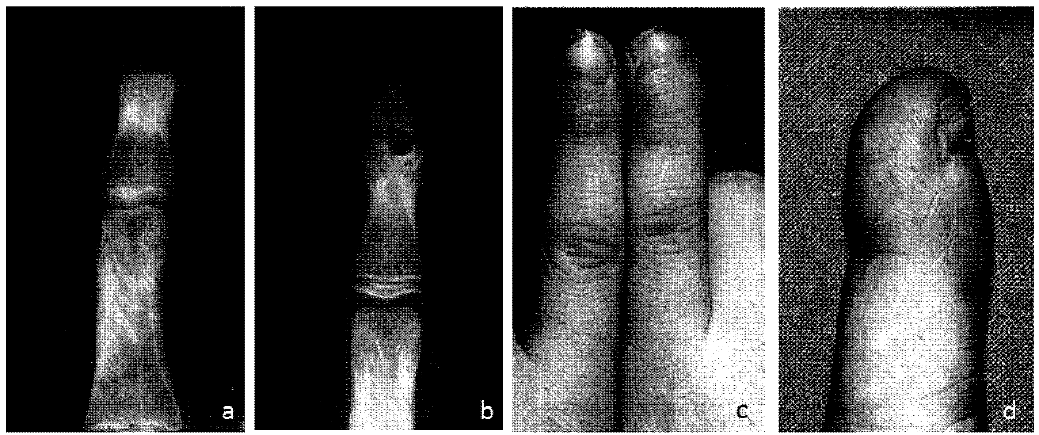
3. Fingertip Regeneration</title>
      <p>Clinical reports from as early as 1932 show that humans are indeed capable of regenerating complex tissues such as the fingertip [<xref ref-type="bibr" rid="B4-cells-01-01313">4</xref>]. Following conservative treatment of fingertip amputations, full restoration of fingerprint, sensation and functionality has been observed with minimal visible scarring (<xref ref-type="fig" rid="cells-01-01313-f002">Figure 2</xref>). Although fingertip regeneration is most commonly described in young children, adults too have demonstrated bone regrowth following conservative treatment of fingertip amputations [<xref ref-type="bibr" rid="B4-cells-01-01313">4</xref>,<xref ref-type="bibr" rid="B5-cells-01-01313">5</xref>]. Amputation resulting in the removal of the distal half of the terminal phalange results in successful regeneration of all components of tissue, including skeletal structure, with no morphological differences to the uninjured digit, although digit length may be shortened. A more proximal amputation, removing more than 2/3 of terminal phalanx, will however illicit no discernible regenerative response, although the wound will heal by cutaneous tissue repair [<xref ref-type="bibr" rid="B6-cells-01-01313">6</xref>,<xref ref-type="bibr" rid="B7-cells-01-01313">7</xref>]. </p>
      <p>Regeneration in the mammalian digit tip is a dynamic three phase process, incorporating reepithelialisation, dedifferentiation and redifferentiation. The first stage is marked by an initial wound healing response with inflammation, leading to the formation of an epidermal layer over the wound [<xref ref-type="bibr" rid="B8-cells-01-01313">8</xref>]. Although not fully investigated, it is likely this first phase closely follows the processes of cutaneous wound healing; inflammation, tissue formation and remodelling, however it is noted that regenerative wounds reepithelialise slower than non-regenerative wounds [<xref ref-type="bibr" rid="B9-cells-01-01313">9</xref>,<xref ref-type="bibr" rid="B10-cells-01-01313">10</xref>]. Wound closure time is variable in distal regenerative amputation injuries, whereas in non-regenerative proximal amputations, a single layer neo epidermis is achieved immediately following the inflammatory response, around four days post injury in mice [<xref ref-type="bibr" rid="B11-cells-01-01313">11</xref>]. Rates of reepithelialisation of the digit tip relate to proximal osteoclast bone degradation of the distal bone stump, resulting in the exposure of the marrow cells to the wound site and provide a route for epidermal migration [<xref ref-type="bibr" rid="B11-cells-01-01313">11</xref>]. It has been suggested that rapid restoration of skin integrity occurs at a cost of scar formation, with reduced structural identity and functionality [<xref ref-type="bibr" rid="B12-cells-01-01313">12</xref>]. The delay in reepithelialisation evident in regenerating digit tips appears to further support this hypothesis. Immediately following re-epithelialisation of the wound, dedifferentiation occurs, with the formation of a blastema under this epidermal layer contiguous with the marrow cavity centrally, and the loose connective tissue peripherally. A blastema is a devascularised, proliferating pool of progenitor cells from which organ formation or regeneration derives [<xref ref-type="bibr" rid="B7-cells-01-01313">7</xref>,<xref ref-type="bibr" rid="B11-cells-01-01313">11</xref>]. Although originally thought to be comprised of a homogenous pool of pluripotent stem cells, as is urodele regeneration, the blastema in regenerative mammalian digit tips has been found to be a heterogeneous pool of fate restricted progenitor cells [<xref ref-type="bibr" rid="B13-cells-01-01313">13</xref>,<xref ref-type="bibr" rid="B14-cells-01-01313">14</xref>]. It is during the final phase, that all tissues of the digit tip, excluding the epidermis redifferentiate from the blastema, however unlike urodele regeneration, transdifferentiation does not occur, with regenerates following fate restricted lineages [<xref ref-type="bibr" rid="B8-cells-01-01313">8</xref>,<xref ref-type="bibr" rid="B13-cells-01-01313">13</xref>].</p><fig id="cells-01-01313-f001" position="float">
        <label>Figure 1</label>
        <caption>
          <p>Regeneration of a salamander forelimb following distal amputation below the elbow (left) or proximal amputation through the humerus (right) (Adapted from [<xref ref-type="bibr" rid="B15-cells-01-01313">15</xref>]). </p>
        </caption>
        <graphic xmlns:xlink="http://www.w3.org/1999/xlink" xlink:href="cells-01-01313-g001.tif"/>
      </fig>
    <fig id="cells-01-01313-f002" position="float">
        <label>Figure 2</label>
        <caption>
          <p>X-rays of patient at admission (<bold>a</bold>) and six months after the injury (<bold>b</bold>) showing regeneration of the distal phalanx. Photographs of the injured finger six months after the injury with left index finger for comparison (<bold>c</bold>) and close up (<bold>d</bold>) (Adapted from [<xref ref-type="bibr" rid="B16-cells-01-01313">16</xref>]). </p>
        </caption>
        <graphic xmlns:xlink="http://www.w3.org/1999/xlink" xlink:href="cells-01-01313-g002.tif"/>
      </fig>
      
      <p>There is a distinct difference between mammalian digit regeneration and limb regeneration in lower order vertebrates, where the same developmental techniques are employed to regrow limbs. This has lead researchers to conclude that digit regeneration seen in mammals has re-evolved from a non-regenerative condition, rather than there being an evolutionarily conserved regeneration capacity [<xref ref-type="bibr" rid="B17-cells-01-01313">17</xref>]. Interestingly implantation of microcarrier beads impregnated with the bone morphogenic proteins, BMP-2 and -7, under the newly formed wound epidermis of proximally amputated digits induces bone regeneration, however bone regrowth occurs via endochondral ossification, appearing to reinitiate the bone development process, rather than stimulating the regenerative response seen in distal digit amputations [<xref ref-type="bibr" rid="B10-cells-01-01313">10</xref>]. Investigations into digit tip regeneration using MRL mice, a strain which exhibits particularly strong regenerative capacity, have revealed the importance not only of the bone morphogenic/TGFβ pathways, critical to developmental limb formation and bone induction, as well as fracture repair, but also genes required for WNT/β-catenin mediated signalling pathways [<xref ref-type="bibr" rid="B8-cells-01-01313">8</xref>]. </p>
    </sec>
    <sec>
      <title>
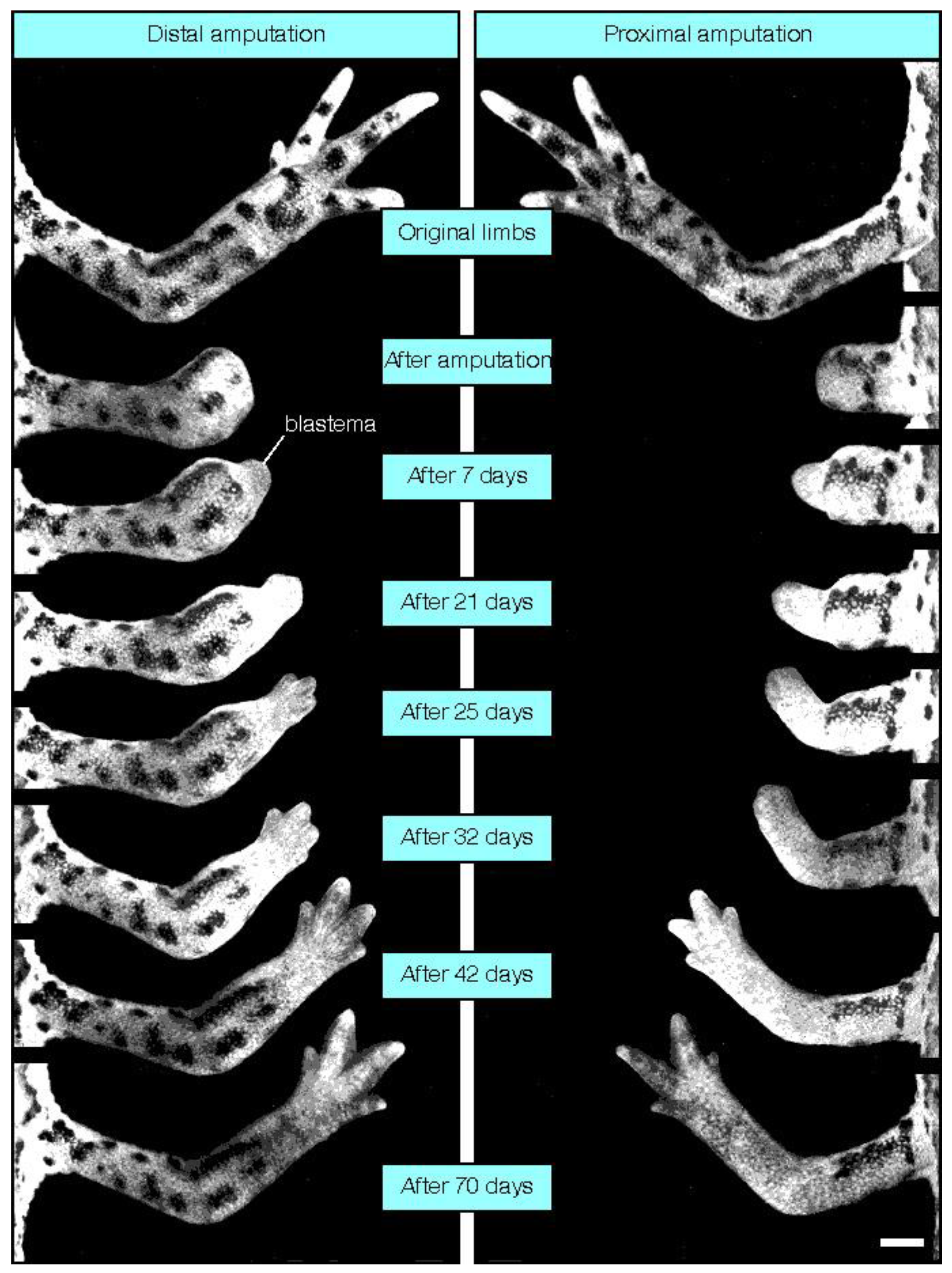
4. Mammalian Regenerative Capacity</title>
      <p>Other potential examples of mammalian regenerative capacity do exist and include hair follicle regeneration which occurs not only as part of normal homeostasis, but also in response to wound healing. Here regeneration relies upon adult somatic stem cells providing a pool of new multipotent cells [<xref ref-type="bibr" rid="B18-cells-01-01313">18</xref>]. Wnt signalling is involved in hair follicle morphogenesis, and both the prolonged activation of β-catenin-dependant pathway, and forced activation of the non-canonical pathway by Wnt-5a has resulted in epidermal appendages such as rudimentary hair follicles, sebaceous glands and epithelial lined cysts forming within the healing wound area [<xref ref-type="bibr" rid="B19-cells-01-01313">19</xref>,<xref ref-type="bibr" rid="B20-cells-01-01313">20</xref>]. The human liver is one of the few internal organs which does display regenerative capacity, being able to restore lost mass following resection, from as little as 25% of its original tissue. This capacity is largely due to proliferation of and differentiation of unipotent hepatocytes [<xref ref-type="bibr" rid="B21-cells-01-01313">21</xref>]. Intraoral mucosal wounds also show regenerative potential, exhibiting rapid remodelling with minimal scar formation. This is due in part to a more optimal wound environment, with regards to moisture and temperature as well as decreased tension across the wound [<xref ref-type="bibr" rid="B22-cells-01-01313">22</xref>]. In addition though, oral mucosal fibroblasts exhibit a phenotype more similar to that seen in fetal wound healing, with enhanced MMP matrix remodelling capability resulting in less scar formation [<xref ref-type="bibr" rid="B23-cells-01-01313">23</xref>]. Being able to capitalise on inherent regenerative capacity of many cells types may enable regeneration of other complex organs and systems.</p>
    </sec>
    <sec>
      <title>
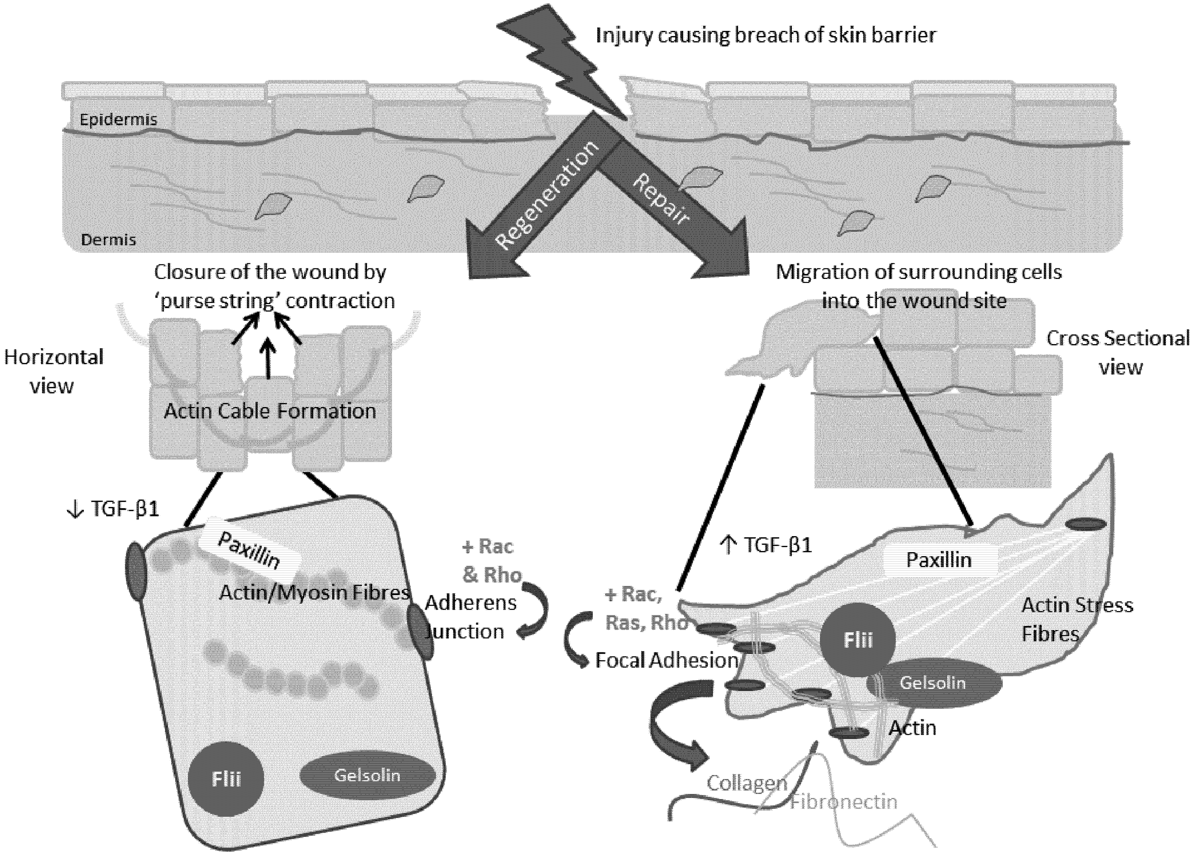
5. The Role of the Actin Cytoskeleton in Regeneration and Repair
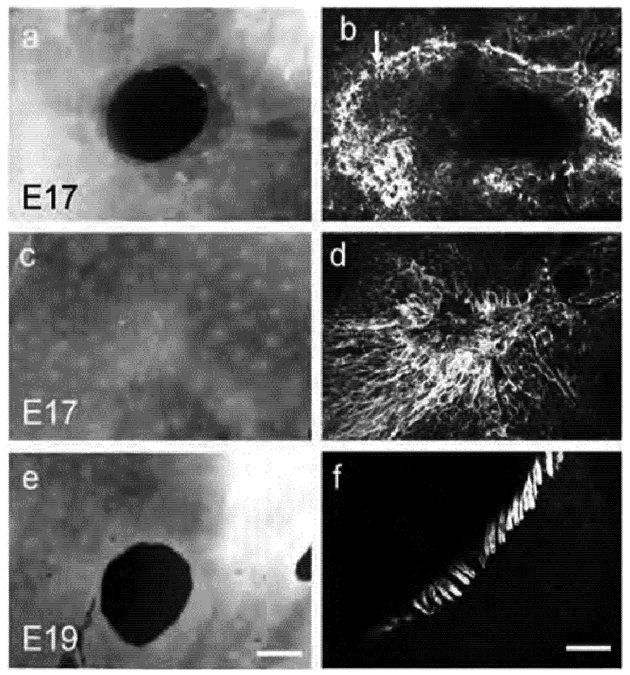
</title>
      <p>From the initial infiltration of inflammatory cells, through the repopulation of the neodermis by fibroblasts and the reestablishment of the epidermis via keratinocyte migration, to the process of angiogenesis and tissue remodelling, effective healing of our skin and tissues relies heavily upon the dynamic reorganisation of the actin cytoskeleton [<xref ref-type="bibr" rid="B24-cells-01-01313">24</xref>,<xref ref-type="bibr" rid="B25-cells-01-01313">25</xref>]. The cytoskeleton is made up of a network of microtubules, actin filaments, intermediate filaments and their associated proteins which are responsible not only for cell shape, but also for controlling cellular adhesion, contraction and motility [<xref ref-type="bibr" rid="B25-cells-01-01313">25</xref>,<xref ref-type="bibr" rid="B26-cells-01-01313">26</xref>]. It is the active assembly and disassembly of filamentous actin and re-organisation of it into functional networks which underpins all of these important cell processes. Adhesion of cells to different surfaces and substrates is particularly required post-wounding when cellular infiltration of the wound site occurs and this is facilitated by integrin-rich focal adhesions connecting cells to underlying substrates as well as cadherin based cell-cell adhesion complexes which occur at the termination of filament bundles [<xref ref-type="bibr" rid="B27-cells-01-01313">27</xref>]. Not only do focal adhesions anchor polymerized actin filament stress fibres, but they facilitate the formation of actin filament bundles which provide contractile forces during cellular migration. Focal adhesion proteins, such as paxillin also serve as platforms for signal transduction which influences the extracellular matrix and facilitates dermal remodelling [<xref ref-type="bibr" rid="B28-cells-01-01313">28</xref>]. Thus, the precise function and regulation of the actin cytoskeleton is critical to the success of the wound healing process and its manipulation may facilitate regenerative healing.</p>
    </sec>
    <sec>
      <title>6. Regenerative Fetal <italic>vs</italic> Reparative Adult Wound Healing</title>
      <p>One of the most researched areas of mammalian regeneration is that of fetal wound repair [<xref ref-type="bibr" rid="B29-cells-01-01313">29</xref>]. Early gestation wounds in fetal skin prior to the third trimester heal without scarring with complete restitution of dermal appendages [<xref ref-type="bibr" rid="B30-cells-01-01313">30</xref>,<xref ref-type="bibr" rid="B31-cells-01-01313">31</xref>]. Differences in fetal vs adult wound repair have been identified including reduced inflammatory responses [<xref ref-type="bibr" rid="B32-cells-01-01313">32</xref>,<xref ref-type="bibr" rid="B33-cells-01-01313">33</xref>], differences in extracellular matrix composition [<xref ref-type="bibr" rid="B34-cells-01-01313">34</xref>] and reduced expression of pro-scarring growth factors eg TGF-β1 [<xref ref-type="bibr" rid="B35-cells-01-01313">35</xref>,<xref ref-type="bibr" rid="B36-cells-01-01313">36</xref>]. However the actin cytoskeleton also appears to play a major role in the regenerative healing observed in the fetus [<xref ref-type="bibr" rid="B29-cells-01-01313">29</xref>]. Fetal wounds reepithelialise very quickly, however, rather than epidermal cells relying upon lammellipodial crawling across a provisional wound matrix the process used in adult wound repair, wound closure occurs by contraction of actin-myosin fibres in ‘purse string’ like manner, drawing the edges of the wound together (<xref ref-type="fig" rid="cells-01-01313-f003">Figure 3</xref>) [<xref ref-type="bibr" rid="B9-cells-01-01313">9</xref>,<xref ref-type="bibr" rid="B29-cells-01-01313">29</xref>,<xref ref-type="bibr" rid="B37-cells-01-01313">37</xref>]. The purse string is formed by rapid polymerisation of filamentous actin some five to six cell back from the wound edge and appears anchored by E-cadherin-mediated adherens junctions at the leading edge to facilitate the coordinated cell movement [<xref ref-type="bibr" rid="B29-cells-01-01313">29</xref>,<xref ref-type="bibr" rid="B31-cells-01-01313">31</xref>,<xref ref-type="bibr" rid="B38-cells-01-01313">38</xref>]. The closure of fetal wound appears less reliant upon the mesenchymal contraction observed in adult wounds, as fetal epidermis is capable of closing a wound <italic>in vitro</italic> in the absence of dermal substrata [<xref ref-type="bibr" rid="B39-cells-01-01313">39</xref>,<xref ref-type="bibr" rid="B40-cells-01-01313">40</xref>]. Indeed fetal wound fibroblasts do not appear to express α-smooth muscle actin (α-SMA) suggesting fetal fibroblasts may not convert to the contractile myofibroblastic phenotype [<xref ref-type="bibr" rid="B41-cells-01-01313">41</xref>] indicating that they are unlikely to be responsible for wound contraction in the fetal wound [<xref ref-type="bibr" rid="B29-cells-01-01313">29</xref>,<xref ref-type="bibr" rid="B41-cells-01-01313">41</xref>]. </p>
      <p>In contrast to the fetus, adult wounds heal by a mechanism of repair which generally leads to scar formation and structurally and functionally diminished skin [<xref ref-type="bibr" rid="B29-cells-01-01313">29</xref>]. In normal skin, the collagen matrix is formed in a basket weave like pattern, allowing movement and stretch; however post injury, wound fibroblasts lay down new collagen in densely packed bundles of thinner fibres, aligned parallel to the epidermis [<xref ref-type="bibr" rid="B42-cells-01-01313">42</xref>]. Due to excessive elastase activity, the elastin content of this new skin is decreased within the dermis and an absence of characteristic rete ridges and normal dermal-epidermal junction can be seen [<xref ref-type="bibr" rid="B43-cells-01-01313">43</xref>]. The resulting scar is marked by a functional loss of elasticity, tensile strength and is prone to contracture. Skin appendages such as hair follicles and sebaceous glands never regenerate and there are clear alterations to the vascularity and nerve supply of the scar and remaining appendages. As such, scar tissue is also marked by alopecia, desiccation and thermal dysregulation [<xref ref-type="bibr" rid="B20-cells-01-01313">20</xref>,<xref ref-type="bibr" rid="B43-cells-01-01313">43</xref>]. Rapid restoration of skin integrity and protection from bacterial onslaught following injury comes with the cost of scar formation and loss of normal structure and function, which are not only unsightly but often require painful and costly revisionary surgeries [<xref ref-type="bibr" rid="B20-cells-01-01313">20</xref>,<xref ref-type="bibr" rid="B41-cells-01-01313">41</xref>]. This is particularly challenging for children post burn injury where their growth places extra demands on their skin and contracture around joints can lead to lifelong disabilities. There remains a great need for a more regenerative response to restore functional properties and original tissue architecture [<xref ref-type="bibr" rid="B20-cells-01-01313">20</xref>]. </p>
	  <fig id="cells-01-01313-f003" position="float">
        <label>Figure 3</label>
        <caption>
          <p>Actin cables are formed in early but not late gestation foetal skin. Wounds created in E17 but not E19 fetal skin, when cultured in DMEM/10%FBS re-epithelialise and close over 72 h. Wounded E17 fetal skin at (<bold>a</bold>) 3 h postwounding and (<bold>c</bold>) 72 h post-wounding. (<bold>e</bold>) wounded E19 fetal skin 72 h post-wounding. Phalloidin-FITC staining of F-actin reveals actin ring around E17 wound 48 (<bold>b</bold>) and 72 h post wounding (<bold>d</bold>). No cables are formed in E19 fetal skin wound (<bold>f</bold>). Magnification bar in (<bold>e</bold>) = 600 μm and in (<bold>f</bold>) = 50 μm. Arrow in (<bold>b</bold>) points to actin cable (Adapted from [<xref ref-type="bibr" rid="B29-cells-01-01313">29</xref>]).</p>
        </caption>
        <graphic xmlns:xlink="http://www.w3.org/1999/xlink" xlink:href="cells-01-01313-g003.tif"/>
      </fig>
      
    </sec>
    <sec>
      <title>7. Involvement of the Actin Cytoskeleton during Regeneration and Repair</title>
      <p>Changes in the expression of proteins associated with the actin cytoskeleton are indicative of the switch between scar-free regeneration and scar-forming repair. Paxillin colocalises with actin in E17 regenerative wounds, but not E19 reparative wounds, indicating a potential role for paxillin in wound regeneration [<xref ref-type="bibr" rid="B31-cells-01-01313">31</xref>]. In contrast, gelsolin is upregulated in E19 embryonic wounds but not in E17 regenerating skin and gelsolin is observed surrounding actin filaments during ‘adult type’ healing but not in the earlier, scar-free wounds [<xref ref-type="bibr" rid="B31-cells-01-01313">31</xref>]. Gelsolin has long been known to regulate both fetal and adult wound healing via its effect upon the actin cytoskeleton and is the founding member of a family of actin-remodelling proteins [<xref ref-type="bibr" rid="B31-cells-01-01313">31</xref>]. The family includes seven cytoskeletal associated proteins: gelsolin, adseverin, villin, capG, advillin, supervillin and flightless I, involved in cellular processes including motility, apoptosis and phagocytosis. All members contain a highly conserved gelsolin-like domain with either 3 or 6 homologous repeats, but may also contain additional domains endowing them with different capabilities [<xref ref-type="bibr" rid="B44-cells-01-01313">44</xref>]. These proteins regulate actin filaments by severing pre-existing filaments and/or capping the filament ends, a process involving the gelsolin domains. After severing, they remain attached to the ‘barbed’ ends of the broken filament, thereby preventing annealing or addition of actin monomers. Actin filaments are subsequently uncapped by interaction with phosphoinositides, leading to rapid actin assembly. This is the first step in enabling cells to reorientate their cytoskeleton to drive changes in motility, adhesion and contraction [<xref ref-type="bibr" rid="B25-cells-01-01313">25</xref>,<xref ref-type="bibr" rid="B45-cells-01-01313">45</xref>]. Gelsolin acts to scavenge actin that is exposed to extracellular spaces or released into the circulation after tissue injury preventing pathogenesis of organ injury at sites removed from the primary insult due to persistence of actin within the microvasculature [<xref ref-type="bibr" rid="B46-cells-01-01313">46</xref>,<xref ref-type="bibr" rid="B47-cells-01-01313">47</xref>].</p>
      <p>Members of the Rho family of small GTPases; Rho, Rac and Cdc42, are largely responsible for regulating the actin cytoskeleton, coordinating the actions of multiple proteins involved in gene transcription and adhesion [<xref ref-type="bibr" rid="B27-cells-01-01313">27</xref>]. The assembly of stress fibres, the contractile actin-myosin filaments and associated focal adhesions are triggered by Rho activation by membrane receptors [<xref ref-type="bibr" rid="B48-cells-01-01313">48</xref>] enabling the stable attachment of the cell to the extracellular matrix and the transmission of force required for remodelling of the dermal matrix [<xref ref-type="bibr" rid="B49-cells-01-01313">49</xref>]. Rac activation is responsible for lammellipodia and membrane ruffle production at the leading edge of the motile cell through the assembly of an actin filament mesh at the cell periphery [<xref ref-type="bibr" rid="B50-cells-01-01313">50</xref>]. Cdc42 triggers the formation of filipodia, another actin based protrusions found at the periphery of the migrating cell [<xref ref-type="bibr" rid="B51-cells-01-01313">51</xref>]. Additionally, Cdc42 acts in concert with Rac to stimulate the assembly of focal complexes [<xref ref-type="bibr" rid="B51-cells-01-01313">51</xref>], the initial, transient adhesions formed at the periphery of the spreading or migrating cell [<xref ref-type="bibr" rid="B52-cells-01-01313">52</xref>]. Ras, another GTPase, also acts as a molecular switch in the regulation of cytoskeletal dynamics, as Ras activates Rac, thereby inducing lammellipodial formation [<xref ref-type="bibr" rid="B50-cells-01-01313">50</xref>]. Rho and Rac are also required for the formation of cadherin-based adherens junctions which link stress fibres from neighbouring cells to form tight cell-cell junctions [<xref ref-type="bibr" rid="B53-cells-01-01313">53</xref>]. It is clear that the dynamic control of the actin cytoskeleton underpins the processes necessary for initiation and progression of the wound healing cascade from cell infiltration of the wound site to remodelling of the dermal scar.</p>
    </sec>
    <sec>
      <title>8. Flightless I in Wound Regeneration and Repair</title>
      <p>Investigations into a more regenerative, scar free wound healing process have revealed the dynamic involvement of the gelsolin family member, Flightless I (Flii). Flii is a member of the gelsolin family of actin-remodelling proteins [<xref ref-type="bibr" rid="B54-cells-01-01313">54</xref>]. Flii was first identified in Drosophila melanogaster where it regulates development of flight muscles and in which Flii mutations result in irregular actin organisation and defective flight muscles [<xref ref-type="bibr" rid="B55-cells-01-01313">55</xref>]. The protein is highly conserved among mammals, where it is developmentally essential, with homozygous loss of Flii exhibiting gastrulation failure, leading to embryonic lethality [<xref ref-type="bibr" rid="B56-cells-01-01313">56</xref>]. Flii associates with many cytoskeletal proteins, colocalising with Ras and Cdc42, molecules involved in cytoskeletal reorganisation as well as localising with actin filaments themselves [<xref ref-type="bibr" rid="B57-cells-01-01313">57</xref>,<xref ref-type="bibr" rid="B58-cells-01-01313">58</xref>]. </p>
      <p>Flii is a negative regulator of wound healing, being shown to affect cell proliferation, motility and matrix production. A decrease in Flii expression, either endogenously in a heterozygous knockout engineered mouse, or via the topical application of a neutralising antibody raised against the LRR domain in murine and porcine wound models results in improved healing [<xref ref-type="bibr" rid="B12-cells-01-01313">12</xref>,<xref ref-type="bibr" rid="B59-cells-01-01313">59</xref>]. Conversely, overexpression of Flii in the mouse resulted in larger scars, with a slower, impaired wound healing response [<xref ref-type="bibr" rid="B12-cells-01-01313">12</xref>]. Flii localises with β-tubulin based structures involved in cell division and within lammellipodia and filopodia associated with migrating cells [<xref ref-type="bibr" rid="B60-cells-01-01313">60</xref>]. A decrease in Flii levels results in increased motility in keratinocytes and fibroblasts <italic>in vitro</italic> and in rates of keratinocyte re-epithelialisation. Decreasing <italic>Flii</italic> expression using siRNA in NIH 3T3 cells also results in decreased intracellular stress fiber formation [<xref ref-type="bibr" rid="B61-cells-01-01313">61</xref>]. A number of studies have shown a cross-talk between pathways involved in cytoskeletal remodelling, cellular adhesion and migration during wound repair, including TGF-β signaling and members of the MAP kinase family, Ras, RhoA, MAPK-ERK kinase and ERK1/2 [<xref ref-type="bibr" rid="B62-cells-01-01313">62</xref>,<xref ref-type="bibr" rid="B63-cells-01-01313">63</xref>,<xref ref-type="bibr" rid="B64-cells-01-01313">64</xref>,<xref ref-type="bibr" rid="B65-cells-01-01313">65</xref>]. Multiple <italic>in vivo</italic> and <italic>in vitro</italic> studies have demonstrated that in mouse fibroblasts, Flii specifically colocalises with cytoskeletal structures connected with a migratory phenotype [<xref ref-type="bibr" rid="B60-cells-01-01313">60</xref>] and associates with both structural and signaling proteins at sites of focal adhesions, including talin, paxillin and vinculin [<xref ref-type="bibr" rid="B66-cells-01-01313">66</xref>]. Flii is associated with actin arcs, membrane ruffles and is present at the leading edge of cells, where it also colocalizes with the GTP-binding proteins Ras, Cdc42 and RhoA that have central roles in regulating cytoskeletal reorganization [<xref ref-type="bibr" rid="B67-cells-01-01313">67</xref>,<xref ref-type="bibr" rid="B68-cells-01-01313">68</xref>]. Association of Flii LRR domain with Ras and Cdc42 proteins suggests possible involvement of Flii in downstream PI3K and MAPK signalling pathways. The PI3K/Akt signalling pathways are known to regulate numerous fundamental cellular functions during wound repair, including cell growth, proliferation, motility and survival and is activated by a variety of extracellular signals [<xref ref-type="bibr" rid="B69-cells-01-01313">69</xref>]. That Flii is endogenously upregulated in response to wounding and yet overexpression leads to impaired wound healing is a quirk similar to that of TGFβ1 in which expression in also upregulated in response to wounding but overexpression leads to increased scar formation [<xref ref-type="bibr" rid="B12-cells-01-01313">12</xref>,<xref ref-type="bibr" rid="B70-cells-01-01313">70</xref>] and indicates the tightly regulated nature of molecules critical to wound healing processes. Indeed in fetal skin, Flii expression in mice increases between embryonic day E17 at which the fetus heals without scar and embryonic day E19, resulting in scar formation [<xref ref-type="bibr" rid="B58-cells-01-01313">58</xref>]. Although wounding transiently increases Flii expression in E17 but not E19 wounds, Flii expression is down regulated in E17 keratinocytes immediately adjacent the wound. Moreover, in E19 wounds, Flii is strongly expressed within the cytoplasm and nucleus of keratinocytes at the leading edge [<xref ref-type="bibr" rid="B58-cells-01-01313">58</xref>]. It appears that an increase in Flii expression is linked with impaired wound healing and a switch to reparative, scar forming wound healing (<xref ref-type="fig" rid="cells-01-01313-f004">Figure 4</xref>). Using a recognized model of hair follicle regeneration, recent studies by Waters <italic>et al.</italic>, have shown that Flii has a positive influence on hair follicle regeneration, which is contrary to its negative influences on wound healing [<xref ref-type="bibr" rid="B71-cells-01-01313">71</xref>]. Regenerated follicles expressing high levels of <italic>Flii</italic>, produced significantly longer terminal hair fibers while <italic>Flii</italic> deficiency resulted in a delayed or impaired regenerative potential of hair follicles [<xref ref-type="bibr" rid="B71-cells-01-01313">71</xref>]. <italic>Flii<sup>+/−</sup></italic> follicles that failed to regenerate the endbulb structures also displayed low expression of markers of normal hair follicle development suggesting a lack in developmental activity [<xref ref-type="bibr" rid="B71-cells-01-01313">71</xref>]. The effect of Flii on cellular adhesion and its involvement in signalling pathways important to hair follicle development, growth and cycling might explain the delayed regeneration of hair follicles in <italic>Flii</italic> deficient mice [<xref ref-type="bibr" rid="B71-cells-01-01313">71</xref>]. Indeed, Flii has been shown to inhibit the Wnt/β-catenin pathway [<xref ref-type="bibr" rid="B72-cells-01-01313">72</xref>] and play a role as a thyroid hormone and estrogen-activated nuclear receptor co-activator [<xref ref-type="bibr" rid="B73-cells-01-01313">73</xref>,<xref ref-type="bibr" rid="B74-cells-01-01313">74</xref>] both of which would have particular significance to hair follicle development. </p>
      <fig id="cells-01-01313-f004" position="float">
        <label>Figure 4</label>
        <caption>
          <p>Schematic diagram of differences in actin cytoskeleton organisation and associated proteins in early gestation, regenerative healing compared to late gestation ‘adult type’ scar forming repair.</p>
        </caption>
        <graphic xmlns:xlink="http://www.w3.org/1999/xlink" xlink:href="cells-01-01313-g004.tif"/>
      </fig>
      <p>Flii not only contains six repeats of the gelsolin-like domain, endowing it with actin binding abilities, but contains the novel feature among gelsolin family members of an amino-terminal leucine rich repeat sequence [<xref ref-type="bibr" rid="B57-cells-01-01313">57</xref>]. Leucine rich repeat motifs are known to mediate protein-protein interactions and it has been suggested this domain enables Flii to link signal transduction to cytoskeletal regulation [<xref ref-type="bibr" rid="B54-cells-01-01313">54</xref>,<xref ref-type="bibr" rid="B75-cells-01-01313">75</xref>]. Flii has both cytoplasmic and nuclear activity where it is a transcriptional coactivator. It is known to regulate transcription of the estrogen and thyroid receptors [<xref ref-type="bibr" rid="B74-cells-01-01313">74</xref>] and has a differential effect upon cell cycle progression [<xref ref-type="bibr" rid="B76-cells-01-01313">76</xref>]. Flii is secreted into human plasma and has immunological roles further indicating the critical role for Flii in wound healing [<xref ref-type="bibr" rid="B77-cells-01-01313">77</xref>]. Flii interacts with CARM1 and GRIP1 which are positive transcriptional regulators of the β-catenin: TCF/LEF complex within the canonical Wnt signalling pathway to inhibit β-catenin dependant transcription [<xref ref-type="bibr" rid="B72-cells-01-01313">72</xref>,<xref ref-type="bibr" rid="B74-cells-01-01313">74</xref>,<xref ref-type="bibr" rid="B78-cells-01-01313">78</xref>] Interestingly the cytoplasmic protein known as the murine Flightless Associated Protein 1 (FLAP1) or its human homologue Leucine Rich Repeat In FLII Interacting Protein 1/2 (LRRFIP1/2) activate this transcription pathway. FLAP1 increases IFN-β expression by enhancing β-catenin activation [<xref ref-type="bibr" rid="B79-cells-01-01313">79</xref>]. LRRFIP binds Dishevelled complexed with GSK-3β, which prevents GSK-3β phosphorylation of β-catenin, subsequently leading to increased transcription of downstream β-catenin targets [<xref ref-type="bibr" rid="B80-cells-01-01313">80</xref>]. It appears that Flii exerts its action upon the canonical WNT pathway by disrupting the binding of FLAP1/LRRFIP1/2 and β-catenin [<xref ref-type="bibr" rid="B72-cells-01-01313">72</xref>].</p>
      <p>Both Flii expression and Wnt signalling is closely linked to the switch from fetal scarless to scarring healing. Wnt signalling is induced at embryonic day 19.5 in mice, but not before, whilst Flii expression is increased significantly at embryonic day 19 [<xref ref-type="bibr" rid="B58-cells-01-01313">58</xref>,<xref ref-type="bibr" rid="B81-cells-01-01313">81</xref>]. The involvement of Flii and the Wnt signalling pathways in the switch between fetal scar free healing and wound repair, as well as hair follicle regeneration combined with the dynamic interactions between Flii and both the canonical and noncanonical signalling pathways, suggests a potential role in regeneration as well as a possible target for therapeutic manipulation leading to enhance wound healing outcomes.</p>
    </sec>
    <sec>
      <title>9. Conclusion</title>
      <p>Although mammals have limited regenerative capacity when compared to the epimorphic regeneration seen in lower order vertebrates and invertebrates, there remains instances in which regenerative healing can occur, returned full structure and function to the injured tissue. As well as select tissues displaying this capacity, such as the liver, oral mucosa, hair follicles and the tissues of the fingertip, fetal wounds heal with scar free restoration, due in a large part, to differential organization of the cellular cytoskeleton. This network of actin filaments and microtubules which give the cell its structure are dynamically regulated to facilitate adhesion, motility and contraction as well as division, processes fundamental to all processes of wound healing. Differences in expression levels and localization of cytoskeletal proteins of the gelsolin family between regenerative healing, such as in fetal skin, and the more common adult type, scar forming healing process, has highlighted the importance of cytoskeletal regulation in tissue regeneration. The dynamic involvement of family member, Flii appears critical to the regulation of wound healing and is a key player in the drive towards regenerative healing. Further insights into the events important for tissue regeneration will identify further targets for therapeutic development. To be able to drive the body towards the replacement of native tissues, rather than merely repairing the injury offers great potential to improve the quality of life of humans worldwide. Understanding the differences between regeneration and repair offers the greatest prospect that we will one day be able to stimulate this regenerative process and identifying the role of the actin cytoskeleton in this process is likely to be a critical step towards this goal.</p>
    </sec>
  </body>
  <back>
    <ack>
      <title>Acknowledgements</title>
      <p>AJC is supported by an NHMRC Senior Research Fellowship (#1002009).</p>
    </ack>
    <ref-list>
      <title>References</title>
      <ref id="B1-cells-01-01313">
        <label>1.</label>
        <citation citation-type="journal">
          <person-group person-group-type="author">
            <name>
              <surname>Morasso</surname>
              <given-names>M.I.</given-names>
            </name>
          </person-group>
          <article-title>The influence of flightless i: Regeneration versus wound healing</article-title>
          <source>J. Invest. Dermatol.</source>
          <year>2011</year>
          <volume>131</volume>
          <fpage>816</fpage>
          <lpage>817</lpage>
          <pub-id pub-id-type="doi">10.1038/jid.2011.8</pub-id>
        </citation>
      </ref>
      <ref id="B2-cells-01-01313">
        <label>2.</label>
        <citation citation-type="journal">
          <person-group person-group-type="author">
            <name>
              <surname>Sanchez Alvarado</surname>
              <given-names>A.</given-names>
            </name>
          </person-group>
          <article-title>Regeneration in the metazoans: Why does it happen?</article-title>
          <source>Bioessays</source>
          <year>2000</year>
          <volume>22</volume>
          <fpage>578</fpage>
          <lpage>590</lpage>
          <pub-id pub-id-type="doi">10.1002/(SICI)1521-1878(200006)22:6&lt;578::AID-BIES11&gt;3.0.CO;2-#</pub-id>
        </citation>
      </ref>
      <ref id="B3-cells-01-01313">
        <label>3.</label>
        <citation citation-type="journal">
          <person-group person-group-type="author">
            <name>
              <surname>Maiorana</surname>
              <given-names>V.C.</given-names>
            </name>
          </person-group>
          <article-title>Tail autonomy, functional conflicts and their resolution by a salamander</article-title>
          <source>Nature</source>
          <year>1997</year>
          <volume>2265</volume>
          <fpage>533</fpage>
          <lpage>535</lpage>
        </citation>
      </ref>
      <ref id="B4-cells-01-01313">
        <label>4.</label>
        <citation citation-type="journal">
          <person-group person-group-type="author">
            <name>
              <surname>McKim</surname>
              <given-names>L.H.</given-names>
            </name>
          </person-group>
          <article-title>Regeneration of the distal phalanx</article-title>
          <source>Can. Med. Assoc. J.</source>
          <year>1932</year>
          <volume>26</volume>
          <fpage>549</fpage>
          <lpage>550</lpage>
        <pub-id pub-id-type="pmid">20318716</pub-id></citation>
      </ref>
      <ref id="B5-cells-01-01313">
        <label>5.</label>
        <citation citation-type="journal">
          <person-group person-group-type="author">
            <name>
              <surname>Soderberg</surname>
              <given-names>T.</given-names>
            </name>
            <name>
              <surname>Nystrom</surname>
              <given-names>A.</given-names>
            </name>
            <name>
              <surname>Hallmans</surname>
              <given-names>G.</given-names>
            </name>
            <name>
              <surname>Hulten</surname>
              <given-names>J.</given-names>
            </name>
          </person-group>
          <article-title>Treatment of fingertip amputations with bone exposure. A comparative study between surgical and conservative treatment methods</article-title>
          <source>Scand. J. Plast. Reconstr. Surg. Suppl.</source>
          <year>1983</year>
          <volume>17</volume>
          <fpage>147</fpage>
          <lpage>152</lpage>
          <pub-id pub-id-type="doi">10.3109/02844318309013111</pub-id>
        </citation>
      </ref>
      <ref id="B6-cells-01-01313">
        <label>6.</label>
        <citation citation-type="journal">
          <person-group person-group-type="author">
            <name>
              <surname>Neufeld</surname>
              <given-names>D.A.</given-names>
            </name>
            <name>
              <surname>Zhao</surname>
              <given-names>W.</given-names>
            </name>
          </person-group>
          <article-title>Bone regrowth after digit tip amputation in mice is equivalent in adults and neonates</article-title>
          <source>Wound Repair Regen.</source>
          <year>1995</year>
          <volume>3</volume>
          <fpage>461</fpage>
          <lpage>466</lpage>
        <pub-id pub-id-type="doi">10.1046/j.1524-475X.1995.30410.x</pub-id><pub-id pub-id-type="pmid">17147657</pub-id></citation>
      </ref>
      <ref id="B7-cells-01-01313">
        <label>7.</label>
        <citation citation-type="journal">
          <person-group person-group-type="author">
            <name>
              <surname>Han</surname>
              <given-names>M.</given-names>
            </name>
            <name>
              <surname>Yang</surname>
              <given-names>X.</given-names>
            </name>
            <name>
              <surname>Lee</surname>
              <given-names>J.</given-names>
            </name>
            <name>
              <surname>Allan</surname>
              <given-names>C.H.</given-names>
            </name>
            <name>
              <surname>Muneoka</surname>
              <given-names>K.</given-names>
            </name>
          </person-group>
          <article-title>Development and regeneration of the neonatal digit tip in mice</article-title>
          <source>Dev. Biol.</source>
          <year>2008</year>
          <volume>315</volume>
          <fpage>125</fpage>
          <lpage>135</lpage>
          <pub-id pub-id-type="doi">10.1016/j.ydbio.2007.12.025</pub-id>
        </citation>
      </ref>
      <ref id="B8-cells-01-01313">
        <label>8.</label>
        <citation citation-type="journal">
          <person-group person-group-type="author">
            <name>
              <surname>Chadwick</surname>
              <given-names>R.B.</given-names>
            </name>
            <name>
              <surname>Bu</surname>
              <given-names>L.</given-names>
            </name>
            <name>
              <surname>Yu</surname>
              <given-names>H.</given-names>
            </name>
            <name>
              <surname>Hu</surname>
              <given-names>Y.</given-names>
            </name>
            <name>
              <surname>Wergedal</surname>
              <given-names>J.E.</given-names>
            </name>
            <name>
              <surname>Mohan</surname>
              <given-names>S.</given-names>
            </name>
            <name>
              <surname>Baylink</surname>
              <given-names>D.J.</given-names>
            </name>
          </person-group>
          <article-title>Digit tip regrowth and differential gene expression in mrl/mpj, dba/2, and c57bl/6 mice</article-title>
          <source>Wound Repair Regen.</source>
          <year>2007</year>
          <volume>15</volume>
          <fpage>275</fpage>
          <lpage>284</lpage>
          <pub-id pub-id-type="doi">10.1111/j.1524-475X.2007.00216.x</pub-id>
        </citation>
      </ref>
      <ref id="B9-cells-01-01313">
        <label>9.</label>
        <citation citation-type="journal">
          <person-group person-group-type="author">
            <name>
              <surname>Singer</surname>
              <given-names>A.J.</given-names>
            </name>
            <name>
              <surname>Clark</surname>
              <given-names>R.A.</given-names>
            </name>
          </person-group>
          <article-title>Cutaneous wound healing</article-title>
          <source>N. Engl. J. Med.</source>
          <year>1999</year>
          <volume>341</volume>
          <fpage>738</fpage>
          <lpage>746</lpage>
          <pub-id pub-id-type="doi">10.1056/NEJM199909023411006</pub-id>
        </citation>
      </ref>
      <ref id="B10-cells-01-01313">
        <label>10.</label>
        <citation citation-type="journal">
          <person-group person-group-type="author">
            <name>
              <surname>Yu</surname>
              <given-names>L.</given-names>
            </name>
            <name>
              <surname>Han</surname>
              <given-names>M.</given-names>
            </name>
            <name>
              <surname>Yan</surname>
              <given-names>M.</given-names>
            </name>
            <name>
              <surname>Lee</surname>
              <given-names>E.C.</given-names>
            </name>
            <name>
              <surname>Lee</surname>
              <given-names>J.</given-names>
            </name>
            <name>
              <surname>Muneoka</surname>
              <given-names>K.</given-names>
            </name>
          </person-group>
          <article-title>Bmp signaling induces digit regeneration in neonatal mice</article-title>
          <source>Development</source>
          <year>2010</year>
          <volume>137</volume>
          <fpage>551</fpage>
          <lpage>559</lpage>
          <pub-id pub-id-type="doi">10.1242/dev.042424</pub-id>
        </citation>
      </ref>
      <ref id="B11-cells-01-01313">
        <label>11.</label>
        <citation citation-type="journal">
          <person-group person-group-type="author">
            <name>
              <surname>Fernando</surname>
              <given-names>W.A.</given-names>
            </name>
            <name>
              <surname>Leininger</surname>
              <given-names>E.</given-names>
            </name>
            <name>
              <surname>Simkin</surname>
              <given-names>J.</given-names>
            </name>
            <name>
              <surname>Li</surname>
              <given-names>N.</given-names>
            </name>
            <name>
              <surname>Malcom</surname>
              <given-names>C.A.</given-names>
            </name>
            <name>
              <surname>Sathyamoorthi</surname>
              <given-names>S.</given-names>
            </name>
            <name>
              <surname>Han</surname>
              <given-names>M.</given-names>
            </name>
            <name>
              <surname>Muneoka</surname>
              <given-names>K.</given-names>
            </name>
          </person-group>
          <article-title>Wound healing and blastema formation in regenerating digit tips of adult mice</article-title>
          <source>Dev. Biol.</source>
          <year>2011</year>
          <volume>350</volume>
          <fpage>301</fpage>
          <lpage>310</lpage>
          <pub-id pub-id-type="doi">10.1016/j.ydbio.2010.11.035</pub-id>
        </citation>
      </ref>
      <ref id="B12-cells-01-01313">
        <label>12.</label>
        <citation citation-type="journal">
          <person-group person-group-type="author">
            <name>
              <surname>Cowin</surname>
              <given-names>A.J.</given-names>
            </name>
            <name>
              <surname>Adams</surname>
              <given-names>D.H.</given-names>
            </name>
            <name>
              <surname>Strudwick</surname>
              <given-names>X.L.</given-names>
            </name>
            <name>
              <surname>Chan</surname>
              <given-names>H.</given-names>
            </name>
            <name>
              <surname>Hooper</surname>
              <given-names>J.A.</given-names>
            </name>
            <name>
              <surname>Sander</surname>
              <given-names>G.R.</given-names>
            </name>
            <name>
              <surname>Rayner</surname>
              <given-names>T.E.</given-names>
            </name>
            <name>
              <surname>Matthaei</surname>
              <given-names>K.I.</given-names>
            </name>
            <name>
              <surname>Powell</surname>
              <given-names>B.C.</given-names>
            </name>
            <name>
              <surname>Campbell</surname>
              <given-names>H.D.</given-names>
            </name>
          </person-group>
          <article-title>Flightless i deficiency enhances wound repair by increasing cell migration and proliferation</article-title>
          <source>J. Pathol. Bacteriol.</source>
          <year>2007</year>
          <volume>211</volume>
          <fpage>572</fpage>
          <lpage>581</lpage>
          <pub-id pub-id-type="doi">10.1002/path.2143</pub-id>
        </citation>
      </ref>
      <ref id="B13-cells-01-01313">
        <label>13.</label>
        <citation citation-type="journal">
          <person-group person-group-type="author">
            <name>
              <surname>Lehoczky</surname>
              <given-names>J.A.</given-names>
            </name>
            <name>
              <surname>Robert</surname>
              <given-names>B.</given-names>
            </name>
            <name>
              <surname>Tabin</surname>
              <given-names>C.J.</given-names>
            </name>
          </person-group>
          <article-title>Mouse digit tip regeneration is mediated by fate-restricted progenitor cells</article-title>
          <source>Proc. Natl. Acad. Sci. USA</source>
          <year>2011</year>
          <volume>108</volume>
          <fpage>20609</fpage>
          <lpage>20614</lpage>
        <pub-id pub-id-type="doi">10.1073/pnas.1118017108</pub-id><pub-id pub-id-type="pmid">22143790</pub-id></citation>
      </ref>
      <ref id="B14-cells-01-01313">
        <label>14.</label>
        <citation citation-type="journal">
          <person-group person-group-type="author">
            <name>
              <surname>Rinkevich</surname>
              <given-names>Y.</given-names>
            </name>
            <name>
              <surname>Lindau</surname>
              <given-names>P.</given-names>
            </name>
            <name>
              <surname>Ueno</surname>
              <given-names>H.</given-names>
            </name>
            <name>
              <surname>Longaker</surname>
              <given-names>M.T.</given-names>
            </name>
            <name>
              <surname>Weissman</surname>
              <given-names>I.L.</given-names>
            </name>
          </person-group>
          <article-title>Germ-layer and lineage-restricted stem/progenitors regenerate the mouse digit tip</article-title>
          <source>Nature</source>
          <year>2011</year>
          <volume>476</volume>
          <fpage>409</fpage>
          <lpage>413</lpage>
        <pub-id pub-id-type="pmid">21866153</pub-id><pub-id pub-id-type="doi">10.1038/nature10346</pub-id></citation>
      </ref>
      <ref id="B15-cells-01-01313">
        <label>15.</label>
        <citation citation-type="book">
          <person-group person-group-type="author">
            <name>
              <surname>Goss</surname>
              <given-names>R.J.</given-names>
            </name>
          </person-group>
          <source>Principles of regeneration</source>
          <publisher-name>Academic Press</publisher-name>
          <publisher-loc>New York, NY, USA</publisher-loc>
          <year>1969</year>
        </citation>
      </ref>
      <ref id="B16-cells-01-01313">
        <label>16.</label>
        <citation citation-type="journal">
          <person-group person-group-type="author">
            <name>
              <surname>Vidal</surname>
              <given-names>P.</given-names>
            </name>
            <name>
              <surname>Dickson</surname>
              <given-names>M.G.</given-names>
            </name>
          </person-group>
          <article-title>Regeneration of the distal phalanx. A case report</article-title>
          <source>J. Hand. Surg.</source>
          <year>1993</year>
          <volume>18</volume>
          <fpage>230</fpage>
          <lpage>233</lpage>
        </citation>
      </ref>
      <ref id="B17-cells-01-01313">
        <label>17.</label>
        <citation citation-type="journal">
          <person-group person-group-type="author">
            <name>
              <surname>Muneoka</surname>
              <given-names>K.</given-names>
            </name>
            <name>
              <surname>Allan</surname>
              <given-names>C.H.</given-names>
            </name>
            <name>
              <surname>Yang</surname>
              <given-names>X.</given-names>
            </name>
            <name>
              <surname>Lee</surname>
              <given-names>J.</given-names>
            </name>
            <name>
              <surname>Han</surname>
              <given-names>M.</given-names>
            </name>
          </person-group>
          <article-title>Mammalian regeneration and regenerative medicine</article-title>
          <source>Birth Defects Res. C Embryo Today</source>
          <year>2008</year>
          <volume>84</volume>
          <fpage>265</fpage>
          <lpage>280</lpage>
          <pub-id pub-id-type="doi">10.1002/bdrc.20137</pub-id>
        </citation>
      </ref>
      <ref id="B18-cells-01-01313">
        <label>18.</label>
        <citation citation-type="journal">
          <person-group person-group-type="author">
            <name>
              <surname>Jahoda</surname>
              <given-names>C.A.</given-names>
            </name>
            <name>
              <surname>Oliver</surname>
              <given-names>R.F.</given-names>
            </name>
            <name>
              <surname>Reynolds</surname>
              <given-names>A.J.</given-names>
            </name>
            <name>
              <surname>Forrester</surname>
              <given-names>J.C.</given-names>
            </name>
            <name>
              <surname>Horne</surname>
              <given-names>K.A.</given-names>
            </name>
          </person-group>
          <article-title>Human hair follicle regeneration following amputation and grafting into the nude mouse</article-title>
          <source>J. Invest. Dermatol.</source>
          <year>1996</year>
          <volume>107</volume>
          <fpage>804</fpage>
          <lpage>807</lpage>
        <pub-id pub-id-type="doi">10.1111/1523-1747.ep12330565</pub-id><pub-id pub-id-type="pmid">8941664</pub-id></citation>
      </ref>
      <ref id="B19-cells-01-01313">
        <label>19.</label>
        <citation citation-type="journal">
          <person-group person-group-type="author">
            <name>
              <surname>Gat</surname>
              <given-names>U.</given-names>
            </name>
            <name>
              <surname>DasGupta</surname>
              <given-names>R.</given-names>
            </name>
            <name>
              <surname>Degenstein</surname>
              <given-names>L.</given-names>
            </name>
            <name>
              <surname>Fuchs</surname>
              <given-names>E.</given-names>
            </name>
          </person-group>
          <article-title>De novo hair follicle morphogenesis and hair tumors in mice expressing a truncated beta-catenin in skin</article-title>
          <source>Cell</source>
          <year>1998</year>
          <volume>95</volume>
          <fpage>605</fpage>
          <lpage>614</lpage>
          <pub-id pub-id-type="doi">10.1016/S0092-8674(00)81631-1</pub-id>
        </citation>
      </ref>
      <ref id="B20-cells-01-01313">
        <label>20.</label>
        <citation citation-type="journal">
          <person-group person-group-type="author">
            <name>
              <surname>Fathke</surname>
              <given-names>C.</given-names>
            </name>
            <name>
              <surname>Wilson</surname>
              <given-names>L.</given-names>
            </name>
            <name>
              <surname>Shah</surname>
              <given-names>K.</given-names>
            </name>
            <name>
              <surname>Kim</surname>
              <given-names>B.</given-names>
            </name>
            <name>
              <surname>Hocking</surname>
              <given-names>A.</given-names>
            </name>
            <name>
              <surname>Moon</surname>
              <given-names>R.</given-names>
            </name>
            <name>
              <surname>Isik</surname>
              <given-names>F.</given-names>
            </name>
          </person-group>
          <article-title>Wnt signaling induces epithelial differentiation during cutaneous wound healing</article-title>
          <source>BMC Cell Biol.</source>
          <year>2006</year>
          <volume>7</volume>
          <fpage>4</fpage>
          <pub-id pub-id-type="doi">10.1186/1471-2121-7-4</pub-id>
        </citation>
      </ref>
      <ref id="B21-cells-01-01313">
        <label>21.</label>
        <citation citation-type="journal">
          <person-group person-group-type="author">
            <name>
              <surname>Michalopoulos</surname>
              <given-names>G.K.</given-names>
            </name>
            <name>
              <surname>DeFrances</surname>
              <given-names>M.C.</given-names>
            </name>
          </person-group>
          <article-title>Liver regeneration</article-title>
          <source>Science</source>
          <year>1997</year>
          <volume>276</volume>
          <fpage>60</fpage>
          <lpage>66</lpage>
          <pub-id pub-id-type="doi">10.1126/science.276.5309.60</pub-id>
        </citation>
      </ref>
      <ref id="B22-cells-01-01313">
        <label>22.</label>
        <citation citation-type="journal">
          <person-group person-group-type="author">
            <name>
              <surname>Thomas</surname>
              <given-names>D.W.</given-names>
            </name>
            <name>
              <surname>Harding</surname>
              <given-names>K.G.</given-names>
            </name>
          </person-group>
          <article-title>Wound healing</article-title>
          <source>Br. J. Surg.</source>
          <year>2002</year>
          <volume>89</volume>
          <fpage>1203</fpage>
          <lpage>1205</lpage>
        <pub-id pub-id-type="doi">10.1046/j.1365-2168.2002.02181.x</pub-id><pub-id pub-id-type="pmid">12296885</pub-id></citation>
      </ref>
      <ref id="B23-cells-01-01313">
        <label>23.</label>
        <citation citation-type="journal">
          <person-group person-group-type="author">
            <name>
              <surname>Stephens</surname>
              <given-names>P.</given-names>
            </name>
            <name>
              <surname>Davies</surname>
              <given-names>K.J.</given-names>
            </name>
            <name>
              <surname>Occleston</surname>
              <given-names>N.</given-names>
            </name>
            <name>
              <surname>Pleass</surname>
              <given-names>R.D.</given-names>
            </name>
            <name>
              <surname>Kon</surname>
              <given-names>C.</given-names>
            </name>
            <name>
              <surname>Daniels</surname>
              <given-names>J.</given-names>
            </name>
            <name>
              <surname>Khaw</surname>
              <given-names>P.T.</given-names>
            </name>
            <name>
              <surname>Thomas</surname>
              <given-names>D.W.</given-names>
            </name>
          </person-group>
          <article-title>Skin and oral fibroblasts exhibit phenotypic differences in extracellular matrix reorganization and matrix metalloproteinase activity</article-title>
          <source>Br. J. Dermatol.</source>
          <year>2001</year>
          <volume>144</volume>
          <fpage>229</fpage>
          <lpage>237</lpage>
          <pub-id pub-id-type="doi">10.1046/j.1365-2133.2001.04006.x</pub-id>
        </citation>
      </ref>
      <ref id="B24-cells-01-01313">
        <label>24.</label>
        <citation citation-type="journal">
          <person-group person-group-type="author">
            <name>
              <surname>Abreu-Blanco</surname>
              <given-names>M.T.</given-names>
            </name>
            <name>
              <surname>Watts</surname>
              <given-names>J.J.</given-names>
            </name>
            <name>
              <surname>Verboon</surname>
              <given-names>J.M.</given-names>
            </name>
            <name>
              <surname>Parkhurst</surname>
              <given-names>S.M.</given-names>
            </name>
          </person-group>
          <article-title>Cytoskeleton responses in wound repair</article-title>
          <source>Cell. Mol. Life Sci.</source>
          <year>2012</year>
          <fpage>2469</fpage>
          <lpage>2483</lpage>
        </citation>
      </ref>
      <ref id="B25-cells-01-01313">
        <label>25.</label>
        <citation citation-type="journal">
          <person-group person-group-type="author">
            <name>
              <surname>Cowin</surname>
              <given-names>A.</given-names>
            </name>
          </person-group>
          <article-title>Role of the actin cytoskeleton in wound healing and scar formation</article-title>
          <source>Primary Intention</source>
          <year>2006</year>
          <volume>14</volume>
          <fpage>39</fpage>
          <lpage>42</lpage>
        </citation>
      </ref>
      <ref id="B26-cells-01-01313">
        <label>26.</label>
        <citation citation-type="journal">
          <person-group person-group-type="author">
            <name>
              <surname>Desai</surname>
              <given-names>A.</given-names>
            </name>
            <name>
              <surname>Mitchison</surname>
              <given-names>T.J.</given-names>
            </name>
          </person-group>
          <article-title>Microtubule polymerization dynamics</article-title>
          <source>Annu. Rev. Cell Dev. Biol.</source>
          <year>1997</year>
          <volume>13</volume>
          <fpage>83</fpage>
          <lpage>117</lpage>
          <pub-id pub-id-type="doi">10.1146/annurev.cellbio.13.1.83</pub-id>
        </citation>
      </ref>
      <ref id="B27-cells-01-01313">
        <label>27.</label>
        <citation citation-type="journal">
          <person-group person-group-type="author">
            <name>
              <surname>Hall</surname>
              <given-names>A.</given-names>
            </name>
          </person-group>
          <article-title>Rho gtpases and the actin cytoskeleton</article-title>
          <source>Science</source>
          <year>1998</year>
          <volume>279</volume>
          <fpage>509</fpage>
          <lpage>514</lpage>
          <pub-id pub-id-type="doi">10.1126/science.279.5350.509</pub-id>
        </citation>
      </ref>
      <ref id="B28-cells-01-01313">
        <label>28.</label>
        <citation citation-type="journal">
          <person-group person-group-type="author">
            <name>
              <surname>Turner</surname>
              <given-names>C.E.</given-names>
            </name>
          </person-group>
          <article-title>Paxillin interactions</article-title>
          <source>J. Cell. Sci.</source>
          <year>2000</year>
          <volume>113</volume>
          <fpage>4139</fpage>
          <lpage>4140</lpage>
        <pub-id pub-id-type="pmid">11069756</pub-id></citation>
      </ref>
      <ref id="B29-cells-01-01313">
        <label>29.</label>
        <citation citation-type="journal">
          <person-group person-group-type="author">
            <name>
              <surname>Martin</surname>
              <given-names>P.</given-names>
            </name>
          </person-group>
          <article-title>Wound healing--aiming for perfect skin regeneration</article-title>
          <source>Science</source>
          <year>1997</year>
          <volume>276</volume>
          <fpage>75</fpage>
          <lpage>81</lpage>
          <pub-id pub-id-type="doi">10.1126/science.276.5309.75</pub-id>
        </citation>
      </ref>
      <ref id="B30-cells-01-01313">
        <label>30.</label>
        <citation citation-type="journal">
          <person-group person-group-type="author">
            <name>
              <surname>Stelnicki</surname>
              <given-names>E.J.</given-names>
            </name>
            <name>
              <surname>Chin</surname>
              <given-names>G.S.</given-names>
            </name>
            <name>
              <surname>Gittes</surname>
              <given-names>G.K.</given-names>
            </name>
            <name>
              <surname>Longaker</surname>
              <given-names>M.T.</given-names>
            </name>
          </person-group>
          <article-title>Fetal wound repair: Where do we go from here?</article-title>
          <source>Semin. Pediatr. Surg.</source>
          <year>1999</year>
          <volume>8</volume>
          <fpage>124</fpage>
          <lpage>130</lpage>
        <pub-id pub-id-type="pmid">10461325</pub-id></citation>
      </ref>
      <ref id="B31-cells-01-01313">
        <label>31.</label>
        <citation citation-type="journal">
          <person-group person-group-type="author">
            <name>
              <surname>Cowin</surname>
              <given-names>A.J.</given-names>
            </name>
            <name>
              <surname>Hatzirodos</surname>
              <given-names>N.</given-names>
            </name>
            <name>
              <surname>Teusner</surname>
              <given-names>J.T.</given-names>
            </name>
            <name>
              <surname>Belford</surname>
              <given-names>D.A.</given-names>
            </name>
          </person-group>
          <article-title>Differential effect of wounding on actin and its associated proteins, paxillin and gelsolin, in fetal skin explants</article-title>
          <source>J. Invest. Dermatol.</source>
          <year>2003</year>
          <volume>120</volume>
          <fpage>1118</fpage>
          <lpage>1129</lpage>
          <pub-id pub-id-type="doi">10.1046/j.1523-1747.2003.12231.x</pub-id>
        </citation>
      </ref>
      <ref id="B32-cells-01-01313">
        <label>32.</label>
        <citation citation-type="journal">
          <person-group person-group-type="author">
            <name>
              <surname>Hopkinson-Woolley</surname>
              <given-names>J.</given-names>
            </name>
            <name>
              <surname>Hughes</surname>
              <given-names>D.</given-names>
            </name>
            <name>
              <surname>Gordon</surname>
              <given-names>S.</given-names>
            </name>
            <name>
              <surname>Martin</surname>
              <given-names>P.</given-names>
            </name>
          </person-group>
          <article-title>Macrophage recruitment during limb development and wound healing in the embryonic and foetal mouse</article-title>
          <source>J. Cell. Sci.</source>
          <year>1994</year>
          <volume>107</volume>
          <fpage>1159</fpage>
          <lpage>1167</lpage>
        <pub-id pub-id-type="pmid">7929625</pub-id></citation>
      </ref>
      <ref id="B33-cells-01-01313">
        <label>33.</label>
        <citation citation-type="journal">
          <person-group person-group-type="author">
            <name>
              <surname>Cowin</surname>
              <given-names>A.J.</given-names>
            </name>
            <name>
              <surname>Brosnan</surname>
              <given-names>M.P.</given-names>
            </name>
            <name>
              <surname>Holmes</surname>
              <given-names>T.M.</given-names>
            </name>
            <name>
              <surname>Ferguson</surname>
              <given-names>M.W.</given-names>
            </name>
          </person-group>
          <article-title>Endogenous inflammatory response to dermal wound healing in the fetal and adult mouse</article-title>
          <source>Dev. Dyn.</source>
          <year>1998</year>
          <volume>212</volume>
          <fpage>385</fpage>
          <lpage>393</lpage>
          <pub-id pub-id-type="doi">10.1002/(SICI)1097-0177(199807)212:3&lt;385::AID-AJA6&gt;3.0.CO;2-D</pub-id>
        </citation>
      </ref>
      <ref id="B34-cells-01-01313">
        <label>34.</label>
        <citation citation-type="journal">
          <person-group person-group-type="author">
            <name>
              <surname>Longaker</surname>
              <given-names>M.T.</given-names>
            </name>
            <name>
              <surname>Peled</surname>
              <given-names>Z.M.</given-names>
            </name>
            <name>
              <surname>Chang</surname>
              <given-names>J.</given-names>
            </name>
            <name>
              <surname>Krummel</surname>
              <given-names>T.M.</given-names>
            </name>
          </person-group>
          <article-title>Fetal wound healing: Progress report and future directions</article-title>
          <source>Surgery</source>
          <year>2001</year>
          <volume>130</volume>
          <fpage>785</fpage>
          <lpage>787</lpage>
          <pub-id pub-id-type="doi">10.1067/msy.2001.114145</pub-id>
        </citation>
      </ref>
      <ref id="B35-cells-01-01313">
        <label>35.</label>
        <citation citation-type="journal">
          <person-group person-group-type="author">
            <name>
              <surname>Cowin</surname>
              <given-names>A.J.</given-names>
            </name>
            <name>
              <surname>Hatzirodos</surname>
              <given-names>N.</given-names>
            </name>
            <name>
              <surname>Holding</surname>
              <given-names>C.A.</given-names>
            </name>
            <name>
              <surname>Dunaiski</surname>
              <given-names>V.</given-names>
            </name>
            <name>
              <surname>Harries</surname>
              <given-names>R.H.</given-names>
            </name>
            <name>
              <surname>Rayner</surname>
              <given-names>T.E.</given-names>
            </name>
            <name>
              <surname>Fitridge</surname>
              <given-names>R.</given-names>
            </name>
            <name>
              <surname>Cooter</surname>
              <given-names>R.D.</given-names>
            </name>
            <name>
              <surname>Schultz</surname>
              <given-names>G.S.</given-names>
            </name>
            <name>
              <surname>Belford</surname>
              <given-names>D.A.</given-names>
            </name>
          </person-group>
          <article-title>Effect of healing on the expression of transforming growth factor beta(s) and their receptors in chronic venous leg ulcers</article-title>
          <source>J. Invest. Dermatol.</source>
          <year>2001</year>
          <volume>117</volume>
          <fpage>1282</fpage>
          <lpage>1289</lpage>
          <pub-id pub-id-type="doi">10.1046/j.0022-202x.2001.01501.x</pub-id>
        </citation>
      </ref>
      <ref id="B36-cells-01-01313">
        <label>36.</label>
        <citation citation-type="journal">
          <person-group person-group-type="author">
            <name>
              <surname>Ferguson</surname>
              <given-names>M.W.</given-names>
            </name>
          </person-group>
          <article-title>Skin wound healing: Transforming growth factor beta antagonists decrease scarring and improve quality</article-title>
          <source>J. Interferon Res.</source>
          <year>1994</year>
          <volume>14</volume>
          <fpage>303</fpage>
          <lpage>304</lpage>
          <pub-id pub-id-type="doi">10.1089/jir.1994.14.303</pub-id>
        </citation>
      </ref>
      <ref id="B37-cells-01-01313">
        <label>37.</label>
        <citation citation-type="journal">
          <person-group person-group-type="author">
            <name>
              <surname>Jacinto</surname>
              <given-names>A.</given-names>
            </name>
            <name>
              <surname>Martinez-Arias</surname>
              <given-names>A.</given-names>
            </name>
            <name>
              <surname>Martin</surname>
              <given-names>P.</given-names>
            </name>
          </person-group>
          <article-title>Mechanisms of epithelial fusion and repair</article-title>
          <source>Nat. Cell Biol.</source>
          <year>2001</year>
          <volume>3</volume>
          <fpage>E117</fpage>
          <lpage>123</lpage>
          <pub-id pub-id-type="doi">10.1038/35074643</pub-id>
        </citation>
      </ref>
      <ref id="B38-cells-01-01313">
        <label>38.</label>
        <citation citation-type="journal">
          <person-group person-group-type="author">
            <name>
              <surname>Danjo</surname>
              <given-names>Y.</given-names>
            </name>
            <name>
              <surname>Gipson</surname>
              <given-names>I.K.</given-names>
            </name>
          </person-group>
          <article-title>Actin 'purse string' filaments are anchored by e-cadherin-mediated adherens junctions at the leading edge of the epithelial wound, providing coordinated cell movement</article-title>
          <source>J. Cell. Sci.</source>
          <year>1998</year>
          <volume>111</volume>
          <fpage>3323</fpage>
          <lpage>3332</lpage>
        <pub-id pub-id-type="pmid">9788874</pub-id></citation>
      </ref>
      <ref id="B39-cells-01-01313">
        <label>39.</label>
        <citation citation-type="journal">
          <person-group person-group-type="author">
            <name>
              <surname>Belford</surname>
              <given-names>D.A.</given-names>
            </name>
          </person-group>
          <article-title>The mechanism of excisional fetal wound repair in vitro is responsive to growth factors</article-title>
          <source>Endocrinology</source>
          <year>1997</year>
          <volume>138</volume>
          <fpage>3987</fpage>
          <lpage>3996</lpage>
          <pub-id pub-id-type="doi">10.1210/en.138.9.3987</pub-id>
        </citation>
      </ref>
      <ref id="B40-cells-01-01313">
        <label>40.</label>
        <citation citation-type="journal">
          <person-group person-group-type="author">
            <name>
              <surname>Ihara</surname>
              <given-names>S.</given-names>
            </name>
            <name>
              <surname>Motobayashi</surname>
              <given-names>Y.</given-names>
            </name>
            <name>
              <surname>Nagao</surname>
              <given-names>E.</given-names>
            </name>
            <name>
              <surname>Kistler</surname>
              <given-names>A.</given-names>
            </name>
          </person-group>
          <article-title>Ontogenetic transition of wound healing pattern in rat skin occurring at the fetal stage</article-title>
          <source>Development</source>
          <year>1990</year>
          <volume>110</volume>
          <fpage>671</fpage>
          <lpage>680</lpage>
        <pub-id pub-id-type="pmid">2088714</pub-id></citation>
      </ref>
      <ref id="B41-cells-01-01313">
        <label>41.</label>
        <citation citation-type="journal">
          <person-group person-group-type="author">
            <name>
              <surname>Amadeu</surname>
              <given-names>T.P.</given-names>
            </name>
            <name>
              <surname>Coulomb</surname>
              <given-names>B.</given-names>
            </name>
            <name>
              <surname>Desmouliere</surname>
              <given-names>A.</given-names>
            </name>
            <name>
              <surname>Costa</surname>
              <given-names>A.M.</given-names>
            </name>
          </person-group>
          <article-title>Cutaneous wound healing: Myofibroblastic differentiation and in vitro models</article-title>
          <source>Int. J. Low. Extrem. Wounds</source>
          <year>2003</year>
          <volume>2</volume>
          <fpage>60</fpage>
          <lpage>68</lpage>
          <pub-id pub-id-type="doi">10.1177/1534734603256155</pub-id>
        </citation>
      </ref>
      <ref id="B42-cells-01-01313">
        <label>42.</label>
        <citation citation-type="journal">
          <person-group person-group-type="author">
            <name>
              <surname>Dallon</surname>
              <given-names>J.</given-names>
            </name>
            <name>
              <surname>Sherratt</surname>
              <given-names>J.</given-names>
            </name>
            <name>
              <surname>Maini</surname>
              <given-names>P.</given-names>
            </name>
            <name>
              <surname>Ferguson</surname>
              <given-names>M.</given-names>
            </name>
          </person-group>
          <article-title>Biological implications of a discrete mathematical model for collagen deposition and alignment in dermal wound repair</article-title>
          <source>IMA J. Math. Appl. Med. Biol.</source>
          <year>2000</year>
          <volume>17</volume>
          <fpage>379</fpage>
          <lpage>393</lpage>
        <pub-id pub-id-type="doi">10.1093/imammb/17.4.379</pub-id><pub-id pub-id-type="pmid">11270750</pub-id></citation>
      </ref>
      <ref id="B43-cells-01-01313">
        <label>43.</label>
        <citation citation-type="journal">
          <person-group person-group-type="author">
            <name>
              <surname>O'Kane</surname>
              <given-names>S.</given-names>
            </name>
          </person-group>
          <article-title>Wound remodelling and scarring</article-title>
          <source>J Wound Care</source>
          <year>2002</year>
          <volume>11</volume>
          <fpage>296</fpage>
          <lpage>299</lpage>
        <pub-id pub-id-type="pmid">12360763</pub-id></citation>
      </ref>
      <ref id="B44-cells-01-01313">
        <label>44.</label>
        <citation citation-type="journal">
          <person-group person-group-type="author">
            <name>
              <surname>Silacci</surname>
              <given-names>P.</given-names>
            </name>
            <name>
              <surname>Mazzolai</surname>
              <given-names>L.</given-names>
            </name>
            <name>
              <surname>Gauci</surname>
              <given-names>C.</given-names>
            </name>
            <name>
              <surname>Stergiopulos</surname>
              <given-names>N.</given-names>
            </name>
            <name>
              <surname>Yin</surname>
              <given-names>H.L.</given-names>
            </name>
            <name>
              <surname>Hayoz</surname>
              <given-names>D.</given-names>
            </name>
          </person-group>
          <article-title>Gelsolin superfamily proteins: Key regulators of cellular functions</article-title>
          <source>Cell. Mol. Life Sci.</source>
          <year>2004</year>
          <volume>61</volume>
          <fpage>2614</fpage>
          <lpage>2623</lpage>
          <pub-id pub-id-type="doi">10.1007/s00018-004-4225-6</pub-id>
        </citation>
      </ref>
      <ref id="B45-cells-01-01313">
        <label>45.</label>
        <citation citation-type="journal">
          <person-group person-group-type="author">
            <name>
              <surname>Sun</surname>
              <given-names>H.Q.</given-names>
            </name>
            <name>
              <surname>Yamamoto</surname>
              <given-names>M.</given-names>
            </name>
            <name>
              <surname>Mejillano</surname>
              <given-names>M.</given-names>
            </name>
            <name>
              <surname>Yin</surname>
              <given-names>H.L.</given-names>
            </name>
          </person-group>
          <article-title>Gelsolin, a multifunctional actin regulatory protein</article-title>
          <source>J. Biol. Chem.</source>
          <year>1999</year>
          <volume>274</volume>
          <fpage>33179</fpage>
          <lpage>33182</lpage>
          <pub-id pub-id-type="doi">10.1074/jbc.274.47.33179</pub-id>
        </citation>
      </ref>
      <ref id="B46-cells-01-01313">
        <label>46.</label>
        <citation citation-type="journal">
          <person-group person-group-type="author">
            <name>
              <surname>Lee</surname>
              <given-names>W.M.</given-names>
            </name>
            <name>
              <surname>Galbraith</surname>
              <given-names>R.M.</given-names>
            </name>
          </person-group>
          <article-title>The extracellular actin-scavenger system and actin toxicity</article-title>
          <source>N. Engl. J. Med.</source>
          <year>1992</year>
          <volume>326</volume>
          <fpage>1335</fpage>
          <lpage>1341</lpage>
          <pub-id pub-id-type="doi">10.1056/NEJM199205143262006</pub-id>
        </citation>
      </ref>
      <ref id="B47-cells-01-01313">
        <label>47.</label>
        <citation citation-type="journal">
          <person-group person-group-type="author">
            <name>
              <surname>Mounzer</surname>
              <given-names>K.C.</given-names>
            </name>
            <name>
              <surname>Moncure</surname>
              <given-names>M.</given-names>
            </name>
            <name>
              <surname>Smith</surname>
              <given-names>Y.R.</given-names>
            </name>
            <name>
              <surname>Dinubile</surname>
              <given-names>M.J.</given-names>
            </name>
          </person-group>
          <article-title>Relationship of admission plasma gelsolin levels to clinical outcomes in patients after major trauma</article-title>
          <source>Am. J. Respir. Crit. Care Med.</source>
          <year>1999</year>
          <volume>160</volume>
          <fpage>1673</fpage>
          <lpage>1681</lpage>
        <pub-id pub-id-type="pmid">10556139</pub-id></citation>
      </ref>
      <ref id="B48-cells-01-01313">
        <label>48.</label>
        <citation citation-type="journal">
          <person-group person-group-type="author">
            <name>
              <surname>Ridley</surname>
              <given-names>A.J.</given-names>
            </name>
            <name>
              <surname>Hall</surname>
              <given-names>A.</given-names>
            </name>
          </person-group>
          <article-title>The small gtp-binding protein rho regulates the assembly of focal adhesions and actin stress fibers in response to growth factors</article-title>
          <source>Cell</source>
          <year>1992</year>
          <volume>70</volume>
          <fpage>389</fpage>
          <lpage>399</lpage>
          <pub-id pub-id-type="doi">10.1016/0092-8674(92)90163-7</pub-id>
        </citation>
      </ref>
      <ref id="B49-cells-01-01313">
        <label>49.</label>
        <citation citation-type="journal">
          <person-group person-group-type="author">
            <name>
              <surname>Gardel</surname>
              <given-names>M.L.</given-names>
            </name>
            <name>
              <surname>Schneider</surname>
              <given-names>I.C.</given-names>
            </name>
            <name>
              <surname>Aratyn-Schaus</surname>
              <given-names>Y.</given-names>
            </name>
            <name>
              <surname>Waterman</surname>
              <given-names>C.M.</given-names>
            </name>
          </person-group>
          <article-title>Mechanical integration of actin and adhesion dynamics in cell migration</article-title>
          <source>Annu. Rev. Cell Dev. Biol.</source>
          <year>2010</year>
          <volume>26</volume>
          <fpage>315</fpage>
          <lpage>333</lpage>
          <pub-id pub-id-type="doi">10.1146/annurev.cellbio.011209.122036</pub-id>
        </citation>
      </ref>
      <ref id="B50-cells-01-01313">
        <label>50.</label>
        <citation citation-type="journal">
          <person-group person-group-type="author">
            <name>
              <surname>Ridley</surname>
              <given-names>A.J.</given-names>
            </name>
            <name>
              <surname>Paterson</surname>
              <given-names>H.F.</given-names>
            </name>
            <name>
              <surname>Johnston</surname>
              <given-names>C.L.</given-names>
            </name>
            <name>
              <surname>Diekmann</surname>
              <given-names>D.</given-names>
            </name>
            <name>
              <surname>Hall</surname>
              <given-names>A.</given-names>
            </name>
          </person-group>
          <article-title>The small gtp-binding protein rac regulates growth factor-induced membrane ruffling</article-title>
          <source>Cell</source>
          <year>1992</year>
          <volume>70</volume>
          <fpage>401</fpage>
          <lpage>410</lpage>
          <pub-id pub-id-type="doi">10.1016/0092-8674(92)90164-8</pub-id>
        </citation>
      </ref>
      <ref id="B51-cells-01-01313">
        <label>51.</label>
        <citation citation-type="journal">
          <person-group person-group-type="author">
            <name>
              <surname>Nobes</surname>
              <given-names>C.D.</given-names>
            </name>
            <name>
              <surname>Hall</surname>
              <given-names>A.</given-names>
            </name>
          </person-group>
          <article-title>Rho, rac, and cdc42 gtpases regulate the assembly of multimolecular focal complexes associated with actin stress fibers, lamellipodia, and filopodia</article-title>
          <source>Cell</source>
          <year>1995</year>
          <volume>81</volume>
          <fpage>53</fpage>
          <lpage>62</lpage>
          <pub-id pub-id-type="doi">10.1016/0092-8674(95)90370-4</pub-id>
        </citation>
      </ref>
      <ref id="B52-cells-01-01313">
        <label>52.</label>
        <citation citation-type="journal">
          <person-group person-group-type="author">
            <name>
              <surname>Wozniak</surname>
              <given-names>M.A.</given-names>
            </name>
            <name>
              <surname>Modzelewska</surname>
              <given-names>K.</given-names>
            </name>
            <name>
              <surname>Kwong</surname>
              <given-names>L.</given-names>
            </name>
            <name>
              <surname>Keely</surname>
              <given-names>P.J.</given-names>
            </name>
          </person-group>
          <article-title>Focal adhesion regulation of cell behavior</article-title>
          <source>Biochim. Biophys. Acta</source>
          <year>2004</year>
          <volume>1692</volume>
          <fpage>103</fpage>
          <lpage>119</lpage>
          <pub-id pub-id-type="doi">10.1016/j.bbamcr.2004.04.007</pub-id>
        </citation>
      </ref>
      <ref id="B53-cells-01-01313">
        <label>53.</label>
        <citation citation-type="journal">
          <person-group person-group-type="author">
            <name>
              <surname>Millan</surname>
              <given-names>J.</given-names>
            </name>
            <name>
              <surname>Cain</surname>
              <given-names>R.J.</given-names>
            </name>
            <name>
              <surname>Reglero-Real</surname>
              <given-names>N.</given-names>
            </name>
            <name>
              <surname>Bigarella</surname>
              <given-names>C.</given-names>
            </name>
            <name>
              <surname>Marcos-Ramiro</surname>
              <given-names>B.</given-names>
            </name>
            <name>
              <surname>Fernandez-Martin</surname>
              <given-names>L.</given-names>
            </name>
            <name>
              <surname>Correas</surname>
              <given-names>I.</given-names>
            </name>
            <name>
              <surname>Ridley</surname>
              <given-names>A.J.</given-names>
            </name>
          </person-group>
          <article-title>Adherens junctions connect stress fibres between adjacent endothelial cells</article-title>
          <source>BMC Biol.</source>
          <year>2010</year>
          <volume>8</volume>
          <fpage>11</fpage>
        <pub-id pub-id-type="doi">10.1186/1741-7007-8-11</pub-id><pub-id pub-id-type="pmid">20122254</pub-id></citation>
      </ref>
      <ref id="B54-cells-01-01313">
        <label>54.</label>
        <citation citation-type="journal">
          <person-group person-group-type="author">
            <name>
              <surname>Liu</surname>
              <given-names>Y.T.</given-names>
            </name>
            <name>
              <surname>Yin</surname>
              <given-names>H.L.</given-names>
            </name>
          </person-group>
          <article-title>Identification of the binding partners for flightless i, a novel protein bridging the leucine-rich repeat and the gelsolin superfamilies</article-title>
          <source>J. Biol. Chem.</source>
          <year>1998</year>
          <volume>273</volume>
          <fpage>7920</fpage>
          <lpage>7927</lpage>
          <pub-id pub-id-type="doi">10.1074/jbc.273.14.7920</pub-id>
        </citation>
      </ref>
      <ref id="B55-cells-01-01313">
        <label>55.</label>
        <citation citation-type="journal">
          <person-group person-group-type="author">
            <name>
              <surname>Claudianos</surname>
              <given-names>C.</given-names>
            </name>
            <name>
              <surname>Campbell</surname>
              <given-names>H.D.</given-names>
            </name>
          </person-group>
          <article-title>The novel flightless-i gene brings together two gene families, actin-binding proteins related to gelsolin and leucine-rich-repeat proteins involved in ras signal transduction</article-title>
          <source>Mol. Biol. Evol.</source>
          <year>1995</year>
          <volume>12</volume>
          <fpage>405</fpage>
          <lpage>414</lpage>
        <pub-id pub-id-type="pmid">7739382</pub-id></citation>
      </ref>
      <ref id="B56-cells-01-01313">
        <label>56.</label>
        <citation citation-type="journal">
          <person-group person-group-type="author">
            <name>
              <surname>Campbell</surname>
              <given-names>H.D.</given-names>
            </name>
            <name>
              <surname>Fountain</surname>
              <given-names>S.</given-names>
            </name>
            <name>
              <surname>McLennan</surname>
              <given-names>I.S.</given-names>
            </name>
            <name>
              <surname>Berven</surname>
              <given-names>L.A.</given-names>
            </name>
            <name>
              <surname>Crouch</surname>
              <given-names>M.F.</given-names>
            </name>
            <name>
              <surname>Davy</surname>
              <given-names>D.A.</given-names>
            </name>
            <name>
              <surname>Hooper</surname>
              <given-names>J.A.</given-names>
            </name>
            <name>
              <surname>Waterford</surname>
              <given-names>K.</given-names>
            </name>
            <name>
              <surname>Chen</surname>
              <given-names>K.S.</given-names>
            </name>
            <name>
              <surname>Lupski</surname>
              <given-names>J.R.</given-names>
            </name>
            <etal/>
          </person-group>
          <article-title>Fliih, a gelsolin-related cytoskeletal regulator essential for early mammalian embryonic development</article-title>
          <source>Mol. Cell. Biol.</source>
          <year>2002</year>
          <volume>22</volume>
          <fpage>3518</fpage>
          <lpage>3526</lpage>
          <pub-id pub-id-type="doi">10.1128/MCB.22.10.3518-3526.2002</pub-id>
        </citation>
      </ref>
      <ref id="B57-cells-01-01313">
        <label>57.</label>
        <citation citation-type="journal">
          <person-group person-group-type="author">
            <name>
              <surname>Davy</surname>
              <given-names>D.A.</given-names>
            </name>
            <name>
              <surname>Ball</surname>
              <given-names>E.E.</given-names>
            </name>
            <name>
              <surname>Matthaei</surname>
              <given-names>K.I.</given-names>
            </name>
            <name>
              <surname>Campbell</surname>
              <given-names>H.D.</given-names>
            </name>
            <name>
              <surname>Crouch</surname>
              <given-names>M.F.</given-names>
            </name>
          </person-group>
          <article-title>The flightless i protein localizes to actin-based structures during embryonic development</article-title>
          <source>Immunol. Cell Biol.</source>
          <year>2000</year>
          <volume>78</volume>
          <fpage>423</fpage>
          <lpage>429</lpage>
          <pub-id pub-id-type="doi">10.1046/j.1440-1711.2000.00926.x</pub-id>
        </citation>
      </ref>
      <ref id="B58-cells-01-01313">
        <label>58.</label>
        <citation citation-type="journal">
          <person-group person-group-type="author">
            <name>
              <surname>Lin</surname>
              <given-names>C.H.</given-names>
            </name>
            <name>
              <surname>Waters</surname>
              <given-names>J.M.</given-names>
            </name>
            <name>
              <surname>Powell</surname>
              <given-names>B.C.</given-names>
            </name>
            <name>
              <surname>Arkell</surname>
              <given-names>R.M.</given-names>
            </name>
            <name>
              <surname>Cowin</surname>
              <given-names>A.J.</given-names>
            </name>
          </person-group>
          <article-title>Decreased expression of flightless i, a gelsolin family member and developmental regulator, in early-gestation fetal wounds improves healing</article-title>
          <source>Mamm. Genome</source>
          <year>2011</year>
          <volume>22</volume>
          <fpage>341</fpage>
          <lpage>352</lpage>
          <pub-id pub-id-type="doi">10.1007/s00335-011-9320-z</pub-id>
        </citation>
      </ref>
      <ref id="B59-cells-01-01313">
        <label>59.</label>
        <citation citation-type="journal">
          <person-group person-group-type="author">
            <name>
              <surname>Jackson</surname>
              <given-names>J.E.</given-names>
            </name>
            <name>
              <surname>Kopecki</surname>
              <given-names>Z.</given-names>
            </name>
            <name>
              <surname>Adams</surname>
              <given-names>D.H.</given-names>
            </name>
            <name>
              <surname>Cowin</surname>
              <given-names>A.J.</given-names>
            </name>
          </person-group>
          <article-title>Flii neutralizing antibodies improve wound healing in porcine preclinical studies</article-title>
          <source>Wound Repair Regen.</source>
          <year>2012</year>
          <volume>20</volume>
          <fpage>523</fpage>
          <lpage>536</lpage>
        <pub-id pub-id-type="pmid">22672080</pub-id></citation>
      </ref>
      <ref id="B60-cells-01-01313">
        <label>60.</label>
        <citation citation-type="journal">
          <person-group person-group-type="author">
            <name>
              <surname>Davy</surname>
              <given-names>D.A.</given-names>
            </name>
            <name>
              <surname>Campbell</surname>
              <given-names>H.D.</given-names>
            </name>
            <name>
              <surname>Fountain</surname>
              <given-names>S.</given-names>
            </name>
            <name>
              <surname>de Jong</surname>
              <given-names>D.</given-names>
            </name>
            <name>
              <surname>Crouch</surname>
              <given-names>M.F.</given-names>
            </name>
          </person-group>
          <article-title>The flightless i protein colocalizes with actin- and microtubule-based structures in motile swiss 3t3 fibroblasts: Evidence for the involvement of pi 3-kinase and ras-related small gtpases</article-title>
          <source>J. Cell Sci.</source>
          <year>2001</year>
          <volume>114</volume>
          <fpage>549</fpage>
          <lpage>562</lpage>
        <pub-id pub-id-type="pmid">11171324</pub-id></citation>
      </ref>
      <ref id="B61-cells-01-01313">
        <label>61.</label>
        <citation citation-type="journal">
          <person-group person-group-type="author">
            <name>
              <surname>Higashi</surname>
              <given-names>T.</given-names>
            </name>
            <name>
              <surname>Ikeda</surname>
              <given-names>T.</given-names>
            </name>
            <name>
              <surname>Murakami</surname>
              <given-names>T.</given-names>
            </name>
            <name>
              <surname>Shirakawa</surname>
              <given-names>R.</given-names>
            </name>
            <name>
              <surname>Kawato</surname>
              <given-names>M.</given-names>
            </name>
            <name>
              <surname>Okawa</surname>
              <given-names>K.</given-names>
            </name>
            <name>
              <surname>Furuse</surname>
              <given-names>M.</given-names>
            </name>
            <name>
              <surname>Kimura</surname>
              <given-names>T.</given-names>
            </name>
            <name>
              <surname>Kita</surname>
              <given-names>T.</given-names>
            </name>
            <name>
              <surname>Horiuchi</surname>
              <given-names>H.</given-names>
            </name>
          </person-group>
          <article-title>Flightless-i (fli-i) regulates the actin assembly activity of diaphanous-related formins (drfs) daam1 and mdia1 in cooperation with active rho gtpase</article-title>
          <source>J. Biol. Chem.</source>
          <year>2010</year>
          <volume>285</volume>
          <fpage>16231</fpage>
          <lpage>16238</lpage>
        <pub-id pub-id-type="doi">10.1074/jbc.M109.079236</pub-id><pub-id pub-id-type="pmid">20223827</pub-id></citation>
      </ref>
      <ref id="B62-cells-01-01313">
        <label>62.</label>
        <citation citation-type="journal">
          <person-group person-group-type="author">
            <name>
              <surname>Begum</surname>
              <given-names>R.</given-names>
            </name>
            <name>
              <surname>Nur</surname>
              <given-names>E.K.M.S.</given-names>
            </name>
            <name>
              <surname>Zaman</surname>
              <given-names>M.A.</given-names>
            </name>
          </person-group>
          <article-title>The role of rho gtpases in the regulation of the rearrangement of actin cytoskeleton and cell movement</article-title>
          <source>Exp. Mol. Med.</source>
          <year>2004</year>
          <volume>36</volume>
          <fpage>358</fpage>
          <lpage>366</lpage>
        <pub-id pub-id-type="doi">10.1038/emm.2004.47</pub-id><pub-id pub-id-type="pmid">15365255</pub-id></citation>
      </ref>
      <ref id="B63-cells-01-01313">
        <label>63.</label>
        <citation citation-type="journal">
          <person-group person-group-type="author">
            <name>
              <surname>Bhowmick</surname>
              <given-names>N.A.</given-names>
            </name>
            <name>
              <surname>Ghiassi</surname>
              <given-names>M.</given-names>
            </name>
            <name>
              <surname>Bakin</surname>
              <given-names>A.</given-names>
            </name>
            <name>
              <surname>Aakre</surname>
              <given-names>M.</given-names>
            </name>
            <name>
              <surname>Lundquist</surname>
              <given-names>C.A.</given-names>
            </name>
            <name>
              <surname>Engel</surname>
              <given-names>M.E.</given-names>
            </name>
            <name>
              <surname>Arteaga</surname>
              <given-names>C.L.</given-names>
            </name>
            <name>
              <surname>Moses</surname>
              <given-names>H.L.</given-names>
            </name>
          </person-group>
          <article-title>Transforming growth factor-beta1 mediates epithelial to mesenchymal transdifferentiation through a rhoa-dependent mechanism</article-title>
          <source>Mol. Biol. Cell</source>
          <year>2001</year>
          <volume>12</volume>
          <fpage>27</fpage>
          <lpage>36</lpage>
        <pub-id pub-id-type="pmid">11160820</pub-id></citation>
      </ref>
      <ref id="B64-cells-01-01313">
        <label>64.</label>
        <citation citation-type="journal">
          <person-group person-group-type="author">
            <name>
              <surname>Hall</surname>
              <given-names>A.</given-names>
            </name>
            <name>
              <surname>Nobes</surname>
              <given-names>C.D.</given-names>
            </name>
          </person-group>
          <article-title>Rho gtpases: Molecular switches that control the organization and dynamics of the actin cytoskeleton</article-title>
          <source>Philos. Trans. R Soc. Lond B Biol. Sci.</source>
          <year>2000</year>
          <volume>355</volume>
          <fpage>965</fpage>
          <lpage>970</lpage>
          <pub-id pub-id-type="doi">10.1098/rstb.2000.0632</pub-id>
        </citation>
      </ref>
      <ref id="B65-cells-01-01313">
        <label>65.</label>
        <citation citation-type="journal">
          <person-group person-group-type="author">
            <name>
              <surname>Wang</surname>
              <given-names>W.</given-names>
            </name>
            <name>
              <surname>Koka</surname>
              <given-names>V.</given-names>
            </name>
            <name>
              <surname>Lan</surname>
              <given-names>H.Y.</given-names>
            </name>
          </person-group>
          <article-title>Transforming growth factor-beta and smad signalling in kidney diseases</article-title>
          <source>Nephrology</source>
          <year>2005</year>
          <volume>10</volume>
          <fpage>48</fpage>
          <lpage>56</lpage>
          <pub-id pub-id-type="doi">10.1111/j.1440-1797.2005.00334.x</pub-id>
        </citation>
      </ref>
      <ref id="B66-cells-01-01313">
        <label>66.</label>
        <citation citation-type="journal">
          <person-group person-group-type="author">
            <name>
              <surname>Kopecki</surname>
              <given-names>Z.</given-names>
            </name>
            <name>
              <surname>Arkell</surname>
              <given-names>R.</given-names>
            </name>
            <name>
              <surname>Powell</surname>
              <given-names>B.C.</given-names>
            </name>
            <name>
              <surname>Cowin</surname>
              <given-names>A.J.</given-names>
            </name>
          </person-group>
          <article-title>Flightless i regulates hemidesmosome formation and integrin-mediated cellular adhesion and migration during wound repair</article-title>
          <source>J. Invest. Dermatol.</source>
          <year>2009</year>
          <volume>129</volume>
          <fpage>2031</fpage>
          <lpage>2045</lpage>
          <pub-id pub-id-type="doi">10.1038/jid.2008.461</pub-id>
        </citation>
      </ref>
      <ref id="B67-cells-01-01313">
        <label>67.</label>
        <citation citation-type="journal">
          <person-group person-group-type="author">
            <name>
              <surname>Ben-Ze'ev</surname>
              <given-names>A.</given-names>
            </name>
          </person-group>
          <article-title>Cytoskeletal and adhesion proteins as tumor suppressors</article-title>
          <source>Curr. Opin. Cell. Biol.</source>
          <year>1997</year>
          <volume>9</volume>
          <fpage>99</fpage>
          <lpage>108</lpage>
          <pub-id pub-id-type="doi">10.1016/S0955-0674(97)80158-5</pub-id>
        </citation>
      </ref>
      <ref id="B68-cells-01-01313">
        <label>68.</label>
        <citation citation-type="journal">
          <person-group person-group-type="author">
            <name>
              <surname>Kaibuchi</surname>
              <given-names>K.</given-names>
            </name>
            <name>
              <surname>Kuroda</surname>
              <given-names>S.</given-names>
            </name>
            <name>
              <surname>Amano</surname>
              <given-names>M.</given-names>
            </name>
          </person-group>
          <article-title>Regulation of the cytoskeleton and cell adhesion by the rho family gtpases in mammalian cells</article-title>
          <source>Annu. Rev. Biochem.</source>
          <year>1999</year>
          <volume>68</volume>
          <fpage>459</fpage>
          <lpage>486</lpage>
          <pub-id pub-id-type="doi">10.1146/annurev.biochem.68.1.459</pub-id>
        </citation>
      </ref>
      <ref id="B69-cells-01-01313">
        <label>69.</label>
        <citation citation-type="journal">
          <person-group person-group-type="author">
            <name>
              <surname>Wilkes</surname>
              <given-names>M.C.</given-names>
            </name>
            <name>
              <surname>Mitchell</surname>
              <given-names>H.</given-names>
            </name>
            <name>
              <surname>Penheiter</surname>
              <given-names>S.G.</given-names>
            </name>
            <name>
              <surname>Dore</surname>
              <given-names>J.J.</given-names>
            </name>
            <name>
              <surname>Suzuki</surname>
              <given-names>K.</given-names>
            </name>
            <name>
              <surname>Edens</surname>
              <given-names>M.</given-names>
            </name>
            <name>
              <surname>Sharma</surname>
              <given-names>D.K.</given-names>
            </name>
            <name>
              <surname>Pagano</surname>
              <given-names>R.E.</given-names>
            </name>
            <name>
              <surname>Leof</surname>
              <given-names>E.B.</given-names>
            </name>
          </person-group>
          <article-title>Transforming growth factor-beta activation of phosphatidylinositol 3-kinase is independent of smad2 and smad3 and regulates fibroblast responses via p21-activated kinase-2</article-title>
          <source>Cancer Res.</source>
          <year>2005</year>
          <volume>65</volume>
          <fpage>10431</fpage>
          <lpage>10440</lpage>
        <pub-id pub-id-type="doi">10.1158/0008-5472.CAN-05-1522</pub-id><pub-id pub-id-type="pmid">16288034</pub-id></citation>
      </ref>
      <ref id="B70-cells-01-01313">
        <label>70.</label>
        <citation citation-type="journal">
          <person-group person-group-type="author">
            <name>
              <surname>Shah</surname>
              <given-names>M.</given-names>
            </name>
            <name>
              <surname>Foreman</surname>
              <given-names>D.M.</given-names>
            </name>
            <name>
              <surname>Ferguson</surname>
              <given-names>M.W.</given-names>
            </name>
          </person-group>
          <article-title>Neutralising antibody to tgf-beta 1,2 reduces cutaneous scarring in adult rodents</article-title>
          <source>J. Cell. Sci.</source>
          <year>1994</year>
          <volume>107</volume>
          <fpage>1137</fpage>
          <lpage>1157</lpage>
        <pub-id pub-id-type="pmid">7929624</pub-id></citation>
      </ref>
      <ref id="B71-cells-01-01313">
        <label>71.</label>
        <citation citation-type="journal">
          <person-group person-group-type="author">
            <name>
              <surname>Waters</surname>
              <given-names>J.M.</given-names>
            </name>
            <name>
              <surname>Lindo</surname>
              <given-names>J.E.</given-names>
            </name>
            <name>
              <surname>Arkell</surname>
              <given-names>R.M.</given-names>
            </name>
            <name>
              <surname>Cowin</surname>
              <given-names>A.J.</given-names>
            </name>
          </person-group>
          <article-title>Regeneration of hair follicles is modulated by flightless i (flii) in a rodent vibrissa model</article-title>
          <source>J. Invest. Dermatol.</source>
          <year>2011</year>
        </citation>
      </ref>
      <ref id="B72-cells-01-01313">
        <label>72.</label>
        <citation citation-type="journal">
          <person-group person-group-type="author">
            <name>
              <surname>Lee</surname>
              <given-names>Y.H.</given-names>
            </name>
            <name>
              <surname>Stallcup</surname>
              <given-names>M.R.</given-names>
            </name>
          </person-group>
          <article-title>Interplay of fli-i and flap1 for regulation of beta-catenin dependent transcription</article-title>
          <source>Nucleic. Acids Res.</source>
          <year>2006</year>
          <volume>34</volume>
          <fpage>5052</fpage>
          <lpage>5059</lpage>
          <pub-id pub-id-type="doi">10.1093/nar/gkl652</pub-id>
        </citation>
      </ref>
      <ref id="B73-cells-01-01313">
        <label>73.</label>
        <citation citation-type="journal">
          <person-group person-group-type="author">
            <name>
              <surname>Archer</surname>
              <given-names>S.K.</given-names>
            </name>
            <name>
              <surname>Behm</surname>
              <given-names>C.A.</given-names>
            </name>
            <name>
              <surname>Claudianos</surname>
              <given-names>C.</given-names>
            </name>
            <name>
              <surname>Campbell</surname>
              <given-names>H.D.</given-names>
            </name>
          </person-group>
          <article-title>The flightless i protein and the gelsolin family in nuclear hormone receptor-mediated signalling</article-title>
          <source>Biochem. Soc. Trans.</source>
          <year>2004</year>
          <volume>32</volume>
          <fpage>940</fpage>
          <lpage>942</lpage>
          <pub-id pub-id-type="doi">10.1042/BST0320940</pub-id>
        </citation>
      </ref>
      <ref id="B74-cells-01-01313">
        <label>74.</label>
        <citation citation-type="journal">
          <person-group person-group-type="author">
            <name>
              <surname>Lee</surname>
              <given-names>Y.H.</given-names>
            </name>
            <name>
              <surname>Campbell</surname>
              <given-names>H.D.</given-names>
            </name>
            <name>
              <surname>Stallcup</surname>
              <given-names>M.R.</given-names>
            </name>
          </person-group>
          <article-title>Developmentally essential protein flightless i is a nuclear receptor coactivator with actin binding activity</article-title>
          <source>Mol. Cell. Biol.</source>
          <year>2004</year>
          <volume>24</volume>
          <fpage>2103</fpage>
          <lpage>2117</lpage>
          <pub-id pub-id-type="doi">10.1128/MCB.24.5.2103-2117.2004</pub-id>
        </citation>
      </ref>
      <ref id="B75-cells-01-01313">
        <label>75.</label>
        <citation citation-type="journal">
          <person-group person-group-type="author">
            <name>
              <surname>Fong</surname>
              <given-names>K.S.</given-names>
            </name>
            <name>
              <surname>de Couet</surname>
              <given-names>H.G.</given-names>
            </name>
          </person-group>
          <article-title>Novel proteins interacting with the leucine-rich repeat domain of human flightless-i identified by the yeast two-hybrid system</article-title>
          <source>Genomics</source>
          <year>1999</year>
          <volume>58</volume>
          <fpage>146</fpage>
          <lpage>157</lpage>
          <pub-id pub-id-type="doi">10.1006/geno.1999.5817</pub-id>
        </citation>
      </ref>
      <ref id="B76-cells-01-01313">
        <label>76.</label>
        <citation citation-type="journal">
          <person-group person-group-type="author">
            <name>
              <surname>Seward</surname>
              <given-names>M.E.</given-names>
            </name>
            <name>
              <surname>Easley</surname>
              <given-names>C.A.T.</given-names>
            </name>
            <name>
              <surname>McLeod</surname>
              <given-names>J.J.</given-names>
            </name>
            <name>
              <surname>Myers</surname>
              <given-names>A.L.</given-names>
            </name>
            <name>
              <surname>Tombes</surname>
              <given-names>R.M.</given-names>
            </name>
          </person-group>
          <article-title>Flightless-i, a gelsolin family member and transcriptional regulator, preferentially binds directly to activated cytosolic camk-ii</article-title>
          <source>FEBS Lett.</source>
          <year>2008</year>
          <volume>582</volume>
          <fpage>2489</fpage>
          <lpage>2495</lpage>
          <pub-id pub-id-type="doi">10.1016/j.febslet.2008.06.037</pub-id>
        </citation>
      </ref>
      <ref id="B77-cells-01-01313">
        <label>77.</label>
        <citation citation-type="journal">
          <person-group person-group-type="author">
            <name>
              <surname>Lei</surname>
              <given-names>N.</given-names>
            </name>
            <name>
              <surname>Franken</surname>
              <given-names>L.</given-names>
            </name>
            <name>
              <surname>Ruzehaji</surname>
              <given-names>N.</given-names>
            </name>
            <name>
              <surname>Offenhauser</surname>
              <given-names>C.</given-names>
            </name>
            <name>
              <surname>Cowin</surname>
              <given-names>A.J.</given-names>
            </name>
            <name>
              <surname>Murray</surname>
              <given-names>R.Z.</given-names>
            </name>
          </person-group>
          <article-title>Flightless, secreted through a late endosome/lysosome pathway, binds lps and dampens cytokine secretion</article-title>
          <source>J. Cell. Sci.</source>
          <year>2012</year>
        </citation>
      </ref>
      <ref id="B78-cells-01-01313">
        <label>78.</label>
        <citation citation-type="journal">
          <person-group person-group-type="author">
            <name>
              <surname>Stallcup</surname>
              <given-names>M.R.</given-names>
            </name>
            <name>
              <surname>Kim</surname>
              <given-names>J.H.</given-names>
            </name>
            <name>
              <surname>Teyssier</surname>
              <given-names>C.</given-names>
            </name>
            <name>
              <surname>Lee</surname>
              <given-names>Y.H.</given-names>
            </name>
            <name>
              <surname>Ma</surname>
              <given-names>H.</given-names>
            </name>
            <name>
              <surname>Chen</surname>
              <given-names>D.</given-names>
            </name>
          </person-group>
          <article-title>The roles of protein-protein interactions and protein methylation in transcriptional activation by nuclear receptors and their coactivators</article-title>
          <source>J. Steroid Biochem. Mol. Biol.</source>
          <year>2003</year>
          <volume>85</volume>
          <fpage>139</fpage>
          <lpage>145</lpage>
          <pub-id pub-id-type="doi">10.1016/S0960-0760(03)00222-X</pub-id>
        </citation>
      </ref>
      <ref id="B79-cells-01-01313">
        <label>79.</label>
        <citation citation-type="journal">
          <person-group person-group-type="author">
            <name>
              <surname>Yang</surname>
              <given-names>P.</given-names>
            </name>
            <name>
              <surname>An</surname>
              <given-names>H.</given-names>
            </name>
            <name>
              <surname>Liu</surname>
              <given-names>X.</given-names>
            </name>
            <name>
              <surname>Wen</surname>
              <given-names>M.</given-names>
            </name>
            <name>
              <surname>Zheng</surname>
              <given-names>Y.</given-names>
            </name>
            <name>
              <surname>Rui</surname>
              <given-names>Y.</given-names>
            </name>
            <name>
              <surname>Cao</surname>
              <given-names>X.</given-names>
            </name>
          </person-group>
          <article-title>The cytosolic nucleic acid sensor lrrfip1 mediates the production of type i interferon via a beta-catenin-dependent pathway</article-title>
          <source>Nat. Immunol.</source>
          <year>2010</year>
          <volume>11</volume>
          <fpage>487</fpage>
          <lpage>494</lpage>
        <pub-id pub-id-type="pmid">20453844</pub-id></citation>
      </ref>
      <ref id="B80-cells-01-01313">
        <label>80.</label>
        <citation citation-type="journal">
          <person-group person-group-type="author">
            <name>
              <surname>Liu</surname>
              <given-names>J.</given-names>
            </name>
            <name>
              <surname>Bang</surname>
              <given-names>A.G.</given-names>
            </name>
            <name>
              <surname>Kintner</surname>
              <given-names>C.</given-names>
            </name>
            <name>
              <surname>Orth</surname>
              <given-names>A.P.</given-names>
            </name>
            <name>
              <surname>Chanda</surname>
              <given-names>S.K.</given-names>
            </name>
            <name>
              <surname>Ding</surname>
              <given-names>S.</given-names>
            </name>
            <name>
              <surname>Schultz</surname>
              <given-names>P.G.</given-names>
            </name>
          </person-group>
          <article-title>Identification of the wnt signaling activator leucine-rich repeat in flightless interaction protein 2 by a genome-wide functional analysis</article-title>
          <source>Proc. Natl. Acad. Sci. USA</source>
          <year>2005</year>
          <volume>102</volume>
          <fpage>1927</fpage>
          <lpage>1932</lpage>
        <pub-id pub-id-type="doi">10.1073/pnas.0409472102</pub-id><pub-id pub-id-type="pmid">15677333</pub-id></citation>
      </ref>
      <ref id="B81-cells-01-01313">
        <label>81.</label>
        <citation citation-type="journal">
          <person-group person-group-type="author">
            <name>
              <surname>Carre</surname>
              <given-names>A.L.</given-names>
            </name>
            <name>
              <surname>James</surname>
              <given-names>A.W.</given-names>
            </name>
            <name>
              <surname>MacLeod</surname>
              <given-names>L.</given-names>
            </name>
            <name>
              <surname>Kong</surname>
              <given-names>W.</given-names>
            </name>
            <name>
              <surname>Kawai</surname>
              <given-names>K.</given-names>
            </name>
            <name>
              <surname>Longaker</surname>
              <given-names>M.T.</given-names>
            </name>
            <name>
              <surname>Lorenz</surname>
              <given-names>H.P.</given-names>
            </name>
          </person-group>
          <article-title>Interaction of wingless protein (wnt), transforming growth factor-beta1, and hyaluronan production in fetal and postnatal fibroblasts</article-title>
          <source>Plast. Reconstr. Surg.</source>
          <year>2010</year>
          <volume>125</volume>
          <fpage>74</fpage>
          <lpage>88</lpage>
        <pub-id pub-id-type="pmid">20048602</pub-id></citation>
      </ref>
    </ref-list>
  </back>
</article>
