Article

Foliar Application of Cu Nanoparticles Modified the Content of Bioactive Compounds in Moringa oleifera Lam

by
Antonio Juárez-Maldonado
1,
Hortensia Ortega-Ortíz
2,
Gregorio Cadenas-Pliego
2,
Jesús Valdés-Reyna
1,
José Manuel Pinedo-Espinoza
3,
César Uriel López-Palestina
4 and
Alma Delia Hernández-Fuentes
5,*
1
Departamento de Botánica, Universidad Autónoma Agraria Antonio Narro, Saltillo, Coahuila 25315, Mexico
2
Centro de Investigación en Química Aplicada, Saltillo, Coahuila 25294, Mexico
3
Unidad Académica de Agronomía, Universidad Autónoma de Zacatecas, Zacatecas, Zacatecas 98000, Mexico
4
Tecnológico Nacional de México-Instituto Tecnológico de Roque, Celaya, Guanajuato 38118, Mexico
5
Instituto de Ciencias Agropecuarias, Universidad Autónoma del Estado de Hidalgo, Tulancingo, Hidalgo 43600, Mexico
*
Author to whom correspondence should be addressed.
Agronomy 2018, 8(9), 167; https://doi.org/10.3390/agronomy8090167
Submission received: 17 July 2018 / Revised: 17 August 2018 / Accepted: 28 August 2018 / Published: 29 August 2018
(This article belongs to the Special Issue Nanotechnology Applications in Agriculture System)

Abstract

:
Moringa oleifera Lam is a plant that has recently gained importance as a food because of its nutritional value and bioactive compound content and because practically all the organs are usable. The use of nanoparticles has appeared as an alternative to increase bioactive compounds in plants. The goal of this work was to determine if the application of copper nanoparticles would increase the content of bioactive compounds and antioxidant capacity in M. oleifera. Copper (Cu) nanoparticles were applied to the leaves at four different times throughout crop growth. The biocompounds were analyzed after the second, third, and fourth applications. The results show that application of Cu nanoparticles has a beneficial effect on the accumulation of bioactive compounds in M. oleifera leaves. In addition, the antioxidant capacity and carotenoid and chlorophyll contents in the leaves of M. oleifera increased after Cu nanoparticles application. The same effect was not observed in the fruit of M. oleifera. Here, the bioactive compound contents diminished. Therefore, the use of Cu nanoparticles can be an important alternative to improve the quality of this plant, particularly that of the leaves.

1. Introduction

Moringa oleifera Lam is a plant that has recently gained importance as a food because of its nutritional value and bioactive compounds content [1]. In addition, practically all organs of this plant can be used—from the root, stem, leaves, to the flowers and fruits [2]. Different applications have been reported in the agriculture, fodder, biofuels, and water treatment industries [1]. However, its use in human nutrition is the most relevant, since it has considerable health benefits due to the high number of bioactive compounds (phenols, flavonoids, vitamin C, carotenoids, etc.) that these plants contain [2]. Although bioactive compounds contained within the different organs of the M. oleifera are well identified [1], few studies describe how to increase the content.
An alternative to induce a higher concentration of bioactive compounds in M. oleifera may be the use of nanoparticles. Due to their size (<100 nm), these nanoparticles have unique characteristics in terms of shape, high surface area, load, chemical properties, solubility, and degree of agglomeration. Their multifunctionality has enabled their use in various areas of engineering, cosmetic industry, medicine, and the agricultural sector [3,4]. The use of nanoparticles in the agricultural sector has already been reported as sources of fertilizers that increase crop yields, mitigate environmental pressures, and/or increase the nutraceutical quality of plants and fruits, thus generating functional foods [5,6,7,8].
In addition, studies have evaluated the effects of nanoparticles on increasing bioactive compounds. Copper nanoparticles (Cu NPs) have been applied in different important crops, such as tomatoes [9,10] and jalapeño peppers [11], in which an increase in the main bioactive compounds was demonstrated. Specifically, foliar application of Cu NPs increased the content of biocompounds including phenols, β-carotene, and vitamin C in tomato [12]. On the other hand, the application of Cu NPs via the root system increased the content of biocompounds, such as lycopene and vitamin C, but also increased the enzymatic activity of superoxide dismutase (SOD), catalase (CAT), ascorbate peroxidase (APX), glutathione peroxidase (GPX), and phenylalanine ammonia lyase (PAL) in tomato [13]. In Vigna radiata, an increase was observed in chlorophyll content, enzymatic activity, photosynthetic activity, and assimilation of nitrogen (N) by application of Cu NPs [14]. In Triticum aestivum L., the Cu NPs induced the accumulation of antioxidants such as proline [15]. The results observed in biocompounds occur because the Cu NPs and their concentrations have a stimulatory effect related to the induction of antioxidant activity [16]. Furthermore, others positive effects have been observed by applying Cu NPs to increase growth and yield in tomato [9,10] or increase fruit weight in Cucumis sativus [17].
Considering the above, the objective of this study was to determine if the application of copper nanoparticles would increase the content of bioactive compounds in and the antioxidant capacity of M. oleifera.

2. Materials and Methods

2.1. Crop Growth

To obtain the Moringa (Moringa oleifera Lam.) plants, direct sowing was carried out in 1 L black polyethylene bags. They were allowed to grow to a height of 20 cm (30 days) and were subsequently transplanted into 12 L black polyethylene bags. A mixture of forest soil and litter was used as a substrate in a 1:1 ratio in volume base (Figure 1). Two irrigations per week were carried out, applying 500 mL per pot or bag. The nutrient solution used by Steiner [12] was used for the nutrition of the crop. The pH of the nutrient solution was adjusted with sulfuric acid to 6.5 each time the nutrient solution was prepared.

2.2. Treatments

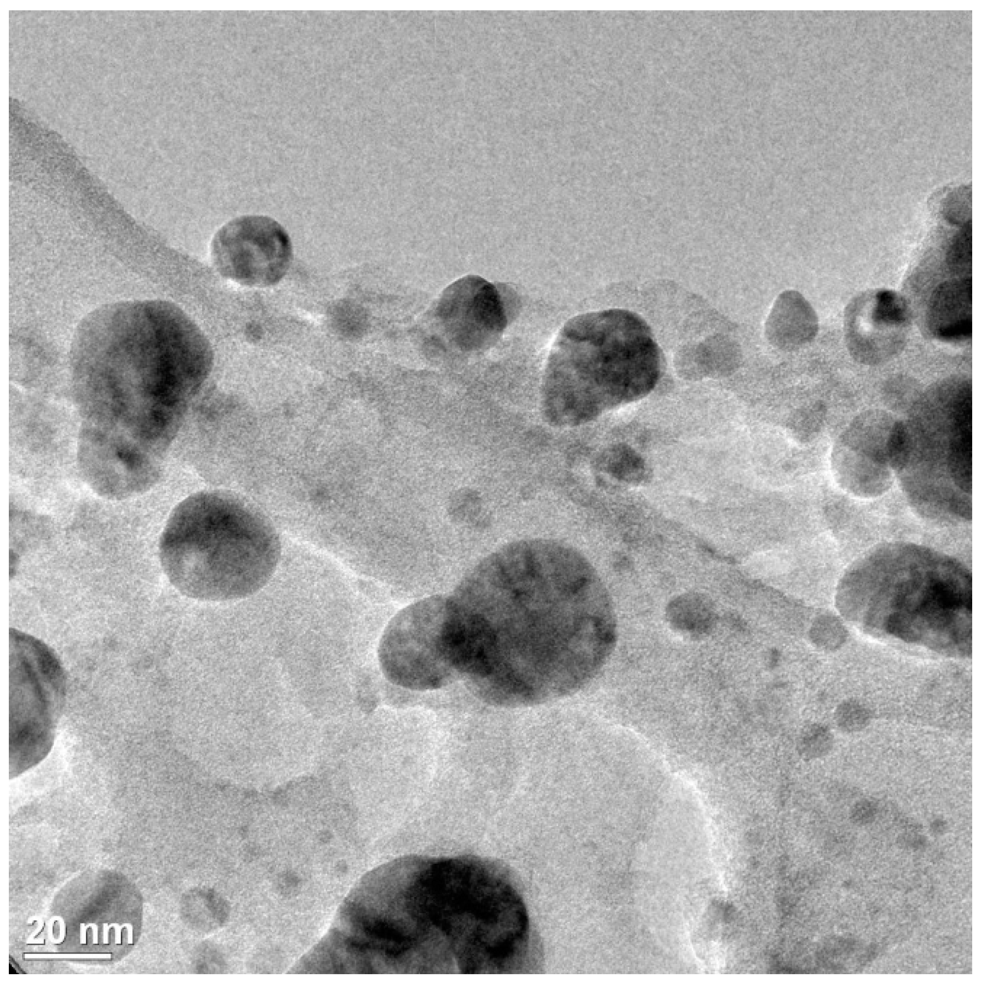
For the experiment, the following treatments were considered: plants without application of Cu nanoparticles (T0), plants with application of Cu nanoparticles at 25 mg L−1 (T1), and plants with application of Cu nanoparticles at 100 mg L−1 (T2). Four foliar applications of Cu nanoparticles occurred every 15 days at 15, 30, 45, and 60 days after transplanting (dat). Each application of Cu NPs was performed by uniformly spraying the entire plant, and approximately 25 mL per plant was used. The copper nanoparticles applied were synthesized in the Applied Chemistry Research Center (Coahuila, Mexico); the process was carried out in a 2 L Parr Reactor equipped with temperature control and mechanical stirrer. A total of 150 mL of glycerol and 24 g of 3-aminopropionic acid dissolved in distilled water were stirred at 350 rpm at 70 °C. Subsequently, 30 g of CuSO4·5H2O dissolved in distilled water were added and the reaction temperature was increased slowly until it reached 180–190 °C. The reaction was carried out with a nitrogen current to remove H2O from the reaction system. The resulting solution was allowed to cool under a nitrogen atmosphere. The Cu NPs were separated by centrifugation, washed with distilled water and methanol, and dried at 70 °C in a vacuum oven. The average particle size was calculated using the Debye-Scherrer equation, yielding a value of 48.3 nm [13,14]. Transmission electron microscopy (TEM) (FEI Titan high-resolution electron microscope, Thermo Scientific, Waltham, MA, USA) micrographs showed nanoparticles of spherical morphology with particle diameters between 20 nm and 50 nm (Figure 2). Figure 3 shows the X-ray diffraction (XRD) patterns of Cu nanoparticles. Three important peaks were detected, reflections corresponding to angle 2θ: 43.6°, 50.8°, and 74.4°, corresponding to the crystalline planes (111), (200), and (220) associated with diffraction patterns of elemental copper.
Three days after the second, third, and fourth foliar application, the leaves were harvested to perform different analyses. For these analyses, the leaves were selected and we verified that they were not physically damaged and that they were uniform in maturity (intense green) using portable measurement carried out by a Chlorophyll Meter (SPAD 502, Minolta, NJ, USA).

2.3. Reactives

Phenolphthalein was purchased from Hycel de México, S.A. of C.V. (Mexico, D.F., Mexico); sodium hydroxide (NaOH), sodium carbonate, and aluminum trichloride, were obtained from J.T. Baker S.A. of C.V. (Avantor Performance Materials, Center Valley, PE, USA). Gallic acid, quercetin, 2,2′-diphenyl-1-picrylhydrazyl (DPPH), 2,2′-azino-bis (3-ethylbenzthiazolin-6-sulfonic acid) (ABTS), Trolox (6-hydroxy-2,5,8-tetramethylchroman-2-carboxylic acid), Folin-Ciocalteau reagent, TPTZ (2,4,6-tri [2-pyridyl]-s-triazine), and ferric chloride were purchased from Sigma-Aldrich Química SA of C.V (St. Louis, MO, USA). Methyl alcohol, ethanol, and methanol were purchased from Química Meyer (Química Suastes S.A. de C.V. Tlahúac, México, D.F., México) and potassium persulfate from Laboratorios Reasol S.A. of C.V. (Tecamac, State of Mexico, Mexico). All aqueous solutions were prepared with Milli-Q® filtered water (resistivity > 18 cm MU) (Millipore, Bedford, MA, USA).

2.4. Sample Preparation

For the evaluation of chlorophyll a, b, total chlorophyll, and bioactive compounds, from each cut-off date, the moringa leaves were subjected to deep-freezing at −70 °C (Thermo Scientific 303 Ultra Freezer). Subsequently, they were lyophilized in a Labconco freeze dryer (Labconco, Model 79480, Kansas City, MO, USA) at a vacuum pressure of 133 × 10−3 mBar and at a temperature of −40 °C. After being lyophilized, the leaves were milled in a knife mill (RTSCH GM 200, Haan, Germany) at 9000 rpm for 50 s until a fine powder of 150 microns was obtained.

2.5. Ascorbic Acid

The determination of ascorbic acid was carried out according to Klein and Perry [18]. A 0.1 g sample of lyophilized Moringa leaves was mixed with metaphosphoric acid (0.1 g L−1). The samples were subjected to an ultrasonic bath (Ultrasonic Cleaner, Mod. 32V118A, IL, USA) for 40 min at intervals of 10 min with 5 min of rest and at a frequency of 40 kHz. Subsequently the samples were centrifuged at 15,000× g for 10 min (Thermo Scientific centrifuge, Mod. ST 16R, Waltham, MA, USA). From the supernatant, 1 mL was taken and 2,6-dichlorophenol-indophenol was mixed, and subsequently absorbance was measured at 515 nm. The analyses were performed in triplicate and the results are expressed as mg of ascorbic acid per 100 g of dry weight.

2.6. Total Phenols

Total phenolic content was determined using the Folin-Ciocalteu method described by Waterman and Mole [19]. A 0.1 g sample of lyophilized powder was taken, then 10 mL of methanol was added. The samples were subjected to an ultrasonic bath (Lab Safety Supply, Mod. 32V118A, Janesville, WI, USA), for 40 min at intervals of 10 min with 5 min of rest and at a frequency of 40 kHz. Subsequently they were centrifuged at 15,000× g for 10 min (Thermo Scientific centrifuge, Mod. ST 16R, Waltham, MA, USA). From the supernatant, 0.5 mL was taken and 5 mL of the Folin–Ciocalteu reagent diluted 50% with distilled water was added; it was left to rest for 7 min, then later, 4 mL of 7.5% sodium carbonate was added and left to react in complete darkness for 1 h. Subsequently, the absorbance at 725 nm was measured in a spectrophotometer (model 6715 UV/Vis, Jenway, Techne Inc., Staffordshire, UK), using methanol as target. A calibration curve was created with a standard solution of gallic acid at a concentration (1000 mg L−1). The results are expressed in milligrams equivalents of gallic acid per 100 gm of dry weight (mg EGA 100 g−1 DW).

2.7. Determination of Flavonoids

The determination of the flavonoid content was done according to Rosales et al. [20]. A 0.1 g lyophilized sample was weighed and mixed with 10 mL of pure methanol. The samples were subjected to an ultrasonic bath (Ultrasonic Cleaner, Mod. 32V118A, Freeport, IL, USA) for 40 min at intervals of 10 min with 5 min of rest and at a frequency of 40 kHz. Subsequently, they were centrifuged at 15,000× g for 10 min (Thermo Scientific centrifuge, Mod. ST 16R, Waltham, MA, USA). After 0.5 mL of the extract was added, plus 0.15 mL of 5% NaNO2, the mixture was allowed to stand for 5 min in the dark. After that time, 0.15 mL of AlCl3·6H2O and 1 mL NaOH were added and allowed to stand for 15 min. The absorbance was subsequently measured at 415 nm in a spectrophotometer (model 6715 UV/Vis, Jenway, Techne Inc., Staffordshire, UK). The total flavonoid content was determined using a standard curve of quercetin. The results are expressed in milligrams equivalents of quercetin per 100 gm of dry weight (mg EQ 100 g−1 DW).

2.8. Determination of Antioxidant Activity

A total of 0.1 g of lyophilized sample were weighed and diluted in 10 mL methanol. The sample was subjected to an ultrasonic bath (Lab Safety Supply, Mod. 32V118A, Janesville, WI, USA), for 40 min at 10 min intervals with 5 min of rest and at a frequency of 40 kHz. Subsequently, they were centrifuged at 15,000× g for 10 min (Thermo Scientific centrifuge, Mod. ST 16R, Waltham, MA, USA). The supernatant was used for the determination of antioxidant activity by ABTS assays [2,2′-azino-bis (3-ethylbenzthiazolin-6-sulfonic acid)], DPPH (2,2-diphenyl-1-picrylhydrazyl), and of the ferric/antioxidant reducing power (FRAP).
The antioxidant activity using ABTS was determined following the method described by Re et al. [21]. The radical ABTS •+ was produced in the following manner: 10 mL of ABTS solution at 7 mM was prepared and mixed with 10 mL of potassium persulfate at 2.45 mM; the mixture was kept in constant agitation and at room temperature in darkness for 16 h. The solution of ABTS •+ was diluted in methanol until an absorbance of 0.7 ± 0.02 at 734 nm was obtained. A 100 μL sample was mixed with 3.9 mL of the diluted ABTS •+ solution and allowed to stand for 6 min. The absorbance was then measured in a spectrophotometer (model 6715 UV/Vis, Jenway, Techne Inc., Staffordshire, UK) at 754 nm. To obtain results, a Trolox standard curve was prepared. The results are expressed as μmol equivalents of Trolox per gram of dry weight (μmol ET g−1 DW).
The antioxidant activity determined by the DPPH method was obtained according to Brand-Williams et al. [22]. A 6 × 10⁻⁵ M DPPH methanol solution was prepared, which was placed in constant agitation for 2 h in complete darkness. Subsequently, 0.3 mL of the extract was taken and 2.7 mL of the methanolic solution was added with DPPH; it was stirred for 15 s and was left to stand in complete darkness for a period of 1 h at 4 °C. Afterward, the absorbance was measured in a spectrophotometer (model 6715 UV/Vis, Jenway, Techne Inc., Staffordshire, UK) at 515 nm. To obtain results, a Trolox standard curve was prepared. The results are expressed as μmol equivalents of Trolox per gram of dry weight (μmol ET g−1 DW).
For the analysis of the ferric/antioxidant reducing power [23], the FRAP reagent was prepared by maintaining it at 37 °C by mixing acetate buffer (0.3 M pH 3.6) with a 10 mM solution of TPTZ (2,4,6 tripyridyl-s-triazine) in 40 mM HCl, and a 20 mM solution of FeCl36H2O, in a ratio of 10:1:1. The assay solutions were prepared by mixing 2.25 mL of the FRAP reagent with 0.3 mL of the extract mixture with ethanol; they were then incubated in the dark for 30 min at room temperature and then the absorbance was measured at 593 nm. The results are expressed as μmol equivalents of Trolox per gram of dry weight (μmol ET g−1 DW).

2.9. Carotenoids

The isochromatic fractions of red carotenoids (R = capsanthin and capsorubin) and yellow carotenoids (A = β-carotene, β-cryptoxanthin, and zeaxanthin) of the total carotenoids were evaluated according to the method reported by Hornero-Méndez and Minguez-Mosquera [24]. The lyophilized sample (100 mg) was mixed with 10 mL of acetone. Subsequently, the samples were subjected to an ultrasonic bath (Lab Safety Supply, Mod. 32V118A, Janesville, WI, USA) for 40 min at intervals of 10 min with 5 min of rest and at a frequency of 40 kHz. They were then centrifuged at 15,000× g for 10 min (Thermo Scientific centrifuge, Mod. ST 16R, Waltham, MA, USA). The supernatant was removed and the absorbance was recorded at 472 (yellow) and 508 (red) nm using a spectrophotometer (model 6715 UV/Vis, Jenway, Techne Inc., Staffordshire, UK). The measurements of red and yellow carotenoids were expressed as milligrams per 100 gm of dry weight (mg 100 g-1 DW).

2.10. Determination of Chlorophyll

The determination of chlorophyll content was performed according to the method of Witham and Blaydes [25] with modifications. Chlorophyll was extracted from lyophilized samples by placing a 100 mg sample in 10 mL acetone. The samples were subjected to an ultrasonic bath (Lab Safety Supply, Mod. 32V118A, Janesville, WI, USA) for 40 min at intervals of 10 min with 5 min of rest and at a frequency of 40 kHz. Subsequently they were centrifuged at 15,000× g for 10 min (Thermo Scientific centrifuge, Mod. ST 16R, Waltham, MA, USA). The supernatant was separated and then the absorbance at 645 and 663 nm was measured in a spectrophotometer (model 6715 UV/Vis, Jenway, Techne Inc., Staffordshire, UK) and the calculations were performed by the following equations:
  Chlorophyll   a   ( mg   g 1   DW ) = ( 12.7 × A b s 663 2.69 × A b s 645 ) × V 100 × W
  Chlorophyll   b   ( mg   g 1   DW ) = ( 22.9 × A b s 645 4.68 × A b s 663 ) × V 100 × W
  Total   Chlorophylls   ( mg   g 1   DW ) = ( 20.2 × A b s 645 + A b s 645 ) × V 100 × W
where Abs is recorded observance at the wavelength specified as a subscript, V is final volume of the chlorophyll-acetone extract, and DW is the dry weight (g) of the sample used.

2.11. Statistical Analysis

An analysis of variance was performed considering a 3 × 3 factorial design completely randomized, where one factor was the different doses of Cu NPs (0, 25, and 100 mg L−1), and the second factor was the number of applications (2, 3, or 4). A means test of Fisher Least Significant Difference was performed to group the means with p ≤ 0.05 considered statistically significant.

3. Results and Discussion

The results show that the application of Cu NPs induced a greater accumulation of antioxidant compounds as well as increased the antioxidant capacity of the leaves of M. oleifera (Table 1). The dose of 25 mg L−1 Cu NPs was superior to the control in terms of content of all the evaluated antioxidant compounds (phenols, flavonoids, and vitamin C), as well as in terms of antioxidant capacity (ABTS, DPPH, and FRAP). The dose of 100 mg L−1 Cu NPs exceeded the control in the content of flavonoids and vitamin C, as well as in the antioxidant capacity, ABTS and FRAP, whereas no effect was generated in phenol content or antioxidant capacity (DPPH). With the application of 25 mg L−1 Cu NPs, the content of phenols and flavonoids in the leaves of M. oleifera increased by 4.3% and 3.6%, respectively, whereas the content of vitamin C increased by 170%. The same dose also increased the antioxidant capacity of ABTS, DPPH, and FRAP by 21%, 2.7% and 6.3%, respectively.
The highest content of phenols (20.80 mg GAE g DW) and flavonoids (42.20 mg QE g DW) observed in leaves of M. oleifera (Table 1) was higher than reported by Vats and Gupta [26] (9.58 mg GAE g DW and 2.3 mg QE g DW, respectively). However, it was slightly lower than that reported by Sreelatha and Padma [27] in mature leaves of M. oleifera (45.81 mg GAE g DW and 27 mg QE g DW, respectively). The highest content of vitamin C (5.84 mg AA g DW) observed in this study was higher than that reported by Vats and Gupta [26] (2.80 mg AA g DW); however, it was lower than that reported by Sreelatha and Padma [27] in mature leaves of M. oleifera (6.60 mg AA g DW).
The number of applications was also a factor that significantly affected the concentration of antioxidant compounds as well as the antioxidant capacity of M. oleifera leaves (Table 1). The content of phenols and flavonoids was higher when only two applications of NPs were made, whereas after the third and fourth applications, the content decreased. In terms of vitamin C content, the opposite was observed: as the number of applications increased, do did the vitamin C. As for the antioxidant capacity, ABTS was higher with three applications of Cu NPs, whereas DPPH was higher with only two applications. FRAP antioxidant capacity was affected with three applications of NPs, since they induced lower antioxidant capacity, with two or four applications being better.
The interactions between the dose factors of Cu NPs and the number of applications also showed statistical differences in all the evaluated antioxidant variables (Table 1). The content of phenols was higher with two applications of 25 or 100 mg L−1 of Cu NPs, whereas the flavonoids were higher with only two applications of 100 mg L−1 of Cu NPs. Three applications of 25 mg L−1 of Cu NPs induced greater accumulation of vitamin C, as well as a higher ABTS antioxidant capacity. The DPPH and FRAP antioxidant capacity was higher with only two applications of 25 mg L−1 of Cu NPs. Therefore, a low dose of Cu NPs (25 mg L−1) applied twice by foliar route was the most efficient for increasing the bioactive compounds and antioxidant capacity of the leaves of M. oleifera.
The results obtained clearly indicate that the application of Cu NPs induces a higher content of antioxidant compounds (phenols, flavonoids, and vitamin C) in the leaves of M. oleifera, which is consistent with the observed increase in antioxidant capacity (ABTS, DPPH, and FRAP) (Table 1). Phenols work as antioxidants due to the reduction of free radical levels in cells [28,29]. In addition, they can function as markers under conditions of abiotic stress [30]. Flavonoids are also metabolites that act as antioxidants, protecting plants from oxidative stress, since they have the capacity for scavenger reactive oxygen species (ROS) such as H2O2 and singlet oxygen generated under stress conditions [31,32]. Vitamin C has many cellular functions in plants, which are mostly linked to the molecule’s ability to donate electrons—a characteristic that allows it to act as an antioxidant by sequestering ROS, preventing or minimizing their damage [33]. In addition, it can regenerate the glycoprotein and tocopherol radicals, and act as a cofactor for many enzymes such as APX [34].
Similar effects have been reported for the application of Cu NPs in tomato: foliar application increased the content of phenols, β-carotene, and vitamin C, and also increased antioxidant capacity [12]. Application via root increased the content of lycopene and vitamin C [13]. The application of Cu NPs also induced the accumulation of antioxidants as proline in Triticum aestivum L. [15]. In jalapeño pepper, the application of Cu NPs in chitosan hydrogels induced the accumulation of capsaicin and increased antioxidant capacity [11]. This indicates that the increase of antioxidant compounds is related to the activation of the antioxidant defense system of plants caused by oxidative stress, which in turn is a result of the application of Cu NPs in cells [11,35]. This is because the application of Cu NPs can generate some stress in the plants, resulting in an increase of EROs, which in turn activates the antioxidant defense mechanism that results in the production of a series of enzymatic antioxidant compounds [35,36]. The final result is an increase in the production of antioxidant compounds such as ascorbate, glutathione, carotenoids, and flavonoids, among others [37]. Therefore, the accumulation of antioxidant compounds and increase in antioxidant capacity in the leaves of M. oleifera can be induced effectively with the application of Cu NPs foliar route.
The contents of red, yellow, and chlorophyll carotenoids in the leaves of M. oleifera were also affected by the application of Cu NPs (Table 2). The red carotenoids increased as the dose of Cu NPs used increased, reaching up to 50% more. The yellow carotenoids were higher with 25 mg L−1 of Cu NPs (8.6%); however, the higher doses of NPs generated a decrease.
The number of applications of Cu NPs also had a significant effect on the carotenoids (Table 2). The red carotenoids decreased as the number of applications of NPs increased, obtaining the best result with only two applications. The yellow carotenoids presented higher contents with three applications of Cu NPs. This indicates a tendency for carotenoids to decrease as the number of applications of Cu NPs increases.
Considering the number of applications and doses of Cu NPs, the best results in terms of content of red carotenoids were obtained with only two applications of 25 mg L−1 of Cu NPs; however, there were no statistically significant differences from the control. In content of yellow carotenoids, the best result was observed with three applications of 25 mg L−1 of Cu NPs. With the exception of the yellow carotenoids, in the other carotenoids, it was clearly observed that two applications of 25 mg L−1 of Cu NPs generated the best results (Table 2).
Carotenoids are a group of more than 600 pigments (carotenes and xanthophylls) from plants and other organisms with a diversity of functions, either in photosynthetic processes of plants and even potentially benefiting human health [38,39]. In plants, they function as accessory pigments for light harvesting and as photoresists during photosynthesis, in addition to being antioxidants [39]. Therefore, the increase in these pigments in plants is a favorable response, since it can be directly related to the increase in antioxidant capacity [40]. In the particular case of M. oleifera, the increase in carotenoid content (red and yellow) observed (Table 2) indicates higher nutraceutical quality and greater antioxidant capacity, as shown in Table 1. Due to the antioxidant function of carotenoids [40], it is possible that its increase was due to the activation of the antioxidant defense system of the plant originated by the application of Cu NPs [11,35], as previously mentioned. When this system is activated, antioxidant compounds such as carotenoids are generated [37]. However, derived from the diversity of carotenoids [38,39], each specific type may present different responses, as observed in this study where red carotenoids increased with higher doses of NPs, whereas yellow carotenoids were higher at lower doses (Table 2).
Only chlorophyll increased with 25 mg L−1 of NPs of Cu, with content 11.9% higher than the control, whereas the content of chlorophyll b and total chlorophyll did not change with application of Cu NPs (Table 2). The number of applications of Cu NPs also had a significant effect on chlorophyll. Two or three applications of NPs generated the largest accumulation, whereas four applications decreased the content of chlorophyll. The optimal number of applications and dose of Cu NPs on chlorophyll (a, b, and total) was two applications of 25 mg L−1 of Cu NPs. These results are consistent with Pradhan et al. [14]. They applied Cu NPs in Vigna radiata and observed an increase in chlorophyll content and therefore photosynthetic activity was modified. However, an opposite effect was reported after the application of Cu NPs, since it decreased the chlorophyll in Chlamydomonas reinhardtii green algae [41,42] as well as in Coriandrum sativum [43].
Regarding the content of antioxidants in the fruits of M. oleifera, the application of NPs generated negative effects, since it decreased the concentration of phenols, flavonoids, and vitamin C, and decreased the antioxidant capacity of ABTS and DPPH (Table 3). This was confirmed by the interactions of the dose factors of Cu NPs and the number of applications, where no treatment with NPs induced an increase in antioxidant compounds or antioxidant capacity. The concentration of phenols and flavonoids decreased up to 14% and 9%, respectively, with a 25 mg L−1 dose of Cu NPs. Vitamin C decreased further with the 100 mg L−1 dose of Cu NPs, presenting 38% less than the control. Finally, the ABTS and DPPH antioxidant capacity decreased in a greater proportion with the dose of 25 mg L−1 of Cu NPs, by 34% and 26%, respectively, which is a lower antioxidant capacity than the control.
Regarding the number of applications, three applications generated the greatest decrease in bioactive compounds (phenols and flavonoids) and antioxidant capacity (ABTS and DPPH). Only for vitamin C did four applications of Cu NPs generate the lowest concentration, whereas the FRAP antioxidant capacity was not affected by the number of applications (Table 3). Therefore, only two applications of Cu NPs would be most suitable, since the biocompounds in the fruits of M. oleifera would be more conserved.
The content of carotenoids in the fruits of M. oleifera was also negatively affected by the application of Cu NPs. The contents of red, yellow, and chlorophyll carotenoids were higher in the control (Table 4). The content of red carotenoids decreased by 43% compared to the control when 25 mg L−1 of Cu NPs were applied. The content of yellow carotenoids was affected only by the application of 100 mg L−1 of Cu NPs, decreasing 28% in comparison to the control. As for chlorophyll (a, b, and total), the application of 25 mg L−1 of Cu NPs negatively affected the concentration, decreasing chlorophyll a by 38%, chlorophyll b by 43%, and total chlorophyll by 42% in comparison to the control.
The number of applications also had a negative influence: as application number increased, the content of red carotenoids and chlorophylls decreased (Table 4). Only the content of yellow carotenoids increased with four applications of Cu NPs.
Interactions between the number of applications and the doses of NPs showed that treatments that did not include the application of NPs did not generate positive effects on the content of red carotenoids and chlorophyll (Table 4). Only in the content of yellow carotenoids did four applications of Cu NPs (25 and 100 mg L−1) have a positive effect on this variable.
In general, there is a clear trend that the foliar application of Cu NPs to M. oleifera plants induces a decrease in the biocompounds in the fruits of this plant. However, the concentrations of biocompounds, such as total phenols, β-carotene, and lycopene, were higher in the leaves of M. oleifera than in the rest of the organs [26]. In addition, the leaf of M. oleifera represents the largest proportion of biomass in the plant; therefore, the increase in biocompounds observed in this organ is more important than a decrease in the fruits.

4. Conclusions

The foliar application of Cu NPs increased the content of bioactive compounds as well as the antioxidant capacity in the leaves of M. oleifera, so the use of these NPs can be an important option to improving the quality of this plant.
A dose of 25 mg L−1 Cu NPs applied via foliar twice during the development of M. oleifera culture seems to be the optimal dose, since it consistently increased the content of carotenoids, phenols, flavonoids, vitamin C, and antioxidant capacity (ABTS, DPPH, and FRAP) in the leaves of the plant. However, in the fruits of M. oleifera, the application of Cu NPs generated the opposite effect—the content of all the bioactive compounds and antioxidant capacity decreased.

Author Contributions

A.D.H.-F., A.J.-M. and J.V.-R. conceived and designed the experiments; J.M.P.-E. and C.U.L.-P. performed the analysis of laboratory and field experiments; H.O.-O. and G.C.-P. contributed reagents and materials. All authors were responsible for processing information and manuscript writing. All authors read and approved the final manuscript.

Funding

This research received no external funding.

Conflicts of Interest

The authors declare no conflict of interest.

References

  1. Velázquez-Zavala, M.; Peón-Escalante, I.E.; Zepeda-Bautista, R.; Jiménez-Arellanes, M.A. Moringa (Moringa oleifera Lam.): Potential uses in agriculture, industry and medicine. Rev. Chapingo Ser. Hortic. 2016, 22, 95–116. [Google Scholar] [CrossRef]
  2. Gupta, S.; Jain, R.; Kachhwaha, S.; Kothari, S.L. Nutritional and medicinal applications of Moringa oleifera Lam.-Review of current status and future possibilities. J. Herb. Med. 2017, 1–11. [Google Scholar] [CrossRef]
  3. McNeil, S.E. Nanotechnology for the biologist. J. Leukoc. Biol. 2005, 78, 585–594. [Google Scholar] [CrossRef] [PubMed]
  4. Naderi, N.; Karponis, D.; Mosahebi, A.; Seifalian, A.M. Nanoparticles in wound healing; from hope to promise, from promise to routine. Front. Biosci. (Landmark Ed.) 2018, 23, 1038–1059. [Google Scholar] [CrossRef] [PubMed]
  5. Capaldi Arruda, S.C.; Diniz Silva, A.L.; Moretto Galazzi, R.; Antunes Azevedo, R.; Zezzi Arruda, M.A. Nanoparticles applied to plant science: A review. Talanta 2015, 131, 693–705. [Google Scholar] [CrossRef] [PubMed]
  6. Liu, R.; Lal, R. Potentials of engineered nanoparticles as fertilizers for increasing agronomic productions. Sci. Total Environ. 2015, 514, 131–139. [Google Scholar] [CrossRef] [PubMed]
  7. Mishra, S.; Keswani, C.; Abhilash, P.C.; Fraceto, L.F.; Singh, H.B. Integrated Approach of Agri-nanotechnology: Challenges and Future Trends. Front. Plant Sci. 2017, 8, 1–12. [Google Scholar] [CrossRef] [PubMed]
  8. Thiruvengadam, M.; Rajakumar, G.; Chung, I.M. Nanotechnology: Current uses and future applications in the food industry. 3 Biotech 2018, 8, 74. [Google Scholar] [CrossRef] [PubMed]
  9. Juarez-Maldonado, A.; Ortega-Ortíz, H.; Pérez-Labrada, F.; Cadenas-Pliego, G.; Benavides-Mendoza, A. Cu Nanoparticles absorbed on chitosan hydrogels positively alter morphological, production, and quality characteristics of tomato. J. Appl. Bot. Food Qual. 2016, 89, 183–189. [Google Scholar] [CrossRef]
  10. Hernández, H.H.; Benavides-Mendoza, A.; Ortega-Ortiz, H.; Hernández-Fuentes, A.D.; Juárez-Maldonado, A. Cu Nanoparticles in chitosan-PVA hydrogels as promoters of growth, productivity and fruit quality in tomato. Emir. J. Food Agric. 2017, 29. [Google Scholar] [CrossRef]
  11. Pinedo-Guerrero, Z.H.; Hernández-Fuentes, A.D.; Ortega-Ortiz, H.; Benavides-Mendoza, A.; Cadenas-Pliego, G. Cu nanoparticles in hydrogels of chitosan-PVA affects the characteristics of post-harvest and bioactive compounds of jalapeño pepper. Molecules 2017, 22, 926. [Google Scholar] [CrossRef] [PubMed]
  12. Hernández-Fuentes, A.D.; López-Vargas, E.R.; Pinedo-Espinoza, J.M.; Campos-Montiel, R.G.; Valdés-Reyna, J.; Juárez-Maldonado, A. Postharvest behavior of bioactive compounds in tomato fruits treated with Cu nanoparticles and NaCl stress. Appl. Sci. 2017, 7, 980. [Google Scholar] [CrossRef]
  13. Hernández-Hernández, H.; González-Morales, S.; Benavides-Mendoza, A.; Ortega-Ortiz, H.; Cadenas-Pliego, G.; Juárez-Maldonado, A. Effects of chitosan–PVA and Cu nanoparticles on the growth and antioxidant capacity of tomato under saline stress. Molecules 2018, 23, 178. [Google Scholar] [CrossRef] [PubMed]
  14. Pradhan, S.; Patra, P.; Mitra, S.; Dey, K.K.; Basu, S.; Chandra, S.; Palit, P.; Goswami, A. Copper nanoparticle (CuNP) nanochain arrays with a reduced toxicity response: A biophysical and biochemical outlook on Vigna radiata. J. Agric. Food Chem. 2015, 63, 2606–2617. [Google Scholar] [CrossRef] [PubMed]
  15. Zhang, Z.; Ke, M.; Qu, Q.; Peijnenburg, W.J.G.M.; Lu, T.; Zhang, Q.; Ye, Y.; Xu, P.; Du, B.; Sun, L.; et al. Impact of copper nanoparticles and ionic copper exposure on wheat (Triticum aestivum L.) root morphology and antioxidant response. Environ. Pollut. 2018, 239, 689–697. [Google Scholar] [CrossRef] [PubMed]
  16. Fu, P.P.; Xia, Q.; Hwang, H.-M.; Ray, P.C.; Yu, H. Mechanisms of nanotoxicity: Generation of reactive oxygen species. J. Food Drug Anal. 2014, 22, 64–75. [Google Scholar] [CrossRef] [PubMed] [Green Version]
  17. Hong, J.; Wang, L.; Sun, Y.; Zhao, L.; Niu, G.; Tan, W.; Rico, C.M.; Peralta-Videa, J.R.; Gardea-Torresdey, J.L. Foliar applied nanoscale and microscale CeO2 and CuO alter cucumber (Cucumis sativus) fruit quality. Sci. Total Environ. 2016, 563, 904–911. [Google Scholar] [CrossRef] [PubMed]
  18. Klein, B.P.; Perry, A.K. Ascorbic Acid and Vitamin A Activity in Selected Vegetables from Different Geographical Areas of the United States. J. Food Sci. 1982, 47, 941–945. [Google Scholar] [CrossRef]
  19. Waterman, P.G.; Mole, S. Analysis of Phenolic Plant Metabolites; Blackwell Scientific: Boston, MA, USA, 1994; ISBN 0632029692. [Google Scholar]
  20. Rosales, M.A.; Cervilla, L.M.; Sánchez-Rodríguez, E.; Rubio-Wilhelmi, M.D.M.; Blasco, B.; Ríos, J.J.; Soriano, T.; Castilla, N.; Romero, L.; Ruiz, J.M. The effect of environmental conditions on nutritional quality of cherry tomato fruits: Evaluation of two experimental Mediterranean greenhouses. J. Sci. Food Agric. 2011, 91, 152–162. [Google Scholar] [CrossRef] [PubMed]
  21. Re, R.; Pellegrini, N.; Proteggente, A.; Pannala, A. Antioxidant activity applying an improved ABTS radical cation decolorization assay. Free Radic. Biol. 1999, 9, 1231–1237. [Google Scholar] [CrossRef]
  22. Brand-Williams, W.; Cuvelier, M.E.; Berset, C.L.W.T. Use of a Free Radical Method to Evaluate Antioxidant Activity. Food Sci. Technol. 1995, 28, 25–30. [Google Scholar] [CrossRef]
  23. Benzie, I.F.F.; Strain, J.J. The ferric reducing ability of plasma (FRAP) as a measure of “antioxidant power”: The FRAP assay. Anal. Biochem. 1996, 239, 70–76. [Google Scholar] [CrossRef] [PubMed]
  24. Hornero-Méndez, D.; Minguez-Mosquera, M.I. Rapid spectrophotometric determination of red and yellow isochromic carotenoid fractions in paprika and red pepper oleoresins. J. Agric. Food Chem. 2001, 49, 3584–3588. [Google Scholar] [CrossRef] [PubMed]
  25. Witham, F.H.; Blaydes, D.F.; Devlin, R.M. Experiments in Plant Physiology; Van Nostrand Reinhold: New York, NY, USA, 1971. [Google Scholar]
  26. Vats, S.; Gupta, T. Evaluation of bioactive compounds and antioxidant potential of hydroethanolic extract of Moringa oleifera Lam. from Rajasthan, India. Physiol. Mol. Biol. Plants 2017, 23, 239–248. [Google Scholar] [CrossRef] [PubMed] [Green Version]
  27. Sreelatha, S.; Padma, P.R. Antioxidant activity and total phenolic content of Moringa oleifera leaves in two stages of maturity. Plant Foods Hum. Nutr. 2009, 64, 303–311. [Google Scholar] [CrossRef] [PubMed]
  28. Klunklin, W.; Savage, G. Effect on quality characteristics of tomatoes grown under well-watered and drought stress conditions. Foods 2017, 6, 56. [Google Scholar] [CrossRef] [PubMed]
  29. Pourmorad, F.; Hosseinimehr, S.J.; Shahabimajd, N. Antioxidant activity, phenol and flavonoid contents of some selected Iranian medicinal plants. Afr. J. Biotechnol. 2006, 5, 1142–1145. [Google Scholar] [CrossRef]
  30. Ren, S.C.; Sun, J.T. Changes in phenolic content, phenylalanine ammonia-lyase (PAL) activity, and antioxidant capacity of two buckwheat sprouts in relation to germination. J. Funct. Foods 2014, 7, 298–304. [Google Scholar] [CrossRef]
  31. Hernández, I.; Alegre, L.; Van Breusegem, F.; Munné-Bosch, S. How relevant are flavonoids as antioxidants in plants? Trends Plant Sci. 2009, 14, 125–132. [Google Scholar] [CrossRef] [PubMed]
  32. Hollman, P.C.H.; Arts, I.C.W. Flavonols, flavones and flavanols-nature, occurrence and dietary burden. J. Sci. Food Agric. 2000, 80, 1081–1093. [Google Scholar] [CrossRef]
  33. Padayatty, S.J.; Katz, A.; Wang, Y.; Eck, P.; Kwon, O.; Lee, J.H.; Chen, S.; Corpe, C.; Dutta, A.; Dutta, S.K.; et al. Vitamin C as an Antioxidant: Evaluation of Its Role in Disease Prevention. J. Am. Coll. Nutr. 2003, 22, 18–35. [Google Scholar] [CrossRef] [PubMed]
  34. Muzolf-Panek, M.; Kleiber, T.; Kaczmarek, A. Effect of increasing manganese concentration in nutrient solution on the antioxidant activity, vitamin C, lycopene and polyphenol contents of tomato fruit. Food Addit. Contam. Part A 2017, 34, 379–389. [Google Scholar] [CrossRef] [PubMed]
  35. Rizwan, M.; Ali, S.; Qayyum, M.F.; Ok, Y.S.; Adrees, M.; Ibrahim, M.; Zia-ur-Rehman, M.; Farid, M.; Abbas, F. Effect of metal and metal oxide nanoparticles on growth and physiology of globally important food crops: A critical review. J. Hazard. Mater. 2017, 322, 2–16. [Google Scholar] [CrossRef] [PubMed]
  36. Shobha, G.; Moses, V.; Ananda, S. Biological synthesis of copper nanoparticles and its impact: A review. Int. J. Pharm. Sci. Invent. 2014, 3, 2319–6718. [Google Scholar]
  37. Seminario, A.; Song, L.; Zulet, A.; Nguyen, H.T.; González, E.M.; Larrainzar, E. Drought stress causes a reduction in the biosynthesis of ascorbic acid in soybean plants. Front. Plant Sci. 2017, 8, 1–10. [Google Scholar] [CrossRef] [PubMed]
  38. Young, A.; Lowe, G. Carotenoids—Antioxidant Properties. Antioxidants 2018, 7, 28. [Google Scholar] [CrossRef] [PubMed]
  39. Nisar, N.; Li, L.; Lu, S.; Khin, N.C.; Pogson, B.J. Carotenoid metabolism in plants. Mol. Plant 2015, 8, 68–82. [Google Scholar] [CrossRef] [PubMed]
  40. Liu, H.; Mao, J.; Yan, S.; Yu, Y.; Xie, L.; Hu, J.G.; Li, T.; Abbasi, A.M.; Guo, X.; Liu, R.H. Evaluation of carotenoid biosynthesis, accumulation and antioxidant activities in sweetcorn (Zea mays L.) during kernel development. Int. J. Food Sci. Technol. 2018, 53, 381–388. [Google Scholar] [CrossRef]
  41. Saison, C.; Perreault, F.; Daigle, J.C.; Fortin, C.; Claverie, J.; Morin, M.; Popovic, R. Effect of core-shell copper oxide nanoparticles on cell culture morphology and photosynthesis (photosystem II energy distribution) in the green alga, Chlamydomonas reinhardtii. Aquat. Toxicol. 2010, 96, 109–114. [Google Scholar] [CrossRef] [PubMed]
  42. Melegari, S.P.; Perreault, F.; Costa, R.H.R.; Popovic, R.; Matias, W.G. Evaluation of toxicity and oxidative stress induced by copper oxide nanoparticles in the green alga Chlamydomonas reinhardtii. Aquat. Toxicol. 2013, 142–143, 431–440. [Google Scholar] [CrossRef] [PubMed]
  43. Zuverza-Mena, N.; Medina-Velo, I.A.; Barrios, A.C.; Tan, W.; Peralta-Videa, J.R.; Gardea-Torresdey, J.L. Copper nanoparticles/compounds impact agronomic and physiological parameters in cilantro (Coriandrum sativum). Environ. Sci. Process. Impacts 2015, 17, 1783–1793. [Google Scholar] [CrossRef] [PubMed]
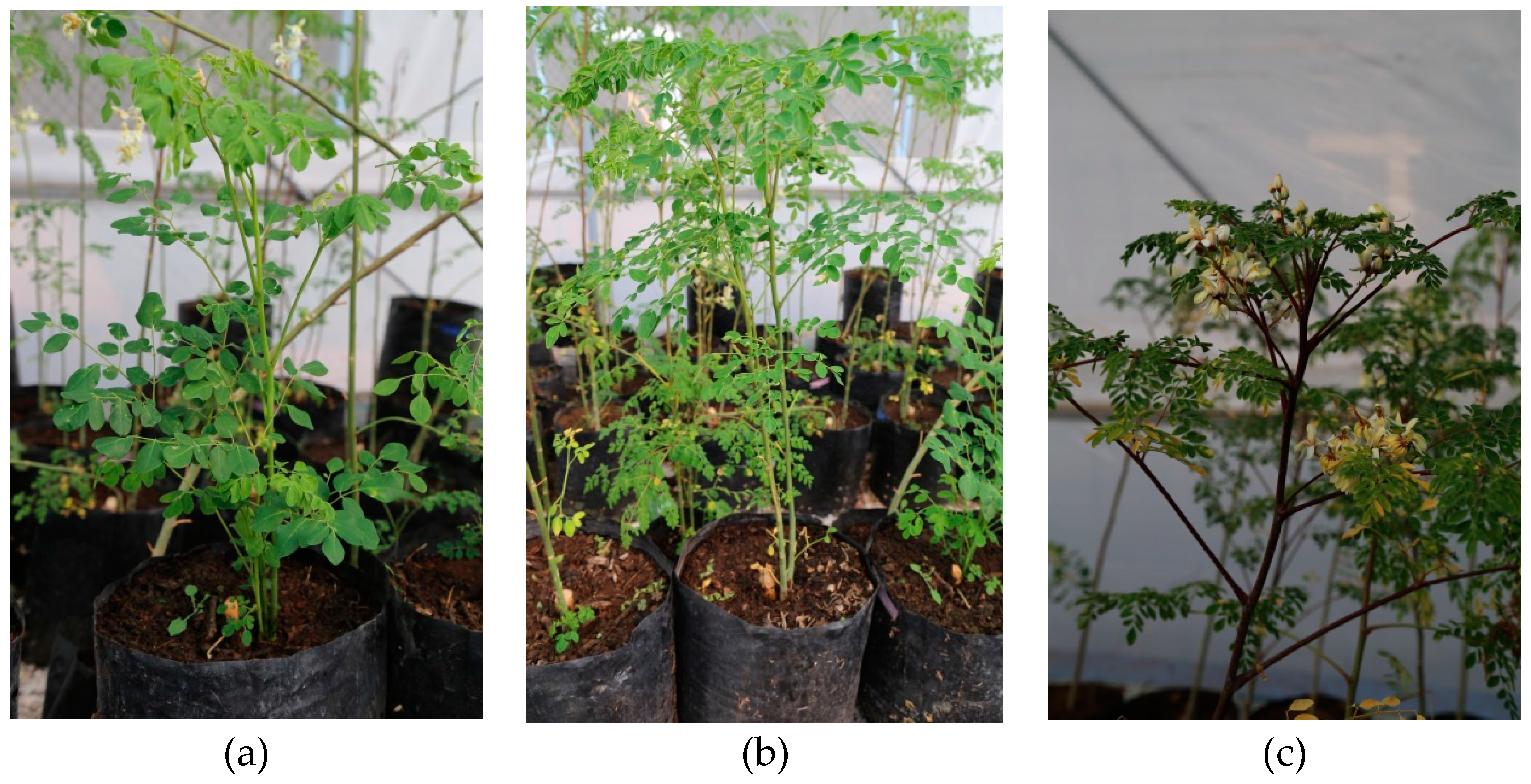
Figure 1. Moringa oleifera plants at different stages of growth: (a) at transplant, (b) 30 days after transplanting, and (c) 60 days after transplanting.
Agronomy 08 00167 g001
Figure 2. Spherical morphology and diameters between 20 and 50 nm of copper nanoparticles (Cu NPs) determined by transmission electron microscopy (TEM).
Agronomy 08 00167 g002
Figure 3. X-ray diffraction (SRD) patterns of Cu NPs.
Agronomy 08 00167 g003
Table 1. Antioxidant content in leaves of Moringa oleifera.
TreatmentPhenols (mg GAE g−1 DW)Flavonoids (mg QE g−1 DW)Vitamin C (mg AA g−1 DW)ABTS (mg T g−1 DW)DPPH (mg T g−1 DW)FRAP (mg T g−1 DW)
Cu NPs 1019.93 b40.73 b2.16 c37.99 c29.50 b40.56 b
2520.80 a42.20 a5.84 a45.94 a30.31 a43.13 a
10020.18 b42.19 a3.50 b41.99 b29.64 b43.42 a
Apps 2221.76 a42.89 a1.37 b39.97 b30.52 a42.74 a
319.55 b41.47 b4.87 a49.04 a29.55 b41.33 b
419.58 b40.76 c5.27 a36.90 c29.40 b43.04 a
T0 320.76 b39.72 ef0.52 f30.47 g30.06 bc41.22 e
2 app 25 mg L−122.58 a42.11 c1.59 ef46.58 bc31.03 a46.53 a
2 app 100 mg L−121.96 a46.84 a2.00 de42.87 d30.46 ab40.47 fg
T019.58 cd43.58 b0.55 f45.45 c29.61 cde39.84 g
3 app 25 mg L−119.47 d41.70 cd11.35 a52.97 a29.88 bcd39.97 fg
3 app 100 mg L−119.61 cd39.12 f2.71 d48.71 b29.15 ef44.17 c
T019.44 d38.89 f5.40 bc38.04 e28.84 f40.63 ef
4 app 25 mg L−120.35 bc42.80 bc4.59 c38.27 e30.03 bc42.88 d
4 app 100 mg L−118.97 d40.59 de5.81 b34.40 f29.32 def45.62 b
1 Doses of Cu NPs applied (mg L−1); 2 Number of applications of Cu NPs performed; 3 Interaction between doses of Cu NPs and number of applications. T0: control; EGA: Equivalents of gallic acid; EQ: Equivalents of quercetin; AA: Equivalents of ascorbic acid; T: Equivalents of Trolox; DW: Dry weight. Different letters per column indicate statistical differences according to Least Significant Differences Fisher test (p ≤ 0.05).
Table 2. Content of carotenoids and chlorophyll in the leaves of Moringa oleifera.
TreatmentRed C. (mg 100 g−1 DW)Yellow C. (mg 100 g−1 DW)Chl a (mg g−1 DW)Chl b (mg g−1 DW)Total Chl (mg g−1 DW)
Cu NPs 100.52 c1.51 b56.09 b35.71 a49.41 a
250.68 b1.64 a62.74 a37.29 a52.94 a
1000.78 a1.38 c57.93 b37.79 a51.82 a
Apps 220.95 a1.30 c60.06 a41.91 a56.13 a
30.60 b1.66 a61.72 a40.06 a55.04 a
40.42 c1.58 b54.99 b28.81 b43.01 b
T0 31.04 a1.20 f61.37 b41.30 ab56.02 ab
2 app 25 mg L−11.01 a1.47 d68.14 a45.93 a62.26 a
2 app 100 mg L−10.82 b1.24 ef50.67 d38.51 ab50.11 b
T00.29 de1.74 b55.54 c42.52 ab55.20 ab
3 app 25 mg L−10.67 c1.94 a69.43 a38.82 ab56.45 ab
3 app 100 mg L−10.85 b1.29 e60.17 b38.84 ab53.47 b
T00.23 e1.61 c51.36 d23.30 d37.01 c
4 app 25 mg L−10.36 d1.52 d50.64 d27.13 cd40.13 c
4 app 100 mg L−10.67 c1.61 c62.96 b36.01 bc51.89 b
1 Doses of Cu NPs applied (mg L−1); 2 Number of applications of Cu NPs performed; 3 Interaction between doses of Cu NPs and number of applications. T0: Control; C: Carotenoids; Chl: Chlorophylls; DW: Dry weight. Different letters per column indicate statistical differences according to Least Significant Differences Fisher test (p ≤ 0.05).
Table 3. Antioxidant content in fruits of Moringa oleifera.
TreatmentPhenols (mg GAE g−1 DW)Flavonoids (mg QE g−1 DW)Vitamin C (mg AA g−1 DW)ABTS (mg T g−1 DW)DPPH (mg T g−1 DW)FRAP (mg T g−1 DW)
Cu NPs 1015.07 a15.83 a2.83 a25.36 a23.08 a20.82 a
2512.98 c14.38 c2.27 b16.64 c17.15 c19.31 a
10013.37 b15.22 b1.76 c20.12 b17.85 b18.82 a
Apps 2214.18 a15.31 a2.80 a25.53 a21.02 a20.77 a
312.81 b14.66 b2.34 b17.50 b16.34 c18.67 a
414.43 a15.47 a1.72 c21.09 a20.73 b19.50 a
T0 315.80 a16.02 a2.90 b28.95 a25.94 a24.80 a
2 app 25 mg L−112.37 e13.40 c2.97 ab18.96 c17.0 f18.31 a
2 app 100 mg L−114.36 b16.52 a2.52 c22.67 bc20.04 c19.21 a
T013.66 cd14.73 b3.16 a22.10 bc18.39 e19.90 a
3 app 25 mg L−112.51 e14.69 b2.37 c11.50 d15.09 g18.34 a
3 app 100 mg L−112.25 e14.55 b1.50 d18.91 c15.53 g17.76 a
T015.74 a16.75 a2.43 c25.02 ab24.93 b17.76 a
4 app 25 mg L−114.07 bc15.05 b1.46 d19.47 c19.27 d21.26 a
4 app 100 mg L−113.48 d14.59 b1.26 d18.79 c17.98 e19.48 a
1 Doses of Cu NPs applied (mg L−1); 2 Number of applications of Cu NPs performed; 3 Interaction between doses of Cu NPs and number of applications. T0: Control; EGA: Equivalents of gallic acid; EQ: Equivalents of quercetin; AA: Equivalents of ascorbic acid; T: Equivalents of Trolox; DW: Dry weight. Different letters per column indicate statistical differences according to Least Significant Differences Fisher test (p ≤ 0.05).
Table 4. Content of carotenoids and chlorophyll in fruits of Moringa oleifera.
TreatmentRed C. (mg 100 g−1 DW)Yellow C. (mg 100 g−1 DW)Chl a (mg g−1 DW)Chl b (mg g−1 DW)Total Chl (mg g−1 DW)
Cu NPs 1029.59 a4.95 a4.52 a7.68 a8.19 a
2516.99 c4.68 ab2.81 b4.42 c4.78 c
10025.31 b3.56 b4.09 a6.33 b6.86 b
Apps 2232.29 a3.57 b4.87 a8.50 a9.02 a
323.51 b3.55 b3.52 b5.69 b6.13 b
416.08 c6.07 a3.03 c4.23 c4.69 c
T0 328.14 b4.68 bc4.59 b8.22 bc8.69 bc
2 app 25 mg L−127.12 b2.81 cd4.10 bc6.98 c7.44 c
2 app 100 mg L−141.62 a3.24 cd5.93 a10.30 a10.94 a
T039.93 a5.99 b5.50 a9.59 ab10.18 ab
3 app 25 mg L−114.57 d3.00 cd2.36 e3.73 de4.03 ef
3 app 100 mg L−116.04 cd1.67 d2.72 de3.76 de4.18 def
T020.68 c4.19 c3.49 cd5.23 d5.71 d
4 app 25 mg L−19.27 e8.23 a1.98 e2.56 e2.88 f
4 app 100 mg L−118.28 cd5.77 b3.61 c4.91 d5.47 de
1 Doses of Cu NPs applied (mg L−1); 2 Number of applications of Cu NPs performed; 3 Interaction between doses of Cu NPs and number of applications. T0: Control; C: Carotenoids; Chl: Chlorophylls; DW: Dry weight. Different letters per column indicate statistical differences according to Least Significant Differences Fisher test (p ≤ 0.05).